Failure analysis of cemented Metal-on-Metal total hip
replacements from a single centre cohort
Michael Bryant
a*, Michael Ward
b, Richard Farrar
c, Robert Freeman
c, Ken Brummitt
c,
John Nolan
d, Anne Neville
aa – Institute of Engineering Thermofluids Surfaces and Interfaces, University of Leeds, UK
b – Institute for Materials Research, University of Leeds, UK c - DePuy International, Leeds, UK
d - Norfolk and Norwich teaching hospital, Nowrich, UK
Received Date Line (to be inserted by Production) (8 pt)
Abstract
This paper presents the failure analysis of cemented metal on metal (MoM) total hip replacements (THR) from a single centre cohort. Between 1997 and 2008, 652 Ultima TPS™ femoral stems with the Ultima MoM articulation (DePuy International, Leeds) were implanted. At 2 – 9 years follow up, 90 revisions had been performed. Upon removal extensive fretting-corrosion of the cemented portions of the femoral stems was seen, resulting in high metal ion levels. In order to fully understand the factors resulting in failure of these cemented MoM THR optical light microscopy, Scanning Electron Microscopy with integrated Energy Dispersive X-ray spectroscopy (SEM/EDX) and X-ray Photo-electron spectroscopy (XPS) were utilised in order assess the surface morphology and chemistry of retrieved MoM THR and surface chemistry of the films found on the surface. Gross slip, plastic deformation and directionality of the surface were extensively seen on the proximal surfaces of the retrievals. A more corrosive phenomenon was in the distal regions of the stem, demonstrating a seemingly inter-granular attack. SEM/EDX analysis demonstrated that black films present commonly seen on the highly worn regions of the cemented portions of the femoral stem were a complex mixture of chromium oxide, protein and amorphous carbon. XPS demonstrated films were Cr, O rich, mixed with organic material.
Keywords: Fretting-corrosion, Femoral Stems, Retrieval study, Case study
1. Introduction
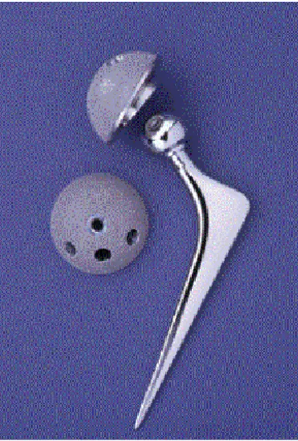
Acetabular outer shell with three holes for supplementary fixation was used. Figure 1 demonstrates the system that was implanted in the cases presented by Donell et al [1].
Figure 1 – Ulitma TPS™ MoM total hip replacement used in the Norwich cohort
In 2008, 90 out of a total series of 652 Ultima TPS™ hip replacement systems metal–on–metal hip replacements had to undergo early revision at the Norfolk and Norwich University Hospital. 17 hips required revision for periprosthetic fracture, with early dislocation in 3 and late dislocations in 16. Infection was found in 10 hips. 44 required revision for extensive, symptomatic, peri-articular soft tissue necrosis of which 35 had normal plain radiographs. The femoral component was cemented with either a plain PMMA bone cement or antibiotic cement containing either the Gentamicin antibiotic or Erythromycin and Colistin. Dramatic corrosion of generally solidly fixed femoral stems was frequently observed on the retrieved cemented part of the femoral component. Blackening of the cemented portions of the stems and staining of the PMMA bone cement, thought to be metallic debris, was also found at revision. It was thought that the necrosis of the surrounding tissue was associated with the release of potentially toxic metal ions such as cobalt and chromium from the stem surface as the bearing surfaces were macroscopically clean of any wear or abrasion. As of 2012, 132 revisions from 652 hips have been conducted. Of the hips that were revised, 169 had at least one abnormal MARS MRI scan and two thirds of revisions for symptomatic ARMD of which three quarters with normal X-rays. 24 hips required revision for peri-prosthetic fractures. As a result of this, a Medical Device Alert MDA/2007/054 dated 14 June 2007 was issued detailing unexplained pain when the Ultima TPS femoral stem was using in conjunction with the 28mm Ulitma MoM articulation.
In this study, 105 Ultima TPS MoM THR were retrieved from the Norfolk and Norwich teaching hospital between 2008-2012. Each stem underwent a sterilisation wash procedure before the analysis was undertaken. Each implant was individually placed in a cardboard surgical tray, sealed in a polythene bag and numbered in numerical order to ensure patient data was kept confidential. Any handling of the implants was performed using latex surgical gloves to avoid contamination. Initially, each stem was macroscopically observed in order to assess the location and extent of fretting-corrosion found on the femoral stem. The location of the fretting corrosion was determined through the use of Gruen zones as outlined by Gruen et al (Figure 2). Each stem was then graded in terms of surface observation and coverage (Table 1). Due to the complex interactions and synergy’s associated with wear and corrosion a grading system was developed that allowed the wear and corrosion to be assessed with the same grading criteria.
Figure 2 – Location and orientation of Gruen zones used to map fretting corrosion in this study
Table 1 – Observational and grading criteria used in this study
No. Scoring Observations/Grading Coverage
1 Low Slight marking of the surface. Less than 10% of the cemented portions affected affected by corrosion/wear
2 Low-Moderate Scoring or dulling of the surface. Between 10% and 25% of the cemented portions show sign of Corrosion/wear. 3 Moderate Definite abrasion/corrosion of the surface. Indication of black oxide debris. 25% to 50% of thecemented portions effected by
corrosion/wear.
4 Moderate-High Pitting and scarring on the surface. Indication of black oxide debris. 50-75% of the cemented portions affected by Corrosion/wear.
5 High Severe surface damage and corrosion debris. Indication of black oxide debris. >75% of the cemented portions affected by severe corrosion/wear.
X-ray photoelectron spectroscopy (XPS) was further utilised to establish the exact chemical composition of any debris or deposit found on the surface. XPS surveys were initially conducted, followed by high resolution XPS scans on the elements found on the surface. Although X-rays penetrate to a depth of several micrometres, ejected photoelectrons generally come from only the first several nanometres of material making the analysis of the passive film and bulk substrate possible. In order to assess the variation of composition and to mitigate any contamination effects each sample was subjected to a 40 sec argon-ion etch it order to displace around 15nm of surface material. Using Licensed CasaXPS fitting software, the peak height, peak area, position of the peaks and concentration of species present on the surface was determined. All the results in this paper use the standard format and units (CPS versus BE). Values of binding energies, FWHM and relative sensitivity factors were taken from literature and applied to the data received [2-7].
3. Results
3.1 Macroscopic observations and grading
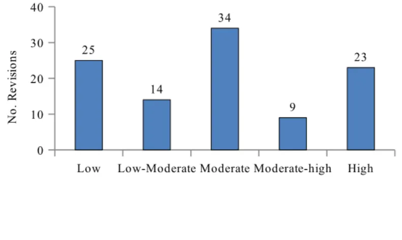
Visual analysis indicated that a wide variety of fretting-corrosion damage was seen on surfaces of the retrieved THRs. On visual inspection, fretting-corrosion of various degrees was observed; with assessment and grading of the retrieved cohort demonstrating a spread in the variety of surface morphologies observed (Figure 3). High amounts of fretting-corrosion were typically seen in Gruen zone 1, 2, 6 and 7. Regions of localised corrosion were also seen in the proximal regions of the cemented femoral stems. Areas of black deposit were also commonly seen on the surfaces of the femoral stems in the areas that appeared to demonstrate high amounts of fretting-corrosion. Although some areas of black deposit were observed in samples that scored a low and low-moderate grades an increased occurrence of blackening of the stem surface was seen with increasing amounts of fretting corrosion.
Low Low-Moderate Moderate Moderate-high High 0 10 20 30 40 25 14 34 9 23 N o . R ev is io n s
Figure 3 – Number of retrieved stems vs. observation/grading stated in table 1.
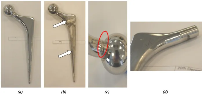
visual grades. Figure 4b demonstrates the blackening of the femoral stem discussed above. At a macroscopic level, the black deposit appeared to have a flaky textured appearance, typically being found in regions of high wear and corrosion. Impingement of varying levels was also seen on the retrieved cohort, typically being seen on the proximal potions of the femoral stem neck (Figure 4c). Analysis of the cohort data identified that impingent of the femoral neck occurred in 65% of the retrieved cohort almost solely in implants utilising the 10° augmented Ø28mm CoCrMo liner. Instances of fretting-crevice corrosion were also noticed at the modular interface (Figure 4d). However it was not seen on all retrieved trunions. It is interesting to note that this corrosive attacked seemed limited to the opening of the taper-trunion interface suggesting a purely corrosive attack with no damage being observed on the trunion in the proximal direction.
(a) (b) (c) (d)
Figure 4- typical macroscopic observation of (a) retrieved femoral stem with low and (b) high observation grading (White arrow indicates areas of black deposits) (c) evidence of impingement (d) corrosion at the modular taper interface
3.2 Microscopic results
(a) (b) (c)
(d) (e) (f)
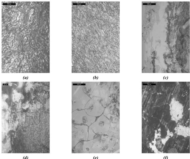
Figure 5 – Light microscope images of typical surface morphology in (a-b) the anterolateral and posteromedial regions of Gruen zone 1and 7 (c-d) Gruen zone 2 and 6 (e-f) Gruen zone 4
clean bulk material.
(a) (b)
(c) (d)
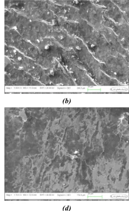
Figure 6 – SEM images of (a) plastically deformed surfaces in Gruen Zone 7 (b) surface morphology in Gruen zone 2 demonstrating the retention of debris within the grooves valleys of the plastically deformed surfaces (c) areas of black deposit seen in Gruen zone 2 and 6. Note the crazy paving like structure of the film on the surface (d) corrosion attack in Gruen zone 4. Not the loss of material in the absence of any indication of directionality on the surface.
(a) (b) (c)
Figure 7 – EDX mapping of (a) Electron image (b) Co (c) Cr (d) c (e) O (f) N the deposit commonly seen on femoral stems demonstrating moderate to high fretting-corrosion grading.
SEM analysis was further conducted on a selected number of trunions from retrieved cohort in order to investigate what effect crevice corrosion at the modular taper interfaces has on the overall degradation of MoM THR. A clear and distinct difference at the interfaces could be seen between material that had remained passive or un-corroded and material that had become active (Figure 8a). A slight directionality of the surface was also seen suggesting that micro motion at the interface may further influence the dissolution rates at these interfaces. Under macroscopic conditions, the proximal regions of the taper appeared to be relatively clean and free from any sign of localised corrosion. However under SEM conditions, a flaky surface appearance was observed (Figure 8b). It is difficult to identify what results in the flaky appearance. However the presence of the original machined taper surface (indicated by white arrow and circled area) suggests that the material below the flaky top surface may result from the initiation and propagation of localised corrosion.
(a) (b)
Figure 8 – SEM evidence of fretting-corrosion at the (a) distal regions of the taper-trunion interfaces and (b) proximal regions of theretrieved Ultima TPS femoral stems
3.3 XPS observations
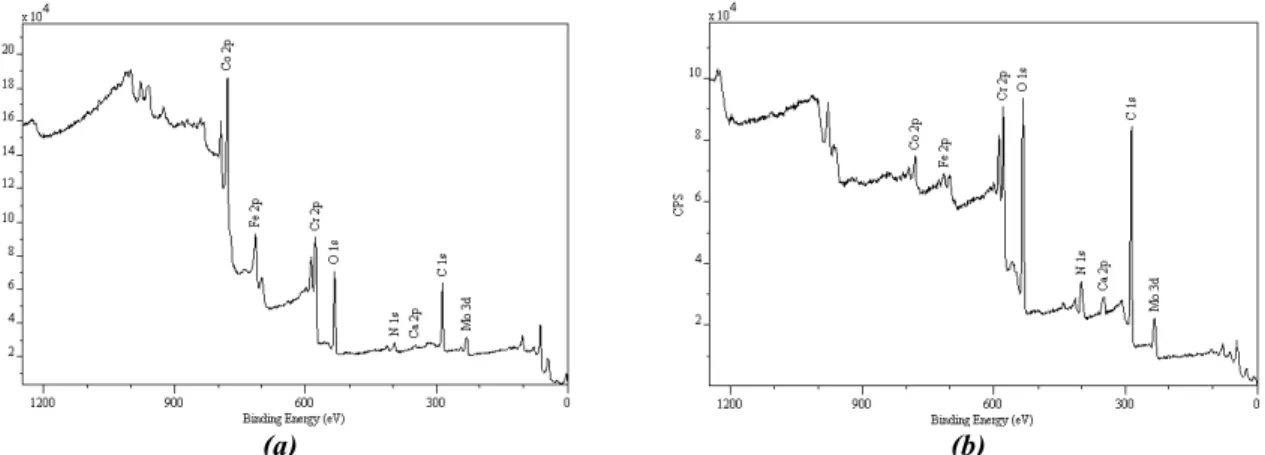
(a) (b) Figure 9 – General XPS survey spectra obtained for the (a) clean (b) deposited areas
Table 2 – Obtained chemical composition from XPS analysis of (a) clean and (b) deposited areas
Name Peak Position (eV)
at %
C 1s 284.516 60.79
Ca 2p 347.216 0.96
Co 2p 778.016 5.37
Cr 2p 574.116 6.94
Fe 2p 712.816 2.71
Mo 3d 227.416 1.48
N 1s 398.516 7.41
O 1s 530.516 14.32
Name Peak Position (eV)
at %
C 1s 284.916 66.76
Ca 2p 347.316 1.58
Co 2p 777.916 1.07
Cr 2p 576.216 4.45
Fe 2p 229.316 0.99
Mo 3d 398.816 6.99
N 1s 530.616 18.16
O 1s 284.916 66.76
(a) Clean area (b) Deposited areas
4 Discussion
4.1 Surface Morphology
In this study, all retrieved femoral stems demonstrated signs of fretting-corrosion on the cemented portions of the femoral stem. However closer examination of the retrievals demonstrated that, in fact, there were two complex degradation mechanisms taking place, fretting-corrosion and crevice corrosion. Fretting-corrosion has been observed in a number of clinical cases on both polished and matte femoral stems [10-13]. However interest into the interaction existing at the stem-cement interfaces has been renewed due to the cases presented by Donell et al and Bolland et al [1, 14]. Recent case studies have demonstrated that the stem-cement interface can play a major influence in the longevity of THR, with the stem-stem-cement interface being linked with high levels of metal ion release resulting in extensive soft tissue necrosis. Howell et al [15] presented a comprehensive study, conducting SEM and 3-D interferometery analysis on both retrieved polished and matte femoral stems, identifying that polished and matte femoral stems present two very different wear mechanisms. Polished stems were seen to exhibit signs of ductile wear accompanied by pitting of the surface, a similar morphology observed in this cohort (Figure 5-6), whilst matte stems presented evidence of abrasive or truncation wear [15]. Although the wear mechanism was seen to differ with different surface finishes the location of wear, typically in the anterolateral and posteromedial aspects of the stem, was unaffected suggesting that the torsional loading is the prominent factor.
4.2 Surface Chemistry
The importance of tribo-chemistry in orthopaedics was first recognised by Wimmer [16], who highlighted the role proteins play at the bearing interfaces. Although the majority of studies to date demonstrate and characterise the complex tribo-chemical reactions taking place at the articulation surface, this study is one of a few to utilise advanced surface microscopy and surface chemistry analytical techniques in order to understand the tribo-corrosion and tribo-chemical interactions at the stem-cement interface. A study by Walczak et al [17] observed the surface chemistry and surface morphology of deposits found on Charnley and Müller prosthesis and also retrieved PMMA bone cement. They proposed that a multilayer film existed on the surface with different elemental chemistry. An initial Cr-rich layer was seen on the surface of the sample, accompanied by an iron rich plaque and a third chloride/sulphur plaque; however provided no cross-sectional evidence to support this. They also observed that the debris found on the surface of the retrieved PMMA bone cement had a similar chemical composition to the deposits found on the retrieved femoral components.
Liao et al [20] further suggested that these films may be graphitic due to the sheer stresses and pressures experienced in-vivo.
4.3 Interactions between fretting-corrosion and surface chemistry
The interactions between fretting and corrosion have been well documented. Fretting-corrosion occurs when two materials under load are subject to minute relative motion within an aqueous environment. The contact between the two materials causes mechanical wear and material transfer at the surfaces. However when typically passive alloys are immersed in fluids and subjected to fretting, removal of the passive film occurs exposing the reactive metallic substrate to the aqueous environment resulting in oxidation of the metallic substrate and metallic debris. It is thought that the repetitive separation and mating of the surfaces, as a result of physical activity, will induce a ploughing and compressive action of the CoCrMo metallic substrate resulting in the formation fretting-corrosion product. A mechanical mixing of the fretting-corrosion products will also occur as a pumping action, similar to the mechanisms described by Willert et al [21], allowing fresh synovial fluid will to freely enter the interfaces as they become separated. It is thought this mechanical mixing in combination with repetitive mechanical ploughing of the metallic surface and compression of fretting debris that result in the dense black deposits being formed within the stem-cement interface.
It is also important to consider the effects of pure electrochemical crevice corrosion. Analysis of the retrieved femoral stems demonstrated that a corrosive attack was seen to exist towards the distal portion of the cemented femoral stem. Willert [21] highlighted that the stem-cement interfaces were ideal conditions for the initiation and propagation of localised crevice corrosion due to its restrictive geometry. It was proposed that as subsidence and partial debonding of the femoral stem occurred, the site at which crevice corrosion occurs would migrate distally, reducing the oxygen available for passivation, resulting in a locally aggressive environment (pH 2-3) being established and rapid chemical dissolution of the alloy.
5 Conclusions
References
[1] S.Donell, C.Darrah, J.Nolan, J.Wimhurst, A.Toms, T.Barker, C.Case, J.Tucker Early failure of the Ultima metal – on – metal total hip replacement in the presence of normal plain radiographs. Journal of bone and joint surgery [Br] 2010;92:1501-8.
[2] I.Milosev, H.Strehblow The composition of the surface passive film formed on
CoCrMo alloy in simulated physiological solution. Electrochemica Acta
2003;48:2767-74.
[3] Milosíev I, Remskar M In vivo production of nanosized metal wear debris formed by tribochemical reaction as confirmed by high-resolution TEM and XPS analyses. J Biomed Mater Res 2009;91A:1100-10.
[4] A.Kocija, I.Milosev, B.Pihlar Colbalt-based alloys for orthopeadic applications
studied by electrochemical and XPS analysis. Journal of Materials Science:
Materials in Medicine 2004;15:643-50.
[5] A.Hodgson, S.Kurz, S.Virtanen, V.Fervel, C.Olsson, S.Mischler Passive and transpassive behaviour of CoCrMo in simulated biological solutions. Electrochemica Acta 2004;49:2167-78.
[6] J.Moulder, W.Stickle, P.Sobol, K.Bomben Handbook of X-ray Photoelectron Spectroscopy, 1 ed: Perkin-Elmer corporation, 1992.
[7] N.Fairley CasaXP Manual, 2009.
[8] R.B.Waterhouse Fretting Corrosion: International series of monographs on materials science and technology, Pergamon press, 1972.
[9] H.Willert, L.Broback, G.Buchhorn, P.Jemsen, G.Koster, I.Lang, P.Ochsner, R.Schenk Crevice corrosion of cemented titanium alloy stems in total hip replacements. Clin Orthop Relat Res 1996;333:333-51.
[10] P.Yates, B.Burston, E.Whitley, G.Bannister Collarless polished tapered stem. J Bone Joint Surg Br 2007;90-B:16-22.
[11] G.Gie, R.Ling Loosening and the Mirgation of the Exeter THR. Journal of Bone and Joint Surgery(Br) 1994;76.
[12] J.Fowler, G.Gie, A.Lee, R.Ling Experience with the Exeter total hip replacement scince 1970. Orthopaedic clinics of North America 2011;19:477-89.
[14] B.Bolland DC, D.Langton, J.Millington, N.Arden, J.Latham High Failure rates with a large-diameter hybrid metal-on-metal total hip replacement. Journal of Bone and Joint Surgery [Br] 2011;93:608-15.
[15] Howell JR, Blunt LA, Doyle C, Hooper RM, Lee AJC, Ling RSM In Vivo Surface Wear Mechanisms of Femoral Components of Cemented Total Hip Arthroplasties: The Influence of Wear Mechanism on Clinical Outcome. Journal of Arthroplasty 2004;19:January.
[16] Wimmer MA, Sprecher C, Hauert R, T+ñger G, Fischer A Tribochemical reaction on metal-on-metal hip joint bearings: A comparison between in-vitro and in-vivo results. Wear 2003;255:1007-14.
[17] J.Walczak, F.Shahgaldi, F.Heatley In vivo corrosion of 316L stainless steel hip implants: morphology and elemental composistions of corrosion products. Biomaterials 1998;19:229-37.
[18] Yan Y, Neville A, Dowson D Biotribocorrosion of CoCrMo orthopaedic implant materialsGÇöAssessing the formation and effect of the biofilm. Tribology International 2007;40:1492-9.
[19] Y.Yan, A.Neville, D.Dowson Biotribocorrosion-an appraisal of the time dependence of wear and corrosion interactions: II. Surface analysis . J Phys D: Appl Phys 2006;39:3206.
[20] Liao Y, Pourzal R, Wimmer MA, Jacobs JJ, Fischer A, Marks LD Graphitic Tribological Layers in Metal-on-Metal Hip Replacements. Science 2011;334:1687-90.