_____________________________________________________________________________________________________ *Corresponding author: E-mail: [email protected];
27(6): 1-8, 2018; Article no.JAMMR.43719 ISSN: 2456-8899
(Past name: British Journal of Medicine and Medical Research, Past ISSN: 2231-0614, NLM ID: 101570965)
A Comparative Study of Time Dependent Candidal
Colonisation in Diabetic and Non-diabetic Complete
Denture Patients
Mujtaba Ashraf
1, Mariyam Ali
1, Abhishek Gaur
1, Monika Rajani
1, A. K. Verma
1,
Pratibha Katiyar
1, Kaushik Kumar Pandey
1*and Fauzia Tarannum
11Department of Prosthodontics, Career Post Graduate Institute of Dental Sciences, Lucknow, India.
Authors’ contributions
This work was carried out in collaboration between all authors. Authors Mujtaba Ashraf, Mariyam Ali and AG designed the study, performed the statistical analysis, wrote the protocol and wrote the first draft of the manuscript. Authors MR, AKV and PK managed the analyses of the study. Authors KKP and FT managed the literature searches. All authors read and approved the final manuscript.
Article Information
DOI: 10.9734/JAMMR/2018/43719 Editor(s): (1)Dr. Mohamed Essa, Department of Food Science and Nutrition, Sultan Qaboos University, Oman.
Reviewers: (1)Adam Husein, University of Science, Malaysia. (2)Renata Souto, Federal University of Rio de Janeiro, Brazil. (3)Reginaldo dos Santos Pedroso, Federal University of Uberlândia, Brazil. Complete Peer review History:http://www.sciencedomain.org/review-history/26206
Received 20 June 2018 Accepted 04 September 2018 Published 12 September 2018
ABSTRACT
Introduction: Acrylic dentures are important predisposing factors for oral candidiasis. Duration of wearing dentures in the oral cavity of diabetic and non-diabetic patients can increase Candida colonisation and results in the higher incidence of oral and systemic candidiasis.
Aims and Objective: 1. To compare the changes in Candidal colonisation with time in complete denture prosthesis in diabetics and non-diabetics.2.To evaluate the change in Candidal growth in diabetics and non-diabetic complete denture wearers. 3. To evaluate the relation of time with Candidal growth in complete denture wearers.
Materials and Methods: Three Salivary samples from each 20 edentulous, non-insulin dependent diabetic and 20 edentulous (Group-1), non-diabetic subjects (Group-2) were collected at 3 different time intervals. First sample was collected on the day of denture insertion, the second sample after one week from the day of insertion and third sample, after one month from the day of denture
Ashraf et al.; JAMMR, 27(6): 1-8, 2018; Article no.JAMMR.43719
insertion in sterile containers using oral rinse technique. All samples were cultured directly on Sabouraud agar medium and species identification was done by HiCrome Candida Differential Agar culture methods.
Results: Candidal carriage and colonisation is more in diabetic denture wearer patients than non- diabetic denture wearer patients. Candidal growth in complete denture wearers was significantly increased from Day 1 to last day of 1st week (p=0.006), Day 1 to last day of 1st Month (p=0.005) and last day of 1st Week to last day of 1st Month (p=0.007) in Group 1. There was significant mean change in Group 2 from last day of 1st Week to last day of 1st Month (p=0.001). The common species of Candida isolated in both diabetic and non-diabetic denture wearer patients are: C. albicans, C. glabrata and C. tropicalis.
Keywords: Candidal colonisation; complete dentures; diabetes mellitus.
1. INTRODUCTION
Diabetes mellitus is a common and globally growing health problem with a high morbidity and mortality. The prevalence of diabetes is swiftly increasing over the globe at an alarming rate. According to the International Federation of Diabetes, 415 million adults around the world are suffering from diabetes, and it is estimated that the numbers will reach around 642 million by 2040. India leads the World and stands at the second position after China, with 69 million persons affected by diabetes [1].
Diabetes mellitus is a chronic metabolic disease that occurs when the body cannot produce enough insulin, or cannot use insulin and is characterised by constant increased glucose levels in the blood (hyperglycemia) associated with alterations in carbohydrate, protein and lipid metabolism [2].
Oral candidiasis is an opportunistic fungal infection which is commonly associated with Diabetes [3] all forms of oral candidiasis are considered opportunistic and hence the epithet ‘the disease of the diseased’ has been given to this condition [4].
Due to high rates of diabetes mellitus in the elderly Indian population and to increase our understanding of growth of Candidal colonies, pathogenicity and related complication, this research work was designed for a comparative study of time dependent Candidal colonisation in diabetic and non-diabetic complete denture patients.
1.1 Aims
To compare the changes in Candidal colonisation with time in complete denture prosthesis in diabetics and non-diabetics.
1.2 Objectives
To evaluate change in Candidal growth in diabetics and non-diabetic complete denture wearers.
To evaluate the relation of time with Candidal growth in complete denture wearers.
2. MATERIALS AND METHODS
This prospective, in-vivo study was conducted in the Department of Prosthodontics and Crown & Bridge, Career Post Graduate Institute of Dental Sciences and Hospital and Department of Microbiology, Career Institute of Medical Sciences and Hospital, Lucknow, after taking informed consent from the patients. Approval was obtained from the ethical committee of Career Post Graduate Institute of Dental Sciences and Hospital, Lucknow.
2.1 Patient Selection
Subjects included in this study were having following inclusion criteria; completely edentulous patients, diabetic and non-diabetic subjects aged between 45-75 years. Exclusion criteria were; patients with lost or broken dentures, HIV, Immuno-compromised and highly debilitated patients.
Forty new complete denture wearing patients reporting to the Department of Prosthodontics, fulfilling the inclusion criteria, which were included in the study were divided into 2 groups:-
Group 1: Edentulous, non-insulin dependent diabetic subjects. (FBS>130 mg/dl)*
Group 2: Edentulous, non-diabetic subjects. (FBS<110 mg/dl)*
The diabetic status of the patients was determined by the medical history of the previous diagnosis of diabetes and their fasting blood glucose levels were also determined before the sample collection using colorimetric method. The patients were assessed as non- diabetics on the ground of fasting plasma glucose levels less than 110 mg/dl.
2.2 Fabrication of Dentures
All complete dentures included in this study was fabricated, following standard balanced denture protocol.
2.3 Sample Collection
Samples were collected by the concentrated oral rinse technique as described by Samaranayake et al. [5]. Subjects were instructed not to eat and drink 2 hours prior to the sample collection. Each individual was given 10 ml of sterile phosphate buffer saline solution (0.1M solution with 7.2 pH) and was asked to rinse mouth for 60 seconds with this solution. Then that mouth rinse was collected in sterile containers and was sent to the microbiological laboratory.
Three samples were collected from each subject.
1st sample: On the day of denture insertion
2nd sample: After one week from the day of insertion and
3rd sample: After one month from the day of denture insertion.
2.4 Inoculation and Incubation
Samples into the Culture Media
Oral rinse samples were then inoculated onto Sabouraud Dextrose Agar plates. The SDA plates were prepared according to the manufacturer’s instruction. The oral rinse sample was vortex mixed prior to plating. A drop of oral rinse sample was taken with 4
diameter inoculating loop and the samples were inoculated. In this study, 4 mm loop was taken to standardise the quantity of samples.
were then incubated aerobically for 48 hours at 37°C. The subject was designated as
carrier if there was any growth of
colonies. A semiquantitative estimation of colony count was done and growth was expressed as CFU/ml with dilution factor of 10 [3
of inoculum.
CFU/ml = Colony Forming Units / milliliters.
Ashraf et al.; JAMMR, 27(6): 1-8, 2018; Article no.
The diabetic status of the patients was determined by the medical history of the previous diagnosis of diabetes and their fasting blood glucose levels were also determined before the sample collection using colorimetric method. The diabetics on the ground of fasting plasma glucose levels less than
included in this study was fabricated, following standard balanced denture
Samples were collected by the concentrated oral rinse technique as described by Samaranayake Subjects were instructed not to eat and drink 2 hours prior to the sample collection. Each ml of sterile phosphate buffer saline solution (0.1M solution with 7.2 pH) and was asked to rinse mouth for 60 seconds with this solution. Then that mouth rinse was collected in sterile containers and was sent to the
samples were collected from each subject.
sample: On the day of denture insertion sample: After one week from the day of
sample: After one month from the day
Incubation of
ulture Media
Oral rinse samples were then inoculated onto Sabouraud Dextrose Agar plates. The SDA plates were prepared according to the manufacturer’s instruction. The oral rinse sample was vortex mixed prior to plating. A drop of oral mm internal diameter inoculating loop and the samples were mm loop was taken to standardise the quantity of samples. The plates were then incubated aerobically for 48 hours at 37°C. The subject was designated as Candidal carrier if there was any growth of Candidal colonies. A semiquantitative estimation of colony and growth was expressed as 3] for 0.001 ml
CFU/ml = Colony Forming Units / milliliters.
2.5 Microscopic Evaluation Colonies
All creamy, pasty colonies on SDA plates (Fig after 48 hrs were picked up and further identification of Candidal species was done by Hi-Crome Candida Differential Agar presumptive budding yeast like cells with pseudo hyphae formation were identified as Candidal species and were subcultured on
Candida Differential Agar.
2.6 Culture on Candida Differential Agar
Small inoculums from isolated Candidal
from Sabouraud’s Dextrose Agar were picked up with a sterile inoculating loop, plated on Crome Candida Differential Agar
incubated at 37°C for 24-48 hours
The different species of Candida
based on the colour displayed by them on Crome Candida Differential Agar [6
C. albicans (CA) = light green C. glabrata (CG)= white pink, purple C. tropicalis (CT) = dark blue to blue gray C. gulliermondii= pale pink
C. kefyr= pink
C. krusei (CK) = pale pink, purple spongy pale edges)
C. parapsilosis= white, pale pink
Fig. 1. Candidal culture on sabouraud dextrose agar
2.7 Statistical Analysis
The Chi-square test was used to compare the categorical variables. Unpaired t-test was used to compare normally distributed variables between
; Article no.JAMMR.43719
Evaluation of Candidal
All creamy, pasty colonies on SDA plates (Fig. 1) hrs were picked up and further identification of Candidal species was done by Crome Candida Differential Agar. All presumptive budding yeast like cells with pseudo hyphae formation were identified as Candidal species and were subcultured on Hi-Crome
Candida Differential Agar
Candidal colonies from Sabouraud’s Dextrose Agar were picked up with a sterile inoculating loop, plated on Hi-Crome Candida Differential Agar and were
(Fig. 2).
Candida were identified displayed by them on
Hi-6,7,8].
= white pink, purple = dark blue to blue gray
= pale pink, purple (rough with
on sabouraud
the groups. Mann-Whitney U test was used to compare non-normal variables between the groups at different time periods. Wilcoxon rank sum test was used to compare the mean change in the continuous variables from Day 1 to subsequent time periods. The p-value<0.05 was considered significant. All the analysis was carried out on SPSS 16.0 version.
Fig. 2. Candidal culture on HiCrome differential agar
3. RESULTS
1. The mean age of patients of Group 1 and Group 2 was 58.45±6.43 and 54.40±7.14 years respectively (Table 1).
2. Candidal growth in complete denture wearers was found to be significantly (p=0.0001) higher in Group 1 compared to Group 2 at Day 1, last day of 1
last day of 1st Month (Table 2).
3. In Group 1 (n=20), 16 (80%), 19 (95%) and 20 (100%) shows positive growth (p 1=0.001) on SDA sample 1, sample 2 and sample 3 respectively (Table
4. Candidal growth in complete denture wearers was significantly increased from Day 1 to last day of 1st week (p=0.006), Day 1 to last day of 1st Month (p=0.005) and last day of 1st Week to last day of 1 Month (p=0.007) in Group 1. T
Table 2. Comparison of Candidal growth in complete denture wearers (SDA) between the groups across the time periods
Time period Group 1
Day 1 40.20±30.57
Week 1 54.90±29.90
Month 1 60.40±30.85
Ashraf et al.; JAMMR, 27(6): 1-8, 2018; Article no.
Whitney U test was used to normal variables between the ods. Wilcoxon rank sum test was used to compare the mean change in the continuous variables from Day 1 to value<0.05 was considered significant. All the analysis was
rome Candida
The mean age of patients of Group 1 and Group 2 was 58.45±6.43 and 54.40±7.14
Candidal growth in complete denture wearers was found to be significantly (p=0.0001) higher in Group 1 compared to Group 2 at Day 1, last day of 1st week and
2).
In Group 1 (n=20), 16 (80%), 19 (95%) and 20 (100%) shows positive growth (p-value on SDA sample 1, sample 2 and
3).
Candidal growth in complete denture wearers was significantly increased from week (p=0.006), Month (p=0.005) Week to last day of 1st Month (p=0.007) in Group 1. There was
significant mean change in Group 2 from last day of 1st Week to last day of 1
(p=0.001) (Table 4).
5. In Group 2 (n=20), 0 (0%), 2 (10%) and 15 (75%) shows positive growth (p value1=0.001) on SDA sample 1, sample 2 and sample 3 respectively.
6. The growth of various Candidal
(CA+CG+CT+CK) was significantly increased from Day 1 to last day of 1 week (p=0.001), Day 1 to last day of 1 Month (p=0.0001) and last day of 1
to last day of 1st Month (p=0.0001) in Group 1. There was a significant mean change in Group 2 from last day of 1 Week to last day of 1st Month (p=0.001) (Table 5).
Table 1. The age distribution between the groups
Groups Age in years (Mean±SD)
Group 1 58.45±6.43
Group 2 54.40±7.14
p-value 0.06
4. DISCUSSION
India leads the world with largest number of diabetic subjects earning the dubious distinction of being termed the ‘diabetes capital of the world. The intrusion of Western culture into the lives of traditional indigenous communities has also devastating results in terms of the rise in diabetes and related metabolic disorders [
Candida is an opportunistic pathogen, resulting in disease when the host-commensal relationship is disturbed [3]. Oral infection with
yeast-like fungus) is a commoninfection, especially in theelderly people [10].
caused by Candida sp. is associated with local and systemic factors too. The use of a dental prosthesis is indispensable for function and esthetic rehabilitation of edentulous pat The adhesion of microorganisms to a surface is a prerequisite for the colonisation of that surface [11].
Comparison of Candidal growth in complete denture wearers (SDA) between the groups across the time periods
Group 2 p-value
0.00±0.00 -
1.75±5.44 0.0001*
15.40±14.91 0.0001*
Mann-Whitney U test, *Significant
; Article no.JAMMR.43719
significant mean change in Group 2 from Week to last day of 1st Month
In Group 2 (n=20), 0 (0%), 2 (10%) and 15 (75%) shows positive growth
(p-on SDA sample 1, sample 2
Candidal colonies (CA+CG+CT+CK) was significantly increased from Day 1 to last day of 1st week (p=0.001), Day 1 to last day of 1st Month (p=0.0001) and last day of 1st Week
Month (p=0.0001) in was a significant mean change in Group 2 from last day of 1st
Month (p=0.001)
The age distribution between the
Age in years (Mean±SD)
India leads the world with largest number of diabetic subjects earning the dubious distinction of being termed the ‘diabetes capital of the world. The intrusion of Western culture into the lives of traditional indigenous communities has also had devastating results in terms of the rise in diabetes and related metabolic disorders [9].
is an opportunistic pathogen, resulting commensal relationship Oral infection with Candida (a s) is a commoninfection, ]. The infection sp. is associated with local The use of a dental prosthesis is indispensable for function and esthetic rehabilitation of edentulous patients.
The adhesion of microorganisms to a surface is a prerequisite for the colonisation of that
Comparison of Candidal growth in complete denture wearers (SDA) between the
value
Ashraf et al.; JAMMR, 27(6): 1-8, 2018; Article no.JAMMR.43719
Table 3. Comparison of SDA growth between the groups
Group 1 (n=20) Group 2 (n=20) p-value
No. % No. %
Day 1
Growth 16 80.0 0 0.0 0.001*
No growth 4 20.0 20 100.0
Week 1
Growth 19 95.0 2 10.0 0.001*
No growth 1 5.0 18 90.0
Month 1
Growth 20 100.0 15 75.0 0.01*
No growth 0 0.0 5 25.0
Chi-square test, *Significant
Table 4. Comparison of mean change in Candidal growth in complete denture wearers (SDA) from Day 1 to subsequent time periods in groups
Time period Group 1 Group 2
Mean change p-value Mean change p-value
Day 1 to Week 1 14.70±21.52 0.006* 1.75±5.44 -
Day 1 to Month 1 20.20±21.60 0.005* 15.40±14.91 -
Week 1 to Month 1 20.20±21.60 0.007* 13.65±15.25 0.001*
Wilcoxon Rank sum test, *Significant
Table 5. Comparison of growth of various Candidal colonies (CA+CG+CT+CK) between the groups across the time periods
Time period Group 1 Group 2 p-value
Day 1 13.15±8.93 0.00±0.00 -
Week 1 18.35±8.36 1.25±4.55 0.0001*
Month 1 29.60±11.77 10.20±8.10 0.0001*
Mann-Whitney U test, *Significant
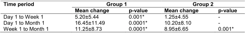
Table 6. Comparison of mean change in growth of various Candidal colonies (CA+CG+CT+CK) from Day 1 to subsequent time periods in groups
Time period Group 1 Group 2
Mean change p-value Mean change p-value
Day 1 to Week 1 5.20±5.44 0.001* 1.25±4.55 -
Day 1 to Month 1 16.45±11.49 0.0001* 10.20±8.10 -
Week 1 to Month 1 11.25±8.73 0.0001* 8.95±6.65 0.001*
Wilcoxon Rank sum test, *Significant
There have been many studies on the adhesion of Candidaal bicans to denture acrylic resin, caused by the association of the commensal, opportunist pathogen yeast with denture-induced stomatitis. The presence of a denture in the oral cavity, associated with the local alterations of the oral mucosa and the systemic complications, may render the denture wearer patient with diabetes even more prone to Candidal infection. A significantly higher incidence of Candida infection and increased levels of Candida spp. were found in diabetic patients wearing removable dentures.
The results of the present study can be explained on the basis of two factors:
a) Local: acrylic prosthesis b) Systemic: diabetes.
Acrylic dentures act as a predisposing factor in
the occurrence of oral Candidal infection. The dentures can act as a reservoir of infection
Ashraf et al.; JAMMR, 27(6): 1-8, 2018; Article no.JAMMR.43719
The adhesion of Candida depends on the microporosity present on the inner surface of the denture. Substrate surface properties, such as surface charge, surface free energy, hydrophobicity and roughness of denture have all been reported to influence the Candidal colonisation. Acrylic resins are hydrophilic, pervious and exhibit more water sorption. This water sorption may help the Candida cells to adhere or to even penetrate the surface of acrylic resin. Surface irregularities of acrylic resin act as a factor in the entrapment of microorganisms [9,13].
The most frequently used primary isolation medium for Candida is SDA [14] which, although permitting growth of Candida, suppresses the growth of many species of oral bacteria due to its low pH. Typically SDA is incubated aerobically at 37°C for 24–48 hours. Candida develop as cream, convex colonies on SDA and differentiation between species is rarely possible. It is estimated that more than one Candida species occurs in approximately 10% of oral samples and in recent years the ability to detect non-albicans species has become increasingly important. As a result, it has been recommended that SDA should be used in combination with a second differential medium [15].
Hi-Crome agar was used as a second differential medium for Candida sp. in this study. On Hi-Crome agar, the differentiation is done on the basis of strongly contrasted colony colors produced by reactions of species specific enzymes with a proprietary chromogenic substrate. CHROMagar is recommended as a useful isolation medium capable of the presumptive identification of the yeast species most commonly isolated from clinical material and facilitating recognition of mixed yeast cultures [6,7].
The Candida carriage rate in oral cavity was found to be different in the various studies done earlier. This could be due to the different methods of sampling used, for example: oral swabs, rinse using water or buffer. The oral rinse technique with phosphate buffer solution was used for sampling in the present study since this is known to be a sensitive technique for estimating the oral Candidal carriage. Also CHROMagar shows 100% specificity and 100% sensitivity when compared to Sabouraud Dextrose Agar and conventional methods [16,17,8,18,19,20].
In the present study, mean age of the Group 1(Non-insulin dependent diabetic subjects) and Group 2 (Non-diabetic subjects) was 58.45±6.43 and 54.40±7.14 years respectively. There was no significant (p>0.05) difference in the age between the groups showing comparability of the groups in terms of age.
In Group 1 and Group 2 (n=40) the prevalence of Candida increases in Sample 1, Sample 2 and sample 3, to 40%, 52.5% and 87.5% respectively. Candida has affinity for the acrylic surface of dentures and the organism can be opportunistic, which can be explained by the fact that dentures decrease the flow of oxygen and saliva to the underlying tissue producing a local acidic and anaerobic micro-environment that favours yeast overgrowth [3].
In the present study, the number of CFU of Candida is found to be more in diabetic denture wearer subjects as compared to the non-diabetic denture wearer. These results are similar to the few previous studies done evaluating the same factor [21]. Daniluk T et al. [22] had earlier evaluated the occurrence rate of oral C. albicans in denture wearer patients. The growth of various Candida species on CA was found to be significantly (p=0.0001) higher in Group 1 compared to Group 2 at Week 1 and Month 1.
Positive culture of Candida enrolled on the denture of all diabetic subjects (100%) but this was seen in (75%) of non-diabetics after 1 month use of denture (p=0.01). Similar observation was noted in a study by Kamran MHL [3] also revealed that diabetes mellitus increased the colonisation of Candida in denture and mouth. The predisposition of the diabetics to infections by pathogenic fungal species has been explained in terms of enhancement of yeast growth by elevated tissue fluid glucose levels. Moreover, the presence of a high concentration of salivary glucose combined with low salivary secretion may enhance growth of yeasts and their adherence in epithelial oral cells [12].
Ashraf et al.; JAMMR, 27(6): 1-8, 2018; Article no.JAMMR.43719
of 41% found by a mouthwash technique. That study also depicted that C. albicans was not uniformly distributed in the mouths of healthy people. Also the risk factors for oral Candidal infection in diabetic patients are complex. Some authors emphasise the role of local factors such as salivary flow rates and pH, poor glycemic control and low salivary buffering capacity [3,4,16].
C. albicans was the commonest yeast found on subjects, followed by C. glabrata and C. tropicalis. Same pattern was also observed by Sanitá et al. [23] and Vanden Abbeele A et al. [24].
5. CONCLUSION
Within the limitations of this in vivo study, the following conclusions were drawn:
1. Candidal carriage and colonisation (based on the CFU) is more in diabeticdenture wearer patients than the non- diabetic denture wearers.
2. In Group 1, shows positive growth on SDA sample 1, sample 2 and sample 3 respectively.
In Group 2 (n=20), 0 (0%), 2 (10%) and 15 (75%) shows positive growth (p-value1=0.001) on SDA sample 1, sample 2 and sample 3 respectively. Thus, Candidal growth is dependent of time.
3. The common species of Candida isolated in both diabetic and non-diabetic denture wearer patients are: C. albicans, C. glabrata and C. tropicalis.
CONSENT
Consent was taken individually from all patients.
ETHICAL APPROVAL
Ethical approval was taken from institutional ethical research cell committee. Ethical approval certificate number is CPGIDSH/562/17.
COMPETING INTERESTS
Authors have declared that no competing interests exist.
REFERENCES
1. Akhtar SN, Dhillon P. Prevalence of diagnosed diabetes and associated risk
factors: Evidence from the large-scale surveys in India. J Soc Health Diabetes. 2017;5:28-35.
2. International Diabetes Federation (IDF). IDF Diabetes Atlas. 6th ed.; 2013. Available:http://www.diabetesatlas.org/ 3. Lotfi-Kamran MH, Jafari AA, Falah-Tafti A,
Tavakoli E, Falahzadeh MH. Candida colonization on the denture of diabetic and non-diabetic patients. Dent Res J (Isfahan). 2009;6(1):23-7.
4. Jorgensen Ejvind Budtz. Clinical aspects of candida infection in denture wearers. J Am Dent Assoc. 1978;96:474-9.
5. Samaranayake LP, MacFarlane TW, Latney, Ferguson MM. A comparison of oral rinse and imprint sampling techniques for the detection of yeast, coliform and
Staphylococcus aureus carriage in the oral cavity. J Oral Pathol.
1986;15:386-388.
6. Odds FC, Bernaerts R. CHROMagar Candida, a new differential isolation medium for presumptive identification of clinically important Candida species. J ClinMicrobiol. 1994;32(8):1923-9.
7. Beighton D, et al. Use of CHROMagar Candida medium for isolation of yeasts from dental samples. J Clin Microbiol. 1995;33(11):3025-7.
8. Nayak S, et al. Comparative study of Candida by conventional and CHROMagar method in non-denture and denture wearers by oral rinse technique. Indian J Dent Res. 2012;23(4):490-7.
9. Sharma R, Sunder Raj S, Vinod K, Reddy YG, Vela D, Bailoor D. Comparison of oral health indicators in type 2 diabetes mellitus patients and controls. J Indian Acad Oral Med Radiol. 2011;23:168–172.
10. Iacipino AM, William F, Wathen. Oral Candidal infection and denture stomatitis: A comprehensive review. J Am Dent Assoc. 1992;123:46-51.
11. Darwazeh AMG, Lamey PJ, Samarnayake LP, Macfarlane TW, Fisher BM, Macrury SM, Maccuish AC. The relationship between colonization, secretor status and in- vitro adhesion of Candida albicans to buccal epithelial cells from diabetics. Journal of Medical Microbiology. 1990;33: 43-49.
Ashraf et al.; JAMMR, 27(6): 1-8, 2018; Article no.JAMMR.43719
13. Pereira-Cenci T, Del BelCury AA, Crielaard W, Ten Cate JM. Development of Candida-associated denture stomatitis: New insights. J Appl Oral Sci. 2008;16(2): 86-94.
14. Odds FC. Sabouraud('s) agar. J. Med. Vet. Mycol. 1991;29:355-359.
15. Pereira-Cenci T, Cury AA, Cenci MS, Rodrigues-Garcia RC. In vitro Candida colonization on acrylic resins and denture liners: Influence of surface free energy, roughness, saliva, and adhering bacteria. Int J Prosthodont. 2007;20:308-10.
16. Belazi M, Velegraki A, Fleva A, et al. Candidal overgrowth in diabetic patients: Potential predisposing factors. Mycoses. 2005;48(3):192-6.
17. Kumar BV, Padshetty NS, Bai KY, Rao MS. Prevalence of Candida in the oral cavity of diabetic subjects. J Assoc Physicians India. 2005;53:599-602. 18. Kalapala L, Raghunath V, Ajay Bernard
Reginald. Salivary factors, Candidal colonization and strain diversity in type II diabetics. NJL Med. 2015;4(4):52-58.
19. Williams DW, Lewis MAO. Isolation and identification of Candida from the oral cavity. Oral Diseases. 2000;6(1):3–11. 20. Verran J, Maryan C. Retention of Candida
albicans on acrylic resin and silicone of different surface topography. J Prosthet Dent. 1997;77:535-539.
21. Tapper Jones LM, Aldred MJ, Walker DM, Hayes TM. Candidal infections and population of Candida albicans in mouths of diabetics. J Clin Pathol. 1981;34:706-711.
22. Daniluk T. Occurrence rate of oral Candida albicans in denture wearer patients. Adv Med Sci. 2006;51(1):77-80.
23. Sanitá PV, Pavarina AC, Giampaolo ET, et al. Candida spp. prevalence in well controlled type 2 diabetic patients with denture stomatitis. Oral Surg Oral Med Oral Pathol Oral Radiol Endod. 2011;111: 726–733.
24. Vanden Abbeele A, de Mil H, Ahariz M, Perraudin JP, Beyer I, Courtois P. Denture contamination by yeasts in the elderly. Gerodontology. 2008;25(4):222-8.
_________________________________________________________________________________
© 2018 Ashraf et al.; This is an Open Access article distributed under the terms of the Creative Commons Attribution License (http://creativecommons.org/licenses/by/4.0), which permits unrestricted use, distribution, and reproduction in any medium, provided the original work is properly cited.
Peer-review history: