9-24-2015 12:00 AM
A Randomized Clinical Trial to Compare the Effect of Non
A Randomized Clinical Trial to Compare the Effect of Non
Operative Treatment With and Without Autologous Conditioned
Operative Treatment With and Without Autologous Conditioned
Plasma (ACP) on Healing and Function in Patients with Achilles
Plasma (ACP) on Healing and Function in Patients with Achilles
Tendon Ruptures
Tendon Ruptures
Tyler M. Pratt
The University of Western Ontario
Supervisor Dr. Dianne Bryant
The University of Western Ontario Joint Supervisor Dr. Kevin Willits
The University of Western Ontario
Graduate Program in Health and Rehabilitation Sciences
A thesis submitted in partial fulfillment of the requirements for the degree in Master of Science © Tyler M. Pratt 2015
Follow this and additional works at: https://ir.lib.uwo.ca/etd
Part of the Medical Anatomy Commons, Orthopedics Commons, Physical Therapy Commons,
Physiotherapy Commons, Podiatry Commons, and the Sports Medicine Commons
Recommended Citation Recommended Citation
Pratt, Tyler M., "A Randomized Clinical Trial to Compare the Effect of Non Operative Treatment With and Without Autologous Conditioned Plasma (ACP) on Healing and Function in Patients with Achilles Tendon Ruptures" (2015). Electronic Thesis and Dissertation Repository. 3242.
https://ir.lib.uwo.ca/etd/3242
This Dissertation/Thesis is brought to you for free and open access by Scholarship@Western. It has been accepted for inclusion in Electronic Thesis and Dissertation Repository by an authorized administrator of
by
Tyler Martin Pratt
Graduate Program in Health and Rehabilitation Science
A thesis submitted in partial fulfillment of the requirements for the degree of
Masters of Science
The School of Graduate and Postdoctoral Studies The University of Western Ontario
London, Ontario, Canada
© Tyler Pratt, 2015
ii
The purpose of this study was to determine whether there is evidence to support conducting a larger randomized control trial (RCT) to compare the non-operative treatment of Achilles tendon (AT) ruptures with or without Autologous Conditioned Plasma (ACP®). Twenty-four patients were randomized to receive an ACP® or saline injection within seven days and again at two weeks post-rupture. Tendon healing was longitudinally evaluated using ultrasound (US) and magnetic resonance imaging (MRI). Functional and patient-reported quality of life measures were also assessed. A small sample size limited our ability to detect statistically significant differences. The mean rankings of healing were slightly higher in the ACP® group at most time points for MRI and US. Functional and patient-reported outcomes were also slightly higher in the ACP® group at most time points. The consistency of these preliminary results suggests that there is evidence to support conducting a larger RCT.
Keywords
iii
This randomized control trial was designed by Drs. Willits, Bryant, and Spouge. I was responsible for patient identification and recruitment; patient scheduling for clinic,
ultrasound, and magnetic resonance imaging; and data collection and analysis. I wrote the original draft of this thesis document. Drs. Bryant and Willits made comments and
iv
I would like to thank my co-supervisors, Drs. Dianne Bryant and Kevin Willits, for their continued support and guidance throughout my Master’s at Western University. Additionally, I would like to thank the following people for their support and assistance over the past two years:
• Dr. Alison Spouge for her expertise and assistance with the imaging portion of this
study.
• Nicole Kaniki and Alliyah Remtulla for helping me transition into graduate school,
and helping with various aspects of my study.
• Marsha, Kathy, and Anne for their help with accommodating study patients, and for
their kindness and friendship in clinic.
• Sam Malian for his support and friendship before and during our Master’s studies
• Special thanks to my girlfriend, Nicole Fields, for constantly believing in me and
motivating me to achieve my goals.
• Finally, I would like to sincerely thank my parents for their unconditional love and
v
Abstract ... ii
Co-Authorship Statement ... iii
Acknowledgments ... iv
Table of Contents ... v
List of Tables ... ix
List of Figures ... x
List of Appendices ... xiii
Appendix A: Research Ethics Board Approval ... xiii
Appendix B:Letter of Information and Consent ... xiii
Appendix C:Image and Table Permissions ... xiii
Appendix D:MR Imaging Sequences ... xiii
Acronyms ... xiv
Chapter 1 ... 1
1 Introduction ... 1
Chapter 2 ... 3
2 Literature Review ... 3
2.1 Anatomy of the Achilles Tendon ... 3
2.2 Achilles Tendon Rupture ... 4
2.2.1 Epidemiology ... 4
2.2.2 Etiology ... 6
2.2.3 Mechanism of Injury ... 8
2.2.4 Clinical Presentation ... 9
2.3 Healing Following Achilles Tendon Rupture ... 13
vi
2.4.1 Operative Treatment ... 23
2.4.2 Non-Operative Treatment ... 24
2.4.3 Operative vs. Non-Operative Treatment: A Review of the Literature ... 25
2.5 Rehabilitation: Immobilization versus Early Mobilization ... 28
2.5.1 Controlled Early Motion ... 28
2.5.2 Controlled Early Weight-Bearing ... 30
2.5.3 Controlled Early Motion and Weight-Bearing ... 31
2.6 A Novel Non-Operative Option: Platelet-rich Plasma ... 33
2.6.1 Platelet Biology ... 34
2.6.2 Preparation of PRP ... 37
2.6.3 Delivery of PRP ... 40
2.6.4 PRP in the Treatment of Orthopedic Injuries ... 41
2.6.5 PRP in the Treatment of Achilles Tendon Ruptures: A Review of the Literature ... 42
2.7 Summary ... 51
Chapter 3 ... 53
3 Objectives ... 53
3.1 Primary Objective ... 53
3.2 Secondary Objectives ... 53
3.3 Overall Study Objective ... 53
Chapter 4 ... 54
4 Methods ... 54
4.1 Study Design ... 54
vii
4.3.2 Intervention Protocol ... 55
4.3.3 Autologous Conditioned Plasma (ACP®) ... 55
4.3.4 Rehabilitation ... 57
4.4 Primary Outcome – Healing ... 58
4.4.1 Ultrasound ... 58
4.4.2 MRI ... 59
4.5 Secondary Outcomes ... 60
4.5.1 Isokinetic Strength ... 60
4.5.2 Range of Motion ... 61
4.5.3 Calf Circumference ... 62
4.5.4 Leppilahti Score ... 62
4.5.5 Ankle-Hindfoot Scale (AOFAS) ... 62
4.5.6 Achilles Tendon Total Rupture Score (ATRS) ... 63
4.6 Plan for Analysis ... 64
Chapter 5 ... 66
5 Results ... 66
5.1 Participant Flow ... 66
5.2 Demographic Information ... 68
5.3 Injection Information ... 69
5.3.1 “PAW” Classification ... 69
5.4 Location of Tendon Rupture ... 70
5.5 Primary Outcome ... 70
5.5.1 Estimated Percent Healing ... 70
viii
5.7 Re-Ruptures and Adverse Events ... 81
6 Discussion ... 82
6.1 Study Strengths ... 91
6.2 Study Limitations ... 92
7 Conclusion ... 92
7.1 Directions for Future Research ... 93
References ... 94
ix
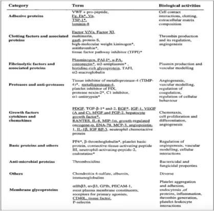
reported pro-angiogenic properties are underlined while those with anti-angiogenic potential
are also identified (*). ... 36
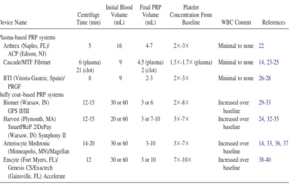
Table 2 Plasma-Based and Buffy Coat-Based PRP Systems ... 39
Table 3 Timeline for all study outcome measures ... 64
Table 4 Patient demographics ... 68
Table 5 White blood cell and platelet counts of patients' blood and ACP samples ... 69
Table 6 Ultrasound and MRI agreement on ordinal ratings of estimated percent healing at 6, 12, and 24 weeks ... 77
Table 7 American Orthopedic Foot and Ankle Society score, Achilles Tendon Total Rupture Score at 6, 12, and 24 weeks and Leppilahti score at 24 weeks post-rupture ... 78
Table 8 Range of motion and calf circumference measurements at 6, 12, and 24 weeks post-rupture ... 80
x
Figure 1 Calf muscles and Achilles tendon ... 4
Figure 2 Ultrasonographic appearance of a normal Achilles tendon ... 11
Figure 3 Ultrasonographic appearance of a ruptured Achilles tendon ... 12
Figure 4 Follow-up scan at 29 months demonstrating a healed tendon with resolution of the hematoma (arrows). The overlapping healed ends remain visible ... 16
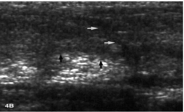
Figure 5 Follow-up scan at 15 months demonstrating a healed tendon with no discernable gap. The hypoechoic band (white arrows) and the slight bulge along the anterior margin of the tendon (black arrows) define the site of prior injury. There is mild residual distortion of the normal fibrillar architecture ... 17
Figure 6 Follow-up scan at nine months demonstrating the healed tendon with a subtle hypoechoic band indicating the site of previous rupture (white arrows). Note the irregular contour along the anterior tendon (black arrows) ... 18
Figure 7 Full thickness tear of the Achilles tendon (T1-weighted sagital MRI). There is discontinuity of the tendon with retraction of the proximal portion (red arrow). The distal tendon is tendinopathic and appears thickened and of increased signal intensity (white arrow). ... 20
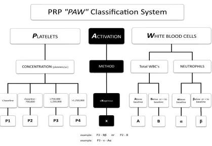
Figure 8 PRP "PAW" Classification System ... 40
Figure 9 Centrifuge ... 56
Figure 10 Three-layer ACP ... 57
Figure 11 Biodex Dynamometer ... 61
Figure 12 Participant flow through the trial ... 68
xi
Figure 15 Estimated percent healing at six weeks post injection with ACP or saline based on ultrasound imaging (left) and MRI (right) ... 73
Figure 16 Estimated percent healing at eight weeks post injection with ACP or saline based on ultrasound imaging ... 74
Figure 17 Estimated percent healing at 12 weeks post injection with ACP or saline based on ultrasound imaging (left) and MRI (right) ... 75
Figure 18 Estimated percent healing at 24 weeks post injection with ACP or saline based on ultrasound imaging (left) and MRI (right) ... 76
Figure 19 Baseline US of an acute Achilles tendon rupture (inflammatory phase). Long axis view at the rupture site shows discontinuity of tendon fibres (T) with a gap (arrows
demarcate proximal and distal ends of the torn fibres) and a small anachoic fluid collection (*) likely related to a hematoma. The Torn tendon fibres are hypoechoic, mildly redundant and show loss of the normal fibrillar pattern ... 86
Figure 20 Ultrasound image at six weeks (proliferative phase) in the same patient as Figure 19. The previous anechoic fluid collection at the tear site has resorbed. A prominent gap is evident at the site of the tear (arrows) filled with echogenic tissue (*) which may correspond to granulation tissue. The tendon fibres adjacent to the tear are thickened, disorganized and hypoechoic, potentially corresponding to persistent edema and/or fibrous tissue ... 87
Figure 21 MR image at six weeks (same patient). Sagittal fast inversion recovery image (water weighted) shows a complete tear of the Achilles tendon with a residual gap at the site of the tear (arrow) marked by increased signal intensity. Increased fluid manifested by high signal intensity likely corresponding to extensive edema is present, most conspicuous within the proximal aspect of the tendon (*). ... 88
xii
xiii
Appendix A: Research Ethics Board Approval
Appendix B:Letter of Information and Consent
Appendix C:Image and Table Permissions
xiv
PRP – platelet rich plasma
US – ultrasound
Chapter 1
1
Introduction
Rupture of the Achilles tendon (AT) is one of the most common tendinous injuries within the adult population. Throughout the last two decades, the incidence of AT rupture has significantly increased as a result of more adults participating in high-demand sports1–3. Tissue healing and regeneration in the AT is a slow process which results in a slow return of normal function. Management of AT ruptures can be broadly classified into operative and non-operative treatment, although there is considerable debate regarding which treatment is optimal as each has unique advantages and disadvantages. In the past, operative treatment has been associated with a lower risk for re-rupture at the expense of a higher risk of surgical and post-surgical complications, whereas non-operative
treatment is associated with a lower risk of complications but a higher risk for re-rupture.
However, in 2010, Willits et al4 published the results of a randomized control trial that
reported similar functional and patient-reported outcomes, as well as similar re-rupture rates between operatively and non-operatively treated patients. They hypothesized that the similarity in re-rupture rates was attributed to the early mobilization rehabilitation protocol. A recent meta-analysis of the literature by Soroceanu et al (2012)5 yielded similar conclusions regarding re-rupture rates when the non-operative management included early mobilization.
Platelet-rich plasma (PRP) is a generic term describing products that contain above-average amounts of platelets in a small volume of plasma. PRP has been used
One limitation of PRP research is the lack of standardization of the intervention, preparation methods and contents of the PRP. Different preparation methods yield PRP samples with different platelet concentrations and leukocyte concentrations. Some samples also require an exogenous platelet activator. To determine the most effective method of PRP preparation and administration, this information must be reported and more head to head comparisons evaluated7.
To date, there has been one study that has examined the effectiveness of ACP® for the non-operative management of AT ruptures8. The results of this study did not support a measurable clinical or functional benefit of ACP® when compared to non-operative management without ACP®. The notable limitations of this study include the use of a retrospective control group, and an ACP® injection period spanning 14 days from the time of rupture. There is anecdotal evidence that suggests that earlier PRP injections (i.e. at the time of surgery, less than seven days post-rupture) may yield more favorable functional and histochemical results9–12. This may be related to the contents of the PRP and because the PRP may be most effective during the inflammatory phase of tendon healing. One of the significant characteristics of this phase of healing is the activation of platelets at the site of tendon injury, which can last up to seven days post-injury13–15.
Chapter 2
2
Literature Review
2.1
Anatomy of the Achilles Tendon
A tendon is a type of connective tissue that is interposed between muscle and bone. Tendons are comprised primarily of collagen (specifically type I collagen) and elastin embedded in a proteoglycan-water matrix, with collagen and elastin accounting for 65-80% and 2-4% of the tendon’s dry mass, respectively16. The fibro-elastic nature of the tendon makes it resistant to mechanical loads, thus allowing the force generated within the muscle to be transmitted to bone, ultimately enabling movement about a joint16.
Figure 1 Calf muscles and Achilles tendon
Medical Illustration Copyright © 2015 Nucleus Medical Media
The average length of the AT is 15 cm, but can range from 11 to 26 cm20,22,23. At its proximal origin the AT is rounded, but flattens out at the distal insertion. The mean width of the AT is 6.8 cm (4.5-8.6 cm) at its origin and gradually decreases distally. The AT inserts at the midpoint of the posterior surface of the calcaneus, with a mean width of 3.4 cm (2.0-4.8 cm)20,23.
2.2
Achilles Tendon Rupture
2.2.1
Epidemiology
Although the AT is the most commonly ruptured tendon in the body24, few studies have examined the epidemiology of AT ruptures. Leppilahti et al (1996)25 detailed the
between 1979 to 1986, to 12 ruptures per 105 individuals between 1987-1994.The peak age-specific annual rupture incidence was in individuals aged 30-39 (r=0.74, p<0.001). The rupture ratio of males to females was 5.5:1. Additionally, 81% of ruptures occurred during sporting activities with 88% of those occurring during ball games.
Similarly, Möller et al (1996) studied the incidence of AT rupture in Malmö, Sweden from 1987-‐1991. They reported 153 diagnosed cases during this span. The average annual age incidence per 105 was calculated for each 10-‐year age interval. The
highest incidence was found in the 30-‐39 year age group with an incidence of 30.4 per 105 individuals. There were significantly more men (132) than women (21) that
were diagnosed with AT rupture. Sports injuries (98) were more common than other injuries (55), with badminton, soccer, and tennis being the sport most often leading to AT rupture.
A literature review conducted by Leppilahti & Orava (1998)1 found similar results to the
previous study conducted by Leppilahti et al (1996)25 with respect to the average rupture ratio of males to female (6:1) and the age of peak incidence (30-40). Additionally, they found that an average of 75% of AT ruptures were sports-related, especially recreational sports demanding sudden acceleration and jumping. Within this group, an average of 8 to 20% of AT ruptures occurred in elite athletes, 75% in recreational athletes, and 10 to 12% did not take part in any sports.
Suchak et al (2005)26 retrospectively examined the incidence of AT rupture in the North
women, respectively. The percentage of ruptures associated with sporting activities was 78.6%.
Gwynne-Jones et al (2011)27 conducted an epidemiological study in Dunedin, New Zealand. They found an incidence of approximately 24 per 105 individuals over an 8.5 year period. Out of 363 AT rupture patients, 197 (54%) were males and 166 (46%) were females. They also found an equal gender distribution up until the age of 50, but there were significantly more males (73%, p=0.0007) in the age group 51-60 years. The mean age of males was significantly higher than females [41.2 years (SD, 9.6) vs. 37.6 years (SD, 9.3), p=0.0004]. The majority (78.5%) of AT ruptures occurred during sporting events, with 31% of these attributed to netball.
More recently, Lantto et al (2014)28 examined the epidemiology of AT rupture over a 33
year period in Oulu, Finland from 1979 to 2011. There were 528 ruptures in 515 patients (456 male, 59 female) with a mean age of 43 years for all patients (SD 13, range 19-90). Sports-related injuries accounted for 70% of cases. The overall incidence increased from 2.1 (95% CI 0.3-7.7) per 105 person-years in 1979 to 21.5 (95% CI 14.6-30.6) per 105 person-years in 2011. For each 11-year period, the incidence of ruptures increased in all age groups; in the first 11-year period (1979-1989) the highest incidence was in the 40-49 years old age group, but for the next two 11-year periods (1990-2000, 2001-2011) the highest incidence was in the 30-39 year old age group.
2.2.2
Etiology
There are two main theories advocated for the cause of, or predisposition to, AT rupture
within the orthopaedic literature: the degenerative theory and the mechanical theory1,2.
tendon vascularity17,29, iatrogenic effects from drug therapies (corticosteroids, fluoroquinolones, anabolic steroids)15,16,32, hyperthermia-associated rupture33, and underlying genetic and autoimmune conditions34,35.
2.2.2.1
The Degenerative Theory
The degenerative theory suggests that chronic degeneration of the tendon leads to a rupture without excessive loads being applied2. Arner et al (1959)36 were the first to report degenerative changes in patients with AT rupture, however, the histologic specimens were obtained more than two days post-rupture. Davidsson & Salo (1969)37 reported marked degeneration in the AT of two patients whom were operated on the day of their injury, indicating that these changes had developed prior to rupture. Kannus & Jozsa (1991)38 suggested that degenerative changes are common in the tendons of people greater than 35 years of age, and that these changes can be associated with spontaneous rupture. Additionally, failure of the extracellular matrix has been proposed to lead to intratendinous degeneration. Jozsa et al (1989)39 observed fibronectin on the torn surfaces
of ruptured ATs. Fibronectin is normally located in basement membranes, and binds more readily to denatured collagen than to normal collagen, indicating pre-existing collagen denaturation40.
2.2.2.2
The Mechanical Theory
tendon42. These circumstances are likely common in many athletic movements requiring a rapid push-off.
It has been demonstrated that participation in sports plays a significant role in the development of AT problems43. Structural disturbances in lower leg mechanics from overtraining, functional overpronation, and gastrocnemius-soleus insufficiency has been proposed to lead to AT injury. Additionally, repeated microtrauma resulting from
eccentric loading of a fatigued muscle may also play an important role in tendon injury44. Tendon rupture is ultimately a consequence of multiple micro-ruptures which lead to failure of the tendon after reaching a critical point45.
Inglis and Sculco (1981)46 hypothesized that malfunction of the proprioceptive inhibitory
mechanism which prevents excessive or uncoordinated muscle contractions could cause rupture of an otherwise normal tendon. They further proposed that this mechanism explains why athletes who return to or begin training after a period of inactivity are at greater risk for a rupture.
2.2.3
Mechanism of Injury
2.2.4
Clinical Presentation
Patients with acute AT rupture typically describe the sensation of being hit in the posterior aspect of the lower leg at the time of rupture. Some patients report an audible ‘snap’. This is followed by pain, an inability to bear weight, an inability to plantar-flex the ankle, weakness or stiffness in the calf muscle of the affected ankle. Physical
examination may reveal edema and bruising; a palpable gap may be felt along the tendon at the site of the rupture, depending on the amount of swelling present. The calf-squeeze test (Thompson test or Simmonds test) and the Knee-flexion (Matles) test are the most sensitive (0.96, 0.88 respectively) and specific (0.93, 0.85 respectively) tests for diagnosing AT rupture49.
2.2.4.1
Calf-Squeeze (Thompson or Simmonds) Test
The calf-squeeze test is performed with the patient prone on the examination table and the ankles clear over the edge of the table. The examiner squeezes the bulk of the calf muscle, deforming the soleus muscle and causing the overlying Achilles tendon to bow away from the tibia, resulting in plantarflexion of the ankle if the tendon is intact50. Limited or no movement of the ankle compared to the contralateral (i.e. uninjured) ankle indicates a rupture51.
2.2.4.2
Knee Flexion (Matles) Test
2.2.4.3
Imaging
Although clinical examination is generally sufficient for a diagnosis of a full AT rupture, some researchers have reported that more than 20% of full ruptures can be missed
clinically upon initial presentation 53–55. The ability to correctly distinguish a full versus a partial rupture or tendinopathy has clinical implications that directly affect patient care, as the treatment algorithm can be different for each diagnosis56. In a study evaluating the ultrasonographic differentiation of full versus partial Achilles tendon ruptures, Hartgerink et al (2001)56 reported the following results: sensitivity=100%; specificity=83%;
accuracy=92%; positive predictive value=88%; negative predictive value=100%. Evidently, imaging can be a useful adjunct to clinical evaluation and diagnosis.
2.2.4.3.1
Ultrasonography
Ultrasonography (US) is a medical imaging technique that uses high-frequency sound waves and their echoes to produce a two-dimensional image of anatomical structures57. Superficial tendons, such as the AT, are well suited to evaluation by high-resolution real-time US58. US is a rapid, reliable, and cost-effective means of evaluating the integrity of the AT56,59,60. US has been used increasingly in the investigation of the AT, from the diagnosis of ruptures and tendinopathy to post-treatment followup56,61–63.
Figure 2 Ultrasonographic appearance of a normal Achilles tendon
Reproduced with permission of W.B./Saunders Co. in the format thesis/dissertation via Copyright Clearance Centre (order detail ID: 68044510)
Figure 3 Ultrasonographic appearance of a ruptured Achilles tendon
Reproduced with permission from Hartgerink, Fessell, Jacobson, et al (2001); Radiological Society of North America; 220; 406-412
2.2.4.3.2
Magnetic Resonance Imaging
A magnetic resonance imaging (MRI) scan is a diagnostic imaging procedure whereby powerful magnets and radio waves produce images of the internal structures of the body. MRI is a powerful and versatile diagnostic tool that can provide definitive diagnoses for a wide range of musculoskeletal pathology71. There are several factors that influence the quality of the images obtained, including: lack of motion during the procedure; the signal intensity and resolution of structures; and tissue contrast72. Different MRI sequences or
2.3
Healing Following Achilles Tendon Rupture
Tissue regeneration and turnover in the Achilles tendon is an extremely slow process. It has been demonstrated that the core of the AT are formed during skeletal growth and maturity, with very little regeneration thereafter73. In contrast, the periphery of the tendon
is capable of altering its structure, composition, and mechanical properties in response to mechanical forces through fibroblast-mediated biomechanical signaling. The
physiological and pathological changes associated with this process are not well understood74,75.
2.3.1
Clinical Assessment of Healing
The clinical assessment of AT healing uses many of the same criteria as the clinical diagnosis of AT rupture (i.e. palpation of the tendon, Thompson’s test, Matles test). Additionally, there are functional criteria that clinicians use to subjectively assess the integrity and healing of the tendon such as the unilateral heel-rise test, active and passive range of motion, and tendon elongation measures. During the heel-rise test, the patient is asked to perform a heel-rise on their uninjured leg to establish a baseline, followed by a heel-rise on their injured leg. A successful heel-rise is acknowledged if the heel can be lifted at least 2 cm off of the ground with an extended knee. The heel-rise appears to reflect the general level of subjective and objective healing in AT rupture patients78. Tendon elongation can be assessed in the Matles test position, where if the injured foot falls into neutral or dorsiflexion it is indicative of tendon elongation52. Passive range of motion can also be assessed in this position.
2.3.2
Using Imaging to Assess Tendon Healing
2.3.2.1
Ultrasound
US is frequently used to diagnose tendon injuries, but is less commonly used to assess the state of healing in these structures79. US studies that examined tendon structure after rupture or tears of the Achilles tendon have qualitatively assessed several criteria,
including: the appearance of torn tendon ends; focal areas of echogenicity; peritendinous reactions, such as edema or effusion; hyperemia via colour Doppler US; the presence of calcifications; and the overall appearance of the tendon with respect to thickening or thinning56,58,61,68,80,81. Several of these criteria are associated with other tendon
importance or contribution of each of these criteria to tendon healing after rupture is therefore unknown.
Figure 4 Follow-up scan at 29 months demonstrating a healed tendon with resolution of the hematoma (arrows). The overlapping healed ends remain visible
Figure 5 Follow-up scan at 15 months demonstrating a healed tendon with no discernable gap. The hypoechoic band (white arrows) and the slight bulge along the anterior margin of the tendon (black arrows) define the site of prior injury. There is mild residual distortion of the normal fibrillar architecture
Figure 6 Follow-up scan at nine months demonstrating the healed tendon with a subtle hypoechoic band indicating the site of previous rupture (white arrows). Note the irregular contour along the anterior tendon (black arrows)
Reproduced with permission of Springer-Verlag in the format thesis/dissertation via Copyright Clearance Center (order detail ID: 67331498)
significant difference in the number of positive imaging findings between operatively- and non-operatively treated patients. Furthermore, there was no correlation between the US imaging outcomes and the clinical and functional outcomes of the patients. A
significant limitation of this study is that there was no baseline US images of the tendons which prevents documentation of any changes in appearance between injury and six months post-injury.
2.3.2.2
Magnetic Resonance Imaging
On MRI, a normal AT is generally low signal intensity on all pulse sequences. Some normal ATs may have slightly increased signal intensity near its calcaneal insertion as a result of non-tendinous fatty material interposed between the tendon fibers as it widens. There is a solid vertical line of high signal intensity in the mid-substance of many normal ATs, which likely represents the gastrocsoleus complex and should not be mistaken for pathology85.
The first sign of tendon abnormality on MRI is often an increase in signal intensity, which could represent a combination of fluid, hematoma, partial tears, or tendon lesions. Complete rupture of the AT appears as a focal disruption with absence of the tendon fibers for variable distances85 (Figure 7)86. Healing tendons are characterized by having diffusely abnormal signal intensity and tendon thickening on most MRI sequences. From an imaging standpoint, a tendon can be considered healed when the tendon is
Figure 7 Full thickness tear of the Achilles tendon (T1-weighted sagital MRI). There is discontinuity of the tendon with retraction of the proximal portion (red arrow). The distal tendon is tendinopathic and appears thickened and of increased signal intensity (white arrow).
Reproduced with permission of BIR in the format republish in a thesis/dissertation via Copyright Clearance Center (order detail ID: 67464872)
high signal intensity, n=10) was observed in 19 of 21 tendons at three months post-rupture on proton density- and T2-weighted images. The three patients with the largest tendon lesions at three months on MRI had clinically poor outcomes at three months. Furthermore, patients with an abnormal gait pattern at three months (n=5) had
significantly larger intratendinous lesions than patients who could walk normally (p=0.03). In all patients, the cross-sectional area of the healing AT showed the largest increase between 6 weeks and 12 weeks post-surgery. Of the nine patients who had intermediate signal intensity lesion at three months, only one patient had a lesion present at six months. Of the ten patients who had areas of high signal intensity at six months, nine still had areas of increased signal intensity but were all reduced in size. The authors concluded that MRI provides a precise valuable tool to evaluate the post-surgical internal structure of the AT. A potential limitation of this study is the use of the 0.1 T MRI scanner, which would yield less contrast and detail in anatomic structures and areas of increased signal intensity versus the conventional 1.5 T MRI scanners used in the present study.
enhancement (i.e. abnormal signal intensity) was present in 100%, 73%, and 7% of the tendons at 4, 8, and 12 weeks post-surgery. The authors concluded that the tendon gap visible on MR images can be expected to disappear approximately 12 weeks
post-surgery, and that the time-course of the MRI features of the AT after percutaneous repair can be considered to reflect normal healing.
2.3.2.3
Ultrasound Versus MRI in Assessing Healing of Tendon
Injuries
Both MRI and US are well-suited to tendon imaging and evaluation, but MRI has been considered the gold standard because of its ability to provide a broader anatomic
overview and excellent soft tissue contrast90. However, US is a more rapidly performed
examination, has greater spatial resolution than MRI, and allows for the dynamic
evaluation of the tendon. Furthermore, the advent of US extended-field-of-view imaging allows an image of the entire length of the tendon to be obtained, similar to MRI’s ability to display a large anatomic region. It has been proposed90 that US should best be
considered a focused examination that concentrates on a distinct area of clinical suspicion of pathology, whereas MRI can provide a global assessment of a region of concern.
Several studies that have used both US and MRI to assess various tendon abnormalities have found comparably high levels of accuracy with both modalities56,59,91–94. A recent meta-analysis95 of 65 studies comparing MRI, US, and MR arthrogram of rotator cuff tendon tears found that MRI and US were grossly comparable in both sensitivity (85.5% versus 85.1%, respectively; 95% CI=83.7% to 87.3% versus 83.5% to 86.7%,
however, Connell et al (2004)94 found that MRI was more sensitive than US to follow-up imaging of healing hamstring tendons. To our knowledge, there have been no studies that have longitudinally evaluated the healing AT using both US and MRI.
2.4
Treatment of Achilles Tendon Rupture
AT rupture can be managed by both operative and non-operative strategies, although there is controversy as to which is the optimal treatment. The reason for this controversy is that each treatment has unique advantages and disadvantages. Specifically, operative treatment has been associated with a lower risk for re-rupture but a higher risk for complications (deep infection, deep vein thrombosis, extreme tendon lengthening, scar adhesion, sensibility disturbance), whereas non-operative treatment has previously been associated with a higher risk for re-rupture but lower risk for complications3,96,97. However, there is recent evidence to suggest that the differing rehabilitation protocols between operative and non-operative management can significantly affect re-rupture rate. When accelerated functional rehabilitation is employed, similar re-rupture rates are evident between operative and non-operative treatment5,98–103. There is little consensus regarding differences in functional recovery between operative and non-operative treatment, primarily due to inconsistencies in how this is assessed96.
2.4.1
Operative Treatment
Operative treatment options for AT rupture include open, minimally invasive, and percutaneous repair104. There are several open surgical techniques, but there is no
wounds to pass suture through the tendon. This technique minimizes the risk of
complications associated with open surgical repair but increases the risk for sural nerve injury107,108.
A recent systematic review conducted by Del Buono et al (2014) compared minimally invasive (including percutaneous repair) and open surgical techniques for treatment of AT rupture. They found that minimally invasive surgery yields a lower rate of
complications (re-rupture, deep infections, deep vein thrombosis, and scar adhesion), than open surgery, with comparable functional outcomes (range-of-motion, strength, return to work time), clinical outcomes (pain, calf-circumference) and patient-reported outcomes (AOFAS scale). It was also noted that minimally invasive surgery is less expensive, less time-demanding, and more cosmetically appealing than open repair but future research should address the minimization of iatrogenic complications to the sural nerve during percutaneous repair.
2.4.2
Non-Operative Treatment
2.4.3
Operative vs. Non-Operative Treatment: A Review of the
Literature
A meta-analysis conducted by Bhandari et al (2002)111 reviewed articles comparing operative versus non-operative treatment of AT ruptures in the Medline, Grateful Med, and Cochrane databases from 1969 to 2001. The primary outcome of interest was re-rupture rate, and secondary outcomes included deep infection rates, return to normal function, and spontaneous complaints. Three investigators independently reviewed abstracts and graded study quality. Of 273 citations initially identified, six met all eligibility criteria (n=448 patients). Pooled analysis revealed that surgical repair
significantly reduced the risk of re-rupture when compared with non-operative treatment (3.1% versus 13%, respectively; RR=0.32; 95% CI=0.14 to 0.71; p=0.005). Five studies provided information on infection rates (n=421 patients) and it was demonstrated that infections only occurred in surgically treated patients (4.7%; range 4% to >20%). The relative risk (RR) of infection with surgical repair was 4.6 times greater than the cast treatment (95% CI 1.2 to 17.8; p=0.03). There was no significant difference in return to normal function and spontaneous complaints. The authors could not provide a strong recommendation for surgery based on these results.
12.6% (23/183), respectively (RR=0.27; 95% CI=0.11 to 0.64). With respect to treatment complications (infection, scar adhesion, disturbed skin sensibility) operative treatment was associated with a higher risk compared to non-operative treatment (34.1% versus 2.7%, respectively; RR=10.60; 95% CI=4.82 to 23.28). The authors noted that future prospective, randomized studies should include larger samples, full reporting of outcomes, and blinded assessors in order for more accurate meta-analysis.
More recently, Jiang et al (2012)96 conducted a meta-analysis comparing operative versus non-operative management of AT rupture. Two authors searched six electronic databases (Medline, Embase, Clinical Ovid, BIOSIS, and Cochrane registry of controlled trials) for RCTs between 1980 and 2011. Ultimately, ten trials were recruited for meta-analysis (n=894 patients). The modified Jadad scale was used to assess methodological quality, with eight studies being deemed good or excellent. The primary outcomes of interest were incidences of re-rupture and complications other than re-rupture. Secondary outcomes included the number of patients who returned to pre-injury sports, mean time for sick leave, and results for subgroup analysis (scar adhesion, superficial infection, deep vein thrombosis, sensibility disturbance). The results of the meta-analysis revealed that the operative group had significantly lower re-rupture rate compared to the non-operative group (4.31% versus 9.71%, respectively; RR=0.44; 95% CI=0.26 to 0.74; p=0.002). The operative group demonstrated significantly higher complication rates than the
(RR=7.53; 95% CI=2.55 to 22.18; p=0.0003). There were no significant differences regarding deep infection or extreme Achilles tendon lengthening. Functional assessment could not be assessed in this meta-analysis due to a lack of standardized assessment systems. A significant limitation of this meta-analysis is that the authors did not separately analyze studies that used immobilization versus those that used early
mobilization, which is an important distinction since early mobilization has been shown to equalize outcomes4,5
Soroceanu et al (2012)5 conducted a meta-analysis of RCTs comparing operative versus non-operative treatment of AT rupture. Two independent reviewers screened 615
abstracts within the most commonly used medical databases (Cochrane, Medline, Web of Science, and Embase) and was the first meta-analysis to include foreign language papers. Ten studies were used for meta-analysis (n=826 patients). The main outcomes of interest were re-rupture rate and complications other than re-rupture (infection, skin and tendon necrosis, fistulas, scar adhesion, sural nerve damage, decreased ankle motion, deep vein thrombosis, and pulmonary embolus). Meta-analysis revealed that if functional
rehabilitation with early range-of-motion was employed, there was no significant difference in re-rupture rate between operative and non-operative management (RD=1.7%; p=0.45). However, if the non-operative treatment protocol involved prolonged immobilization the absolute risk reduction (ARR) in the re-rupture risk was 8.8% in favour of the surgical intervention (p=0.001). With respect to other
2.5
Rehabilitation:
Immobilization versus
Early
Mobilization
Over the past decade, rehabilitation has shifted from immobilizing management towards early mobilization112. The concept of mobilization can be further divided into: controlled early motion, controlled early weight-bearing, or a combination of both. Controlled early motion can be achieved with the use of a dynamic brace which permits movement of the ankle, or using a removable brace and instructing patients to periodically remove the brace and perform ankle exercises75. There are varying descriptions of controlled early weight-bearing protocols, however, they generally involve functional bracing of the ankle in an equinus position and weight-bearing immediately post-rupture.
There are several physiological and biomechanical factors that explain the benefits of early mobilization instead of immobilization for rehabilitation protocols113. Compared to immobilization, early mobilization will: increase blood and lymphatic flow to aid in healing114–117; produce tension to stimulate tissue repair for faster healing117–119; produce
tension to improve tissue alignment during healing120–122; limit connective tissue fibrosis119–121,123,124; preserve coordination caused by neuromuscular activation from exercise and movement125,126; preserve ROM to avoid or minimize joint fibrosis120,121,127; maintain proprioceptive functions123,124; produce quicker recovery and return to
activity114,121,123,127. However, in situations where there are severe open wounds or fractures associated with muscle, tendon, or ligament injuries then immobilization may be necessary113.
2.5.1
Controlled Early Motion
Cetti et al (1994)99 prospectively randomized 60 patients who underwent operative repair of AT rupture to be placed in a new mobile cast (n=30) or a rigid below-knee
weight-bearing as-tolerated during the casting period. All patients had their casts removed at six weeks post-rupture. All patients in the rigid cast group reported painful edema of the injured leg daily during the casting period; there were no such reports in the mobile cast group. Twenty-three patients (77%) thought that the mobile cast was
excellent, and six patients (20%) thought that the rigid cast was excellent (p<0.00005). Patients in the rigid cast group had significantly greater calf muscle atrophy upon removal of the cast compared to the mobile cast group (28.7 mm; range = 10-45 mm versus 18 mm; range 8-30 mm, respectively; p=0.0019). Upon removal of the cast at six weeks patients in the mobile cast group had significantly greater active range-of-motion in the ankle compared to the rigid cast group (54.3°; range 30° dorsiflexion to 70° plantar flexion versus 29.5°; range 5° to 60° plantar flexion, respectively; p<0.00001). Ankle mobility remained significantly greater in the mobile cast group at six months and one year (p<0.01). With respect to plantar flexion calf strength, the mobile cast group was significantly stronger at three months, six months, and one year post rupture (p<0.01). There was no statistically significant difference in re-rupture rate between the mobile cast group and rigid cast group. The results of this study suggest early mobilization can lead to favorable outcomes with respect to pain, range of motion, and strength compared to immobilization.
(p<0.05) post-surgery. There were no differences between groups at the final clinical assessment, which ranged from 12 to 24 months post-surgery. There were no significant differences between groups regarding atrophy of calf muscles. The early motion group had significantly better outcomes with respect to subjective result of rehabilitation (p<0.05), scar-adhesion complications (p<0.01), earlier time to resume sports (p<0.001), and earlier time to return to pre-injury sport status (p<0.001). There was one re-rupture in the early motion group, and two in the immobilizing group. The authors concluded that early motion appears to shorten the rehabilitation period while posing no additional adverse risks, suggesting that larger studies are warranted to confirm these findings.
A randomized control trial by Kangas et al (2003)101 compared early functional treatment
(n=25) versus cast immobilization (n=25) in post-operative AT rupture rehabilitation. Early functional treatment involved movement of the ankle from active plantar flexion and active dorsiflexion to neutral while in a brace for six weeks post-surgery, whereas immobilized patients’ ankles were fixed at neutral for six weeks. The isokinetic calf muscle strength scores were excellent in 56%, good in 32%, fair in 8%, and poor in 4% of the patients in the early functional treatment group and excellent in 29%, good in 50%, and fair in 21% of the immobilized patients. There were no significant differences with respect to pain, stiffness, subjective calf muscle weakness, or footwear restrictions
between groups. There was one re-rupture in the early functional treatment group and two in the immobilization group. The authors advocated the use of early functional treatment due to the potential for slightly better strength outcomes while obtaining similar results in all other functional and subjective outcomes.
2.5.2
Controlled Early Weight-Bearing
(n=55). Early weight bearing commenced at two weeks post-surgery, whereas the non-weight bearing group was immobilized and non-non-weight bearing for six weeks post-surgery. At six weeks, the early weight bearing group had significantly higher scores on the RAND-36 domains of physical functioning, social functioning, role-emotional, and vitality scores (p<0.05). Additionally, patients in the early weight bearing group reported fewer limitations of daily activities at six weeks (p<0.001). There were no significant differences between groups at six months. The authors concluded that early weight bearing improves the patients’ quality of life in the early post-operative period with no detrimental effect on long-term recovery.
2.5.3
Controlled Early Motion and Weight-Bearing
Saleh et al (1992)128 conducted a RCT to compare the effects of early motion and weight bearing (i.e. early mobilization) versus cast immobilization in the non-operative
Costa et al (2006)129 conducted two RCTs comparing immediate weight bearing
mobilization versus cast immobilization in patients with AT rupture. In the first RCT, 48 patients consented to operative management of their injury (n=23 randomized to
immediate mobilization group, n=25 to control group). In the second RCT, 48 patients consented to non-operative management of their injury (n=22 randomized to immediate mobilization group, n=26 randomized to control group). Patients in the operative trial began their respective rehabilitation assignments on the first post-operative day. Those in the immediate weight bearing mobilization group were placed in a carbon-fiber orthosis with three 1.5 cm heel-wedges, and those in the immobilization group were placed in a plaster cast. Patients were reviewed every two weeks for an eight week period, where the number of wedges or equinus position of the plaster was reduced at each visit. The cast or orthoses were discontinued at eight weeks. A significant difference was found in favor of the immediate mobilization group versus the control group for the time to return to walking (median=12.5 weeks versus 18 weeks, respectively; 95% CI=10 to 18 versus 18 to 22; p=0.027) and stair climbing (median=13 weeks versus 22 weeks, respectively; 95% CI= 10 to 18 weeks versus 18 to 22 weeks, respectively; p<0.023). There were no significant differences regarding time to return to work or sport, or quality-of-life assessments (EQoL and E5D). There were two re-ruptures in the treatment group due to breaching the rehabilitation protocol. The authors provided further evidence of improved functional outcomes for patients immediately mobilized following operative repair of AT rupture.
significant differences in any of the functional or clinical outcomes for the non-operative trial. The authors suggested, however, that the practical advantages associated with immediate mobilization do not appear to predispose patients to a higher re-rupture rate. They ultimately recommend immediate weight bearing mobilization for all AT rupture patients. It should be noted that, overall, these trials are subject to selection bias as older individuals were recommended non-operative treatment whereas younger and more active individuals were recommended operative treatment, ultimately preventing any between-group comparisons.
More recently, Barfod et al (2014)103 completed a RCT comparing early motion and weight bearing (n=30) to early motion without early weight bearing (n=30) in the non-operative treatment of patients with AT rupture. Both groups remained in a foot orthosis for eight weeks, with controlled early motion commencing at two weeks post-rupture. The only difference was that the intervention group could begin weight bearing
immediately. The results yielded no significant differences between groups with respect to the Achilles Tendon Total Rupture Score (ATRS), heel-rise work, heel-rise height, time to return to work, or time to return to sport. A significant difference was found in favor of the early weight-bearing group for the quality of life during the eight weeks of treatment (p=0.009). There were three ruptures in the treatment group and two re-ruptures in the control group. The authors concluded that immediate weight bearing appears to be a safe recommendation that can improve quality of life during the non-operative treatment period of AT rupture patients.
2.6
A Novel
Non
-
Operative Option:
Platelet
-
rich
Plasma
differ with respect to the commercial preparation system used, the absolute concentration of platelets, the presence or absence of platelet activators, the presence or absence of leukocytes, and the delivery technique to the tissue site7. PRP has been used in several fields of medicine including cardiovascular surgery, plastic surgery, dental surgery, and more recently in the treatment of orthopedic injuries130. Platelets are recognized as the primary source of the growth factors and proteins that are responsible for initiating and regulating tissue healing through the recruitment, proliferation, and differentiation of cells at the site of injury131. The theoretical concept that concentrating platelets at the site of tissue damage could optimize healing mechanisms provides the rationale for the continued research of PRP in the treatment of orthopedic injuries.
2.6.1
Platelet Biology
Platelets, also called “thrombocytes,” are cytoplasmic fragments of megakaryocytes and are formed in the bone marrow. They are round or slightly oval in shape, measuring approximately 2 µm in diameter. Platelets are anucleate but contain organelles and cellular structures such as mitochondria, microtubules, and granules (α, δ, λ)131. The α -granules are most abundant132 and are the primary source of the growth factors and bioactive proteins that are involved in haemostasis and tissue healing133.
Platelets circulating in the blood stream are inactive; they become activated by von Willebrand factor, collagen (exposed endothelium), or thrombin binding to respective platelet receptors which ultimately initiates a haemostatic intracellular signaling
2.6.1.1
Proteins and Growth Factors in PRP and their Roles
Table 1 Platelet α-granule contents and their functional categories. Secreted proteins with reported pro-angiogenic properties are underlined while those with anti-angiogenic potential are also identified (*).
2.6.1.2
Mechanism of Effect on Tissue
Upon platelet activation, the α-granules fuse to the platelet cell membrane where the proteins and growth factors are completed to a bioactive state by the addition of histones and carbohydrate side chains. The secreted growth factors immediately bind to the external surface of cell membranes of cells of the damaged tissue via transmembrane receptors, ultimately initiating an intracellular signaling cascade which results in the expression of genes responsible for cellular proliferation, matrix formation, osteoid production, collagen synthesis, etc. After the initial burst of PRP-related growth factors, the platelets synthesize and secrete additional growth factors for the remaining seven days of their life span. Once the platelet is exhausted and dies off, the macrophage, which has arrived in the region via the vascular in-growth (i.e. angiogenesis) stimulated by the platelets, assumes the function of wound healing regulation by secreting some of the same growth factors as well as others137.
2.6.2
Preparation of PRP
A sample of PRP can be prepared in various pre-treatment settings, including a laboratory, operating room, or a clinic. Once a patient’s blood has been obtained, the autologous PRP sample can be prepared in minutes. There are three methods of preparation to obtain a sample of PRP: gravitational platelet sequestration (GPS), standard cell separators, and autologous selective filtration technology
(plateletpheresis)131.
of preparing PRP, as the centrifugation force may be a critical step in yielding undamaged platelets in the PRP sample131.
The second method, standard cell separators generally use a continuous-flow centrifuge bowl or a continuous-flow disk separation technique and both a hard (fast) and a soft (slow) spin, yielding platelet concentrations from 2 to 4 times baseline138. In this method, a full unit of blood is required from the patient. Most of the red blood cells and platelet-poor plasma can be returned to the patient to maintain circulating volume139.
The final method, selective filtration technology or plateletpheresis depends on a single-use disposable proprietary filter designed to concentrate platelets from whole blood. The platelets are captured on the filter and are then harvested to provide a platelet-rich concentrate (PRC) without the need for centrifugation. This method yields a similar platelet concentration compared to the commercial centrifugation method140.
2.6.2.1
Classification of PRP
Table 2 Plasma-Based and Buffy Coat-Based PRP Systems
Figure 8 PRP "PAW" Classification System
Reprinted from The Journal of Arthroscopy & Related Surgery, 28 (7), Delong, Russel, & Mazzocca, Platelet rich plasma: the PAW classification system, 998-1009, 2012, with permission from Elsevier
2.6.3
Delivery of PRP
formation and for this reason are generally applied manually to the tissues, rather than administered by injection7. Common exogenous platelet activators include thrombin, calcium chloride, and calcium chloride plus thrombin. Endogenous activation occurs via the platelets binding to exposed tissue collagen and, to a lesser extent, needle-induced bleeding from an injection144.
2.6.4
PRP in the Treatment of Orthopedic Injuries
A meta-analysis was conducted by Sheth et al (2012)130 to determine the efficacy of
autologous blood concentrates in the treatment of orthopaedic bone and soft-tissue injuries. They screened 895 potential studies in Medline and Embase databases and identified 23 randomized controlled trials and ten prospective cohort studies that compared PRP or autologous blood concentrates with a control therapy in patients with an orthopaedic injury. Of the 33 studies, 10 examined ligamentous injuries, 14 examined bone injuries, and 9 examined tendinous injuries. A total of 27 different functional outcome measures were used, 11 of which served as the primary outcome of the study. Of the 23 RCTs, six showed that PRP provided a significant functional benefit, 15 demonstrated no difference, one showed that the control provided significant functional benefit, and one did not evaluate functional outcomes. Of the 10 prospective studies, 3 showed PRP provided a significant functional benefit, 6 demonstrated no difference, and 1 showed that the control provided significant functional benefit. There were no
was uncertainty regarding the evidence supporting the use of PRP in the treatment of orthopaedic injuries.
A similar conclusion was reached by Willits et al (2013)6 who reviewed the literature regarding the use of PRP in the treatment of orthopaedic injuries. Many of the studies included in the aforementioned meta-analysis130 were part of this review. Studies were selected for review based on the highest levels of evidence. A total of 17 studies were selected for review: 13 RCTs, two retrospective cohort studies, one prospective cohort study, and one pilot study. Of the 17 studies, 10 examined PRP in the tendon healing, 2 examined PRP in bone healing, 2 examined PRP in muscle healing, and 3 examined PRP in the treatment of intra-articular pathologies. A systematic review and subsequent meta-analysis on the clinical efficacy of PRP in arthroscopic rotator cuff repair was also reviewed. The conclusions of this review suggest no clear indications for the use of PRP in the treatment of musculoskeletal injuries. This is largely due to a lack of homogenous, high level studies, which precludes attempts to compare results across PRP studies.
2.6.5
PRP in the Treatment of Achilles Tendon Ruptures: A
Review of the Literature
2.6.5.1
Animal Trials
In an animal study, Aspenberg & Virchenko (2004)9 examined effect of platelet concentrate (PC) injections on AT repair in an established rat model. Sprague-Dawley rats (n=263) had their AT surgically transected transversely 3 mm proximal to the
or control injection. At eight days, the rats were killed and their AT was removed to undergo mechanical evaluation using a material-testing machine that fixed each end of the tendon between two metal clamps and pulled at a speed of 1mm/s until failure. Peak force, stiffness, and energy uptake was recorded for each mechanical evaluation. After a significant improvement was found for this initial test, a repeat experiment was
performed which did not reach statistical significance. The authors then conducted three additional experiments using the same parameters. Four more experiments were
conducted with different times allotted for healing (11, 14, 21, and 28 days). Mechanical evaluation of the tendons occurred for each of these additional experiments, and
priori that it was their intention to conduct five sets of identical trials, as it seems that they conducted the additional trials due to the second trial not reaching statistical significance. Furthermore, the effect of PC in these trials was highly variable which the authors attribute to premature platelet activation in some batches of PC samples. The authors ultimately concluded that a single PC injection can improve tendon repair in rats and that these effects can be seen up to four weeks post-injection.
Stemming from the results of Aspenberg & Virchenko (2004)9, Virchenko & Aspenberg (2006)10 evaluated the interplay between platelet-concentrate (PC) injections and
cage rats with platelet gel; and normal cage rats with no platelet gel. These results
showed that platelets increased stiffness, reduced area, and increased stress at mechanical failure of the tendon. There was no evidence to suggest that cage activity influenced the tendon’s response to platelets. The authors then combined the results of the activity cage experiments (excluding platelet-treated groups from the aforementioned trial) and found that activity increased force (mean increase=12%; 95% CI=0.8-23%; p=0.04), energy uptake (mean increase=18%; 95% CI=2-34%; p=0.04), and tendon area (mean
increase=24%; 95% CI=4.6-43%; p=0.02). In the final experiment, 40 rats received Botox injections, followed by randomized to platelet or control treatment after tendon transection and were evaluated after five days. These results showed that platelet treatment increased force (mean increase=37%; 95% CI=11-63%; p=0.01), stiffness (mean increase=52%; 95% CI=15-87%; p=0.01), and area (mean increase=64%; 95% CI=-25-154%; p=0.009). In summary, these results suggest that activity (i.e. mechanical loading) increases size (and thereby force and energy) of the tendon and platelets
improve material properties early in the healing process. This appears to occur in sequence, rather than in synergism, as the effects of platelets are absent after 14 days without mechanical loading. This proposed principle, however, does not show directly what time point mechanical loading starts to stimulate repair. With respect to clinical implications in humans, the authors suggest that these results support the use of mobilization in AT rupture rehabilitation and that platelet injections can increase the response of tendon cells to loading of the tendon.
Lyras et al (2009)145 examined the influence of PRP on angiogenesis during the early (i.e. inflammatory and proliferative) phase of healing in 48 rabbits. The rabbits were
on the tendons. Microscopic images of the tendons were obtained and an angiogenesis analysis was performed using an established technique. Vessel density (mean number of vessels per mm2) was significantly higher in the PRP group (mean=495) than the control group (mean=322) at one week (p<0.0001) and at two weeks (mean=1736 versus 309; p<0.0001). At three and four weeks, the control group had significantly higher vessel density than the PRP group (p<0.0001). These results suggest that PRP can significantly increase the vascularity of injured ATs in the early stages of healing, corresponding to the inflammatory and proliferative phases. Given that tendons are poorly vascularized
structures, an increase in vascularity could contribute to more substantial healing and remodeling of the tendon. It is important to note that although these results demonstrate increased vascularity, there was no biomechanical or functional evaluation of these tendons. These results, therefore, do not have any functional implications.
study of how various growth factor expression is altered after PRP injections could lead to a more thorough understanding of how PRP affects the healing process.
Kaux et al (2012) studied the effects of PRP on the healing of surgically transected ATs of 120 Sprague-Dawley rats. The rats received either a PRP injection (n=60) or a control injection (n=60), defined as a “physiological solution”. It was not explicitly stated how the rats were allocated into treatment or control groups. Injections were performed two hours after the tendons had been surgically transected. Biomechanical and histological analyses were performed at 5, 15, and 30 days post-injection. Biomechanical evaluation was performed using a traction-compression testing machine, and values for ultimate tensile strength (UTS) were normalized by accounting for the cross-sectional area of the tissue. The UTS represents the amount of force applied to the tissue at its failure point. Histological evaluation was accomplished by staining sections of the tendons and subsequently obtaining photomicroscopic images which enabled the quantification of collagen content. Additionally, tenocyte proliferation was assessed by quantitiative reverse-transcription polymerase chain reaction (RT-PCR). The biomechanical results showed that the UTS was higher in the PRP group than the control group at all time points, with statistically significant differences at 15 and 30 days (p<0.05). There were no obvious histological differences in tissue organization between groups. There was,
2.6.5.2
Human Trials
2.6.5.2.1
Operative Trials
Sanchez et al (2007) examined the effect of PRP injection and a platelet-rich fibrin matrix (PRFM) scaffold application in 12 athletes who underwent open surgical repair of their AT rupture. Six athletes received the PRP treatment and their outcomes were
retrospectively compared with six athletes who underwent open surgical repair with no PRP augmentation. Identical surgical procedures were performed in each group, with the addition of a PRP injection into tendon fibers after the tendon was sutured followed by a PRFM application. The PRP treatment group experienced significantly earlier restoration of functional range of motion (7 versus 11 weeks; p<0.05), earlier time to running (11 versus 17 weeks; p<0.05), and earlier time to return to normal training activities (14 versus 21 weeks; p<0.05). Additionally, the mean increase in cross-sectional area of the operated tendon was significantly less in the PRP group than the control group (298% ± 90% versus 499% ± 91%, respectively; p=0.009). There were no re-ruptures in either group. The authors concluded that the addition of PRP and PRFM scaffolding in open suture repair of AT ruptures could provide opportunities for enhanced healing and functional recovery, although a larger RCT is warranted to confirm these results
function) in the PRP group than the control group (78 versus 89; p=0.014) at 52 weeks. There was one re-rupture in the PRP group who was lost to follow-up, and no reruptures in the control group. The authors concluded that there was no clinical or functional benefit to PRP treatment in AT surgical repair. One possible explanation for the lack of a treatment effect is the fact that patients were immobilized in a plaster cast for seven weeks post-surgery, as it has been demonstrated in animal studies that there is an
interplay between autologous platelet augmentation and early mechanical loading in AT repair10. A weakness of this study is that there was no control injection, which prevented blinding of the clinical investigators to the treatment group of the patients. Investigator blinding could have been accomplished by having saline injections for the control group, and masking the contents of all syringes with opaque tape.
De Carli et al (2015)148 alternately assigned 30 patients on a case-by-case basis to receive
PRP (n=15) or no PRP (n=15) during surgical repair of AT rupture. Liquid PRP and PRP gel was applied at the suture site in the treatment group. All surgeries were performed within three days of the rupture. Patient-reported outcome measures were assessed at 1, 3, 6, and 24 months and included the VISA-A, FAOS, and VAS. Functional outcome
enables investigators to deduce the allocation to grouping. Additionally, there was no mention of whether or not patients were blinded to group allocation.
2.6.5.2.2
Non Operative Trials
2.7
Summary
The AT is the most commonly ruptured tendon in the human body; occurring more often in men and in individuals between 30 and 39 years of age. There is some evidence that the incidence of AT rupture increases within the last two to three decades. AT ruptures are more likely to occur during sporting activities, especially those that involve rapid, eccentric, impact loading. Additionally, there are several documented risk factors that can potentially predispose an individual to tendon rupture, but consensus is lacking amongst researchers regarding the definitive contributions of these factors.
AT ruptures can be treated operatively or non-operatively, although the optimal treatment remains controversial. Traditionally, operative treatment of AT rupture has been
associated with a lower re-rupture rate compared to non-operative treatment but also a higher complication rate. However, there is evidence to suggest that the rehabilitation protocol, not the initial treatment, has a significant influence on re-rupture rate.
Specifically, when an accelerated functional rehabilitation protocol is followed, re-rupture rates are similar between patients treated operatively and non-operatively.
The effectiveness of PRP in the treatment of musculoskeletal injuries remains controversial because studies evaluating its effectiveness use different preparation methods, ultimately yielding a different PRP product with respect to the platelet
concentration, the presence or absence of leukocytes, and the need for a platelet activator. Furthermore, this information is rarely documented, limiting the ability to make accurate comparisons across studies.