Intense interval training in healthy older adults increases
skeletal muscle [
3H]ouabain-binding site content and
elevates Na
+,K
+-ATPase
a
2isoform abundance in Type II
fibers
Victoria L. Wyckelsma1 , Itamar Levinger1, Robyn M. Murphy2, Aaron C. Petersen1, Ben D. Perry1,3, Christopher P. Hedges1, Mitchell J. Anderson1,4& Michael J. McKenna1
1 Clinical Exercise Science Research Program, Institute of Sport, Exercise and Active Living (ISEAL), Victoria, Australia 2 Department of Biochemistry and Genetics, La Trobe Institute for Molecular Science, La Trobe University, Victoria, Australia 3 Renal Division, Department of Medicine, Emory University, Atlanta, Georgia
4 Baker IDI Heart and Diabetes Institute, Melbourne, Australia
Keywords
Aging, exercise, Na+,K+-pump, potassium, single fiber.
Correspondence
Michael J. McKenna, Clinical Exercise Science Research Program, Institute of Sport, Exercise and Active Living (ISEAL), Victoria University, PO Box 14428, Melbourne, VIC 8001, Australia.
Tel: (+61-3) 9919-4499 Fax: (+61-3) 9919-9480
E-mail: [email protected]
Funding Information
A/Prof Levinger was supported by Future Leader Fellowship (ID: 100040) from the National Heart Foundation of Australia. Dr Aaron Petersen was supported by the Australian Government Collaborative Research Networks program.
Received: 26 February 2017; Accepted: 27 February 2017
doi: 10.14814/phy2.13219
Physiol Rep, 5 (7), 2017, e13219, doi: 10.14814/phy2.13219
Abstract
Young adults typically adapt to intense exercise training with an increased skeletal muscle Na+,K+-ATPase (NKA) content, concomitant with reduced extracellular potassium concentration [K+] during exercise and enhanced exer-cise performance. Whether these changes with longitudinal training occur in older adults is unknown and was investigated here. Fifteen older adults (69.4 3.5 years, meanSD) were randomized to either 12 weeks of intense interval training (494 min at 90–95% peak heart rate), 3 days/week (IIT,n =8); or no exercise controls (n=7). Before and after training, partici-pants completed an incremental cycle ergometer exercise test until a rating of perceived exertion of 17 (very hard) on a 20-point scale was attained, with measures of antecubital venous [K+]v. Participants underwent a resting muscle
biopsy prior to and at 48–72 h following the final training session. After IIT, the peak exercise work rate (25%), oxygen uptake (16%) and heart rate (6%) were increased (P<0.05). After IIT, the peak exercise plasma [K+]vtended to
rise (P=0.07), while the rise in plasma [K+]v relative to work performed
(nmol.L 1.J 1) was unchanged. Muscle NKA content increased by 11% after IIT (P<0.05). Single fiber measurements, increased in NKA a2 isoform in
Type II fibers after IIT (30%, P<0.05), with no changes to the other iso-forms in single fibers or homogenate. Thus, intense exercise training in older adults induced an upregulation of muscle NKA, with a fiber-specific increase in NKA a2 abundance in Type II fibers, coincident with increased muscle
NKA content and enhanced exercise performance.
Introduction
Sustained skeletal muscle contractions require repeated action potential (AP) propagation along sarcolemmal and t-tubular membranes. During exercise, the cellular Na+ influx and K+ efflux can be large and lead to
The Na+,K+-ATPase (NKA) protein is critical in regulat-ing these Na+/K+fluxes in skeletal muscle and thus acts to preserve muscle membrane excitability and contractile function (McKenna et al. 2008; Clausen 2013). In human skeletal muscle, three NKA isoforms are expressed for each of the catalytica(a1-3) and the regulatoryb(b1-3) subunits
(Murphy et al. 2004), with acsubunit known as FXYD1 is an important regulator of NKA activity (Bibert et al. 2008). In rodent skeletal muscle, thea1isoform is important for
basal Na+/K+regulation, whereas thea2isoform is
primar-ily responsible for regulating the large Na+/K+fluxes dur-ing muscle contraction (Radzyukevich et al. 2013; Manoharan et al. 2015) and is the most abundant a iso-form (Hansen 2001; He et al. 2001); the role for thea3
iso-form in skeletal muscle is unclear. Theb1isoform is critical
in NKA integration into the cell membrane (Geering 2001) and plays a key role in regulating NKA enzymatic activity (Lavoie et al. 1997; Blanco and Mercer 1998).
The NKA content can be measured in human muscle by the [3H]ouabain-binding site content, since ouabain binds with high affinity to theaisoforms in human cells (Nør-gaard et al. 1984; Wang et al. 2001). In young adults, the muscle NKA content,aandbisoforms are highly adaptable to high-intensity physical exercise training (Mohr et al. 2007; Iaia et al. 2008; Bangsbo et al. 2009). Typically, in young adults, the NKA content is increased by around 15% with different training types (McKenna et al. 1996). Older adults who had trained for 12–17 years had a 30–40% higher muscle NKA content than untrained older adults (Klitgaard and Clausen 1989); but as this was a cross-sec-tional study, it remains unclear whether this substantial dif-ference was due to training per se. In aged rats, fiber-type specific increases were found after endurance training for the NKAa1, a2and b1isoforms in red and white muscles
(Ng et al. 2003). The functional implications of upregu-lated muscle NKA content include an enhanced capacity to regulate Na+/K+ fluxes across muscle membranes during contractions, with improved K+ regulation and reduced fatigability (Nielsen et al. 2004; McKenna et al. 2008). Lit-tle is known about the training effects on K+ regulation during exercise in older adults. A cross-sectional study reported a greater rate of rise in venous K+concentration [K+] during exercise in older than young adults (Ford et al.
1993); this might reflect underlying muscle NKA differ-ences, since the terbutaline-induced K+uptake into fore-arm muscle was reduced in the aged (Ford et al. 1995).
Aging is associated with a gradual loss of muscle mass and strength (Deschenes 2004) which may be attributed to alterations of both muscle and motor unit morphology (Hunter et al. 2016). These affect muscle fiber-type and recruitment of these muscle fibers. To date, there are con-flicting results in regard to the effects of aging on NKA in human skeletal muscle. Muscle NKA content did not
differ between aged and young adults in three studies (Klitgaard and Clausen 1989; McKenna et al. 2012; Wyck-elsma et al. 2016), while another found a lower content in individuals aged 68–81 years compared to 55–68 years (Perry et al. 2013). We recently reported that with aging the muscle NKA a1 isoform was upregulated in Type I
fibers (Wyckelsma et al. 2016). Skeletal muscle is hetero-geneous in nature, in broad terms being comprised of slow-twitch, oxidative (Type I) and/or fast-twitch oxida-tive or glycolytic (Type II) muscle fibers. The proportion of a given fiber-type is dependent on, among other fac-tors, an individual’s age as there is a progressive decline in Type II fibers with age (Evans and Lexell 1995). Any differences in the extent of fiber loss or transition between fiber types may contribute to the conflicting results for NKA content in whole muscle homogenates from aged individuals. The sensitivity of current methods prevents the determination of muscle NKA content in individual fibers, as measured by [3H]ouabain-binding site content. However, the measurement of NKA isoform protein abundances in single muscle fibers is possible (Thomassen et al. 2013; Wyckelsma et al. 2015, 2016). In particular, such measurements provide vital information when con-ducted alongside whole muscle homogenates, which will contain a mixed fiber population of unknown propor-tions, in muscle obtained from elderly individuals.
High intensity interval training (HIT) is time-efficient and easily implemented (Gibala et al. 2012) and induces marked muscle metabolic adaptations in young adults (Gibala et al. 2012; Cochran et al. 2014; Gillen et al. 2014), and middle-aged individuals (Levinger et al. 2014). However, the effect of intense interval training (IIT) on NKA content and isoforms in older adults is not clear. We therefore tested the hypothesis that muscle NKA con-tent, as well as key NKA isoforms in whole muscle homo-genates and in single fibers will increase with intense exercise training in older adults. We also hypothesized that training in older adults will enhance exercise perfor-mance associated with a reduction in plasma [K+] during exercise. We also explored the suitability of IIT for train-ing apparently healthy older adults.
Methods
Participants
were excluded from the study if they had any of the fol-lowing conditions: Diabetes (Type I or II), chronic heart disease, severe hypertension (systolic 160–179 mmHg sys-tolic; diastolic 100–109 mmHg), severely overweight/obese (BMI> 30), an uncontrolled metabolic disease (such as uncontrolled diabetes) and/or cardiovascular disease. Par-ticipants with any pre-existing injury impeding their abil-ity to perform exercise were also excluded from the study. Finally, the final exclusion criteria included those who were on medications known to affect the NKA, including salbutamol (5). Both males and females were included as our group has previously published no sex differences in skeletal muscle NKA content in healthy young adults (Murphy et al. 2007).
Physical characteristics of the 15 older adults who com-pleted the study (nine male, six female) who comcom-pleted the study were; age 69.43.5 years, height 170.8 10.4 cm, body mass 75.2 13.0 kg and BMI 21.62.6 kg.m 2 (meanSD); their self-reported physical activity duration was 8.22.6 h per week. The questionnaire asked partici-pants about the amount of exercise performed in the previ-ous 7 days and included all activities ranging from structured exercise through to incidental exercise including household tasks. Each category was graded into a different category of perceived difficulty, which ranged from moder-ate, to hard and to very hard. The importance of recruiting older adults as participants who maintain physical activity has recently been published (Lazarus and Harridge 2016). Our recruitment of active older adults allowed us to exam-ine the direct effects of IIT in an older population to mini-mize possible confounding effects of prolonged sedentary behaviors on muscle in addition to adverse effects of aging per se. The study was approved by the Victoria University Human Research Ethics Committee and conforms to the Declaration of Helsinki.
Experimental overview
Participants were randomized into either intense-interval training (IIT,n =8, two females, six males) or a no exer-cise control group (CON, n= 7, four females, three males) undertook an initial familiarization session com-prising a sign- and symptom-limited incremental exercise test on a cycle ergometer. On a separate day participants underwent a resting muscle biopsy and repeated the incremental exercise test with venous blood sampling to measure plasma [K+], and with measurement of the peak exercise heart rate (HRpeak) to determine target heart rate
for training sessions. The muscle biopsy and incremental exercise test with blood sampling were repeated following the IIT or CON period. The post-training biopsy was taken 48–72 h following the final training session and the exercise test was performed 24–48 h following the biopsy.
Sign- and symptom-limited incremental exercise test
The protocol for the familiarization and invasive incre-mental exercise tests were identical except that a short-ened test was conducted for the familiarization session. Exercise tests were conducted on an electronically braked cycle ergometer (Excalibur Sport, Lode, The Netherlands) and blood pressure was measured before and during exercise by auscultation using a standard mercury sphyg-momanometer. Participants commenced cycling at 20 W at a cadence between 50 and 70 revolutions per minute (rpm), with workrate increased every min by 20 W for males and 10 W for females. To minimize potential adverse risks, each exercise test was sign- and symptom-limited, and was continued until a symptom-limited end-point, defined for the familiarization as when participants advised a rating of perceived exertion (RPE) of 13, corre-sponding to a reading of “somewhat hard”, and for the experimental test as when a RPE of 17 (“very hard”) from a 20-point scale (Borg 1982) was attained. Partici-pants were asked to indicate their RPE at 45 sec of each minute during exercise; when an RPE of 17 was indicated participants were then asked to complete the final 15 sec of that exercise bout. Before, during and after the experi-mental test cardiac rhythm and HR were continuously monitored via a 12-lead electrocardiogram. The highest heart rate achieved during peak exercise (at RPE 17) was defined as the HRpeak. Blood pressure was taken
manu-ally at rest, throughout exercise and for up to 10 min of recovery.
During exercise, participants breathed through a Hans-Rudolph 3-way non-rebreathing valve, with expired air passing through flexible tubing into a mixing chamber. Expired volume was measured using a ven-tilometer (KL Engineering Sunnyvale, CA) and mixed expired O2 and CO2 contents were analyzed by rapidly
responding gas analyzers (S-3lA/II and CD-3A analyzers, Ametek, PA). The gas analyzers were calibrated immedi-ately prior to each test using commercially prepared gas mixtures and the ventilometer was calibrated prior to each test using a standard 3 L syringe. The pulmonary oxygen uptake (VO2) was calculated each 15 sec during
exercise and the highest VO2 at peak exercise (RPE
17) averaged over 30 sec and was termed peak VO2
(VO2peak). When exercising at RPE-17, participants were
Blood sampling and analysis
A 20-gauge catheter was inserted into an antecubital vein, attached to a 30 cm extension tube; the participant then rested supine for 10 min before a resting blood sample was obtained. Blood was sampled during cycling exercise in the final 15 sec of the last exercise workrate that corre-sponded to RPE 17. Two microliter of whole blood was sampled in a heparin-coated syringe and analyzed imme-diately for plasma [K+] using an automated analyzer (Rapid point 405, Siemens Medical Solutions and Diag-nostics, NY). To enable comparisons after training, the rise in [K+] during exercise above rest (Δ[K+]v) was
calcu-lated and normalized to cumulative work (Δ[K+]v.work 1
ratio) and expressed as nmol.L 1.J 1 (McKenna et al. 1993).
Intense interval training
Participants trained under full supervision, three times per week for 12 weeks, with all training conducted on a mechanically braked cycle ergometer (Monark 868, Vans-bro, Sweden). A standardized 3 min warm up followed by 1 min of passive rest on the cycle ergometer was con-ducted prior to every training session. The training was based on a previous protocol (Wisloff et al. 2007) and comprised four bouts of 4-minute exercise intervals per-formed at an intensity corresponding to 90–95% HRpeak,
interspersed by 4 min intervals; during each interval par-ticipants performed active recovery, cycling at 50–60% HRpeak. Each training session was followed by a 5 min
cool down. Throughout the training session heart rate was recorded by a heart rate monitor (RS800sd, Polar Electro Oy, Kempele, Finland). Progressive overload was implemented into the training program, by increasing the workrate as required to ensure that participants reached their target HR. Participants in the control group were asked to continue with their regular daily activities for the 12 week period. After this they also received 12 weeks of IIT, as an incentive to assist recruitment into the study and then to remain in the CON group for the entire 12 week period. No post-training measurements were conducted on the CON group following this additional period as it would have been unreasonable to ask the par-ticipants in the CON group to undergo a greater number of invasive measurements and exercise tests than the IIT group.
Resting muscle biopsy and single fiber separation
A resting muscle biopsy was taken prior to commence-ment of the training program and at 48–72 h following
completion of the final training session. In the control group, consent for the post-intervention biopsy could only be obtained from five participants. After an injection of a local anesthetic into the skin and fascia (Xylocaine 1%, AstraZeneca, Australia) a small incision was made and a vastus lateralis muscle sample was taken using a biopsy needle with suction. The muscle was rapidly blot-ted on filter paper to remove excess blood and~15 mg of muscle was placed in a petri dish on ice coated with paraffin oil for single fiber isolation. The remaining mus-cle was immediately frozen in liquid nitrogen and stored at 80°C until analysis of [3H]ouabain-binding site con-tent and whole muscle homogenate NKA isoforms via western blots. Approximately 30–40 single fiber segments (~3–5 mm in length) were separated from the fresh mus-cle under a dissecting microscope using jeweller’s forceps as previously described (Murphy 2011). All individual fiber segments were placed in separate microfuge tubes, containing 10 lL of 19 solubilizing buffer (0.125 mol/L Tris-HCI, 10% glycerol, 4% SDS, 4 mol/L urea, 10% mercaptoethanol and 0.001% bromophenol blue, pH 6.8) diluted 2:1 with 19Tris.Cl (pH 6.8) and stored at 80°C until western blotting analyses. We were unsuccessful in obtaining Type II fibers from three of the eight partici-pants, thereby the sample size for the isoform measure-ments was reduced in Type II fibers.
Whole muscle homogenate preparation
A whole muscle homogenate was prepared as described earlier (Murphy et al. 2006). Briefly, a small portion of whole muscle (15–30 mg) was accurately weighed and homogenized on ice (1:20 wt:vol) in a Na-EGTA solution (165 mmol/L Na+, 50 mmol/L EGTA, 90 mmol/L HEPES, 1 mmol/L free Mg2+ (10.3 mmol/L total Mg2+), 8 mmol/L total ATP, 10 mmol/L creatine phosphate, pH 7.10) with a protease inhibitor cocktail (PIC, Complete; Roche Diagnostics, Sydney, Australia). The homogenate was then diluted to 33lg muscle wet weight using a 39 SDS solution (0.125 mol/L Tris-HCI, 10% glycerol, 4% SDS, 4 mol/L urea, 10% mercaptoethanol and 0.001% bromophenol blue, pH 6.8). Finally, samples were further diluted to 2.5lg wet weight muscle.lL 1 using 39 SDS
solution diluted 2:1 with 19Tris.Cl (pH 6.8).
loaded on every gel in the single fiber analyses. This pro-vided knowledge of the linear relationships and appropri-ate upper and lower detection limits for the samples being measured for each NKA isoform. The calibration curve did not reach complete upper limit saturation, however, all samples fell within the limits of our calibra-tion curve. On each gel the calibracalibra-tion curve showed good linearity which the vast majority of gels for each NKA isoform averaging an r2 between ~0.98 and 0.99. The range was betweenr20.93 and 1.0.
Western blotting
Western blots were performed to determine the NKA iso-form protein abundance in single skeletal muscle fiber segments. In isolated fibers from five of the eight partici-pants, individual fibers were divided and allocated across two gels allowing for the analysis of multiple isoforms in the same fiber, as previously described (Wyckelsma et al. 2016). Single fiber data were only included from partici-pants when both pre- and post-training fibers of the same fiber-type could be collected from the biopsy samples; hence a different number of participant (N) and fiber numbers analyzed (n) are presented for the different isoforms.
The western blotting technique has been described pre-viously (Murphy 2011; Wyckelsma et al. 2015). Briefly, single fibers were loaded onto gels with calibration curves derived from addition of graduated amounts of whole human muscle homogenate. Denatured protein samples were separated on a 26-well, 10% or 4–15% Criterion TGX Stain Free gel (Bio-Rad Laboratories, Hercules, CA) and electrophoresis run for 45 min at 200 V. Images of the Stain Free gels were obtained following UV activation of the gel using a Stain Free Imager (BioRad). From these gels, the abundant muscle protein, myosin was used as an indicator of total protein (Murphy and Lamb 2013). Using a wet transfer protocol, protein was transferred to nitrocellulose membrane at 100 V for 30 min. Mem-branes were incubated in Miser solution (Pierce, Rock-ford, IL) and following quick washes in milli-Q water, blocked in 5% skim milk powder in Tris-buffered saline-Tween (TBST). Membranes were cut into three portions (>120 kDa, 120–70 kDa, <70 kDa) and each section was incubated with antibodies diluted in 1% bovine serum albumin in phosphate-buffered saline with 0.025% Tween and 0.02% NaN3. Details of antibodies are: NKAa1 (a6F,
Developmental Studies Hybridoma Bank (DSHB), mouse, monoclonal, 1:750), NKA a2 (07-674, Millipore, rabbit,
polyclonal, 1:500), NKA b1 (MA3-930, Affinity
Biore-agents, mouse, monoclonal, 1:500), Myosin Heavy Chain IIa (MHCIIa) (A4.74, DSHB, mouse, monoclonal, IgG, 1:200), Myosin Heavy Chain I (MHCI) (A4.840, DSHB,
mouse, monoclonal, IgM, 1:200). Membranes were incu-bated overnight at 4°C and 2 h at room temperature, all with rocking. After washing and incubating with a sec-ondary antibody and following TBST washes, the mem-brane was coated with chemiluminescent substrate (West Femto, ThermoScientific, IL). Images were taken using Image Lab software (Bio-Rad). The positions of molecular mass markers were captured under white light prior to chemiluminescent imaging without moving the membrane. Membranes were then washed in TBST and re-probed using a different antibody with a different animal host.
Each single fiber gel contained 10 Pre-and 10 Post-training fibers; when loading fibers it was unknown whether these fibers expressed the MHC I or II isoform and thus an uneven number of Type I and II fibers from before and after training were loaded, in some instances there were no Type II fibers on a gel and thus the sample size for type II fibers is reduced compared to Type I fibers. On homogenate gels, 4 lL (10 lg muscle wet weight) of homogenate was loaded in each lane. Single fiber NKA isoform abundance was measured in samples from all eight participants in the IIT group but was not measured for the CON group. Whole muscle homogenate samples from before and after IIT and before and after CON group were run on their own gel each with a 3–5 point calibration curve ranging from ~3 to 32lg muscle wet weight. NKA isoform abundance was measured in whole muscle homogenate from eight participants from the IIT group and from four participants in the CON group.
Western blotting analysis
For data analysis, the density of a given protein was obtained for each muscle sample and expressed relative to the calibration curve and then normalized to the total protein of their respective lane, which was also expressed relative to its standard curve. This allowed us to deter-mine the relationship between total protein density on the stain free gel and our proteins of interest. For single fiber analysis, the linearity of each gel showed a minimum
r2 ≥0.9465 and for homogenate gels the linearity showed r2 ≥0.9650.
We had difficulties obtaining consistent data for the b2
and b3 isoforms, as previously reported (Wyckelsma et al.
2015, 2016).
[3H]ouabain-binding site content
muscle (Nørgaard et al. 1984; Petersen et al. 2012). Briefly, each sample was washed for 2910 min at 37°C in vanadate buffer (250 mmol/L sucrose, 10 mmol/L Tris-HCl, 3 mmol/L MgSO4, 1 mmol/L NaVO4; pH 7.3).
Following washing, samples were incubated for 2 h at 37°C in vanadate buffer with the addition of [3 H]oua-bain (1.6lCi.mL 1 and 10–6mol/L, PerkinElmer Inc., Boston, MA). The muscle was then placed in ice-cold vanadate solution for 4930 min to remove any unbound [3H]ouabain. The muscle samples were divided into four ~5 mg pieces, which were individually blotted on filter paper and weighed before being soaked in 500 lL 5% trichloroacetic acid and 0.1 mmol/L ouabain for ~20 h. Following this, 2.5 mL of scintillation cocktail (Opti-Fluor, Packard, PerkinElmer Inc., Boston, MA) was added before liquid scintillation counting of [3H]ouabain. The content of [3H]ouabain-binding site content was cal-culated on the basis of the sample wet weight and speci-fic activity of the incubation buffer and samples, and expressed as pmol.g wet wt 1. The final [3H]ouabain content was then calculated accounting for unspecific binding, correction factor for impurity of [3H]ouabain, loss of bound [3H]ouabain during washout and incom-plete saturation, as previously described (Nørgaard et al. 1984). The muscle [3H]ouabain-binding analysis was conducted on eight participants from the IIT group and five from the CON group.
Statistical analysis
The single fiber NKA isoform data was not normally dis-tributed and therefore was log-transformed before analy-sis, while all other data was normally distributed and were analyzed as raw data. A univariate nested model analysis (linear mixed model) was performed to detect any fiber-type specific training differences in single fibers. The effects of IIT on physical characteristics (body mass, body mass index, blood pressure), were measured using a two-tailed paired t-test. Due to hypothesized unidirec-tional change after training based on findings from previ-ous studies in young adults (McKenna et al. 1993; Nielsen et al. 2004; Mohr et al. 2007), the effects of IIT on peak exercise variables (workrate, duration to RPE-17, VO2 peak, heart rate, plasma [K+]), as well as on muscle
[3H]ouabain-binding were analyzed using a one-tailed paired t-test with IIT and CON groups analyzed individu-ally. The CON exercise data pre and post was analyzed using a two-tailed paired t-test along with all whole mus-cle homogenate data from both groups. Calculated effect size using Cohen’s d are presented, Cohen’s conventions for effect size were adopted for interpretations, where 0, 0.2, 0.5, and 0.8 are considered trivial, small, moderate and large, respectively (Cohen 1988). Moderate to large
effect size signify a functional effect of an intervention. All statistics were performed on SPSS statistics version 22.0. All data are presented as meanSD. Significance was set asP<0.05.
Results
Compliance to training and adverse responses occurring from IIT
Two of the 15 commencing participants discontinued the training program, the first due to ill health unrelated to this study and the other due to an abnormally high blood pressure response to exercise; their data has not been included here. All other participants completed a mini-mum 83% (30 out of 36) of training sessions. Testing and training were generally well tolerated by these older adults. However, in the first 2 weeks of training five out of ten participants experienced mild vasovagal episodes during the course of training; with no further subsequent incidents.
Physical characteristics
IIT had no effect on resting systolic blood pressure (Pre, 134.017.0 vs. Post, 125.610.3 mmHg, P= 0.13,
d= 0.7) but had no effect on resting diastolic blood pressure (Pre, 80.5 8.1 vs. Post, 80.01.6 mmHg) or body mass (Pre, 74.7 9.3 vs. Post, 74.3 10.2 kg). There were no changes in CON for any of body mass (Pre, 75.2 15.8 vs. Post, 74.9 15.7 kg), systolic (Pre, 132.012.0 vs. Post, 136.0 16.0 mmHg) or diastolic blood pressure (80.310.2 vs. 82.6 7.5 mmHg).
Exercise performance and plasma [K+]
IIT increased each of WRpeak (2523%), total work (J)
(6046%), VO2peak(16 12%), and HRPeak(68%),
(P<0.05) and tended to increase exercise time until a RPE-17 was attained (P=0.057) during the incremental test (Table 1). There were no changes in CON for any performance measures (Table 1).
Following IIT, the peak exercise plasma [K+]
vat
RPE-17 tended to be higher than pre-training (10%, 0.49 mmol/L, P= 0.056, Table 1), with no differences observed in CON (Table 1). TheΔ[K+]v.work 1 ratio did
not change following either IIT or CON (Table 1).
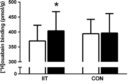
Muscle [3H]ouabain-binding site content
muscle NKA content increased by 11% (P=0.03,
d =0.8), with no changes observed in CON (Fig. 1).
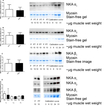
NKA isoform abundances
Analyses in whole muscle homogenates revealed no changes in the NKA a1, a2 or b1 isoforms following
12 weeks of IIT or CON (Fig. 2). Effect size calculations revealed a moderate effect of IIT on the a2 isoform
(d=0.6).
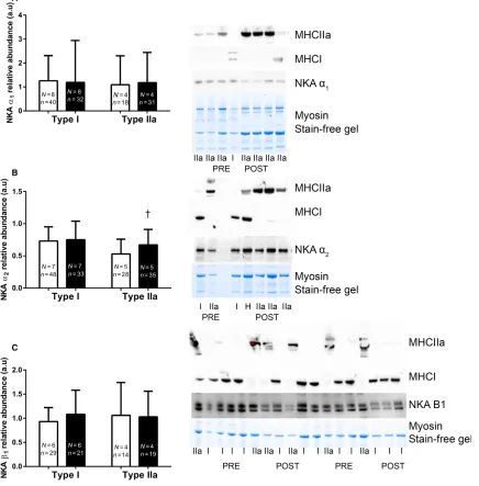
When NKA isoform abundances were measured in sin-gle fiber segments, no changes were seen in the NKA a1
orb1abundance following IIT in either Type I or II fibers
(a1 P>0.05, d =0.2 and 0.4, Type I and II respectively, b1 P<0.05, d =0.4 and 0.0). The a2 abundance was
unchanged after IIT in Type I fibers (P> 0.05, d=0.1), but was increased by 30% in Type II fibers (P<0.05,
d =0.5, Fig. 3).
Discussion
We report that 12 weeks of IIT increased muscle NKA content by 11% in older adults and that fiber-type speci-fic upregulation occurred for the NKA a2 isoform, being
elevated by 30% in Type II fibers after IIT. No changes in NKA isoforms occurred in Type I fibers after IIT. In addition no increases were detected in the NKA the a1, a2 or b1 isoform abundances in whole muscle
homoge-nates after training. While we also found that IIT increased VO2peak by 16% and peak workrate by 11%,
there was no significant reduction in the rise in venous [K+] relative to work performed during incremental exer-cise, suggesting that at least in antecubital venous blood, IIT did not improve circulating K+homeostasis. Adverse responses to IIT in some individuals during the initial 2 weeks indicate that IIT should however, be imple-mented with appropriate caution in healthy older adults.
Intense interval training increases muscle NKA content and isoforms
We show for the first time that 12 weeks of IIT increased muscle [3H]ouabain-binding in healthy older adults. This demonstrates that the NKA remains highly adaptable in skeletal muscle in an aged cohort, and that this occurs
Table 1. The effects of 12 weeks of intense interval training (IIT) in elderly humans aged between 65 and 76 years, on performance and physiological variables including peak potassium, during incremental exercise continued until a RPE-17.
Variable Group Pre Post Pvalue Cohen’s d
WRpeak(W) IIT 145.049.5 181.252.4 P=0.01 1.6
CON 142.046.4 147.140.2 NS 0.0
Work (J) IIT 43,72521,282 70,05031,834 P=0.001 2.1
CON 47,91424,408 47,48624,834 NS 0.0
Time to RPE-17 (min) IIT 7.33.7 9.44.5 NS (P=0.057) 0.6
CON 9.63.1 9.73.1 NS 0.2
HRpeak(b.min 1) IIT 136.216.4 144.314.4 P=0.03 0.7
CON 14111 14214 NS 0.5
VO2peak(mL.kg 1.min 1) IIT 24.75.4 28.75.1 P=0.002 1.4
CON 23.65.3 23.85.3 NS 0.0
[K+]
v peak IIT 4.740.41 5.230.57 NS (P=0.07) 0.0
(mmol.L 1) CON 4.880.33 4.900.42 NS 0.0
Δ[K+]
v.work 1 IIT 21.410.6 17.44.5 NS 0.4
(nmol.L 1.J 1) CON 22.310.9 21.114.7 NS 0.0
Data is expressed as mean+SD;n=8 IIT,n=7 CON.
Figure 1. The effects of 12 weeks of intense interval training (IIT) in elderly humans aged between 65 and 76 years, on the skeletal muscle Na+,K+-ATPase (NKA) content, measured by [3
H]ouabain-binding site content before (Pre, open bars) and after IIT-training (Post, filled bars) IIT or control (CON). Data presented as
even in a “low volume” (i.e., 494 min, totaling 16 min per session) training protocol as utilized here. This increase in NKA content is consistent with an earlier find-ing in a cross-sectional study that reported older adults who had been active for 10–12 years had a 30–40% greater NKA content than inactive older adults (Klitgaard and Clausen 1989). Our data supports the cross-sectional data showing for the first time in elderly individuals undertaking a longitudinal training program, that there was an 11% gain in NKA content after IIT, which typi-cally reported in studies with healthy young adults after various forms of physical training (McKenna et al. 1996). We also note, that gender of the participants had no effect on NKA content and thus their training responses,
this is the first time that this has been reported in older adults in previous findings in healthy young adults (Mur-phy et al. 2007).
A novel finding was the fiber-specific upregulation of the NKA a2 isoform after IIT in older adults, being
increased in Type II fibers only; this is the first time an upregulation of a2 in a fiber-specific manner has been
seen after training in any age group. In rodent muscle, thea2may represent ~80–85% of all aisoforms (Hansen
2001; Clausen 2003) and is particularly important in reg-ulation of Na+/K+exchange and membrane potential dur-ing muscle contractions (He et al. 2001; Radzyukevich et al. 2013). Furthermore, the a2 is typically upregulated
in muscle in response to high-intensity exercise training
Figure 2. The effects of 12 weeks of intense interval training (IIT) in elderly humans aged between 65 and 76 years, on the Na+,K+-ATPase
in healthy young adults, when measured in muscle lysates (Nielsen et al. 2004; Mohr et al. 2007; Bangsbo et al. 2009; Thomassen et al. 2010), although not always (Aughey et al. 2007; Gunnarsson et al. 2013; Wyckelsma
et al. 2015). In aged muscle, Type II fibers undergo the greatest atrophy (Evans and Lexell 1995) and exhibit reductions in specific force compared to young adults (Lamboley et al. 2015). The increased a2 abundance Figure 3. The effects of 12 weeks of intense interval training (IIT) in elderly humans aged between 65 and 76 years, on the Na+,K+-ATPase
(NKA) isoform abundances in Type I and Type II muscle fibers. For each panel the mean+SD data is shown on the left and the representative blots with Type I and Type IIa fibers from before (Pre) and after (Post) HIT shown on the right for each of thea1,a2andb1isoforms,
respectively, with samples from. (A) NKAa1, (B)a2and (C)b1isoform abundances, Pre Type I (open bars), Post Type I (closed bars), Post Type II
(open bars), Post Type II (closed bars). For the representative images, the fiber-type is identified using antibodies specific to MHCI or MHCIIa, the NKA isoform is shown and the abundant muscle protein, myosin is shown on the Stain Free gel image, which indicates the relative amount of tissue loaded in each lane. Analysis involved determining the density of the specific NKA bands and expressing relative to the total protein obtained from the entire StainFree gel. Thea1anda2isoforms migrated at~100 kDa and theb1isoform between~50 and 55 kDa. Each lane
specifically in Type II fibers after IIT may have important implications for maintenance of muscle function in the aged. This is based on research in skeletal muscle a2
knockout mice (ska2 / ) (Radzyukevich et al. 2012),
which exhibited a markedly impaired treadmill incremen-tal running performance, lower tetanic force and more rapid fatigue, compared with wild-type mice (Radzyuke-vich et al. 2012). In rat isolated skeletal muscle, blocking 26% of the NKA with ouabain induced a substantial impairment of contractile force when exposed to high K+ solutions (Clausen and Everts 1991). We recently found no difference ina2abundance in single muscle fibers and
homogenate between old and young humans (Wyckelsma et al. 2016). Thus, the increase in a2 with IIT does not
appear to be a compensatory response to an age-related deficit of NKA isoforms, as suggested in rat muscles (Ng et al. 2003). Rather, the increaseda2in Type II fibers and
[3H]ouabain-binding site content with IIT suggests upreg-ulation of NKAa2may assist the muscle Type II fibers to
undertake repeated contractions and attenuate muscle fatigue in these older adults. This is consistent with improved contractility of aged muscle and enabling the completion of simple everyday tasks requiring repeated muscle contractions in older adults. Whether the excitability of Type II fibers during muscle contractions is improved in the aged following training is of importance and remains to be determined.
An interesting finding was the lack of change in either thea1orb1 isoforms in the single fibers or homogenates
following IIT in older adults. Possible rationales for the lesser adaptability of the a1 and the b1 isoforms with
training, are that the purported importance of the a1 is
primarily under rest conditions (He et al. 2001) and an existing overabundance of thebisoforms relative to thea isoforms in muscle (Lavoie et al. 1997). However, this finding regardinga1 andb1 is also not unexpected, given
the inconsistencies in the reported responses of these NKA isoforms to intense exercise training in young adults. For example, of nine studies in humans that investigated the effects of exercise training on skeletal muscle homogenate/ lysate NKA a1, a2 and b1 isoforms in young adults, an
increase in a1 was reported in only three (Nielsen et al.
2004; Green et al. 2008; Iaia et al. 2008), with a1
unchanged in the remaining six studies (Aughey et al. 2007; Mohr et al. 2007; Bangsbo et al. 2009; Thomassen et al. 2010; Gunnarsson et al. 2013; Wyckelsma et al. 2015). For the a2 isoform, increases were reported after
training in only five of these studies (Nielsen et al. 2004; Mohr et al. 2007; Green et al. 2008; Bangsbo et al. 2009; Thomassen et al. 2010) being unchanged in the other four, whilst increases in the b1 isoform were only reported in
three of these nine studies (Mohr et al. 2007; Green et al. 2008; Wyckelsma et al. 2015) being unchanged in the
other six. Recently, resistance training following a period of muscle disuse resulted in an upregulation ofa1in Type
II fibers and a2 in Type I fibers, but with no changes
detected for either isoform in a crude homogenate (Perry et al. 2016). There does not appear to be a clear link between the type of training or in the training status of individuals in the adaptability of these NKA isoforms in younger adults or whether these analyses utilized whole muscle homogenates or spun muscle lysates.
Our findings revealed differences in the relative changes in the [3H]ouabain-binding site content and thea2
abun-dance after IIT. After IIT, the NKA content measured by [3H]ouabain-binding assay was increased by 11%, the a
2
abundance detected in Type II fibers increased by 30%, whilst the a2 abundance in whole muscle homogenates
and in Type I fibers were unchanged. This firstly suggests that the [3H]ouabain-binding assay may be more sensitive in detecting changes in mixed muscle analyses that are restricted to only one fiber-type, than western blotting conducted with whole muscle homogenates; and secondly, that the [3H]ouabain-binding assay can detect changes restricted to one fiber-type if this change is relatively large. Numerous studies investigating the effects of train-ing, agtrain-ing, injury and disease on NKA content have reported that the relative increases in the protein abun-dance of a2 measured in muscle homogenates were
greater than those when measured with [3 H]ouabain-binding site content, with similar proportional increases reported only five times out of ten studies in the litera-ture (Clausen 2013). Our findings show that our [3H] ouabain-binding responses were greater than the a2, the
opposite to what is normally shown, however this may be simply related to differences in western blot techniques, including the use of a whole homogenate versus lysates. Regardless, this study has shown that the NKA content remains highly adaptable to physical training with age and further expands on our previous work where we speculated that physical activity levels, rather than age, directly affected muscle NKA content (Wyckelsma et al. 2016). This is also consistent with our earlier observation in patients with osteoarthritis that physical activity may play an important role in preserving NKA content anda2
abundance in the aged (Perry et al. 2013).
Physiological adaptations and other practical implications
A further novel finding was the effectiveness of IIT for improving important functional outcomes in healthy older adults. The IIT protocol was effective at increasing each of the VO2peak, WRpeak and HRpeak of older adults
no differences in Δ[K+]v.work 1ratio, suggesting that the
tendency for higher peak [K+]vat the end of exercise after
IIT was simply a function of being able to attain a higher WRpeak after training. This finding differs from other
training studies in young adults, that have demonstrated reduced Δ[K+].work 1 ratio following training, when [K+] was measured during sprint or high-intensity exer-cise (McKenna et al. 1993, 1997; Harmer et al. 2006) or during submaximal exercise (Nielsen et al. 2004), although this was not always the case (McKenna et al. 1997). Thus, despite the improved exercise performance and upregulation of the [3H]ouabain-binding and NKA
a2 in Type II fibers after IIT, no change was seen in the
[K+]v.work 1 ratio. One possible interpretation is that
there was no improvement in the in-vivo K+ regulation in these older individuals after training; however, several factors will have affected these [K+] findings and may make this interpretation invalid. First, blood was sampled from the antecubital vein, draining the relatively inactive forearm muscles during exercise, with the [K+]v being
substantially lower than in arterial blood or in venous blood draining the active musculature (Lindinger 1995; Sejersted and Sjøgaard 2000). Measurement of the femoral venous or arterial [K+] rather than antecubital venous [K+] would have been preferred. However, we judged that in these elderly participants, these more demanding procedures were impractical, may have been difficult to justify ethically, and may have compromised recruitment and retention. Second, for safety reasons the test was only continued until a RPE 17 (“very hard”) was achieved, and thus the exercise was not truly maximal, which would underestimate the peak exercise workrate and plasma [K+] (Medbø and Sejersted 1990). Pre-train-ing, participants reached a HRpeak of ~136 b.min 1
dur-ing exercise, estimated at 88–92% of the age-predicted maximal heart rate (220-age), whereas after training, a HRpeakof~144b.min 1was attained, estimated to
corre-spond to between 93% and 100% of the age-predicted maximal heart rate. This suggests that after IIT, older adults were able to work at a higher workrate and closer to their age-predicted maximal HR. It is unlikely that this results simply from a greater motivation to exercise harder after training. From a practical aspect, the imple-mentation of IIT for the general population of older adults should be performed with great caution due to potential adverse responses during the first few weeks of training. In this study, 50% of the participants exhibited mild vasovagal episodes during the first 2 weeks of train-ing. It is recommended that those who wish to perform IIT should have had recent exposure to at least regular moderate intensity exercise, and have appropriate supervi-sion and monitoring during and after each exercise ses-sion (Levinger et al. 2015).
Conclusions
IIT upregulated NKA a2 isoform in Type II fibers and
this coincided with increases in whole muscle NKA con-tent measured by [3H]ouabain-binding site content. These findings indicate that skeletal muscle from elderly individ-ual remains highly adaptable to exercise training with respect to muscle NKA. Further research is required to determine the functional significance of this increase in NKA a2with training. Whilst IIT improved exercise
per-formance in older adults, it should be implemented with caution due to potential adverse responses.
Acknowledgments
We thank all participants for their contributions to the study.
Conflict of Interest
The authors declare no conflict of interest.
References
Aughey, R. J., K. T. Murphy, S. A. Clark, A. P. Garnham, R. J. Snow, D. Cameron-Smith, et al. 2007. Muscle Na+-K+ -ATPase activity and isoform adaptations to intense interval exercise and training in well-trained athletes. J. Appl. Physiol. 103:39–47.
Bangsbo, J., T. P. Gunnarsson, J. Wendell, L. Nybo, and M. Thomassen. 2009. Reduced volume and increased training intensity elevate muscle Na+-K+pumpa2-subunit expression as well as short- and long-term work capacity in humans. J. Appl. Physiol. 107:1771–1780.
Bibert, S., S. Roy, D. Schaer, J.-D. Horisberger, and K. Geering. 2008. Phosphorylation of phospholemman (FXYD1) by protein kinases A and C modulates distinct Na, K-ATPase Isozymes. J. Biol. Chem. 283:476–486.
Blanco, G., and R. W. Mercer. 1998. Isozymes of the Na-K-ATPase: heterogeneity in structure, diversity in function. Am. J. Physiol. Renal Physiol. 275:F633–F650.
Borg, G. A. 1982. Psychological bases of perceived exertion. Med. Sci. Sports Exerc. 14:377–381.
Clausen, T. 2003. Na+-K+pump regulation and skeletal muscle contractility. Physiol. Rev. 83:1269–1324.
Clausen, T. 2013. Quantification of Na+,K+pumps and their transport rate in skeletal muscle: functional significance. J. Gen. Physiol. 142:327–345.
Clausen, T., and M. E. Everts. 1991. K(+)-induced inhibition of contractile force in rat skeletal muscle: role of active Na (+)-K+transport. Am. J. Physiol. 261:C799–C807. Cochran, A. J. R., M. E. Percival, S. Tricarico, J. P. Little, N.
acute but different chronic muscle adaptations. Exp. Physiol. 99:782–791.
Cohen, J. 1988. Statistical power analysis for the behavioral sciences. L. Erlbaum Associates, Hillsdale, NJ
Deschenes, M. R. 2004. Effects of aging on muscle fibre type and size. Sports Med. 34:809–824.
Dutka, T. L., and G. D. Lamb. 2007. Na+-K+pumps in the transverse tubular system of skeletal muscle fibers preferentially use ATP from glycolysis. Am. J. Physiol. Cell Physiol. 293:C967–C977.
Evans, W. J., and J. Lexell. 1995. Human aging, muscle mass, and fiber type composition. J. Gerontol. Series A: Biol. Sci. Med. Sci. 50A:11–16.
Ford, G. A., T. F. Blaschke, R. Wiswell, and B. B. Hoffman. 1993. Effect of aging on changes in plasma potassium during exercise. J. Gerontol. 48:M140–M145. Ford, G. A., W. D. Dachman, T. F. Blaschke, and B. B.
Hoffman. 1995. Effect of aging on beta 2-adrenergic receptor-stimulated flux of K+, PO4, FFA, and glycerol in human forearms. J. Appl. Physiol. 78:172–178.
Geering, K. 2001. The functional role of beta subunits in oligomeric P-type ATPases. J. Bioenerg. Biomembr. 33:425– 438.
Gibala, M. J., J. P. Little, M. J. MacDonald, and J. A. Hawley. 2012. Physiological adaptations to low-volume, high-intensity interval training in health and disease. J. Physiol. 590:1077–1084.
Gillen, J. B., M. E. Percival, L. E. Skelly, B. J. Martin, R. B. Tan, M. A. Tarnopolsky, et al. 2014. Three minutes of all-out intermittent exercise per week increases skeletal muscle oxidative capacity and improves cardiometabolic health. PLoS ONE 9:e111489.
Green, H. J., E. Bombardier, T. A. Duhamel, R. D. Stewart, A. R. Tupling, and J. Ouyang. 2008. Metabolic, enzymatic, and transporter responses in human muscle during three consecutive days of exercise and recovery. Am. J. Physiol. Regul. Integr. Comp. Physiol. 295:R1238–R1250.
Gunnarsson, T. P., P. M. Christensen, M. Thomassen, L. R. Nielsen, and J. Bangsbo. 2013. Effect of intensified training on muscle ion kinetics, fatigue development, and repeated short-term performance in endurance-trained cyclists. Am. J. Physiol. Regul. Integr. Comp. Physiol. 305: R811–R821.
Hansen, O. 2001. Thea1 isoform of Na+,K+-ATPase in rat soleus and extensor digitorum longus. Acta Physiol. Scand. 173:335–341.
Harmer, A. R., P. A. Ruell, M. J. McKenna, D. J. Chisholm, S. K. Hunter, J. M. Thom, et al. 2006. Effects of sprint training on extrarenal potassium regulation with intense exercise in Type 1 diabetes. J. Appl. Physiol. (1985) 100:26–34. He, S., D. Shelly, A. Moseley, P. James, J. James, R. Paul, et al.
2001. The a1- and a2-isoforms of Na-K-ATPase play different roles in skeletal muscle contractility. Am. J. Physiol. Regul. Integr. Comp. Physiol. 281:R917–R925.
Hunter, S. K., H. M. Pereira, and K. G. Keenan. 2016. The aging neuromuscular system and motor performance. J. Appl. Physiol. 121:982–995.
Iaia, F. M., M. Thomassen, H. Kolding, T. Gunnarsson, J. Wendell, T. Rostgaard, et al. 2008. Reduced volume but increased training intensity elevates muscle Na+-K+pump 1-subunit and NHE1 expression as well as short-term work capacity in humans. AJP: Regul. Integr. Comp. Physiol. 294: R966–R974.
Klitgaard, H., and T. Clausen. 1989. Increased total
concentration of Na-K pumps in vastus lateralis muscle of old trained human subjects. J. Appl. Physiol. 67:2491–2494. Lamboley, C. R., V. L. Wyckelsma, T. L. Dutka, M. J.
McKenna, R. M. Murphy, and G. D. Lamb. 2015. Contractile properties and sarcoplasmic reticulum calcium content in type I and type II skeletal muscle fibres in active aged humans. J. Physiol. 593:2499–2514.
Lavoie, L., R. Levenson, P. Martin-Vasallo, and A. Klip. 1997. The molar ratios of alpha and beta subunits of the Na+ -K+-ATPase differ in distinct subcellular membranes from rat skeletal muscle. Biochemistry 36:7726–7732.
Lazarus, N. R., and S. D. R. Harridge. 2016. Declining performance of master athletes: silhouettes of the trajectory of healthy human ageing? J. Physiol. doi: 10.1113/JP272443 Levinger, I., G. Jerums, N. K. Stepto, L. Parker, F. R. Serpiello,
G. K. McConell, et al. 2014. The effect of acute exercise on undercarboxylated osteocalcin and insulin sensitivity in obese men. J. Bone Miner. Res. 29:2571–2576. Levinger, I., C. S. Shaw, N. K. Stepto, S. Cassar, A. J.
McAinch, C. Cheetham, et al. 2015. What doesn’t kill you makes you fitter: a systematic review of high-intensity interval exercise for patients with cardiovascular and metabolic diseases. Clin. Med. Insights Cardiol. 9:53–63. Lindinger, M. I. 1995. Potassium regulation during exercise
and recovery in humans: implications for skeletal and cardiac muscle. J. Mol. Cell. Cardiol. 27:1011–1022. Manoharan, P., T. L. Radzyukevich, H. Hakim Javadi, C. A.
Stiner, J. A. Landero Figueroa, J. B. Lingrel, et al. 2015. Phospholemman is not required for the acute stimulation of Na+-K+-ATPasea2-activity during skeletal muscle fatigue. Am. J. Physiol. Cell Physiol. 309:C813–C822.
McKenna, M. J., T. A. Schmidt, M. Hargreaves, L. Cameron, S. L. Skinner, and K. Kjeldsen. 1993. Sprint training increases human skeletal muscle Na(+)-K(+)-ATPase concentration and improves K+regulation. J. Appl. Physiol. 75:173–180.
McKenna, M. J., A. R. Harmer, S. F. Fraser, and J. L. Li. 1996. Effects of training on potassium, calcium and hydrogen ion regulation in skeletal muscle and blood during exercise. Acta Physiol. Scand. 156:335–346.
McKenna, M. J., J. Bangsbo, and J.-M. Renaud. 2008. Muscle K+, Na+, and Cl disturbances and Na+-K+pump inactivation: implications for fatigue. J. Appl. Physiol. 104:288–295.
McKenna, M. J., B. D. Perry, F. R. Serpiello, M. K. Caldow, P. Levinger, D. Cameron-Smith, et al. 2012. Unchanged [3H] ouabain binding site content but reduced Na+-K+pump
a2-protein abundance in skeletal muscle in older adults. J. Appl. Physiol. 113:1505–1511.
Medbø, J. I., and O. M. Sejersted. 1990. Plasma potassium changes with high intensity exercise. J. Physiol. 421:105–122. Mohr, M., P. Krustrup, J. J. Nielsen, L. Nybo, M. K.
Rasmussen, C. Juel, et al. 2007. Effect of two different intense training regimens on skeletal muscle ion transport proteins and fatigue development. Am. J. Physiol. Regul. Integr. Comp. Physiol. 292:R1594–R1602.
Murphy, R. M. 2011. Enhanced technique to measure proteins in single segments of human skeletal muscle fibers: fiber-type dependence of AMPK-alpha1 and -beta1. J. Appl. Physiol. 110:820–825.
Murphy, R. M., and G. D. Lamb. 2013. Important considerations for protein analyses using antibody based techniques: down-sizing Western blotting up-sizes outcomes. J. Physiol. 591:5823–5831.
Murphy, K. T., R. J. Snow, A. C. Petersen, R. M. Murphy, J. Mollica, J. S. Lee, et al. 2004. Intense exercise up-regulates Na+,K+ -ATPase isoform mRNA, but not protein
expression in human skeletal muscle. J. Physiol. 556:507–519. Murphy, R. M., E. Verburg, and G. D. Lamb. 2006. Ca2+
activation of diffusible and bound pools ofl-calpain in rat skeletal muscle. J. Physiol. 576:595–612.
Murphy, K. T., R. J. Aughey, A. C. Petersen, S. A. Clark, C. Goodman, J. A. Hawley, et al. 2007. Effects of endurance training status and sex differences on Na+,K+-pump mRNA expression, content and maximal activity in human skeletal muscle. Acta Physiol. 189:259–269.
Ng, Y.-C., M. Nagarajan, K. N. Jew, L. C. Mace, and R. L. Moore. 2003. Exercise training differentially modifies age-associated alteration in expression of Na+-K+-ATPase subunit isoforms in rat skeletal muscles. Am. J. Physiol. Regul. Integr. Comp. Physiol. 285:R733–R740.
Nielsen, J. J., M. Mohr, C. Klarskov, M. Kristensen, P. Krustrup, C. Juel, et al. 2004. Effects of high-intensity intermittent training on potassium kinetics and performance in human skeletal muscle. J. Physiol. 554:857–870.
Nørgaard, A., K. Kjeldsen, and T. Clausen. 1984. A method for the determination of the total number of 3H-ouabain binding sites in biopsies of human skeletal muscle. Scand. J. Clin. Lab. Invest. 44:509–518.
Perry, B. D., P. Levinger, F. R. Serpiello, M. K. Caldow, D. Cameron-Smith, J. R. Bartlett, et al. 2013. The effects of osteoarthritis and age on skeletal muscle strength,
Na+-K+-ATPase content, gene and isoform expression. J. Appl. Physiol. 115:1443–1449.
Perry, B. D., V. L. Wyckelsma, R. M. Murphy, C. H. Steward, M. Anderson, I. Levinger, et al. , 2016. Dissociation between short-term unloading and resistance training effects on skeletal muscle Na+,K+-ATPase, muscle function and fatigue in humans. J. Appl. Physiol. (1985) 121:1074–1086. Petersen, A. C., M. J. Leikis, L. P. McMahon, A. B. Kent, K. T.
Murphy, X. Gong, et al. 2012. Impaired exercise
performance and muscle Na(+), K(+)-pump activity in renal transplantation and haemodialysis patients. Nephrol. Dial. Transplant. 27:2036–2043.
Radzyukevich, T. L., J. C. Neumann, T. N. Rindler, N. Oshiro, D. J. Goldhamer, J. B. Lingrel, et al. 2012. Tissue-specific role of the Na,K-ATPase 2 isozyme in skeletal muscle. J. Biol. Chem. 288:1226–1237.
Radzyukevich, T. L., J. C. Neumann, T. N. Rindler, N. Oshiro, D. J. Goldhamer, J. B. Lingrel, et al. 2013. Tissue-specific role of the Na,K-ATPase alpha2 isozyme in skeletal muscle. J. Biol. Chem. 288:1226–1237.
Sejersted, O. M., and G. Sjøgaard. 2000. Dynamics and consequences of potassium shifts in skeletal muscle and heart during exercise. Physiol. Rev. 80:1411–1481. Thomassen, M., P. M. Christensen, T. P. Gunnarsson, L.
Nybo, and J. Bangsbo. 2010. Effect of 2-wk intensified training and inactivity on muscle Na+-K+pump expression, phospholemman (FXYD1) phosphorylation, and
performance in soccer players. J. Appl. Physiol. 108:898–905. Thomassen, M., R. M. Murphy, and J. Bangsbo. 2013.
Fibre type-specific change in FXYD1 phosphorylation during acute intense exercise in humans. J. Physiol. 591: 1523–1533.
Wang, J., J. B. Velotta, A. A. McDonough, and R. A. Farley. 2001. All human Na+-K+-ATPasea-subunit isoforms have a similar affinity for cardiac glycosides. Am. J. Physiol. Cell Physiol. 281:C1336–C1343.
Wisloff, U., A. Stoylen, J. P. Loennechen, M. Bruvold, O. Rognmo, P. M. Haram, et al. 2007. Superior cardiovascular effect of aerobic interval training versus moderate
continuous training in heart failure patients: a randomized study. Circulation 115:3086–3094.
Wyckelsma, V. L., M. J. McKenna, F. R. Serpiello, C. R. Lamboley, R. J. Aughey, N. K. Stepto, et al. 2015. Single-fiber expression and Single-fiber-specific adaptability to short-term intense exercise training of Na+-K+-ATPase alpha- and beta-isoforms in human skeletal muscle. J. Appl. Physiol. 118:699–706.