R E V I E W
Cold agglutinin disease: current challenges and
future prospects
This article was published in the following Dove Press journal: Journal of Blood Medicine
Sigbjørn Berentsen1 Alexander Röth2 Ulla Randen3 Bernd Jilma4
Geir E Tjønnfjord5–7
1Department of Research and Innovation,
Haugesund Hospital, Haugesund,
Norway;2Department of Hematology,
West German Cancer Center, University Hospital Essen, University of
Duisburg-Essen, Duisburg-Essen, Germany;3Department of
Pathology, Akershus University Hospital,
Lørenskog, Norway;4Department of
Clinical Pharmacology, Medical University
of Vienna, Vienna, Austria;5Department
of Haematology, Oslo University
Hospital, Oslo, Norway;6KG Jebsen’s
Center for B-cell Malignancies, University
of Oslo, Oslo, Norway;7Institute of
Clinical Medicine, University of Oslo, Oslo, Norway
Abstract: Cold agglutinin disease (CAD) is a complement-dependent, classical pathway-mediated immune hemolytic disease, accounting for 15–25% of autoimmune hemolytic anemia, and at the same time, a distinct clonal B-cell lymphoproliferative disorder of the bone marrow. The disease burden is often high, but not all patients require pharmacological treatment. Several therapies directed at the pathogenic B-cells are now available. Rituximab plus bendamustine or rituximab monotherapy should be considered first-line treatment, depending on individual patient characteristics. Novel treatment options that target the classical complement pathway are under development and appear very promising, and the C1s inhibitor sutimlimab is currently being investigated in two clinical Phase II and III trials. These achievements have raised new challenges and further prospects, which are discussed. Patients with CAD requiring therapy should be considered for clinical trials.
Keywords: autoimmune hemolytic anemia, cold agglutinin disease, lymphoproliferative, complement, complement inhibitors, therapy
Introduction
Over the last 15 years, considerable progress has been made in the understanding
and treatment of chronic cold agglutinin disease (CAD). CAD accounts for 15–25%
of cases of autoimmune hemolytic anemia (AIHA) and affects mainly elderly or
middle-aged people, but has been reported in patients as young as 30.1–4 The
prevalence in Northern Europe has been estimated to be 16 cases per million inhabitants and the incidence rate to be 1 per million per year, both of which are
probably slight underestimations.3
CAD, also known as primary CAD, is a clonal B-cell lymphoproliferative
disorder (LPD).5–8 This well-defined clinico-pathological entity should be
distin-guished from secondary cold agglutinin syndrome (CAS), a similar cold hemolytic
syndrome that occasionally complicates specific infections (typicallyMycoplasma
pneumoniae pneumonia or Epstein–Barr virus infection) or malignancies
(espe-cially aggressive lymphoma).9Only CAD will be further addressed here.
The involved autoantibodies, known as cold agglutinins (CAs), bind to their
antigen at an optimum temperature of 3–4°C, but are also able to react at a higher
temperature, depending on the thermal amplitude (TA). The TA is defined as the
highest temperature at which the CA will bind to its antigen.10–12 CA activity is
assessed by the titer, expressed as the inverse value of the highest serum dilution at which agglutination can be detected. CAs in CAD are monoclonal, usually of the
immunoglobulin (Ig) Mκclass and, in the vast majority of cases, specific for the
Correspondence: Sigbjørn Berentsen Department of Research and Innovation, Haugesund Hospital, 5504 Haugesund, Haugesund 2170, Norway
Tel +475 273 2000
Email [email protected]
Journal of Blood Medicine
Dove
press
open access to scientific and medical research
Open Access Full Text Article
Journal of Blood Medicine downloaded from https://www.dovepress.com/ by 118.70.13.36 on 24-Aug-2020
erythrocyte surface carbohydrate antigen termed I.3,12–15 Complement activation via the classical pathway is
essen-tial for subsequent hemolysis.16–21
Recent advances in CAD include the demonstration of a distinct clonal B-cell LPD, the documentation of effec-tive B-cell-directed therapies, and the development of novel complement-directed therapies. This review will focus on these achievements, which also raise some challenges.
Clinical presentation
According to a descriptive study, the median age of patients with CAD is 76 years and the median age at
presentation is 67 years (lower range, 30 years).3 In the
same cohort, median hemoglobin (Hb) level was 8.9 g/dL with a lower and upper tertile of 8.0 g/dL and 10.4 g/dL, respectively. There are several reports of Hb levels as low
as 4 g/dL.3,4,10,22 At the other end of the spectrum,
com-pensated hemolysis with a normal Hb is not
uncommon.3,4,22 Another large series found
hemoglobi-nuria in 15% of the patients.4 An association between
low ambient temperatures and worsening of hemolysis
has been documented in individual patients.8,23
According to our data, exacerbations occur in 70% of patients during febrile infections and, at least in some
cases, following major trauma or major surgery.3,24–26
Literature estimates on transfusion dependency show a high degree of variability; the two largest series of unselected patients with well-documented CAD found
that 40–50% had received transfusions for a shorter or
longer period of time. Cold-induced, agglutination-mediated symptoms from the capillary circulation have been recorded in up to 90% of the patients, ranging from
slight acrocyanosis to disabling Raynaud phenomena.3
Less typical clinical features include gangrene, which is uncommon, and cutaneous disorders, sometimes described
as livedo reticularis and sometimes as livedo
racemosa.20,27,28 Such atypical vascular manifestations
should warrant the exclusion of cryoglobulinemia as a differential diagnosis or a concomitant property of the
cold-reactive IgM.3,27,29
Diagnostic criteria
Patients with biochemical evidence of chronic hemolysis are diagnosed with CAD if the direct antiglobulin test (DAT) is strongly positive for complement protein C3d and negative (or only weakly positive) for IgG, combined
with a CA titer of ≥64.3,4,12,30 Usually, the CA titer is
much higher, but it should nonetheless be acknowledged that occasional patients with otherwise typical CAD may
have a CA titer <64.4,30,31An additional diagnostic
criter-ion is the absence of known causes of secondary CAS, for
example, specific infections or aggressive lymphoma.
A bone marrow low-grade clonal LPD (discussed below)
can be demonstrated in a majority of patients, but confi
r-mation of an LPD is sometimes a matter of sensitivity and
not required for diagnosis.5,8,32 A spuriously high mean
corpuscular volume and false reduction in red blood cell counts are regularly seen, making estimated hematocrit
values unreliable, but these are not diagnostic criteria.33
Polyclonal CAs with TA <25–28°C and, mostly, in low
titers are detectable in some healthy individuals, probably as remnants of a phylogenetically ancient, primitive
adap-tive immune system.30,34,35In one study, CAs were found
in 0.3% of a cohort with unrelated disorders.36 Subjects
with detectable CA without hemolysis or circulatory
symp-toms do not have CAD.30,32
It is of critical importance that blood samples for CA
titration, TA determination, Ig quantification,
electrophor-esis, and immunofixation are kept at 37–38°C from
sam-pling to separation of serum or plasma to avoid false low
values and low sensitivity (Table 1).12,30,32 Even
auto-mated cell counts and Hb levels in ethylene diamine
tetra-acetate (EDTA) blood are sometimes difficult to obtain
because of agglutination in the tube. Prewarming at 37°C for up to 2 hrs will usually overcome this problem; if not,
a short preheating at 41°C for 1 min may be tried.37
Pathogenesis
A clonal lymphoproliferative B-cell
disorder
In 1957, Christenson et al characterized thefirst monoclonal
protein ever described, which was a CA from a patient with
CAD.38 Monoclonal IgM withκlight chain restriction was
recurrently found in subsequent studies.13During the 1990s,
a British group demonstrated selective usage of theIGHV4-34
gene segment, followed by the description by a Norwegian group of an abnormal bone marrow B-cell clone in a majority
of patients.39,40 In a subsequent multicenter cohort of 86
Norwegian patients, a clonal LPD was found by histology in
50 (76%) of 66 bone marrow biopsy samples and by flow
cytometry in 36 (90%) of 40 bone marrow aspirates.3
A retrospective single-center study from the USA confirmed
a clonal disorder in 69 (78%) of 89 patients.4More recently, an
Journal of Blood Medicine downloaded from https://www.dovepress.com/ by 118.70.13.36 on 24-Aug-2020
Austrian group was able to show that even patients with no
electrophoretic,flow cytometric, or histologic evidence of an
abnormal B-cell clone had a clonal rearrangement of their
heavy or light Ig chain genes.8
The two large retrospective studies interpreted thesefi
nd-ings as consistent with a heterogeneous group of clonal
disorders.3,4The LPD was most often classified as
lympho-plasmacytic lymphoma (LPL)/Waldenström’s
macroglobuli-nemia (WM), followed by marginal zone lymphoma (MZL)
and unclassified indolent LPD.3 In a more recent,
comprehensive study, however, bone marrow samples from 54 patients with CAD were systematically re-examined by a group of lymphoma pathologists, using a standardized
panel of morphologic, immune histochemical, flow
cyto-metric, and molecular methods.5Thefindings were
consis-tent with a surprisingly homogeneous bone marrow disorder
that we termed“primary CAD-associated LPD”and found to
be distinct from LPL, MZL, and other previously recognized lymphoma entities (Figure 1). Nodular B-cell aggregates were found in biopsy specimens from 40 of 54 patients,
Table 1Cold agglutinin disease: handling of samples
Analysis Material Sampling Handling of sample
Hemoglobin, blood cell counts Blood EDTA vacutainer Prewarm at 37–38°C before analysis if problems
with agglutination Cold agglutinin titer, thermal amplitude,
immunoglobulin quantification,
electro-phoresis, immunefixation
Serum Blood is drawn into
prewarmed vacutainers without additive. Place in warming cabinet or
water bath at 37–38°C
Keep at 37–38°C until serum has been removed
from the clot, after which the sample can be handled at room temperature
Flow cytometry Bone marrow aspirate
(too low sensitivity if performed in peripheral blood)
Add EDTA or heparin Prewarming before analysis will often be suffi
-cient. If not, wash cells at 37–38°C (description:
see reference 12)
Abbreviation:EDTA, ethylenediaminetetraacetic acid.
Figure 1Cold agglutinin-associated lymphoproliferative bone marrow disorder. Bone marrow trephine biopsy showing intraparenchymatous nodular lymphoid lesions (panels A and B, H&E staining, 40× and 200×, respectively). Immunoperoxidase staining for CD20 highlights intraparenchymatous nodular B-cell infiltration (panel C, 200×). Mast cells are not discerned around the nodular lymphoid lesions (panel D, Giemsa staining, 200×).
Notes:Reproduced with permission from Randen U, Trøen G, Tierens A, et al. Primary cold agglutinin-associated lymphoproliferative disease: a B-cell lymphoma of the bone marrow distinct from lymphoplasmacytic lymphoma.Haematologica. 2014;99(3):497–504.5Copyright © 2014 Ferrata Storti Foundation.
Journal of Blood Medicine downloaded from https://www.dovepress.com/ by 118.70.13.36 on 24-Aug-2020
whereas the bone marrow of 14 patients showed only scat-tered clonal B-cells. In the samples showing aggregates,
median lymphoid infiltration was 10% of the intertrabecular
surface (range, 5–80%). Specific features of LPL, such as
paratrabecular growth, fibrosis, lymphoplasmacytoid cell
morphology, or an increased number of mast cells surround-ing the lymphoid aggregates, were not seen. Differences between CAD and LPL were also observed by
immunohis-tochemical and flow cytometric analysis. It is noteworthy
that the MYD88 L265P mutation, previously shown to be
present in approximately 90% of cases of WM/LPL,41could
not be demonstrated in CAD-associated LPD.5Three
subse-quent studies have confirmed the absence or infrequent
occurrence of this mutation in CAD.6,42,43
A British flow cytometry study confirmed significant
immune phenotypic differences in clonal B-cells between
CAD and WM/LPL.6In addition to the well-known,
selec-tive IGHV4-34 heavy chain gene usage in primary CAD, a recent study found a highly restricted light chain gene
usage.42 Clonal rearrangement of the IGKV3-20 gene
sequence was seen in 16 (59%) of 27 patients; seven of these displayed highly homologous CDR3 regions. In another study of 16 patients with well-characterized
pri-mary CAD, usingflow cytometry–assisted cell sorting of
bone marrow followed by whole-exome and targeted
sequencing, recurrent mutations of KMT2D andCARD11
were detected in 11 (69%) and five patients (31%),
respectively.7 In contrast, KMT2D mutations have been
found in 26% of patients with WM/LPL and 5% of indi-viduals with monoclonal IgM-gammopathy of
undeter-mined significance (IgM-MGUS).44
These findings confirm that CAD is a clonal LPD of
the bone marrow in probably every case. This LPD should be regarded as a distinct entity, well characterized by
a total of morphological, immune histochemical, flow
cytometric, and molecular geneticfindings.
A complement-mediated immune
hemolytic anemia
CA binds to its antigen at the erythrocyte surface during passage through the cooler parts of the peripheral circu-lation, causing agglutination of red blood cells and
com-plement activation by the classical pathway.12,16,17,45,46
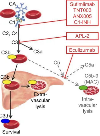
IgM is a potent classical pathway trigger, and the anti-gen-IgM complex binds complement protein C1q. C1 esterase then activates C2 and C4, generating C3
con-vertase which cleaves C3 into C3a and C3b. These steps are critical for the hemolytic process; if they are
blocked, there is no hemolysis.19,21,47 Upon warming
to 37°C in the central circulation, CA detaches from the cells, allowing agglutinated erythrocytes to separate, while C3b remains bound. C3b-opsonized cells are prone to phagocytosis by the mononuclear phagocytic system, mainly in the liver, a process known as
extra-vascular hemolysis18,45,46 On the surface of the
surviv-ing erythrocytes, C3b is cleaved, leavsurviv-ing high numbers of C3d molecules which can be detected by the DAT. Because there are no C3d receptors in the mononuclear phagocyte system, these erythrocytes are protected from
phagocytosis.18 This is probably part of the explanation
why most patients do not develop life-threatening anemia.
In most patients with CAD, the C3b-mediated pha-gocytosis is considered the predominant hemolytic
mechanism during steady-state disease.16,18,48 The
absence, or at least modest role, of terminal pathway activation in stable patients can probably be attributed to the protective effect of CD55 and CD59, which, unlike in paroxysmal nocturnal hemoglobinuria (PNH), are
intact in CAD.16,20 At least in some patients and
situa-tions, however, C3 is supposed to initiate the terminal complement cascade by binding and splitting C5, result-ing in the formation of the C5b-9 complex (membrane
attack complex) and intravascular hemolysis.20,49 The
occurrence of hemoglobinuria in about 15% of the
patients4 and the rather frequent finding of
hemosiderinuria29 support this notion, as does the
ben-eficial effect of C5 inhibition with eculizumab.20,26,49,50
Due to continuous consumption, most patients have low C3 and C4 serum levels, which is probably rate-limiting for
the complement-mediated hemolysis.12,25 During febrile
infections and following major surgery or major trauma, acute phase reaction will increase the production of com-plement proteins. The low levels are replete and
comple-ment activity is enhanced,25 which explains the
exacerbations seen in a majority of CAD patients in such situations.3,24–26
In general, AIHA as well as other diseases with complement-mediated intravascular hemolysis are asso-ciated with an increased risk of venous and arterial
thrombosis.51,52 Such a risk has been well documented
in severely affected CAD patients,20 but it has been
Journal of Blood Medicine downloaded from https://www.dovepress.com/ by 118.70.13.36 on 24-Aug-2020
difficult to show this in unselected cohorts. Registry studies do indicate an increased risk, but these studies have been based on hospitals' diagnosis rather than hard diagnostic criteria and have not distinguished between
CAD and CAS.53,54
Each of the two main components of the pathogenesis–
clonal B-cell lymphoproliferation and classical complement
pathway-dependent hemolysis–provide a rationale for
ther-apeutic approaches.
Therapy
Watchful waiting and
non-pharmacological management
The beneficial effect of keeping warm has been recognized
for more than 50 years.13,30Although the observed benefit
is moderate and largely based on clinical experience, there is no reason to change existing recommendations on pro-tection against cold exposure, in particular of the head,
face, and extremities, and of avoidance of cold
infusions.30,32 To avoid an acute phase reaction-induced
exacerbation of hemolysis, any bacterial infection should
be treated promptly.24,25Transfusions, when indicated, can
safely be given, provided the patient and the extremity used for infusion are kept warm and an in-line blood
warmer is used.30,32 It may be necessary to perform the
screening for irregular antibodies and crossmatching (if required) at 37°C, but the compatibility problems typical for warm-AIHA are not encountered in CAD. Transfusion of complement-rich blood products, eg, plasma, should probably be avoided as this may replete the low C4 and C3 levels and, at least in theory, enhance
complement-mediated hemolysis.25
Traditionally, it has been recommended to rely on non-pharmacological management in most cases and reserve drug therapy for CAD patients with severe hemolytic
anemia.10,55 Although these recommendations have been
based on the poor effect of older therapies and an under-estimation of the clinical symptoms, there is still no hard evidence to support pharmacological treatment in patients with only mild anemia and tolerable cold-induced circula-tory symptoms. Descriptive studies have shown, however,
that drug therapy has been tried in 70–80% of unselected
patients, indicating that previous recommendations may
have been too restrictive.3,4Increasingly, pharmacological
treatment is considered in patients with symptomatic ane-mia, severe cold-induced circulatory symptoms, and/or thrombosis.20,30,32,56,57
Unspeci
fi
c and supportive therapies
At least in some countries, therapy with corticosteroids
remains frequently used in clinical practice.51,57 No trials
have been published, but retrospective series have described low response rates and heterogeneous and
weak response criteria.3,4,51,57 Furthermore, the poor effi
-cacy will often lead to the use of unacceptably high
main-tenance doses in the few responders.3,55 It is, therefore,
widely agreed that corticosteroids should not be used to
treat CAD.30,32,55,56,58,59 Other unspecific immune
sup-pression, for example, with azathioprine, has not proved
beneficial.3,60Therapy with interferon-αwas ineffective in
a cohort of four patients although some conflicting
evi-dence does exist.61–63
Based on theoretical considerations and clinical experi-ence, folic acid supplementation has often been
recom-mended as an adjuvant.30 In a small prospective trial,
erythropoietin was seen to produce a favorable effect in
all of 12 AIHA patients,five of whom had CAD.64It is not
clear whether prophylaxis against thrombosis is indicated in CAD and, if so, for which patients. Until more data have been collected, it seems reasonable to consider antithrombotic prophylaxis in the presence of additional risk factors, severe intravascular hemolysis, or acute exacerbation until the hemolytic process is under control.
B-cell-directed therapies
The first treatment shown to be effective in CAD was
rituximab monotherapy, 375 mg/m2once a week for four
weeks.65–67 A Norwegian prospective uncontrolled trial
described 37 courses of therapy in 27 patients, using strict
response criteria.66 The overall response (OR) rate was
54% and the responders achieved a median increase in Hb levels of 4.0 g/dL. There was only one complete response (CR), and the median time to response (TTR) was 1.5 months. The median response duration was mod-est (11 months), but six of 10 relapsed patients responded to re-treatment. The study drug was well tolerated. A subsequent Danish prospective study reported similar
results.67 Rituximab and fludarabine in combination,
investigated in a prospective trial in 2010, resulted in a 76% OR rate with 20% CR, 56% partial responses (PR), and an estimated median response duration of more
than 66 months, however with some toxicity.68
In 2017, a Nordic group published a prospective,
non-randomized study of rituximab (375 mg/m2 day 1) plus
bendamustine (90 mg/m2day 1 and 2) for four cycles at
28-Journal of Blood Medicine downloaded from https://www.dovepress.com/ by 118.70.13.36 on 24-Aug-2020
day interval in 45 patients.22The OR rate was 71% and the CR rate 40%, with a median Hb increase of 4.4 g/dL in the CR group and 3.9 g/dL among those who achieved PR. The observed response duration was long (less than 10% relapses after a median of 32-month observation). Median TTR was 1.9 months (upper range, 12 months) with an even longer time to optimal response in some patients. The toxicity
profile was favorable as compared with the rituximab plus
fludarabine regimen; neutropenia grade 3–4 was seen in 33%
of the patients; however, only 11% experienced infection with or without neutropenia.
In a recent Italian prospective trial, six of 19 patients responded to bortezomib monotherapy, administered as
a single cycle.69Although this response rate may seem low,
the results should be considered a“proof of principle,”and an extended duration and/or bortezomib-based combination therapy may yield higher response rates.
In an early report, four patients treated with chloram-bucil achieved a reduction in CA titer and IgM serum
concentration, but no significant increase in Hb
levels.40,70Another study describedfive patients who
pro-spectively received cladribine monotherapy, none of
whom responded.71 There is a strong theoretic rationale
for the novel B-cell targeted therapies, of which ibrutinib has become an established treatment for WM/LPL,
show-ing favorable results even in MYD88 L265P-negative
cases.72,73However, there are no published studies
regard-ing the use of these drugs in CAD.
Complement-directed therapies
Complement modulation for treatment of CAD was first
described in 2009 and is undergoing rapid development
(Figure 2).16,49,74The anti-C5 monoclonal antibody,
eculizu-mab, has proved able to inhibit intravascular hemolysis and reduce lactate dehydrogenase levels and transfusion require-ments in a prospective study of 13, mostly transfusion-dependent patients with CA-mediated hemolytic anemia
(12 CAD and one severe CAS).20Only a marginal increase
in Hb level was observed. This is not unexpected, as the C5 inhibition will not affect the C3b-mediated, extravascular hemolysis. Eculizumab has also proved useful as a rapidly acting rescue therapy for severe CAD/CAS and, according to a case report, as prophylaxis against cardiac surgery-induced
exacerbation in CAD.20,26,75,76
Classical pathway inhibition would be expected to do
better. The first available drug for blocking the classical
pathway was plasma-derived C1 esterase inhibitor (C1-INH). Although approved only for hereditary angioedema, which is not a complement-mediated disorder, pharmaco-logical doses of C1-INH were shown to stop complement
activation and hemolysis and efficiently improve anemia
when used as rescue therapy in a patient with a severe,
IgM-mediated warm-AIHA.77,78 Recently, a similar effect
was observed in a patient with acute, severe CAS.21
However, levels of endogenous C1-INH are normal in CAD and CAS; frequently repeated high doses are likely to be required to maintain the effect, and, therefore, C1-INH is probably not attractive as a long-term therapy.
Sutimlimab (previously known as TNT009 or BIVV009)
is a humanized monoclonal antibody against C1s.79 An
in vitro study of TNT003, the murine antibody from which sutimlimab is derived, found complete inhibition of comple-ment activation, C3 deposition, and phagocytosis of erythro-cytes in the presence of patient sera as a source of CA and
normal human serum as a source of complement.19In a recent
clinical Phase IB trial describing 10 patients with CAD, weekly intravenous administration of sutimlimab increased
Hb levels by a median of 1.6 g/dL within thefirst week of
treatment and by 3.9 g/dL within 6 weeks.80 The anti-C1s
antibody immediately abrogated extravascular hemolysis, nor-malizing bilirubin levels within 24 hrs in most patients, and all of six previously transfusion-dependent patients became trans-fusion-free. Pharmacodynamics and pharmacokinetics were
favorable. Hemolytic anemia recurred 3–4 weeks after
CA
C1
C2, C4
C3
C3b C3a Eculizumab
APL-2 Sutimlimab
TNT003 ANX005 C1-INH
Extra-vascular
lysis
C3d
Survival
Intra-vascular
lysis C5b-9 (MAC) C5a C5
C3b
Figure 2Role of the classical and terminal complement pathways in cold agglutinin disease and target levels of complement inhibitors. Black arrows, major pathway; gray/dotted arrows, minor pathway.
Abbreviations:CA, cold agglutinin; C, complement protein; C1-INH, plasma-derived C1-esterase inhibitor; MAC, membrane attack complex.
Journal of Blood Medicine downloaded from https://www.dovepress.com/ by 118.70.13.36 on 24-Aug-2020
discontinuation, but re-introduction of sutimlimab restored the control. Drug-related adverse events were not observed. The
patients were vaccinated against Neisseria meningitidis,
Streptococcus pneumoniae, and Haemophilus influenzae.80 Sutimlimab is now undergoing Phase II and III trials in patients with CAD (ClinicalTrials.gov, NCT03347396 and NCT03347422).
In vitro experiments have shown that ANX005, a humanized monoclonal antibody that inhibits C1q, pre-vents C4 and C3 deposition on human erythrocytes exposed to CAD sera, resulting in a dose-dependent
reduc-tion in hemolysis.81,82 A safety trial in healthy volunteers
has just been completed but has not yet been published (ClinicalTrials.gov, NCT03010046).
APL-2 is a compstatin analog, a pegylated cyclic pep-tide that inhibits C3 and thereby prevents the formation of C3b.83,84 Thus, APL-2, which is developed for subcuta-neous administration, has the potential to block the entire complement system. A safety study in healthy volunteers has not shown a risk of infection thus far, but the experience
is limited and the participants were vaccinated.83 High
efficacy and safety have recently been reported in a Phase
IB trial in PNH.85 APL-2 is currently being studied in
a Phase II trial comprising patients with AIHA, including CAD (ClinicalTrials.gov, NCT03226678).
Challenges and future perspectives
Concepts and de
fi
nitions
Definitions found in the literature are not uniform, which
may create some confusion.30,32,34The terms CAD (or cold
hemagglutinin disease, CHAD) and CAS (or cold hemag-glutinin syndrome, CHAS) have previously been used inter-changeably, but these entities should be distinguished and
defined as explained in the “Introduction“ section.9,20,56
The criteria for CR and PR used in this review are identical to those applied in most prospective trials of B-cell-directed
therapies for CAD,22,66,68,71but these response definitions
are not applicable to treatment with complement
inhibitors.16,20,80Literature- or consensus-based definitions
would be appreciated.
Diagnosis and awareness
In 1970, only 13 patients had been diagnosed with CAD in the whole population of Denmark, which was then 4.93 million
inhabitants.70More recent Nordic population-based estimates
suggest a prevalence of 12–16 cases per million
inhabitants.3,54 These data, together with a discrepancy
between prevalences found even in recent studies, may
indi-cate that CAD is still underdiagnosed.3,54,86Misdiagnosis has
therapeutic consequences because different types of AHIA should be treated differently. Furthermore, even in large
cen-ters, CA titration, Ig quantification, and electrophoresis may
yield misleading low values or even false negative results due to incorrect handling of blood samples. Knowledge of the clinical features seen in many patients may help increase
doctors’ awareness and facilitate correct diagnosis, as may
the correct handling of samples (Table 1).
Descriptive studies
In order to improve knowledge of epidemiology as well as clinical and pathogenetic features, we should recognize the strengths and weaknesses of different methodologies used in descriptive studies of CAD. Good single institution or multicenter studies have the advantage of exact diagnostic
criteria, whereas it may be difficult to design such studies
to be population-based and selection bias may occur.3,4,34
This issue might be avoided by performing registry-based studies, but until now, these studies have utilized
insur-ance-based or hospital diagnosis registries.53,54,57
Therefore, patient inclusion has relied on doctors’or
hos-pitals’ diagnoses rather than well-defined diagnostic
cri-teria. The conflicting data on mortality and frequency of
thromboembolic incidents are typical examples that high-light these methodology issues. Establishing an interna-tional CAD patient registry would probably help overcome these problems.
Evidence-based therapy for CAD:
a challenge?
Because CAD is a rare disease, it is difficult to design and
conduct randomized studies with sufficient statistical
power. Even uncontrolled prospective trials and large ret-rospective series are relatively few, and recommendations found in the literature have often been based on pooled case reports or very small cohorts.
Papers published between 1998 and 2003 illustrate the
problem. Rituximab monotherapy wasfirst described
dur-ing this period, and several sdur-ingle-case observations were
published.65,87–89 A small, prospective trial was reported
in 2001;90two Phase II trials appeared in 2004–2006,66,67
and a larger, retrospective series was published in 2006.3
Altogether, of the 23 cases reported by 2003, responses
had been observed in 21 patients (91%);91,92the two
non-responders were described in 2001 in the only prospective
Journal of Blood Medicine downloaded from https://www.dovepress.com/ by 118.70.13.36 on 24-Aug-2020
cohort.90In the three larger studies (2004–2006), however,
OR rates were between 45% and 58%.66,67 Remissions
were often classified as CR in the case reports, whereas
the systematic studies show that CR following rituximab monotherapy is an uncommon event. The obvious expla-nation for these discrepancies is the well-known fact that response rates calculated from pooled case reports are
likely to be influenced by publication bias and
heteroge-neous or poorly defined response criteria.
Several publications have shown that it is possible to
per-form prospective trials using a multicenter approach.20,22,66–
69,71,80
As spontaneous remissions of chronic CAD are rare,3,4
a given therapy modality can be considered evidence-based if supported by good non-randomized trials. Recent experience indicates that even randomized trials are feasible when
per-formed on an international scale (ClinicalTrials.gov,
NCT03347396 and NCT03347422). We should not accept response data derived from single or pooled case reports as a basis for treatment recommendations.
Due to these unmet needs, patients with CAD requiring treatment should preferably be included in prospective trials whenever possible.
Implementation of achievements
Many patients with CAD requiring therapy are still being treated with corticosteroids. When reading case reports and descriptive studies, we appreciate that clinicians should be more familiar with the existing B-cell-directed therapies in CAD. Although such therapy options should be considered even when no B-cell clone has been
demonstrated,8,32the consequent use of relevant diagnostic
procedures (serum electrophoresis, immunofixation, bone
marrow flow cytometry, and biopsy) in all patients with
AIHA would help to reveal the clonal nature of CAD in
a higher proportion of patients in clinical practice.5,59
Perspectives on B-cell-directed therapies
Rituximab plus bendamustine combination therapy hasproved safe and efficacious and may be used in the first
line for severely affected patients without major
comorbidities.22,32 For other patients requiring treatment,
rituximab monotherapy should be preferred. Like all other therapies described here, these drugs are off-label for CAD in most countries. Several options are available in the second-line setting: rituximab plus bendamustine (if
not used in the first line or if the patient has enjoyed
remission for more than a couple of years after having received the same therapy); repeat rituximab monotherapy
in relapsed patients; bortezomib-based therapy; and
ritux-imab plusfludarabine in selected,fit elderly patients.30,32
Despite this, we should aim at developing less toxic and, if
possible, still more efficacious therapies. The safety and
efficacy of novel targeted B-cell-suppressive agents, such
as ibrutinib, second-generation Bruton tyrosine kinase
inhibitors, and venetoclax should be explored.72 It would
also be worthwhile to study proteasome inhibitor–based
combination therapies, hoping to improve on response
rates achieved by bortezomib monotherapy.69
Future perspectives on complement
modulation
nhibition of the classical complement pathway in CAD is still investigational but very promising, and, currently, the most developed option is therapy with sutimlimab. Further trials and experience can be expected to clarify any remaining safety issues, but until now, studies have not shown a risk of severe
infection provided the patients are properly vaccinated.80
Likewise, a theoretical risk of developing systemic lupus erythematosus as a consequence of proximal complement inhibition has not been confirmed.80,93
However, complement modulation will not improve the cold-induced circulatory symptoms, which are caused by
agglutination and not by complement activation.9The novel
complement inhibitors will probably be expensive. It may also be argued that complement inhibition will have to be
contin-ued indefinitely to maintain its effect, in contrast to
B-cell-directed therapies, which are temporary.22On the other hand,
at least 25% of the patients will fail on chemoimmunotherapy, and some patients have contraindications to such treatment or are reluctant to receive cytotoxic drugs. Furthermore, rapidly acting therapies will be required in the most severe cases and in some patients with acute exacerbation, whereas the TTR
can be long following chemoimmunotherapy.20,22 In such
patients, proximal complement inhibition will possibly be
used as a “bridge” that can allow the transition to
B-cell-directed therapy when remission has been achieved and the condition is stabilized .75,80,94
Conclusion
“Primary”CAD is a complement-driven AIHA and, at the
same time, a clonal B-cell LPD of the bone marrow. The disease burden is often high, but not all patients require treat-ment. For those who do, several B-cell-directed therapeutic options are available. Rituximab plus bendamustine or
ritux-imab monotherapy may be used in thefirst line, depending on
Journal of Blood Medicine downloaded from https://www.dovepress.com/ by 118.70.13.36 on 24-Aug-2020
patient characteristics. The novel complement-directed thera-pies are still investigational but very promising, and the mono-clonal anti-C1s antibody, sutimlimab, has entered clinical Phase II and III trials. Patients with CAD requiring treatment should be included in prospective trials whenever possible.
Acknowledgment
We thank Sarah Ely for review of the language.
Disclosure
S Berentsen has received research support from
Mundipharma, travel support from Alexion and Apellis, lec-ture honoraria from Alexion, Bioverativ, and Janssen-Cilag, and has consulted for Apellis, Bioverativ, Momenta Pharmaceuticals, and True North Therapeutics, outside the submitted work. A Röth has received research support from Alexion and Roche, travel support from Alexion and AbbVie, lecture honoraria from Alexion, Roche, and Novartis, and has consulted for Alexion, Bioverativ, Novartis, and True North Therapeutics, outside the submitted work. U Randen reports
no conflicts of interest. B Jilma has received reimbursement
for travel for presentations and scientific advice from True
North Therapeutics (a Bioverativ Company), outside the sub-mitted work. GE Tjønnfjord has received research support from Mundipharma, Janssen-Cilag and Alexion Pharma, and lecture honoraria from Janssen-Cilag, Alexion Pharma, and Roche Pharma, outside the submitted work.
References
1. Michel M. Classification and therapeutic approaches in autoimmune hemolytic anemia: an update. Expert Rev Hematol. 2011;4 (6):607–618. doi:10.1586/ehm.11.60
2. Sokol RJ, Hewitt S, Stamps BK. Autoimmune haemolysis: an 18-year study of 865 cases referred to a regional transfusion centre.Br Med J (Clin Res Ed). 1981;282(6281):2023–2027.
3. Berentsen S, Ulvestad E, Langholm R, et al. Primary chronic cold agglutinin disease: a population based clinical study of 86 patients. Haematologica. 2006;91(4):460–466.
4. Swiecicki PL, Hegerova LT, Gertz MA. Cold agglutinin disease.Blood. 2013;122(7):1114–1121. doi:10.1182/blood-2013-02-474437
5. Randen U, Trøen G, Tierens A, et al. Primary cold agglutinin-associated lymphoproliferative disease: a B-cell lymphoma of the bone marrow distinct from lymphoplasmacytic lymphoma.Haematologica. 2014;99 (3):497–504. doi:10.3324/haematol.2013.091702
6. de Tute R, Rawstron A, Evans P, Owen R. Cold agglutinin disease is a phenotypically distinct clonal B-cell disorder. 15 International Myeloma Workshop, Rome, Italy: sept 23–26, 2015.Clin Lymphoma Myeloma Leuk. 2015;15:e184.
7. Malecka A, Trøen G, Tierens A, et al. Frequent somatic mutations of KMT2D (MLL2) and CARD11 genes in primary cold agglutinin disease.Br J Haematol. 2018;183(5):838–842. doi:10.1111/bjh.15063
8. Arthold C, Skrabs C, Mitterbauer-Hohendanner G, et al. Cold antibody autoimmune hemolytic anemia and lymphoproliferative disorders: a retrospective study of 20 patients including clinical, hematological, and molecular findings. Wien Klin Wochenschr. 2014;126(11–12):376–382. doi:10.1007/s00508-014-0547-z 9. Berentsen S, Randen U, Tjønnfjord GE. Cold agglutinin-mediated
autoimmune hemolytic anemia. Hematol Oncol Clin North Am. 2015;29(3):455–471. doi:10.1016/j.hoc.2015.01.002
10. Schubothe H. The cold hemagglutinin disease. Semin Hematol. 1966;3(1):27–47.
11. Rosse WF, Adams JP. The variability of hemolysis in the cold agglutinin syndrome.Blood. 1980;56(3):409–416.
12. Ulvestad E, Berentsen S, Bo K, Shammas FV. Clinical immunology of chronic cold agglutinin disease.Eur J Haematol. 1999;63(4):259–266. 13. Harboe M, van Furth R, Schubothe H, Lind K, Evans RS. Exclusive occurrence of K chains in isolated cold haemagglutinins. Scand J Haematol. 1965;2(3):259–266.
14. Stone MJ, McElroy YG, Pestronk A, Reynolds JL, Newman JT, Tong AW. Human monoclonal macroglobulins with antibody activity. Semin Oncol. 2003;30(2):318–324. doi:10.1053/sonc.2003.50077 15. Issitt PD. I blood group system and its relationship to disease.J Med
Lab Technol. 1968;25(1):1–6.
16. Berentsen S. Complement activation and inhibition in autoimmune hemolytic anemia: focus on cold agglutinin disease.Semin Hematol. 2018;55(3):141–149. doi:10.1053/j.seminhematol.2018.04.002 17. Jonsen J, Kåss E, Harboe M. Complement and complement
compo-nents in acquired hemolytic anemia with high titer cold antibodies. Acta Med Scand. 1961;170:725–729.
18. Jaffe CJ, Atkinson JP, Frank MM. The role of complement in the clearance of cold agglutinin-sensitized erythrocytes in man.J Clin Invest. 1976;58(4):942–949. doi:10.1172/JCI108547
19. Shi J, Rose EL, Singh A, et al. TNT003, an inhibitor of the serine protease C1s, prevents complement activation induced by cold agglutinins.Blood. 2014;123(26):4015–4022. doi:10.1182/blood-2014-02-556027 20. Röth A, Bommer M, Hüttmann A, et al. Eculizumab in cold
agglu-tinin disease (DECADE): an open-label, prospective, bicentric, non-randomized phase 2 trial. Blood Adv. 2018;2(19):2543–2549. doi:10.1182/bloodadvances.2018024190
21. Tesfaye A, Broome C. A novel approach for treatment of cold agglutinin syndrome-related severe hemolysis. J Hematol. 2016;5 (1):30–33. doi:10.14740/jh242w
22. Berentsen S, Randen U, Oksman M, et al. Bendamustine plus ritux-imab for chronic cold agglutinin disease: results of a Nordic prospec-tive multicenter trial. Blood. 2017;130(4):537–541. doi:10.1182/ blood-2017-04-778175
23. Lyckholm LJ, Edmond MB. Images in clinical medicine. Seasonal hemolysis due to cold-agglutinin syndrome.N Engl J Med. 1996;334 (7):437. doi:10.1056/NEJM199606133342404
24. Ulvestad E. Paradoxical haemolysis in a patient with cold agglutinin disease.Eur J Haematol. 1998;60(2):93–100.
25. Ulvestad E, Berentsen S, Mollnes TE. Acute phase haemolysis in chronic cold agglutinin disease. Scand J Immunol. 2001;54 (1–2):239–242.
26. Tjønnfjord E, Vengen OA, Berentsen S, Tjønnfjord GE. Prophylactic use of eculizumab during surgery in chronic cold agglutinin disease. BMJ Case Rep. 2017. doi:10.1136/bcr-2016-219066
27. Rørvik K. The syndrome of high-titre cold haemagglutination; a survey and a case report.Acta Med Scand. 1954;148(4):299–308. 28. Shiiya C, Ota M. Cold agglutinin disease presenting as livedo
racemosa.Cmaj. 2017;189(22):E781. doi:10.1503/cmaj.171389 29. Stone MJ, Berentsen S. Hyperviscosity syndrome, cold agglutinin
hemolytic anemia, and cryoglobulinemia. In: Leblond V, Treon SP, Dimopoulos M, editors. Waldenström’s Macroglobulinemia. Switzerland: Springer International Publishing; 2017:171–183.
Journal of Blood Medicine downloaded from https://www.dovepress.com/ by 118.70.13.36 on 24-Aug-2020
30. Hill QA, Stamps R, Massey E, et al. The diagnosis and management of primary autoimmune haemolytic anaemia. Br J Haematol. 2017;176(3):395–411. doi:10.1111/bjh.14478
31. Bendix BJ, Tauscher CD, Bryant SC, Stubbs JR, Jacob EK. Defining a reference range for cold agglutinin titers. Transfusion. 2014;54 (5):1294–1297. doi:10.1111/trf.12453
32. Berentsen S. How I manage patients with cold agglutinin disease.Br J Haematol. 2018;181(3):320–330. doi:10.1111/bjh.15109
33. Bessman JD, Banks D. Spurious macrocytosis, a common clue to erythrocyte cold agglutinins.Am J Clin Pathol. 1980;74(6):797–800. 34. Dacie J. Auto-immune haemolytic anaemia (AIHA): cold-antibody syndromes I: idiopathic types: clinical presentation and haematological and serologicalfindings. In: Dacie J, editor.The Haemolytic Anaemias. Vol. 3. London: Churchill Livingstone; 1992:210–239.
35. Litman GW. Sharks and the origins of vertebrate immunity.Sci Am. 1996;275(5):67–71.
36. Jain MD, Cabrerizo-Sanchez R, Karkouti K, Yau T, Pendergrast JM, Cserti-Gazdewich CM. Seek and you shallfind– but then what do you do? Cold agglutinins in cardiopulmonary bypass and a single-center experience with cold agglutinin screen-ing before cardiac surgery.Transfus Med Rev. 2013;27(2):65–73. doi:10.1016/j.tmrv.2012.12.001
37. La Gioia A, Fumi M, Fiorini F, et al. Short preheating at 41 degrees C leads to a red blood cells count comparable to that in RET channel of Sysmex analysers in samples showing cold agglutination.J Clin Pathol. 2018;71(8):729–734. doi:10.1136/jclinpath-2017-204954 38. Christenson WN, Dacie JV, Croucher BE, Charlwood PA.
Electrophoretic studies on sera containing high-titre cold haemagglu-tinins: identification of the antibody as the cause of an abnormal gamma 1 peak.Br J Haematol. 1957;3(3):262–275.
39. Pascual V, Victor K, Spellerberg M, Hamblin TJ, Stevenson FK, Capra JD. VH restriction among human cold agglutinins. The VH4-21 gene segment is required to encode anti-I and anti-i specificities.J Immunol. 1992;149(7):2337–2344.
40. Berentsen S, Bø K, Shammas FV, Myking AO, Ulvestad E. Chronic cold agglutinin disease of the“idiopathic”type is a premalignant or low-grade malignant lymphoproliferative disease.APMIS. 1997;105(5):354–362. 41. Treon SP, Xu L, Yang G, et al. MYD88 L265P somatic mutation in
Waldenstrom’s macroglobulinemia. N Engl J Med. 2012;367 (9):826–833. doi:10.1056/NEJMoa1200710
42. Malecka A, Trøen G, Tierens A, et al. Immunoglobulin heavy and light chain gene features are correlated with primary cold agglutinin disease onset and activity. Haematologica. 2016;101(9):e361–364. doi:10.3324/haematol.2016.146126
43. Cao XX, Meng Q, Cai H, et al. Detection of MYD88 L265P and WHIM-like CXCR4 mutation in patients with IgM monoclonal gammo-pathy related disease. Ann Hematol. 2017. doi:10.1007/s00277-017-2968-z
44. Varettoni M, Zibellini S, Defrancesco I, et al. Pattern of somatic muta-tions in patients with Waldenstrom macroglobulinemia or IgM mono-clonal gammopathy of undetermined significance. Haematologica. 2017;102(12):2077–2085. doi:10.3324/haematol.2017.172718 45. Schreiber AD, Frank MM. Role of antibody and complement in the
immune clearance and destruction of erythrocytes. I. In vivo effects of IgG and IgM complement-fixing sites. J Clin Invest. 1972;51 (3):575–582. doi:10.1172/JCI106846
46. Schreiber AD, Frank MM. Role of antibody and complement in the immune clearance and destruction of erythrocytes. II. Molecular nature of IgG and IgM complement-fixing sites and effects of their interaction with serum. J Clin Invest. 1972;51(3):583–589. doi:10.1172/JCI106847
47. Jäger U, Gilbert JC, Panicker S, et al. The anti C1s complement antibody TNT009 induces rapid complete remissions of anaemia in patients with primary cold agglutinin disease. 21st Congress of the European Hematology Association; June 9–12; 2016; Copenhagen.
48. Baines AC, Brodsky RA. Complementopathies.Blood Rev. 2017;31 (4):213–223. doi:10.1016/j.blre.2017.02.003
49. Röth A, Hüttmann A, Rother RP, Dührsen U, Philipp T. Long-term efficacy of the complement inhibitor eculizumab in cold agglutinin disease. Blood. 2009;113(16):3885–3886. doi:10.1182/blood-2009-01-196329
50. Gupta N, Wang ES. Long-term response of refractory primary cold agglutinin disease to eculizumab therapy. Ann Hematol. 2014;93 (2):343–344. doi:10.1007/s00277-013-1800-7
51. Barcellini W, Fattizzo B, Zaninoni A, et al. Clinical heterogeneity and predictors of outcome in primary autoimmune hemolytic anemia: a GIMEMA study of 308 patients.Blood. 2014;124(19):2930–2936. doi:10.1182/blood-2014-06-583021
52. Yusuf HR, Hooper WC, Grosse SD, Parker CS, Boulet SL, Ortel TL. Risk of venous thromboembolism occurrence among adults with selected autoimmune diseases: a study among a U.S. cohort of commercial insurance enrollees. Thromb Res. 2015;135(1):50–57. doi:10.1016/j.thromres.2014.10.012
53. Broome C, Cunningham JM, Mullins M, et al. Incidence of throm-boembolic events is increased in a retrospective analysis of a large cold agglutinin disease (CAD) Cohort. 59th annual meeting of the American Society of Hematology, Atlanta, GA. Blood. 2017;130:928.
54. Bylsma L, Ording AG, Frøslev T, et al. The occurrence and survival of cold agglutinin disease in Denmark. 23rd congress of the Eurpopean Hematology Association, Stockholm, June 2018. HemaSphere. 2018;2(S1):513.
55. Dacie J. Treatment and prognosis of cold-antibody AIHA. In: Dacie J, editor. The Haemolytic Anaemias. Vol. 3. London: Churchill Livingstone; 1992:502–520.
56. Michel M, Jäger U. Autoimmune hemolytic anemia. In: Hoffman R, Benz EJ, Silberstein LE, Heslop H, Weitz J, Anastasi J, editors. Hematology: Basic Principles and Practice. 7 ed. Philadelphia, PA: Elsevier; 2017:648–662.
57. Mullins M, Jiang X, Bylsma LC, et al. Cold agglutinin disease burden: a longitudinal analysis of anemia, medications, transfusions, and health care utilization. Blood Adv. 2017;1(13):839–848. doi:10.1182/bloodadvances.2017004390
58. Gertz MA. Management of cold haemolytic syndrome.Br J Haematol. 2007;138(4):422–429. doi:10.1111/j.1365-2141.2007.06664.x 59. Barcellini W, Fattizzo B, Zaninoni A. Current and emerging
treatment options for autoimmune hemolytic anemia. Expert Rev Clin Immunol. 2018;14(10):857–872. doi:10.1080/ 1744666X.2018.1521722
60. Worlledge SM, Brain MC, Cooper AC, Hobbs JR, Dacie JV. Immmunosuppressive drugs in the treatment of autoimmune haemo-lytic anaemia.Proc R Soc Med. 1968;61(12):1312–1315.
61. Hillen HF, Bakker SJ. Failure of interferon-alpha-2b therapy in chronic cold agglutinin disease.Eur J Haematol. 1994;53(4):242–243. 62. O’Connor BM, Clifford JS, Lawrence WD, Logue GL.
Alpha-interferon for severe cold agglutinin disease. Ann Intern Med. 1989;111(3):255–256.
63. Rordorf R, Barth A, Nydegger U, Tobler A. Treatment of severe idiopathic cold-agglutinin diseases using interferon-alpha 2b.Schweiz Med Wochenschr. 1994;124(1–2):56–61.
64. Salama A, Hartnack D, Lindemann HW, Lange HJ, Rummel M, Loew A. The effect of erythropoiesis-stimulating agents in patients with therapy-refractory autoimmune hemolytic anemia. Transfus Med Hemother. 2014;41(6):462–468. doi:10.1159/ 000366244
65. Lee EJ, Kueck B. Rituxan in the treatment of cold agglutinin disease. Blood. 1998;92(9):3490–3491.
66. Berentsen S, Ulvestad E, Gjertsen BT, et al. Rituximab for primary chronic cold agglutinin disease: a prospective study of 37 courses of therapy in 27 patients. Blood. 2004;103(8):2925–2928. doi:10.1182/ blood-2003-10-3597
Journal of Blood Medicine downloaded from https://www.dovepress.com/ by 118.70.13.36 on 24-Aug-2020
67. Schöllkopf C, Kjeldsen L, Bjerrum OW, et al. Rituximab in chronic cold agglutinin disease: a prospective study of 20 patients. Leuk Lymphoma. 2006;47(2):253–260. doi:10.1080/ 10428190500286481
68. Berentsen S, Randen U, Vågan AM, et al. High response rate and durable remissions followingfludarabine and rituximab combina-tion therapy for chronic cold agglutinin disease.Blood. 2010;116 (17):3180–3184. doi:10.1182/blood-2010-06-288647
69. Rossi G, Gramegna D, Paoloni F, et al. Short course of bortezomib in anemic patients with relapsed cold agglutinin disease: a phase 2 pro-spective GIMEMA study.Blood. 2018;132(5):547–550. doi:10.1182/ blood-2018-03-835413
70. Hippe E, Jensen KB, Olesen H, Lind K, Thomsen PE. Chlorambucil treatment of patients with cold agglutinin syndrome.Blood. 1970;35 (1):68–72.
71. Berentsen S, Tjønnfjord GE, Shammas FV, et al. No response to cladribine infive patients with chronic cold agglutinin disease.Eur J Haematol. 2000;65(1):88–90.
72. Treon SP, Tripsas CK, Meid K, et al. Ibrutinib in previously treated Waldenstrom’s macroglobulinemia. N Engl J Med. 2015;372 (15):1430–1440. doi:10.1056/NEJMoa1501548
73. Abeykoon JP, Paludo J, King RL, et al. MYD88 mutation status does not impact overall survival in Waldenstrom macroglobulinemia.Am J Hematol. 2018;93(2):187–194. doi:10.1002/ajh.24955
74. Risitano AM. Current and future pharmacologic complement inhibitors. Hematol Oncol Clin North Am. 2015;29(3):561–582. doi:10.1016/j.hoc.2015.01.009
75. Shapiro R, Chin-Yee I, Lam S. Eculizumab as a bridge to immuno-suppressive therapy in severe cold agglutinin disease of anti-Pr specificity. Clin Case Rep. 2015;3(11):942–944. doi:10.1002/ ccr3.399
76. Makishima K, Obara N, Ishitsuka K, et al. High efficacy of eculizu-mab treatment for fulminant hemolytic anemia in primary cold agglu-tinin disease.Ann Hematol. 2018 Oct 15. doi: 10.1007/s00277-018-3521-4. [Epub ahead of print]
77. Wouters D, Stephan F, Strengers P, et al. C1-esterase inhibitor con-centrate rescues erythrocytes from complement-mediated destruction in autoimmune hemolytic anemia.Blood. 2013;121(7):1242–1244. doi:10.1182/blood-2012-11-467209
78. Wouters D, Zeerleder S. Complement inhibitors to treat IgM-mediated autoimmune hemolysis. Haematologica. 2015;100 (11):1388–1395. doi:10.3324/haematol.2015.128538
79. Bartko J, Schoergenhofer C, Schwameis M, et al. A randomized, first-in-human, healthy volunteer trial of sutimlimab, a humanized antibody for the specific inhibition of the classical complement pathway.Clin Pharmacol Ther. 2018;104(4):655–663. doi:10.1002/ cpt.1111
80. Jäger U, D’Sa S, Schörgenhofer C, et al. Inhibition of complement C1s improves severe hemolytic anemia in cold agglutinin disease: afi rst-in-human trial.Blood. 2019;133(9):893–901. doi:10.1182/blood-2018-06-856930
81. Gertz MA, Qiu H, Kendall L, Saltarelli M, Yednock T, Sankaranarayanan S. ANX005, an inhibitory antibody against C1q, blocks complement activation triggered by cold agglutinins in human disease. 58th meeting of the American Society of Hematology, San Diego, CA.Blood. 2016;128:1265. doi:10.1182/blood-2016-06-724161 82. Lansita JA, Mease KM, Qiu H, Yednock T, Sankaranarayanan S, Kramer S. Nonclinical development of ANX005: a humanized anti-C1q antibody for treatment of autoimmune and neurodegenera-tive diseases. Int J Toxicol. 2017;36(6):449–462. doi:10.1177/ 1091581817740873
83. Grossi FV, Bedwell P, Deschatelets P, et al. APL-2, a complement C3 inhibitor for the potential treatment of paroxysmal nocturnal hemo-globinuria (PNH): phase I data from two completed studies in healthy volunteers. 58th annual meeting of the American Society of Hematology, San Diego, CA. Blood. 2016;128:1251. doi:10.1182/ blood-2016-06-724161
84. Risitano AM, Ricklin D, Huang Y, et al. Peptide inhibitors of C3 activation as a novel strategy of complement inhibition for the treat-ment of paroxysmal nocturnal hemoglobinuria. Blood. 2014;123 (13):2094–2101. doi:10.1182/blood-2013-11-536573
85. Wong RSM, Pullon RWH, Deschatelets P, et al. Inhibition of C3 with APL-2 results in normalisation of markers of intravascular and extravascular hemolysis in patients with paroxysmal noctur-nal hemoglobinuria (PNH). 60th annual meeting of the American Society of Hematology, San Diego, CA. Blood. 2018;132:2314.
86. Hadnagy C. Agewise distribution of idiopathic cold agglutinin disease.Z Gerontol. 1993;26(3):199–201.
87. Sparling TG, Andricevic M, Wass H. Remission of cold hemagglu-tinin disease induced by rituximab therapy. Cmaj. 2001;164 (10):1405.
88. Engelhardt M, Jakob A, Ruter B, Trepel M, Hirsch F, Lubbert M. Severe cold hemagglutinin disease (CHD) successfully treated with rituximab.Blood. 2002;100(5):1922–1923.
89. Gharib M, Poynton C. Complete, long-term remission of refractory idiopathic cold haemagglutinin disease after Mabthera. Br J Haematol. 2002;117(1):248–249.
90. Berentsen S, Tjønnfjord GE, Brudevold R, et al. Favourable response to therapy with the anti-CD20 monoclonal antibody rituximab in primary chronic cold agglutinin disease.Br J Haematol. 2001;115 (1):79–83.
91. Finazzi G. Rituximab in autoimmune cytopenias: for which patients? Haematologica. 2002;87(2):113–114.
92. Camou F, Viallard JF, Pellegrin JL. Rituximab in cold agglutinin disease.Rev Med Interne. 2003;24(8):501–504.
93. Aggarwal R, Sestak AL, D’Souza A, Dillon SP, Namjou B, Scofield RH. Complete complement deficiency in a large cohort of familial systemic lupus erythematosus. Lupus. 2010;19(1):52–57. doi:10.1177/0961203309346508
94. Berentsen S. Cold agglutinins: fending off the attack. Blood. 2019;133(9):885–886. doi:10.1182/blood-2019-01-894303
Journal of Blood Medicine
Dove
press
Publish your work in this journal
The Journal of Blood Medicine is an international, peer-reviewed, open access, online journal publishing laboratory, experimental and clinical aspects of all aspect pertaining to blood based medicine including but not limited to: Transfusion Medicine; Blood collec-tion, Donor issues, Transmittable diseases, and Blood banking logistics; Immunohematology; Artificial and alternative blood based
therapeutics; Hematology; Biotechnology/nanotechnology of blood related medicine; Legal aspects of blood medicine; Historical per-spectives. The manuscript management system is completely online and includes a very quick and fair peer-review system. Visit http://www.dovepress.com/testimonials.php to read real quotes from published authors.
Submit your manuscript here:http://www.dovepress.com/journal-of-blood-medicine-journal
Journal of Blood Medicine downloaded from https://www.dovepress.com/ by 118.70.13.36 on 24-Aug-2020