REGENERATION USING TISSUE ENGINEERING
TECHNIQUES
By
KUANG SHENG LEE
SUBMITTED FOR THE DEGREE OF DOCTOR OF PHILOSOPHY
IN THE UNIVERSITY OF LONDON
NOVEMBER 2001
CENTRE FOR BIOMEDICAL ENGINEERING
INSTITUTE OF ORTHOPAEDICS AND M USCULO-SKELETAL SCIENCE ROYAL FREE AND UN IV ERSITY COLLEGE M EDICAL SCHOOL
ROYAL NATIONAL ORTHOPAEDIC HOSPITAL BRO CK LEY HILL
All rights reserved
INFORMATION TO ALL USERS
The quality of this reproduction is dependent upon the quality of the copy submitted. In the unlikely event that the author did not send a complete manuscript and there are missing pages, these will be noted. Also, if material had to be removed,
a note will indicate the deletion.
uest.
ProQuest U642968
Published by ProQuest LLC(2015). Copyright of the Dissertation is held by the Author. All rights reserved.
This work is protected against unauthorized copying under Title 17, United States Code. Microform Edition © ProQuest LLC.
ProQuest LLC
789 East Eisenhower Parkway P.O. Box 1346
Abstract
Acknowledgement
List o f figures
List o f Tables
m
V VI X Chapter One Chapter Two Chapter Four Chapter Five Chapter Six Appendix I Appendix IIAn Introduction to the treatment of bone tumours
and tissue engineering in bone regeneration
Effects o f short exposure o f fibroblast growth
factor-2 on mesenchymal stem cells
Chapter Three Effects o f chemotherapy on mesenchymal stem cells
Effects o f chemotherapy on adult and immature rat
skeleton
Effects o f chemotherapy on bone regeneration
Use of mesenchymal stem cells to facilitate bone
regeneration in normal and chemotherapy-treated
rats
Chapter Seven General Discussion
Reconstructing segmental bone defects after resection of malignant bone tumours is a
long-standing clinical problem. With the increased knowledge o f mesenchymal stem
cells (MSCs), it may now be possible to reconstruct segmental bone defects using a
tissue engineering approach. Treatment of bone tumours such as osteosarcoma
involves chemotherapy. These chemotherapeutic agents are potent inhibitors o f cell
division and these drugs may affect regeneration o f bone from osteoprogenitor cells.
The ultimate aim o f this study was to investigate the use o f mesenchymal stem cells
for repairing segmental bone defects after tumour resection. The effects of
chemotherapeutic drugs on (1) differentiation of mesenchymal stem cells; (2) natural
repair o f bone defects; (3) regeneration and remodeling of the bones were studied.
The in vitro effects o f fibroblast growth factor-2 on mesenchymal stem cells were
investigated and it was found that there was time and dose-dependent effects o f
fibroblast growth factor-2 on proliferation and chondrogenic differentiation but not
osteogenic differentiation o f mesenchymal stem cells. Secondly, the toxicity of
chemotherapy agents on mesenchymal stem cells was demonstrated. Cisplatin and
doxorubicin significantly inhibited proliferation and osteogenic differentiation of
mesenchymal stem cell. Methotrexate did not inhibit proliferation if the cells were
pre-treated with osteogenic supplements. The systemic influences o f these
quantitatively analysed. Ultimate bending (p=0.018 in adult rats and 0.061 in
immature rats) and torsional strength (p=0.026 in adult rats and p=O.056 in immature
rats) of the femur as well as the ultimate shear strength o f the distal femur physis
(p=0.044) was significantly reduced following chemotherapy. A significant reduction
of cell numbers within the growth plate (p<0.001) and a change of growth plate
morphology was evident. Using dual energy X-ray absorptiometry, radiography and
histology it was demonstrated that bone regeneration was delayed in
chemotherapy-treated rats in a femoral bone defect model. Finally, bone regeneration
in normal and chemotherapy-treated rats was enhanced with mesenchymal stem cells
and injectable fibrin glue scaffolds. It was shown that MSCs with fibrin glue could
remain viable for up to 96 hours in tissue culture. When MSCs were used in
conjunction with fibrin glue in vivo then the effects o f chemotherapy could be
alleviated and bone formation significantly enhanced. These studies demonstrated that
the effect o f systemic administration of chemotherapeutic agents on bone strength,
regeneration and repair and indicated that a tissue engineering approach in patients
undergoing chemotherapy may be beneficial for treating segmental bone defects after
I consider myself very fortunate that I can have the opportunity to join the department and have Professor Gordon Blunn as my PhD supervisor. Thank you to Professor Blunn for devoting so much of your time and effort to the success o f this work. Without your help and patience, it is impossible for an overseas student like myself to finish all the work in two years.
Thank you to Professor Allen Goodship for your joint supervision and contribution to my research.
Thank you to the Ministry o f Education, Taiwan for the financial sponsorship. Thank you to the Committee of Vice Chancellors and Principals, United Kingdom for awarding me an Overseas Research Student Scholarship.
The research was carried out in the Centre for Biomedical Engineering, the Institute o f Orthopaedics and the Royal Veterinary College. I am deeply indebted to members o f staff who have all at one point directly contributed to their time and assistance to this work. Special thanks to Annie Bartram, Mark Harrison, Keith Rayner and Mary Wait in BME, Mike Kayser in the Institute and Gillian Hughes in RVC
I am privileged to work alongside some remarkable people and I acknowledge and am grateful for their contributions and support.
Writing this thesis took so much hard work with many obstacles to conquer. It was only through faith, belief and His grace, mercy and love that it is now complete.
Jesus Christ is the same yesterday today and forever (Hebrews
13:8)-This thesis is dedicated to my wife Catherine, my daughters Elizabeth and
Figure No. Caption Page No.
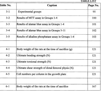
1-1 Stanmore custom-made tumour prosthesis 13
1-2 A diagram showing dififerentiation of human mesenchymal stem cells
40
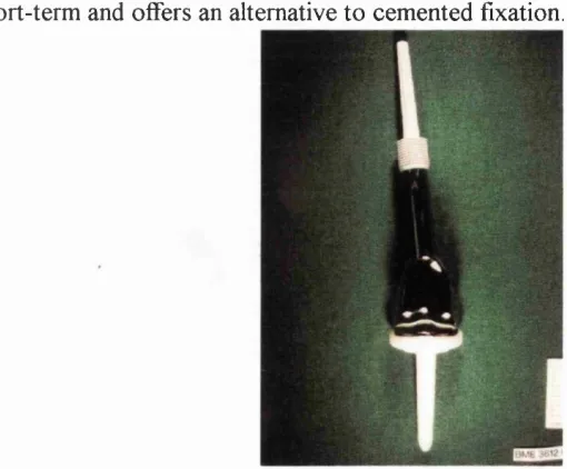
2-1 ANOVA analysis in ^H-Thymidine incorporation assay of MSCs treated with different concentrations of FGF-2 at Day 2
72
2-2 ANOVA analysis in ^H-Thymidine incorporation assay of MSCs treated with different concentrations of FGF-2 at Day 7
73
2-3 ANOVA analysis in ^H-Thymidine incorporation assay of MSCs treated with different concentrations of FGF-2 at Day
14
73
2-4 ANOVA analysis in ^H-Thymidine incorporation assay of MSCs treated with different concentrations of FGF-2 at Day
19
74
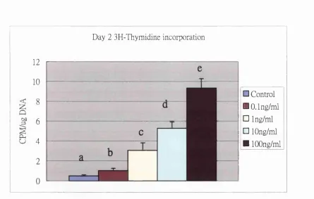
2-5 ANOVA analysis in alkaline phosphatase expression o f MSCs treated with different concentrations of FGF-2 at Day 2
75
2-6 ANOVA analysis in alkaline phosphatase expression of MSCs treated with different concentrations of FGF-2 at Day 7
75
2-7 ANOVA analysis in alkaline phosphatase expression of MSCs treated with different concentrations of FGF-2 at Day 14
76
2-8 ANOVA analysis in alkaline phosphatase expression of MSCs treated with different concentrations of FGF-2 at Day 19
76
2-9 Type 2 collagen immunostaining at Day 11 in 0.1 ng/ml group 77 2-10 Type 2 collagen immunostaining at Day 11 in 1 ng/ml group 77 2-11 Type 2 collagen immunostaining at Day 11 in 10 ng/ml group 78 2-12 Type 2 collagen immunostaining at Day 11 in 100 ng/ml group 78 2-13 Type 2 collagen immunostaining at Day 11 in the control
group
78
2-14 ANOVA analysis in ^H-Thymidine incorporation of MSCs treated with different exposure time of FGF-2 at Day 7
79
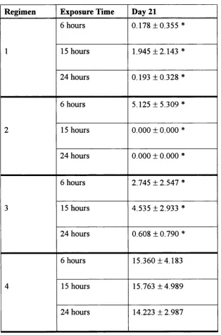
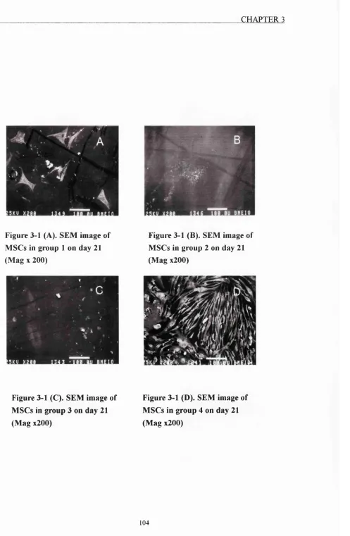
3-1(A) Figure 3-1 (A). SEM image o f MSCs in group 1 on day 21 (Mag x200)
3-l(B) Figure 3-1 (B). SEM image of MSCs in group 2 on day 21 (Mag x200)
104
3-l(C) Figure 3-1 (C). SEM image of MSCs in group 3 on day 21 (Mag x200)
104
3-l(D ) Figure 3-1 (D). SEM image of MSCs in group 4 on day 21 (Mag x200)
104
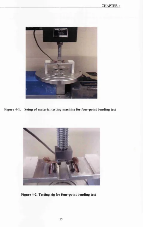
4-1 Setup of material testing machine for four-point bending test 115
4-2 Testing rig for four-point bending test 115
4-3 Testing Rig for ultimate torsion test 116
4-4 Bone fractured under torsional load 116
4-5 Separation of distal femur physis under shear load 116 4-6 An example of load-deformation curve of a four-point bending
test in the adult control group
119
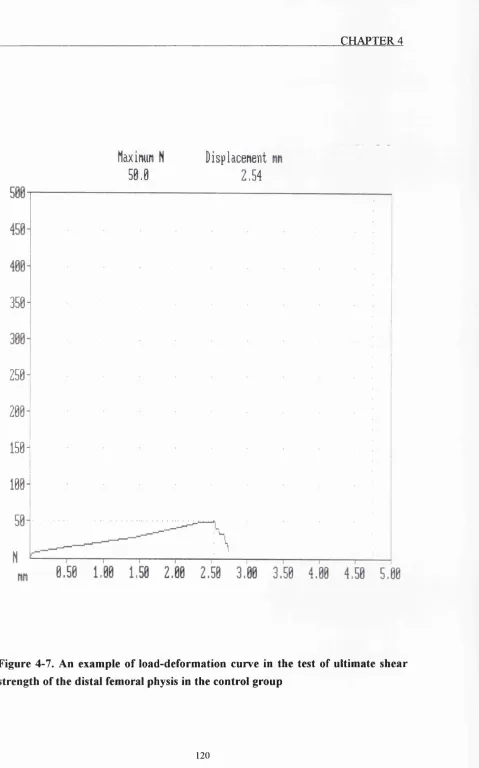
4-7 An example o f load-deformation curve in the test of ultimate shear strength of the distal femoral physis in the control group
120
4-8 An example of histology o f the growth plate in the control group (Mag xlOO)
122
4-9 An example of the histology o f the growth plate in the chemotherapy group (Mag xlOO)
122
5-1 A photograph showing the external skeletal fixator used in this study
133
5-2 DEXA scanning 134
5-3 Mounting device for DEXA scan 134
5-4 A radiograph showing bone regeneration three weeks after the operation in the control group
136
5-5 A radiograph showing bone regeneration three weeks after the operation in the chemotherapy group
136
5-6 A radiograph showing bone regeneration five weeks after the operation in the control group
137
5-7 A radiograph showing bone regeneration five weeks after the operation in the chemotharapy group
5-8 Histology o f the regenerated tissue in the osteotomy gap in the contorl group five weeks after the operation (Mag xl60)
138
5-9 Histology o f the regenerated tissue in the osteotomy gap in the chemotherapy group five weeks after the operation (Mag xl60)
138
5-10 Bone mineral density in the osteotomy gaps o f the rats treated and not treated with chemotherapy
139
6-1 A photograph showing lateral incision for femoral bone exposure
150
6-2 A photograph showing that soft tissues were removed and the femur bone was retrieved
150
6-3 Tisseel® kit 154
6-4 Injection apparatus o f Tisseel® 154
6-5 A photograph showing the external skeletal fixator used in this study
158
6-6 Rat mesenchymal stem cells (Paasage 1, Mag xlOO) 159 6-7 SEM image o f rat mesenchymal stem cells (Mag x330) 159
6-8 Alamar blue assay o f MSCs in fibrin glue 161
6-9 ^H-Thymidine incorporation assay of MSCs in fibrin glue 161 6-10 SEM image o f MSCs in fibrin glue for 24 hours (Mag x600) 162 6-11 SEM image of MSCs in fibrin glue for 96 hours (Mag x600) 162 6-12 Histology o f MSCs in fibrin glue for 24 hours (Mag x 600) 163 6-13 Histology o f MSCs in fibrin glue for 96 hours (Mag x 600) 163
6-14 Histology showing dividing MSCs (Mag x 600) 163
6-15 TEM image of MSCs in fibrin glue for 24 hours (Mag x20,000)
164
6-16 TEM image o f MSCs in fibrin glue for 96 hours (Mag x20,000)
164
6-17 radiograph showing bone regeneration in Group 1 [Mag x20)
6-18 A radiograph showing bone regeneration in Group 2 (Mag x20)
168
6-19 A radiograph showing bone regeneration in Group 3 (Mag x20)
169
6-20 A radiograph showing bone regeneration in Group 4 (Mag x20)
169
6-21 A radiograph showing bone regeneration in Group 5 (Mag x20)
170
6-22 A radiograph showing bone regeneration in Group 6 (Mag x20)
170
6-23 ANOVA analysis o f DEXA scan results (g/cm^) at the osteotomy gap three weeks after the operation
171
6-24 ANOVA analysis of DEXA scan results (g/cm^) at the osteotomy gap five weeks after the operation
171
6-25 A photomicrograph showing bone regeneration in Group 1 (Mag X100)
172
6-26 A photomicrograph showing bone regeneration in Group 2 (Mag xlOO)
172
6-27 A photomicrograph showing bone regeneration in Group 3 (Mag xlOO)
173
6-28 A photomicrograph showing bone regeneration in Group 4 (Mag xlOO)
173
6-29 A photomicrograph showing bone regeneration in Group 5 (Mag X100)
174
6-30 A photomicrograph showing bone regeneration in Group 6 (Mag xlOO)
174
6-31 Quantitative histological analysis of percentage bone formation at the osteotomy gap
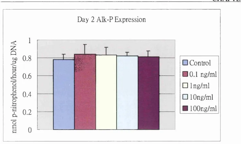
Table No. Caption Page No.
3-1 Experimental groups 95
3-2 Results of MTT assay in Groups 1-4 100
3-3 Results of alamar blue assay in Groups 1-4 101
3-4 Results of alamar blue assay in Groups 5-11 102
3-5 Results of alkaline phosphatase assay in Groups 1-4 103
4-1 Body weight o f the rats at the time of sacrifice (g) 121
4-2 Ultimate bending strength (N) 121
4-3 Ultimate torsional strength (N) 121
4-4 Ultimate shear strength of distal femoral physis (N) 121
4-5 Cell numbers per column in the growth plate 121
Chapter One
AN INTRODUCTION TO THE TREATMENT OF BONE
TUMOURS AND TISSUE ENGINEERING IN BONE
1.1 Limb Salvage for treatment of bone cancer
1.2 Fixation of hydroxyapatite-coating cementless bone tumour implants
1.3 Chemotherapy for malignant bone tumours
1.4 Tissue engineering in bone regeneration 1.4.1 What is tissue engineering? 1.4.2 Mesenchymal stem cells
1.4.3 Scaffolds for bone tissue engineering 1.4.4 Growth factors
The aims of the thesis are to investigate the hypotheses that:
(1) Chemotherapy drugs, at their therapeutic concentrations, are detrimental to
the proliferation and osteogenic differentiation of mesenchymal stem cells.
(2) Chemotherapeutic agents influence the mechanical properties of the skeletal
tissues.
(3) Chemotherapy treatment slows down the rate of bone regeneration.
(4) Use of mesenchymal stem cell-based tissue engineering approach facilitates
bone regeneration when chemotherapy agents are systemically administered.
The rationale for developing the hypotheses will be introduced in the following
sections. Essentially, the motive of this thesis stems from the observation of a clinical
dilemma - reconstruction of bone defects after removal o f bone in patients with
skeletal neoplasm of the extremities. It is hoped that at the end o f this thesis, a
1.1 LIMB SALVAGE FOR TREATMENT OF BONE CANCER
Skeletal neoplasm is a rare but difficult disease (Simon 1991). Historically, the only
treatment of skeletal neoplasm o f the extremities was amputation of the limb. With
the advances in the treatment of bone sarcoma, it was then realised that not all bone
cancers need an amputation. Preservation of the involved limb after surgical removal
o f the tumour has been performed for more than a century for bone sarcomas of low
and moderate grade (Simon 1988). In the past two decades, advances in adjuvant and
neo-adjuvant chemotherapy treatment, in diagnostic imaging and in the surgical
technique for reconstruction of the limbs have led to the performance of limb salvage
surgery for most patients who have high-grade sarcomas (Choong and Sim 1997).
It is important to understand the significance o f grading and staging of bone tumours
because this relates to prognosis. Grading o f the bone tumours means the degree of
malignancy o f the cancer cells while staging o f the bone tumours refers to the extent
o f involvement o f the tumour (Bolling and Beauchamp 1999). Most important of all,
staging and grading are important indictors of prognosis (Bentzen 2001). The higher
the histological grading, the more malignant the tumour cells. The most extensively
used surgical staging system for musculoskeletal sarcomas is the Enneking staging
surgical reconstruction will be because higher surgical grade implies greater
involvement of the tumours and results in wider surgical margins.
The difficulty o f limb salvage not only lies in the size o f the bone defects created after
surgical removal o f the tumours, but also in the locations of the tumours, and in the
age of the patients (Nichter and Menendez 1993). For example, the most common
sites for osteosarcoma, the most common primary malignant bone tumours, are the
distal femur and proximal tibia (Jaffe 1991). Therefore, the distal femoral and
proximal tibial articular surfaces often have to be removed and reconstruction of a
painless, free-mobile joint which is durable is challenging (Lindell and Carroll 1993).
A large number of patients with malignant bone tumours are skeletally immature
(Finn and Simon 1991). Removal of the growth plates of the involved limb will result
in limb length discrepancy as the patient grows and this has to be addressed (Frassica
1997).
When considering whether surgery for limb-salvage is justified, it is usual to consider
long-term oncological results after limb salvage and compare them with the historical
results after amputation. Comparison can be made in four broad areas; (1) the overall
the function of the salvaged limb and its maintenance over a prolonged period of
follow-up and (4) the quality of life in patients (Cannon 1997).
Various methods have been developed for limb salvage reconstruction after resection
of bone tumours. Arthrodesis, particularly for tumours around the knee, has
previously been used. Although the knee joint is not reconstructed, the function o f the
lower extremity can be reasonably preserved. Vascularised or non-vascularised
autologous bone graft, usually part of the fibula, or allograft, is used to reconstruct the
bone stock. Although some succèssftil long-term results have been reported (Salai
1997), non-union, graft fracture, failure of fixation of the graft-host bone junction and
infection are the main problems associated with massive bone grafts (Scarborough
and Helmstedter 1997). Leg length discrepancy, which occurs in skeletally immature
patients, cannot be compensated by arthrodesis. Consequently, arthrodesis of the knee
joint should only be used in selected cases nowadays.
Osteoarticular segmental allograft reconstruction of the resected bone segments is
another method o f reconstruction which has been used previously. The advantage o f
this method is that articular surface cartilage is replaced and long-term success has
reported, however, that although osteoarticular allografting is an alternative to
amputation, it presents unsolved immunologic and preservation problems which make
the prognosis unpredictable (Alho 1991). Scintigraphic results also showed that it
takes a long time for revascularisation o f the osteoarticular allograft (Bar-Sever 1997).
The risk o f graft fracture can not be eliminated and long-term durability of the graft is
still doubtful (Rodl 2000). It has been reported that although the current results of this
technique are adequate, they are imperfect, and research should be directed at
improving the outcome (Mankin 1996). Instability, degeneration, or a fracture near the
articular surface of the graft are the complications o f osteoarticular allografts and
subsequent total knee arthroplasty are sometimes necessary as salvage procedures
(DeGroot and Mankin 2000).
Distraction osteogenesis has also been used for reconstruction o f bone defects in limb
salvage surgery. It has been reported that distraction osteogenesis is useful in limb
salvage surgery for reconstruction o f bone defects (Tsuchiya 1997). However,
distraction osteogenesis can only be used in patients whose articular surfaces can be
preserved because it is impossible to regenerate articular cartilage simply by
mechanical distraction. Hence, the application o f this technique is limited and it can
nature of their disease, require almost immediate rehabilitation to maximise their
quality of life. Due to size of the defects and the complicated technique as well as
patient requirement, distraction osteogenesis is often not suitable for bone tumour
patients.
Extracorporeal irradiation of the resected bone segment which contained tumour with
very high dose irradiation and then re-implanting the segment has also been reported
as a successful method for reconstruction in limb salvage surgery. This technique was
first described by a group of Belgian surgeons (Uyttendaele 1988). Subsequently,
some clinical success in using this technique has been reported (Araki 1999, Hong
2001). Extracorporeal irradiated autograft can be used as an alternative in the
countries where allografts and prostheses are not easily available. However, there are
several disadvantages in using irradiated bones for reconstruction. If the resected bone
segment which contained tumour is irradiated and re-implanted, it is impossible to
perform a thorough pathological examination of the tumour. The information obtained
from pathological examination is important for deciding post-operative adjuvant
chemotherapy regime and for predicting the prognosis. Besides, the mechanical
properties of the bones will be influenced after high-dose irradiation and the strength
technetium-99m-MDP scintigraphy to evaluate the vascularisation and integration of
the irradiated autografts, it is found that revascularisation and partial bone ingrowth
did not lead to a lower complication rate (Van Laere 1998). Finally, animal
experiment showed that articular cartilage underwent degeneration after
extracorporeal irradiation (Sabo 2000).
Autoclaving (Smith and Struhl 1988) and microwave heating (Liebergall 1998) of the
bone segments have also been used. The disadvantages o f autoclaving and microwave
heating are similar to extracorporeal irradiation. The heat emitted during autoclaving
and microwave heating would ftirther damage the autografts (Liebergall 2000). Again,
these techniques should only be used when there are no alternatives.
Allograft-prosthesis composite (APC) has also been used for reconstruction,
particularly, of knee joints (Harris 1994). Compared to other techniques, APC
arthroplasty has many advantages, including restoration o f bone stock, customisation
with conventional implant components, soft tissue attachment of tendons and
ligaments, and preservation of the medullary canal of the host bone. The
disadvantages o f this technique include slow healing in the presence of chemotherapy,
Besides, in a carefully performed sequential cohort study, the results o f this technique
seem to be inferior to massive prosthetic replacement and this comparative study
suggested that limb salvage surgery using a tumour prosthesis has a better and more
predictable outcome (Wunder 2001).
Reconstruction with massive prosthesis has a number o f advantages, including a
relatively simple technique, the modularity the prosthesis offered, immediate function,
and, no worry o f disease transmission. (Morris 1997). In the Royal National
Orthopaedic Hospital Trust, one of the two centres in the United Kingdom that treat
bone tumour patients, custom-made massive prostheses have been used for
reconstruction o f bone defects for more than two decades. The results o f the first 218
cases o f distal femoral custom-made massive prostheses for limb salvage (Unwin
1993) revealed that the overall probability of one of these early prostheses surviving
for 10 years was 65%, and for 20 years it was 53%. For cases o f bone tumour, the
probability o f surviving for 10 years was 68% (not including recurrence), for primary
knee replacement cases it was 32%, for other bony diseases plus trauma it was 86%,
and for cases o f revised massive prostheses it was 53%. It was concluded that this
type o f distal femoral arthroplasty provided good medium-term results, but showed
cement was used for implant fixation. The overall loosening rate was 5% in an
average follow-up period of 58 months. The results o f Stanmore extendible
endoprostheses for the skeletally immature also revealed that aseptic loosening was
the major cause o f implant failure (Unwin 1996a). Aseptic loosening was identified as
the predominant cause of implant related failure in a retrospective study of a
consecutive series o f 168 Stanmore custom made extendible endoprosthetic
replacements used in skeletally immature patients. Most of the replacements were
used in the treatment of bone tumor and the remainder for the revision o f failed
massive endoprosthetic replacements. Since the first Stanmore extendible
endoprosthesis was inserted in 1976, four types of extension mechanisms have been
used. Thirty-eight of the 164 cases with follow-up data were revised, of which 19
were as a result of aseptic loosening. Survival analysis revealed that the overall
probability o f surviving an implant related failure was 0.512 (+/- 0.005) at 5 years,
highlighting the high complication rate of these extendible replacements that required
a revision procedure. Sixteen of the 19 aseptic loosening cases were distal femoral
replacements. The probability o f a patient with a distal femoral replacement surviving
aseptic loosening was 0.773 (+/- 0.008) at 5 years.
(Unwin 1996b), it was found that the probability o f a patient surviving aseptic
loosening for 120 months was 93.8% for a proximal femoral replacement, 67.4% for a
distal femoral prosthesis and 58% for a proximal tibial implant. In patients with distal
femoral replacements the age of the patient at the time o f operation and the percentage
of bone resected were related to the risk of aseptic loosening. Young patients with
distal femoral prostheses in whom a high percentage o f the femur had been replaced
had the poorest prognosis for survival. The percentage of bone removed had a
significant effect in the proximal tibial replacement group, but the age of the patient
did not. By contrast, neither the age nor the percentage of bone removed was a factor
after proximal femoral replacement.
To combat the high incidence of aseptic loosening for young patients and for patients
with failed implants after resection for bone tumors, intramedullary cementless
fixation o f massive tumor implants was investigated (Blunn 2000). These implants
consist of a hydroxyapatite coated titanium stem (Figure 1-1). To date, 47 of these
prostheses have been inserted for the treatment o f primary bone tumors. Radiographs
indicate that the stems are osseointegrated. Radiolucent lines have not been seen
between the implant and the bone. Bone remodelling changes have been observed. In
bone grew to the shoulder of the implant. Bone remodelling was particularly evident
in stems that were coated over their entire surface. In these cases, the implant induced
local bone resorption so that the bone around the mid-stem region became thinner,
with resorption of cortical bone on the periosteal surface and maintenance of bone on
the endosteal surface adjacent to the stem. This effect was attributed to stress
shielding, and a three-dimensional finite element model using loading data obtained
from a telemetry study indicated that, where the stem was bonded to the bone over the
entire surface, stresses in the outer cortex became reduced. In the finite element model,
reducing the region of hydroxyapatite coating to approximately 1/3 of the stem length
reduced the extent of the low-stress area in the outer cortex. Subsequently, prostheses
have been coated with hydroxyapatite over only approximately 1/3 of their stem. This
method of fixing the massive endoprosthesis to the bone is thought to be successful in
the short-term and offers an alternative to cemented fixation.
1.2 FIXATION OF HYDROXYAPATITE-COATING CEMENTLESS
BONE TUMOUR IMPLANTS
A prerequisite for any orthopaedic arthroplasty or implant is permanent fixation to the
surrounding skeleton with no intervening soft tissue (Sun 2001). A successful fixation
should be generated quickly, as soon as possible after surgery. It should be strong,
able to transmit everyday loading activities and it should exhibit life long stability.
Fixation takes place by osseointegration, which was first described by Branemark
(1983) as the intimate contact between a titanium implant interface and the
surrounding bone. The current accepted definition for osseointegration is “contact
established between normal and remodelled bone and an implant surface without the
interposition of non-bone or connective tissue, at the microscopic level” (Mentag
1986)
Prostheses have been implanted into the human body by either cemented or
cementless fixation methods. Although the traditional cemented fixation using
polymethylmethacrylate (PMMA) bone cement can obtain immediate stability
between the implant and bone, this type of prosthesis is not suitable for young, active
patients in whom more stable fixation and bone growth are needed. Problems o f cell
necrosis from the exothermic reaction caused by the heat emitted during
bone are still a concern when using the intramedullary cement (Galante and Jacobs
1992). Thus, cementless fixation, primarily by biological means whereby press-fit
insertion is followed by bone growth into a porous surface, has been developed.
However, there is little histological evidence o f sufficient bone ingrowth in retrieved
uncemented porous-coated prostheses (Pidhorz 1993, Bloebaum 1997) and it has been
shown that bone must be within 50 pm of the porous coating for ingrowth to occur
(Bloebaum 1994). Meanwhile, fibrous rather than bony ingrowth into porous surfaces
has been found, and the loss of endosteal bone still exists (Bloebaum 1994).
Bioactive materials such as HA and bioactive glass can stimulate a direct bond to
form between the implant and the surrounding bone and improve osseointegration
(Geesink 1987). This bone-implant bonding is one o f the most important factors for
implant fixation and function. HA coatings have been shown to achieve a very strong
bond with living bone, even under loaded conditions and with the presence of a gap
(Soballe 1991, Soballe 1992, Soballe 1993).
Cementless fixation with HA coating in Stanmore custom-made tumour prosthesis has
gained encouraging short-term success (Blunn 2000) and it is the current design of
coatings on metallic implants in orthopaedics since the mid-1980’s (Geesink 1989,
Furlong and Osborn 1991). The advantages that are sought in this application include
(1) more rapid fixation and (2) increased and more uniform bone ingrowth and/or
ongrowth at the bone-implant interface (Geesink 1988, Stephenson 1991, Cook 1992).
Most clinical experiences with either weight-bearing or non-weight-bearing models
have shown promising results shortly after the implantation and continued fixation for
up to 10 years (Capello 1997, Donnelly 1997, Magyar 1997, Neilssen 1998).
What is more exciting is that HA coating can enhance bone growth across a gap of
one mm between the bone and the implant in both stable and unstable mechanical
conditions, and it is capable o f limiting the formation o f any fibrous membrane and
converting a motion-induced fibrous membrane into a bony anchorage (Soballe 1991,
Soballe 1992, Soballe 1993). It has also been suggested that HA coatings have sealed
the bone-implant interface, preventing migration of polyethylene particles which may
reduce the incidence o f osteolysis and the subsequent implant failure (Rahbek 1996,
Soballe and Overgaard 1996, Coathup 2001).
Cementless fixation with hydroxyapatite-coated implants is dependent on the
recruitment, proliferation, differentiation and matrix production o f bone cells and
osteoprogenitor precursor cells (Ohgushi and Caplan 1999). Since chemotherapy has
become standard treatment for malignant bone tumours, the influences of
chemotherapy on osteoprogenitor cells should be taken into consideration in terms o f
longevity and stability o f implant fixation o f the massive tumour prostheses. It is
reported that cisplatin, one of the most commonly used chemotherapy drugs for
malignant bone tumours (Souhami 1997), caused a temporary delay in the formation
of new bone around the prosthesis in a canine diaphyseal segment replacement model
(Young 1997). The effects of chemotherapy on osteoprogenitor cells, i.e.,
mesenchymal stem cells (MSCs) which are contained in bone marrow, remain unclear.
It would be valuable to elucidate the consequences o f chemotherapy on MSCs.
Besides hydroxyapatite coating o f the intramedullary stem, there is also a
hydroxyaptite-coating “collar” design in Stanmore MARK 5 custom-made tumour
prostheses (Blunn 2000). The purpose of adding a hydroxyapatite-coating at the
junction o f the prosthesis adjacent to the transection site is to induce extracortical
bone bridging and to increase implant fixation. This has been shown to significantly
reduce the incidence o f aseptic loosening. However, bony bridging occurs in only
collar is limited. These implants are implanted into patients who are receiving
neoadjuvant chemotherapy and this may affect bone formation. So, whether and to
1.3 CHEMOTHERAPY FOR MALIGNANT BONE TUMOURS
It has been proved that chemotherapy substantially improves the disease-free and
overall survival rates in patients with malignant bone tumours (Eilber 1987). In a
prospective, randomized trial (Link 1986), after undergoing surgery, 36 patients were
randomly assigned to a group receiving adjuvant chemotherapy or a group without
adjuvant treatment. At two years the actuarial relapse-free survival was 17 percent in
the control group, similar to that found in studies before 1970 (Miser 1985), and 66
percent in the adjuvant-chemotherapy group (P less than 0.001). Similar results were
observed among 77 additional patients who declined to undergo randomisation but
who elected observation or chemotherapy. The authors concluded that adjuvant
chemotherapy increases the chances of relapse-free survival o f patients with
high-grade osteosarcoma, and recommended that it should be given to all such
patients.
In order to assess the optimum duration o f treatment and the relative contributions o f
the constituent drugs in randomised trials, the European Osteosarcoma Intergroup
(EOI) was formed in 1982 to carry out randomised studies o f sufficient size to allow
investigation o f important features o f treatment (Souhami 1997). In the first EOI trial,
to be efifective as an adjuvant chemotherapy regime for osteosarcoma (Ettinger 1986).
Three hundred and seven patients with osteosarcoma were randomly assigned one or
two regimens of chemotherapy (Bramwell 1992). A regimen of cisplatin and
doxorubicin, given before and after surgery for a total of six cycles to patients with
operable non-metastatic osteosarcoma, produced 5-year survival o f 64% and
progression free o f 57%. These results were similar with those of trials (Link 1986,
Winkler 1984) in which chemotherapy was based on the TIO regimen (Rosen 1982).
The TIO regime, which had been the basis o f previous chemotherapy regimes, was a
combination of seven chemotherapeutic drugs given over a period of 44 weeks. A
report o f the results of TIO regimen showed that 65% o f 279 patients were alive and
free of disease at 8 years. However, non-randomised studies of treatment did not
allow unbiased comparison of outcomes (Souhami 1997) and the EOI undertook a
formal comparison o f a 44-week multi-drug TlO-based regimen with the 18-week
cisplatin and doxorubicin two-drug regimen. The results showed that in the 407
randomised patients, 391 were eligible and have been followed up for at least 4 years.
Toxic effects were qualitatively similar with the two regimens. However, 188 (94%)
of 199 patients completed the six cycles of two-drug treatment, whereas only 97 (51%)
of 192 completed 18 or more of the 20 cycles o f the multi-drug regimen. The
preoperative chemotherapy was about 29% with both regimens and was strongly
predictive of survival. Overall survival was 65% at 3 years and 55% at 5 years in both
groups. It was found that there was no difference in survival between the two-drug
and multi-drug regimens in operable, non-metastatic osteosarcoma. The two-drug
regimen is shorter in duration and better tolerated, and is therefore the preferred
treatment (Souhami 1997).
In Europe the two-drug regimen with cisplatin and doxorubicin has become the gold
standard for treatment of malignant bone tumours such as osteosarcoma. It is
therefore important to understand the toxicity that this regimen causes in the skeletal
tissues of the patients who receive the treatment. Because of the systemic
administration of these drugs, normal skeletal tissues are also exposed and it is not
difficult to imagine that normal skeletal tissues and cells are also affected. However,
the toxic effects o f combined cisplatin and doxorubicin use on skeletal tissues have
not previously been studied. Therefore, it is valuable to quantitatively assess the
detrimental effects of the two-drug regimen on skeletal tissues as well as
mesenchymal stem cells (MSCs). MSCs play an important role in achieving bone
ingrowth around the cementless implants. It is important to quantitatively assess
of new tissue engineering therapies to treat segmental bone defects is healthy and
viable MSCs or bone cells. This may be an extremely valuable tool but the
effectiveness of these new techniques needs to be investigated in realistic
1.4 TISSUE ENGINERING IN BONE REGENERATION
1.4.1 WHAT IS TISSUE ENGINEERING?In order to create tissues and organs for replacement, understanding the processes of
repair and regeneration is essential. The regeneration and repair o f tissues are
fundamentally different processes (Caplan and Goldberg 1999). Regeneration of
tissues readily occurs in embryos, is almost absent in neonates (although some
regenerative events have been reported), and is never observed in adults (Whitby and
Ferguson 1991). Regeneration is a relatively slow process that seems to recapitulate
many but not all the steps that occur in the embryos. In contrast, repair is rapid and
has been evolutionarily selected to minimise the animal’s vulnerability, that is, to get
the animal away from danger as soon as possible (Armstrong and Ferguson 1995).
Likewise, the initial steps of recovery from surgical events or wounding involve an
acute inflammatory response and a sealing off o f the repair site, to minimise the
spread of bacteria or fungal contamination o f the wound site, and to provide the rapid
fill o f that tissue deficit with fibrous or bridging tissue (Thornton 1968). Later, after
the animal is safe, slower turnover process occurs to attempt to remodel the tissue into
its natural state (Caplan and Goldberg 1999). Regeneration o f a tissue re-establishes
the age dependent turnover dynamics and can include the capacity of that regenerated
growth and provides tissue with only short-term benefit.
It was not until the late 80’s that tissue engineering was regarded as an independent
branch of science. The term tissue engineering was initially defined by the attendees
of the first National Science Foundation of the United States sponsored meeting in
1988 as “application of the principles and methods o f engineering and life sciences
toward fundamental understanding of structure-function relationship in normal and
pathologic mammalian tissues and the development of biological substitutes for the
repair and regeneration of tissue or organ function” (Chapekar 2000). In 1993, Langer
and Vacanti summarized the early development in this field and defined tissue
engineering as “an interdisciplinary field that applies the principles o f engineering and
life sciences towards the development of biological substitutes that restore, maintain,
or improve tissues or organ function (Langer and Vacanti 1993). The exercise of
interdigitating these different functional talents into a coherent device has produced
the working definition o f tissue engineering (Brekke and Toth 1998); “Tissue
engineering is an art and science by which synthetic compounds are manipulated into
anatomically and/or functionally specific architectures and, when required, may be
integrated with biologically active agents and/or living cells such that resultant
for recipient tissues”. Consequently, tissue engineering has now emerged as a
potential alternative to tissue or organ transplantation. With this technology, tissue
loss or organ failure can be treated either by implantation of an engineered biological
substitute or alternatively with ex vivo perfijsion systems. The tissue-engineered
products may be fully functional at the time of treatment (e.g., liver assist devices,
encapsulated pancreatic islets), or have potential to integrate and form the expected
functional tissue upon implantation (e.g., chondrocytes embedded in a matrix carrier).
In some cases, biomaterials are modified to enhance migration and attachment of the
specific cell populations, which repair or replace the damaged tissue (Chapekar 2000).
As the definitions suggest, cells are a key to tissue regeneration and repair due to their
proliferation and differentiation, cell-to-cell signalling, biomolecule production, and
formation o f extracellular matrix. The most fundamental and crucial reason is that it is
still impossible to create one single living cell from lifeless materials up to now. The
functionality of an engineered tissue may be structural (e.g., bone, cartilage, skin), or
metabolic (e.g., liver), or both. Cells may be part of an engineered tissue or
alternatively, these cells may be recruited in vivo with the help of biomaterials and/or
biomolecules. When selecting the cellular component o f an engineered product, it is
source. Relatively easy access o f the cell source(s) and abundant supply of target cells
are other considerations. In addition, expansion of these cells without permanently
altering the phenotype and fonction during the expansion phase and without
introduction o f any adventitious and species-specific bacterial/viral agents poses
significant challenges. Finally, when genetically modified cells are used in a
tissue-engineered product, there are additional concerns such as cell transformation by
the vector, vector stabihty, and optimal fonction o f the inserted gene (Chapekar 1996).
Scaffolds are also indispensable in tissue engineering. Actually, tissue-engineering
devices can be looked upon as a three dimensional (3-D) in vivo cell culture system
designed to perform a plethora of vital fonctions required for producing a directed
host response (Brekke and Toth 1998). Both engineering and biologic issues must be
taken into consideration in order to maximise the fonction of the tissue-engineered
constructs in vivo. The gross mechanical properties of the construct, structural
qualities of its internal fabrics and its 3-D architectural geometry are important.
Retention o f mechanical characteristics after implantation and possession of a 3-D
internal geometry enable these devices to maintain a tissue void, or a space, of the
prescribed size and shape for future tissue occupancy. This is especially important in
decades that bone will grow into an adjacent tissue void if that space can be
maintained and protected from encroachment by non-osseous tissue and competing
cell types (Hurley 1959; Linde 1993; Levy 1994). This is known as guided bone
regeneration.
A scaffold’s internal, 3-D geometry links its engineering properties to those qualities
impacting host response. Architecture defines a unit’s internal spatial arrangements
which, in turn, contribute substantially to its mechanical characteristic limits. By
governing the scaffold’s internal spatial arrangements, the architecture design also
determines quantity and shape o f substratum surfaces available for colonization by in
vitro cell seeding or endogenous cell populations. A construct’s apparent density,
number and size of internal chambers and total void volume determine the size of
individual cells and the cell population densities that the unit can accommodate
(Brekke and Toth 1998). Most important of all, the scaffolds must allow the delivery
of morphogenetic and regulatory proteins and peptides (Boden 1995; Hollinger
1996a). Moreover, incorporation o f signal peptides into the material has been
attempted to effectively mimic the extracellular matrix and induce cell migration
(Grzesiak 1997; Rowley 1999). The mechanical strength o f the scaffold material
replace. Moreover, material porosity as well as pore size distribution and continuity
greatly influences the attachment of specific cell types and interaction of the
biomaterials with the host. It is also preferable that the biomaterial degrades in vivo to
minimise the long-term biocompatibility concerns, with the material degradation rate
matching the regeneration rate of the tissue. The resulting degradation products must
be non-toxic to the host. Lastly, vascularisation of the tissue-engineered is critical for
the three-dimensional constructs greater than Imm^ to meet their nutritional and
metabolic requirements (Shea 1999).
Besides cells and scaffolds, cell signalling is also crucial in the tissue engineering
constructs in order to guide the cells in the scaffold to grow and/or differentiate into
desired cell/tissue types. The best known biomolecules are a group of proteins from
transforming growth factor- y3 (TGF- ) superfamily called bone morphogenetic
proteins (BMPs) (Urist 1965; Urist and Strates 1971). There are several strategies for
delivery o f osteoinductive growth factors (Boden 1999). First, extracted human
(Johnson 1988; Johnson 1992; Teixeira and Urist 1998) or animal growth factors
(Sciadini 1997a; Sciadini 1997b) can be delivered with scaffolds or direct injection.
Using recombinant DNA technology, recombinant osteoinductive proteins can be
called osteogenic protein-1, or OP-1 (Asahina 1993; Sampath 1992). The third
strategy for delivery o f cell signalling biomolecules in bone tissue engineering is the
use o f gene therapy. The concept involves the delivery o f the gene or cDNA sequence
for an osteoinductive factor rather than the delivery of the factor itself (Fang 1996;
Riew 1998). Again, various safety and efficacy issues remain to be explored. The
choice o f specific molecules, the correct dose, timing, and sequence of administration,
whether singly or in combination, still must be defined for each application and for
the specific mechanical and biological requirements. The development o f optimal
delivery methods for these molecules is another important question to answer.
1.4.2 MESENCHYMAL STEM CELLS
The middle embryonic layer, the mesoderm, gives rise to all o f the body’s skeletal
elements. The term mesenchyme is derived from the Greek meaning “middle (meso)
infusion” and refers to the ability of mesenchymatous cells to spread and migrate in
early embryonic development between the ectodermal and endodermal layers. It is
generally agreed that in the mesoderm of an embryo a mesenchymal stem cell is a
pleuripotent progenitor cell which divides many times and whose progeny eventually
gives rise to skeletal tissues such as bone, cartilage, tendon and ligament. By
divisions (Caplan 1991). These cells have the capacity for extensive replication
without differentiation, and they possess a multilineage development potential
(Bruder and Fox 1999). Recently, pleuripotent stem cells have been cultured from
human foetal tissue and have shown the ability to give rise to a variety o f cell types
found in embryonic germ layers (Shamblott 1998; Thomson 1998). Many adult
tissues contain populations of stem cells that have the capacity for renewal after
trauma, disease or ageing. The cells may be found within the tissues or in other tissues
that serve as stem cell reservoirs. For example, although bone marrow is the major
source o f adult haematopoietic stem cells (HSCs) that renew circulating blood
elements, these cells can be found in other tissues, such as peripheral blood, as well.
The adult bone marrow also contains mesenchymal stem cells (MSCs), which
contribute to the regeneration of different mesenchymal tissues such as bone, cartilage,
muscle, ligament, tendon and adipose tissues by various investigators (Beresford 1989;
Haynesworth 1992b; Wakitani 1994; Bruder 1998a; Yoo 1998; Johnstone 1999). In
vitro and animal studies have indicated that there is either a multipotent MSG or the
populations are the mixtures o f committed progenitor cells, each with restricted
potential (Wakitani 1995; Bergman 1996; Cassiede 1996; Young 1998; Awad 1999;
Petite 2000). Recently, multi-potential human mesenchymal stem cells (hMSCs),
manipulation, these hMSCs from the same donors could become osteoblasts,
chondrocytes and adipocytes (Pittenger 1999).
As these MSCs are harvested from bone marrow and bone marrow is easily obtained,
the understanding o f bone marrow, in particular, the stromal cell system, is important.
There has been an increasing interest in recent years in the stromal cell system, which
includes the marrow-derived stromal cell that supports haematopoiesis, as well as the
mesenchymal stem cell and its progeny, connective tissue cells such as osteoblasts,
chondrocytes, tenocytes, adipocytes and smooth muscle cells. This was first described
by Owen (1985). Essentially, there are three main cellular systems in the bone marrow;
haematopoietic, endothelial and stromal (with stromal cells loosely referring to the
non-haemopoietic cells o f mesenchymal origin; Deans and Moseley 2000). The
stromal system, as proposed by Owen (1985), was based on an analogy with the
haematopoietic system, in which MSCs reside within the marrow, maintain a level o f
self-renewal, and give rise to cells that can differentiate into different connective
tissue lineages as well as stromal tissues (Owen 1988).
Within the stromal environment, alkaline phosphatase positive (ALP^) reticular cells
are thought to originate from cells that are destined to differentiate into osteoblasts but
are also capable o f forming stroma. The presence o f adipocytes in the postnatal
stroma depends on the stage of skeletal development, age, and the level of
haematopoiesis (Dexter 1982). It has been suggested that ALP^ reticular cells and
adipocytes are alternative phenotypes that are modulated by the marrow environment
(Bianco 1988).
The differentiation potential o f the stromal cell system has also been widely studied
with respect to the mesenchymal connective tissues, in particular, bone tissues (Aubin
1998). Human stromal cells that had been depleted o f circulating haematopoietic cells
by negative immunoselection with antibodies against monocytes/macrophages
(anti-CD 14), endothelial cells (anti-CD31) and lymphocytes (anti-CD 11 a/LFA-1)
were shown to co-express genes characteristic o f the osteoblastic lineage (alkaline
phosphatase, osteocalcin, and osteopontin) and adipocyte lineage (lipoprotein lipase),
indicating that stromal cells were uncommitted precursor cells (Rickard 1996).
Human marrow stromal fibroblasts are capable o f forming colonies in vitro in the
presence of serum and these cells produce at least four growth factors;
platelet-derived growth factor (PDGF), basic fibroblast growth factor (bFGF),
(Kuznetsov 1997a; van den Bos 1997). Pittenger et a l (1999) reported that
approximately one-third of the initially adherent bone marrow-derived stromal
colonies are pleuripotent and capable of differentiation into the osteogenic,
chondrogenic and adipogénie lineages as demonstrated by lineage specific in vitro
assays.
Kuznetsov et al. (1997b) demonstrated that in vivo transplants of all
multicolony-derived marrow stromal fibroblasts derived from multiple in vitro
stromal cell colonies resulted in bone formation, whereas only 58.8% of fibroblasts
formed bone. The same investigators recently presented evidence for a circulating
osteogenic precursor in humans, although the circulating precursor demonstrates
greater variability in clonogenic potential than precursors derived from bone and bone
marrow. That is to say, the circulating precursors not only differentiate into bone cells,
but also are more able to commit to other cell lineages (Kuznetsov 2001). The
reported precursor frequency o f osteoblast precursors in normal human bone marrow
is approximately four per 100,000 nuclear cells (based on ALP^ staining) and appears
to correlate negatively with age and the existence of disease states such as
Mesenchymal stem cell cultures are often defined as the non-haematopoietic adherent
cell population which are obtained by direct plating of bone marrow. Marrow stroma
may have relatively simple or complex cellular compositions depending on the
growth media or plating substrate used (Deans and Moseley 2000). The majority o f
reported tissue culture conditions use relatively undefined media compositions
containing foetal calf serum or other animal sera, this limits the supply o f physiologic
signals required for efficient attachment and differentiation as well as growth.
However, ex vivo culture results in consistently reproducible stromal cell cultures that
have been evaluated in human clinical studies for support of autologous
haematopoietic engraftment (Lazarus 1995).
Standard conditions for generation o f marrow-derived mesenchymal stem cell
cultures have been reported (Bruder 1998b; Majumdar 1998). In brief, a bone marrow
aspirate is collected and processed using density gradient centrifugation, from which
light-density cells are taken and plated at a standard density in a Dulbecco’s modified
essential medium (DMEM) containing foetal calf serum. After allowing two days for
adhesion to non-coated polystyrene, non-adherent cells are removed, and a feeding
schedule established for a 14-day primary expansion period o f adherent colonies. At
expansion through sequential passages. Cells may be expanded by 40 generations and
still retain their multipotent mesenchymal lineage capability, although growth rates
become reduced. The expanded mesenchymal stem cells exhibit a finite lifetime and
do not display properties of immortalised cells (Deans and Moseley 2000).
In terms o f mobilisation o f circulating mesenchymal stem cells, investigators have
tried to detect circulating cells exhibiting the multilineage mesenchymal stem cell
phenotype. It has been described by Chesney et al. (1997) that a fibroblast-like cell,
termed fibrocyte, is found in the circulating blood. These fibrocytes are reported to be
CD34^, CD45^, CD 13 , and to be capable of synthesizing collagen. In addition, these
cells express class II HLA molecules, the co-stimulatory molecules CD80 and CD 86,
and the adhesion molecules CD IIa, CD54 and CD58. These cells are also
demonstrated to induce T-cell responses consistent with a dendritic cell function.
Previous studies from this group have shown that this circulating cell can be recruited
where it aids the tissue repair processes (Bucala 1994). Additional evidence for
circulating mesenchymal stem cell populations is provided by Ferrari et al. (1998),
who reported that the transfer of genetically marked ( galatosidase) bone marrow
could lead to genetically marked muscle cell progenitors in mice. These precursors
mature myocytes, indicating that the mesenchymal muscle progenitor is derived from
a bone marrow-derived precursor or stem cell.
In terms o f the characteristics of human mesenchymal stem cells, hMSC cultures
contain a homogenous population of fibroblast like cells which have a population
doubling time o f 33 hours and exhibit a large but variable ex vivo expansive potential
(Conget and Minguell 1999). It has been reported that while some MSG preparations
can be expanded by over 15 cell doublings, others cease replicating after about 4 cell
doublings (Bruder 1997a, Digirolamo 1999, Phinney 1999). In addition, some
investigators reported that human mesenchymal stem cells could maintain the
potential to differentiate into osteogenic lineage even after 40 doublings in culture.
Cryo-preserved MSCs are also able to differentiate into osteogenic lineage after being
thawed (Bruder 1997a, Jaiswal 1997). hMSCs that are highly expanded (>40
doublings) do loose their multipotentiality and approach senescence and at this stage
numerous apoptotic features are present in culture (Conget and Minguell 1999,
Digirolamo 1999).
Cell cycle studies on isolated human mesenchymal stem cell culture have revealed the
typical o f quiescent (GO) cells. These cells can be isolated by a negative selection
procedure using 5-fluorouracil, which selects a population of more than 90% GO cells,
expressing the gene for ornithine decarboxylase antizyme (Minguell 2000). The
resting condition, together with a selective immunophenotype and the absence of
commitment markers, gives experimental support to the hypothesis that a “rare”
mesenchymal stem cell in the bone marrow is capable of self-renewal and
differentiation into various mesenchymal lineages.
The antigen profile of MSCs is not unique, and antigens are common to mesenchymal,
endothelial, epithelial and muscle cells (Haynesworth 1992a; Galmiche 1993; Conget
and Minguell 1999; Pittenger 1999). Moreover, since hMSCs do not express typical
haematopoietic lineage markers such as CD 14, CD34 and CD45 (Conget and
Minguell 1999; Pittenger 1999), it supports the concept that the bone marrow hosts at
least two main different stem/progenitor cells which can give rise to mature
haematopoietic and mesenchymal cells (Prockop 1997 and Gerson 1999).
The extended cytokine expression profile of hMSCs, which includes several
haematopoietic and non-haematopoietic growth factors, interlukins and chemokines,
inductive and regulatory signals necessary for the development of haematopoietic
cells as well as for stromal cells, including hMSCs themselves (Haynesworth 1996;
Majumdar 1998). It has been postulated that these hMSCs are under the control of
autocrine or juxtacrine loops because of the expression o f the numerous growth factor
and cytokine receptors on them (Pittenger 1999). Additional evidence for the dynamic
function performed by hMSCs in the marrow microenvironment is given by data
revealing their capability to produce and organise an array of extracellular matrix
molecules (Chichester 1993). Moreover, hMSCs express several receptors that are
associated with matrix- and cell-to-cell adhesive interactions (Conget and Minguell
1999; Pittenger 1999), which enable the attachment, adhesion and homing o f MSCs at
the desired locations.
As described above, the in vitro potential o f MSCs to differentiate into osteoblasts,
chondrocytes, myotubes and haematopoietic-supporting stroma, has been
demonstrated (Caplan 1994; Wakitani 1995; Jaiswal 1997; Dennis 1999). However,
the cellular and molecular events associated with differentiation pathways are not
clearly understood. It seems that commitment to the osteo-chondrogenic lineage
requires the gene expression o f Cbfa-1 (Ducy 1997), while commitment into
is evidence that hMSCs commit to osteogenic or adipogénie lineage after being
exposed to osteogenic supplements and thus is regulated by mitogen-activated protein
kinase (Jaiswal 2000). Subsequent maturation along these pathways includes the
expression of alkaline phosphatase, osteopontin, osteocalcin and collagen I in the
osteocytic lineage; collagen II and collagen IX in the chondrocytic lineage; aP2, leptin
and lipoprotein lipase in the adipocytic lineage (Gori 1999; Pittenger 1999). Analysis
at the gene expression level using RT-PCR has shown that hMSCs differentiate in
vitro, according to the stimuli applied, into the desired lineage that are committed and
not capable o f de-differentiation, but not into cells expressing multiple lineages
(Pittenger 1999).
Although diagrams for a hierarchy o f mesenchymal progenitors fi'om a putative
mesenchymal stem cell have been published (Bruder 1994; Caplan and Dennis 1996;
Bruder 1997b, Bordignon 1999), information explaining how gene and gene
transcription are controlled and how this leads to cell differentiation fi*om multipotent
mesenchymal progenitor cells are lacking. More works need to be done to fill in the
gaps on these issues (Minguell 2000). A summary of differentiation of human MSCs
Differentiation o f Human M SCs
-► MYOBLASTS
Mechanotransduction BMPs TGFps Cbfa-1 Fibroblast Adipocyte Osteoblast p Glycerophosphate Abscorbic acid Dexamethasone Hydroxy apatite Chondrocyte PAPAR %2 LPL aP2
Y e \
/ 'I ! i
N.
\
y
/ r
-Mechanotransduction Insulin
Dexamethasone Isobutylxanthine Dexamethasone
Low oxygen tension Indomethacin
Figure 1-2. A diagram showing differentiation of human mesenchymal stem cells.
Recent data have shown that, in addition to adult human bone marrow, umbilical cord
blood is also a source of MSCs (Erices 2000). These cells exhibit an
immunophenotype, a population of quiescent cells and a differentiation potential
similar to that of bone marrow-derived MSCs. The content of hMSCs is higher in
preterm than in term cord blood, and thus has also been observed for haematopoietic
progenitors (Shields and Andrews 1998), which suggests that haematopoietic and
mesenchymal progenitors travel early during development, probably from foetal
haematopoietic sites to the newly formed bone marrow via cord blood (Tavassoli
1991).
Given the promising features of adult stem cells for the development of new cell
therapies (Weissman 2000), researchers have pursued numerous investigations to
which have stimulated therapeutic ultilisation o f MSCs. The first clinical trials have
revealed that systemic infusion o f ex vivo expanded MSCs is feasible and safe in
cancer patients in the short term (Lazarus 1995; Koc 2000). Also, the safety and
influence o f systemic infusion of MSCs on whole body physiology has been explored
(Hayneswoith 1998). However, there is yet no conclusive evidence to support the
contention that transplanted MSCs may have a positive impact on the management of
lymphohaematopoietic or cancer patients (Koc 2000). More positively, increased
growth rate and reduced frequency of bone fractures were also observed in children