CONTINUOUS SENSORIMOTOR CONTROL MECHANISMS IN POSTERIOR PARIETAL CORTEX: FORWARD MODEL ENCODING AND TRAJECTORY
DECODING
Thesis by
Grant Haverstock Mulliken
In Partial Fulfillment of the Requirements for the Degree of
Doctor of Philosophy
CALIFORNIA INSTITUTE OF TECHNOLOGY Pasadena, California
2008
© 2008
Grant Haverstock Mulliken
Dedicated to:
My loving family: THM, SKM and ASM
ABSTRACT
During goal-directed movements, primates are able to rapidly and accurately control a movement despite substantial delay times (more than 200 milliseconds) incurred in the
sensorimotor control loop. To compensate for these large delays, it has been proposed that the brain uses an internal forward model of the arm to estimate current and upcoming states of a movement, which would be more useful for rapid online control. To study
online control mechanisms in the posterior parietal cortex (PPC), we recorded from single neurons while monkeys performed a joystick task. Neurons encoded the static
target direction and the dynamic heading direction of the cursor. The temporal encoding properties of many heading neurons reflected a forward estimate of the current state of the cursor that is neither directly available from passive sensory feedback nor compatible
with outgoing motor commands, and is thus consistent with PPC serving as a forward model for online sensorimotor control. In addition, we found that the space-time tuning functions of these neurons mostly encode straight and approximately instantaneous
trajectories.
Recent advances in cortical prosthetics have focused on recording neural activity in motor cortices and decoding these signals to control the trajectory of a cursor on a computer screen. Building on our encoding results, we demonstrate that
could accurately reconstruct a monkey’s trajectories using only 5 simultaneously recorded PPC neurons. Furthermore, we tested whether we could decode trajectories during closed-loop brain control sessions, in which the real-time position of the cursor
was determined solely by a monkey’s thoughts. The monkey learned to perform brain control trajectories at 80% success rate (for 8 targets) after just 4–5 sessions. This
improvement in behavioral performance was accompanied by a corresponding
enhancement in neural tuning properties (i.e.,, increased tuning depth and coverage of 2D space) as well as an increase in offline decoding performance of the PPC ensemble. This
Figure 4-1 ………..….………... Figure 4-2 ………...………...
Figure 4-3 ………..…….………... Figure 4-4 ………..….…………...
122 127
Chapter 1
INTRODUCTION
This world is but a canvas to our imaginations.
Henry David Thoreau
We continuously integrate and evaluate information sensed in our environment in order to guide our decisions and actions with an aim to maximize our likelihood of survival. Thriving in one’s environment therefore relies upon the ability of neural circuitry to
accurately specify an internal representation of the interaction of oneself and the outside world. For example, the ability to carry out goal-directed activities such as building a
campfire, catching a fish or watercolor painting depend on the operation of a neural model that links our sensory experience of the world with our body’s ability to act competently within it. From a systems neuroscience perspective: where in the brain are
such internal models of sensorimotor experience located, how are they encoded, and can they be measured directly? Second, from both a medical and engineering standpoint: can
A growing body of clinical and psychophysical evidence supports the theory that the brain makes use of an internal model during control of movement; a sensorimotor
representation of the interaction of one’s self with the physical world (M. Kawato et al., 1987; M. I. Jordan, 1995). Two primary types of internal models for sensorimotor control
have been proposed: the forward model and the inverse model. A forward model predicts the sensory consequences of a movement (M. I. Jordan and D. E. Rumelhart, 1992; D. M. Wolpert et al., 1995). That is, it mimics the behavior of a motor system by predicting the
expected, upcoming state of an end effector (e.g. one’s own limb) as a function of the characteristic dynamics of the system as well as stored copies of recently issued motor
commands. Conversely, an inverse model encodes the motor commands necessary to produce a desired outcome (C. G. Atkeson, 1989). That is, an inverse model estimates the set of procedures (e.g. motor commands) that will cause a particular state of the motor
system to occur. While inverse models likely play an important role in sensorimotor control, they will not be discussed further in this thesis and instead emphasis will be placed on the forward model, and in particular the role of the posterior parietal cortex
(PPC) in forward state estimation for motor planning and control.
1.2 Encoding of intention and anticipation in PPC: the seat of the forward model?
PPC is a critical node for bridging sensory and motor representations in the brain. PPC associates multiple sensory modalities (e.g., visual - the dominant input to PPC,
Anatomically, PPC is positioned along the dorsal visual pathway of the brain, also known as the vision-for-action pathway (L. G. Ungerleider and M. Mishkin, 1982; M. A.
Goodale, 1998). Evidence from lesions studies indicates that damage to PPC results in an
inability to link the sensory requirements of a task with the appropriate motor behavior needed to complete it. For example, parietal lesion patients can have difficultly planning
skilled movements, a condition known as apraxia (N. Geshwind and A. R. Damasio, 1985). Impairments from apraxia can range from an inability to properly follow instructions that describe how to make a particular movement to how to coordinate a
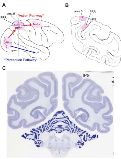
Figure 1-1. The two streams of visual information in the primate brain and reach intention areas of
PPC. (A) The dorsal stream processes visual information for movement and is concerned with how
visual information is used to form an action. The “vision for action” pathway branches from the
occipital lobe to the posterior parietal cortex (PPC), which performs sensorimotor transformations, and
is largely concerned with visual perception, for example the inferotemporal cortex (IT) is critical for
discriminating and recognizing objects. This whole-brain view shows the surface location of the parietal
reach region (PRR) located along the medial bank of the intraparietal sulcus (IPS), which can extend to
depths of up to 10 mm. Moving anterior and slightly anterior, PRR merges into area 5, which is situated
more on the surface of cortex. (B) Coronal section designated by the dashed vertical line in panel (A),
illustrating the convoluted geometry of the IPS as well as the relative depths of PRR and area 5 (C)
Nissl-stained coronal section of the rhesus macaque brain, clearly showing the intraparietal sulcus.
Coronal section was obtained from www.brainmaps.org (S. Mikula et al., 2007).
Numerous neurophysiological studies in monkeys have shed light on the neural correlates of reach planning in PPC. Monkeys have served as a successful model for studying
sensorimotor representations in humans since the two species engage in a variety of similar sensorimotor behaviors. Moreover, functional magnetic resonance imaging (fMRI) studies have provided evidence that PPC’s functional role is similar in both
monkeys and humans (J. F. X. DeSouza et al., 2000; M. F. S. Rushworth et al., 2001; J. D. Connolly et al., 2003). When trained monkeys plan a reach to an illuminated target,
the firing rates of neurons in the medial bank of the intraparietal sulcus (MIP) generally reflect a combination of both sensory and motor parameters (V. B. Mountcastle et al., 1975; D. L. Robinson et al., 1978; R. A. Andersen and C. A. Buneo, 2002). Importantly
however, during a memory period in which the monkey must maintain a reach plan to the remembered location of an extinguished target, elevated neural activity persists in PPC
before the reach is executed, suggesting that these neurons likely encode the intention to reach, rather than the visual stimulus location (L. H. Snyder et al., 1997). Furthermore, neurons in MIP are generally correlated more strongly with the motor goal, and not the
A. Assad, 1999; A. Gail and R. A. Andersen, 2006). Based on these planning studies, it is tempting to hypothesize that memory-period activity might also reflect a prediction of the
sensory consequences of an upcoming arm movement (i.e. the expected endpoint of a reach) derived from efference copy, a signal that is compatible with the output of a
forward model of arm position. More generally, one could interpret the effector-specific segregation of planning activity in the intraparietal sulcus, such as MIP responses
associated with a reach and LIP responses involved in the formulation of a saccade (R. A.
Andersen and C. A. Buneo, 2002), as a prediction of the future state of an effector, the motor command itself or both. Along these lines multiple researchers have also suggested
that the ‘early’ discharge of neurons in area 5 prior to initiation of an arm movement might reflect an efference copy signal fed back to PPC from frontal motor areas (J. Seal et al., 1982; J. F. Kalaska et al., 1983). Interestingly, Seal and colleagues also showed
that area 5 responses that occurred prior to movement onset were generally not sensory in origin and furthermore demonstrated that these early responses persisted even after deafferentation. However, caution should be advised when attempting to infer the causal
flow of information in the parieto-frontal circuit during reach preparation using single-area correlation analyses. Furthermore, it is quite possible that planning and forward
model prediction may be carried out by distinct neural processes within the PPC. Future simultaneous multi-area recordings, combined with micro-stimulation approaches, should shed light on the directional flow of information in these recurrent inter-area circuits
PPC is also a good candidate for a forward model of eye position since a variety of eye behavior related signals, such as saccade and fixation responses have been described in this region (V. B. Mountcastle et al., 1975). Whereas area 7a saccade responses begin
largely after the saccade, area LIP saccade responses tend to occur before, during or after saccades (R. A. Andersen et al., 1987). Interestingly, Duhamel and colleagues also
showed that the receptive fields of neurons in LIP can update their receptive fields before an eye movement occurs (J. R. Duhamel et al., 1992). 44% (16 out of 36) of their LIP sample anticipated the sensory effects of an impending saccade (i.e. a stimulus appearing
in the future location of the receptive field), and adjusted their responses approximately 80 milliseconds (ms) before the saccade was launched. It is conceivable that this
predictive remapping relies upon a forward model of eye position within PPC, which estimates the upcoming eye position from oculomotor commands, though direct evidence of the anticipatory eye position signal itself in PPC has not been reported. Fixation
related activity commonly found in PPC is sensitive for eye position, and this response characteristic is often multiplicatively combined with the sensory and saccade-related activity of single neurons (R. A. Andersen et al., 1987). An eye position signal in PPC
could be derived from proprioceptive feedback from the eye muscles (X. L. Wang et al., 2007) and/or the integration of saccade command signals. It would be interesting to see if
a component of the eye position signal might also provide anticipatory information (ahead of passive sensory feedback) about the current state of the eye position during fixations between saccades. Last, Batista and colleagues showed that neurons in PRR,
Buneo et al., 2002). It would be interesting to test if the reach receptive fields of these PRR neurons also exhibit anticipatory updating just before the eye moves, similar to the
cells found in LIP by Duhamel and colleagues.
1.3 Forward state estimation to compensate for sensory delay times
Skilled individuals are capable of guiding their limb movements with remarkable
accuracy and speed. Examples from athletics are illustrative: hitters in baseball adjust the
trajectory of their bat-swing so as to make contact with a pitched ball arriving in fewer than 500 ms, racquetball players return an oncoming serve traveling at speeds up to 140
miles per hour, a boxer must avoid or block a punch with less than 200 ms to react.
During execution of a goal-directed arm movement, in order to continuously guide the
arm to a target the brain must maintain an estimate of the time-varying state of the arm (e.g. position and velocity of the arm, possibly coded in a variety of potential coordinate frames) and compare that state measurement with the desired state of the movement.
Unfortunately, the human brain, in particular PPC, does not have direct access to the true state of the arm due to delayed and noise-corrupted measurements of the state in the
visual and proprioceptive domains. (e.g. visual signals typically reach sensorimotor association areas of cortex after a delay of approximately 90 ms (S. E. Raiguel et al., 1999), 30 ms in the case of proprioception (N. Petersen et al., 1998). Subsequent
100 ms for proprioceptive control (M. Flanders and P. J. Cordo, 1989) and over 200 ms for visuomotor control (A. P. Georgopoulos et al., 1981; R. C. Miall et al., 1993). These long delay times severely limit a feedback control system’s ability to make rapid
adjustments to an ongoing movement and thus increase the likelihood that a reach trajectory might become erroneous and/or unstable.
Remarkably, despite substantial transmission and processing delays in the sensorimotor control loop, we are able to rapidly and accurately control our movement trajectories.
Therefore, passive sensory feedback must not be the only signal used by the brain to estimate the current state of the arm. Fortunately the brain can also monitor recently
issued motor commands (i.e. efference copy), which could be transmitted centrally (e.g. from frontal motor areas) with little delay time (e.g. one synapse + transmission time < 10 ms) and used by a forward dynamics model to form an estimate of the current or
upcoming state of the arm. (M. I. Jordan and D. E. Rumelhart, 1992; D. M. Wolpert et al., 1995). Thus a forward model’s prediction can be made available immediately to sensorimotor control structures in the brain, well in advance of late-arriving sensory
information. Since the output of the forward dynamics model reflects a best guess as to what the next state of the arm will be, errors due to various sources of noise will
inevitably accumulate over time into this estimate. Therefore it is likely that sensory observations, which arrive at later times, are also continually integrated by the brain in order to update and refine the estimate of the forward dynamics model (R. C. Miall and
In order to estimate the state of the arm at each time step k, the output of the forward dynamics model, (i.e. a priori estimate), is linearly combined with the difference between the output of the observation model (i.e. predicted sensory measurement) and the
actual sensory measurement. This, discrepancy, the ‘sensory innovation’, is then
optimally scaled by the Kalman gain, Kk, to produce an a posterior estimate of the state
of the arm,
−
k xˆ
(
ˆ)
ˆ
ˆ = − + − −
k k k k
k x K y Hx
x . (3)
In brief, discrete state estimation consists of a two-step recursive procedure; such that the forward dynamics model generates an a priori estimate of the state, which is next refined by potentially innovative information gleaned from the sensory input to form the final, a posterior estimate. PPC, specifically the parietal reach region (PRR) and area 5, seems to be a reasonable site for an observer to reside given its large number of feedback
connections from frontal areas (i.e. efference copy) and substantial sensory input from both visual and somatosensory domains (E. G. Jones and T. P. Powell, 1970; P. B.
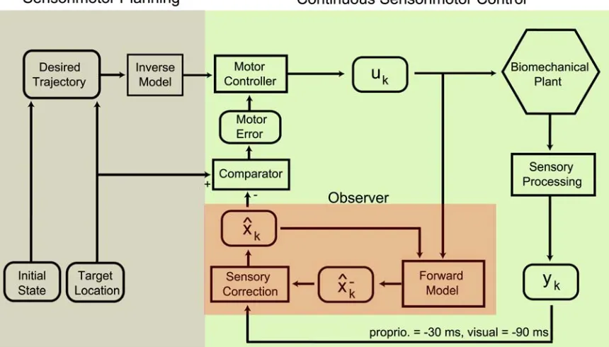
Figure 1-2. Flow diagram illustrating sensorimotor integration for reach planning and online control
(after M. Desmurget and S. Grafton, 2000). Items in rounded boxes denote pertinent sensorimotor
variables; computational processes are contained in rectangular boxes. Prior to a reach, an intended
trajectory is formulated as a function of both the initial state of the arm and the desired endpoint, the
target location. An inverse model is used to determine a set of motor plans that will result in the desired
trajectory. Motor plans are then issued (likely by the primary motor cortex, M1) and subsequently
executed by muscles acting within the physical environment (i.e. biomechanical plant hexagon).
Following movement onset, the path of the arm is continuously monitored and corrected, if necessary, to
ensure successful completion of the reach. Critical to rapid online correction of movement is the forward
model, which generates a rapid a priori estimate of the next state of the arm, , as a function of the previous state and efference copy. Intermittent sensory feedback is used to refine the a priori estimate of the forward dynamics model (observer output). This a posterior current state estimate, , is then evaluated by comparing it with the target location in order to make corrections to subsequent motor
commands.
−
k xˆ
1.3.1 A forward model can be used to cancel sensory reafference
A forward model’s ability to predict the sensory consequences of an action is also useful
to an organism because a given sensory outcome can be produced by a variety of possible causes (R. W. Sperry, 1950; Weiskran.L et al., 1971; G. Claxton, 1975; J. F. A. Poulet
and B. Hedwig, 2003; K. E. Cullen, 2004; J. E. Roy and K. E. Cullen, 2004). In particular, a forward model can provide an internal reference signal that is useful for canceling the sensory effects of self-motion. For example, motion on our retina can occur
because of movement in the physical world (i.e., afference) or because of motion induced by an eye movement itself (reafference). Therefore, in order to correctly perceive the
motion of an external stimulus, the brain must distinguish afferent motion from reafferent motion. A subtractive comparison between a forward model’s estimate of the expected sensory outcome of an eye movement and the actual sensory signals could remove this
retinal shift from our perception (T. Haarmeier et al., 2001). Reafference-canceling mechanisms are likely to be in operation for perception of limb movements as well. A
forward model of the arm could be critical for distinguishing self-generated arm movement from both movement of the environment and movement of one’s arm by an external force.
1.4 Continuous sensorimotor control and state estimation in PPC
Clinical and psychophysical studies in humans have established that PPC is involved not only in specifying movement plans, but also in the execution and control of ongoing movement. For example, it is well known that lesions in parietal cortex often lead to optic
patients have difficulty making rapid and ‘automatic’ corrective movements when
guiding the hand to targets that have been jumped (L. Pisella et al., 2000). Similarly, Grea
and colleagues reported a patient with bi-lateral parietal lesions that was unable to amend her movement to pick up a cylinder after it had been jumped to a new location at
movement onset (H. Grea et al., 2002). Interestingly, instead of making corrective movements during the initial trajectory, the subject needed to perform two distinct movements, one that represented the initial plan and a second movement to reach to the
new location of the cylinder. Using transcranial magnetic stimulation (TMS) applied to the posterior parietal cortex, Desmurget and colleagues were able to transiently disrupt
the ability of most subjects to correct reaching trajectories made to targets that were displaced around the time of movement onset (M. Desmurget et al., 1999). Later, Della-Maggiore and colleagues showed that TMS applied to PPC interfered with the ability of
subjects to adapt to novel force-field environments (V. Della-Maggiore et al., 2004). An intriguing, potentially unifying explanation for all of these deficits, which was originally
suggested by Wolpert and colleagues, is that PPC may serve as an observer, which forms an internal estimate of the state of the arm during movement (D. M. Wolpert et al., 1998a). A failure to accurately maintain this estimate online could result in an inability to
monitor and therefore correct an ongoing movement. Interestingly, Wolpert and colleagues reported a parietal lesion patient who was unable to maintain an internal
disappear, and finally when asked to make slow-pointing movements to peripheral targets while maintaining central fixation, large errors accumulated in her trajectories (although self-paced movements were not impaired).
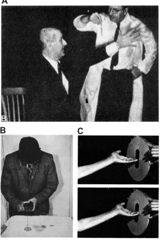
Figure 1-3. Examples of patients with sensorimotor deficits due to parietal injury. (A) A patient with a
symptoms, such that he is unable to direct an arm movement toward a visually cued target (i.e., pour
liquid into a glass) (R. S. Allison et al., 1969). (C) A patient with a left parietal lesion is unable to
properly orient his hand to successfully position it inside the slot (M. T. Perenin and A. Vighetto, 1988).
Evidence that PPC is involved in sensorimotor state estimation also comes from studies of the mental simulation of movement, which presumably activates circuits overlapping
with those engaged during motor control, but while inhibiting execution of a movement itself (K. M. Stephan et al., 1995; J. Decety, 1996; E. Gerardin et al., 2000). When
normal healthy subjects imagine making a goal-directed movement, mental simulation time typically matches the time needed to execute that same movement (F. C. Donders, 1969; J. Decety and F. Michel, 1989). This suggests that the brain is able to maintain a
realistic estimate of the state of the hand over time while imagining a movement, despite sensory feedback being unavailable. Interpreting this finding in the context of observer
theory, this capability suggests that the brain/observer is able to rely entirely upon the output of a forward model to estimate the state of the arm during mental simulation (e.g. Kalman gain in Equation 3 is set to zero) (R. Shadmehr and J. W. Krakauer, 2008).
Interestingly, patients with unilateral motor cortex lesions (A. Sirigu et al., 1995) who show prolonged movement times compared to normal control subjects are still able to
with lesions of the cerebellum (F. A. Kagerer et al., 1998) and of the basal ganglia (P. Dominey et al., 1995) also do not show a difference between simulation and execution times.
While M1, the cerebellum and the basal ganglia do not appear to be critically involved in
the state estimation during simulated movements, PPC does appear to be essential for maintaining an internal representation of the state of the hand that is necessary for representing a consistent relationship between simulated and execution time. Sirigu and
colleagues later reported an impairment in the ability to simulate a movement in patients with right PPC lesions: the time needed to mentally simulate a movement was
significantly different (generally less) than the time necessary to execute that same movement (A. Sirigu et al., 1996). (Note, similar to motor cortex lesion patients, actual execution time was also prolonged compared to control subjects). This inconsistency
suggests that the brain was unable to reliably estimate the state of the hand after damage to PPC. This impairment could be explained by multiple possible failures of the observer
model: 1) an error in the forward dynamics model (i.e. faulty A or B matrices in Equation 1), 2) an error when incorporating sensory feedback into the a priori estimate of the forward dynamics model (i.e. faulty H or K matrices in Equation 3) or 3) a combination of 1 and 2. Based on known strong sensory input to PPC, it is probable that PPC is involved in integrating sensory feedback into the state estimate. However, because visual
complete a movement when simulating it. Therefore, such a systematic decrease in imagined movement duration is more likely to have arisen due to an erroneous a priori
estimate made by a forward dynamics model, whose transition matrices A and B govern the speed at which the arm propagates through space (rather than a faulty observation
model which relates sensory input to the state of the arm using a time-independent model). Therefore, these mental simulation results suggest that PPC is also involved in propagating the state of the arm forward in time using a forward dynamics model
(Equation 1). Assuming PPC incorporates sensory information into the forward model state estimate as well, then PPC would be best described as an observer, as suggested by
Wolpert and colleagues.
Psychophysical and clinical reports have pointed to both the parietal lobe and the
cerebellum as candidate neural substrates for a forward model (R. C. Miall et al., 1993; D. M. Wolpert et al., 1998a; D. M. Wolpert et al., 1998b; S. J. Blakemore and A. Sirigu,
2003). Desmurget and colleagues suggested that PPC encodes a forward model of the arm’s dynamics from which it may also compute an estimate of the motor error (i.e. the difference between the target vector and the movement vector), which could then be
transformed into a corrective motor command by the cerebellum (M. Desmurget and S. Grafton, 2000). While these insights suggest that PPC and the cerebellum may be
Previous encoding studies have shown that area 5 neurons are correlated with a variety of movement and task-related parameters (most notably velocity and target position) during reaching movements made with a manipulandum (J. Ashe and A. P. Georgopoulos, 1994;
B. B. Averbeck et al., 2005). However, these studies concluded that area 5 largely encodes a sensory (i.e. proprioceptive) representation that slightly lags the state of the
movement (i.e. lag time = -30 ms). In Chapter 2 we further investigate the neural representation of online directional control signals in PPC while monkeys perform center-out and obstacle avoidance joystick tasks. We provide new evidence that neurons
in PPC compute an estimate of the current and upcoming states of the cursor (G. H. Mulliken et al., 2008). This finding provides the first direct evidence of a state estimate
that is derived from a forward model and is a starting point for future studies designed to rigorously reverse-engineer the computational mechanisms and circuits involved in forward model control.
1.5 A PPC neural prosthesis: continuous closed-loop control of an end effector
It would be interesting to test if dynamic state estimates in PPC, presumably reflecting
the operation of an observer could be used to causally control an external device, besides our own limbs. During recent years, several groups have leveraged findings from decades
of primate neurophysiology toward the development of an important medical application, a neural prosthesis to assist paralyzed individuals. Paralysis affects millions of people in the United States and can occur as a result of stroke and cervical spine injuries as well as
neurodegenerative disorders such as amyotrophic lateral sclerosis (Lou Gehrig’s
however, these individuals can still think about moving, and thus maintain the capacity to plan and specify instructions for the control of movement. Therefore, a neural prosthesis
could directly read out the desired movement intentions of these patients from regions of the brain not affected by injury or disease. Sensorimotor areas of cortex, particularly
those which are strongly innervated by visual feedback projections (e.g., PPC) represent candidate regions that are potentially useful for driving a neural prosthesis since their primary source of input, visual information, is typically uncompromised after paralysis
(Fig 1-4).
A cortical prosthesis is composed of multiple parts, which constitute a marriage of engineering and scientific advances. Neural signals from a targeted brain region (i.e., spiking activity of neurons and the local field potential (LFP)) are extracted by measuring
the extracellular potential at many different sites in the cortical tissue using metal multi-electrode arrays, which currently can record from on the order of 100 signals
simultaneously. Amplification and filtering of these neural signals occurs proximal to the implant, maximizing the signal-to-noise ratio of each neural measurement.
Signal-processing, including spike-sorting, firing rate calculations and space-time analysis of
LFPs, is then performed before passing these assuaged signals along (e.g., via wireless transmission) to a decoding algorithm. The decoding algorithm then computes an optimal
In general, neurophysiological approaches have emphasized extracting continuous movement information (i.e., trajectories) from motor cortices involved in directly controlling the limb, such as primary motor cortex (M1) (P. R. Kennedy et al., 2000; J.
Wessberg et al., 2000; M. D. Serruya et al., 2002; D. M. Taylor et al., 2002; J. M. Carmena et al., 2003; K. V. Shenoy et al., 2003; S. Musallam et al., 2004; P. G. Patil et
al., 2004; J. R. Wolpaw and D. J. McFarland, 2004; G. Santhanam et al., 2006). However, recently Musallam and colleagues introduced the concept of decoding
cognitive signals from the brain (R. A. Andersen et al., 2004; S. Musallam et al., 2004).
The authors distinguished pure motor-related signals from those involved with the high-level organization of movement. In contrast to signals extracted from M1, which are
more likely to encode movement execution signals that are represented in a
musculoskeletal reference frame, high-level visuomotor signals can be found in earlier stages of the vision-for-action pathway, such as in PPC or PMd. For example, the goal of
a reach in visual coordinates has been decoded successfully from both PPC and PMd neurons (S. Musallam et al., 2004; G. Santhanam et al., 2006). In chapters 3 and 4, we
build on the work of Musallam and colleagues and demonstrate that a PPC prosthesis can also be used to perform online, continuous control of movement. In particular, we
demonstrate that we can successfully read out a trajectory from PPC spiking activity in
real-time, allowing a monkey to control a cursor on a computer screen using his thoughts alone. Presumably, and in contrast to similar approaches in M1, we are decoding from the
ultimately be advantageous for inferring variables beyond just sensorimotor signals, which may include, for example, the expected value of an impending action or one’s
behavioral or cognitive state (S. Musallam et al., 2004).
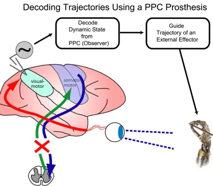
Figure 1-4. A neural prosthesis using PPC for trajectory control. A spinal cord injury can render
communication (afferent and efferent) between somatosensory/motor areas of cortex and the limbs
useless. However, the function of the “vision for action” pathway is still largely intact, which includes
Ungerleider LG, Mishkin M (1982) Two cortical visual systems. In: Analysis of Visual Behavior (Ingle DJ, Goodale MA, Mansfield RJW, eds), pp 549-586. Cambridge, MA: MIT Press.
Wang XL, Zhang MS, Cohen IS, Goldberg ME (2007) The proprioceptive representation of eye position in monkey primary somatosensory cortex. Nat Neurosci 10:640-646.
Weiskrantz.L, Elliott J, Darlington.C (1971) Preliminary Observations on Tickling Oneself. Nature 230:598-599.
Wessberg J, Stambaugh CR, Kralik JD, Beck PD, Laubach M, Chapin JK, Kim J, Biggs J, Srinivasan MA, Nicolelis MAL (2000) Real-time prediction of hand trajectory by ensembles of cortical neurons in primates. Nature 408:361-365.
Wolpaw JR, McFarland DJ (2004) Control of a two-dimensional movement signal by a noninvasive brain-computer interface in humans. Proceedings of the National Academy of Sciences of the United States of America 101:17849-17854. Wolpert DM, Ghahramani Z, Jordan MI (1995) An internal model for sensorimotor
integration. Science 269:1880-1882.
Wolpert DM, Goodbody SJ, Husain M (1998a) Maintaining internal representations the role of the human superior parietal lobe. Nat Neurosci 1:529-533.
Chapter 2
FORWARD ESTIMATION OF MOVEMENT STATE IN PPC1
2.1 Summary
During goal-directed movements, primates are able to rapidly and accurately control an online trajectory despite substantial delay times incurred in the sensorimotor control loop. To address the problem of large delays, it has been proposed that the brain uses an internal forward model of the arm to estimate current and upcoming states of a
movement, which are more useful for rapid online control. To study online control mechanisms in the posterior parietal cortex (PPC), we recorded from single neurons while monkeys performed a joystick task. Neurons encoded the static target direction and the dynamic movement angle of the cursor. The dynamic encoding properties of many movement angle neurons reflected a forward estimate of the state of the cursor that is neither directly available from passive sensory feedback nor compatible with outgoing motor commands, and is consistent with PPC serving as a forward model for online sensorimotor control. In addition, we found that the space-time tuning functions of these neurons were largely separable in the angle-time plane, suggesting that they mostly encode straight and approximately instantaneous trajectories.
2.2 Introduction
The Posterior Parietal Cortex (PPC) lies at the functional interface between sensory and motor representations in the primate brain. Known sensory inputs to PPC arrive from
visual and proprioceptive pathways (Fig. 2-1A). Previous work has suggested how these sensory inputs could be integrated to compute a goal vector in eye-centered coordinates
for an impending reach (L. H. Snyder et al., 1997; A. P. Batista et al., 1999; C. A. Buneo et al., 2002). In addition, psychophysical and clinical studies in humans have clearly established a role for PPC in rapid online updating and correction of continuous
movement (M. Desmurget et al., 1999; L. Pisella et al., 2000; V. Della-Maggiore et al., 2004). In order for a brain area to play an effective role in rapid online control, it would
have to represent an estimate of the state of the movement (position, direction, speed etc.) that is derived from mechanisms other than just sensory feedback, which is generally considered to be too slow to accomplish the task much of the time (R. C. Miall and D. M.
Wolpert, 1996; M. Desmurget and S. Grafton, 2000). Another possible input to PPC is an efference copy signal that relays replicas of recent movement commands from
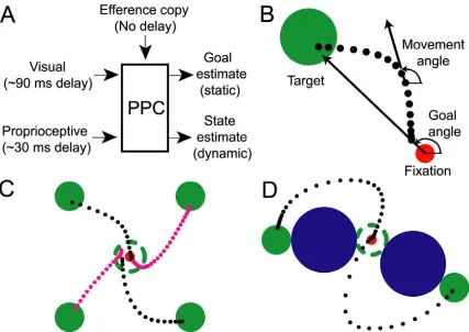
Figure 2-1. Model and experimental design. (A) Diagram of sensorimotor integration for online control in posterior parietal cortex (PPC). Inputs to PPC consist of visual and proprioceptive sensory signals
and potentially an efference copy signal. Plausible PPC outputs to be tested are (1) the static target
direction (goal angle) and (2) the dynamic cursor heading direction (movement angle). (B) Diagram of actual trajectory showing the goal angle and movement angle, and their respective origins of reference.
The filled green and red circles represent the target and fixation point, respectively. (C) Example trajectories for center-out task. The dashed green circle is the starting location of the target and is not
visible once the target has been jumped to the periphery. Dots represent cursor position sampled at 15 ms
intervals along the trajectory (black = monkey 1, magenta = monkey 2). (D) Example trajectories for obstacle task. Targets, fixation points, and cursor representations are identical to center-out task. Blue
filled circle represents the obstacle.
the system dynamics to generate estimates of upcoming states of the effector (otherwise not inferable from late-arriving sensory feedback), which are more suitable for the rapid control of movement (M. I. Jordan and D. E. Rumelhart, 1992; D. M. Wolpert et al.,
1995). Since the output of a forward model reflects a best guess of the next state of the arm in lieu of delayed sensory feedback, it is also likely that sensory observations that
arrive at later times are continually integrated as well by the online controller in order to improve the estimate of the forward model as time goes by (G. C. Goodwin and K. S. Sin, 1984).
In addition, the output of a forward model can be used to create an internal estimate of
the sensory consequences of a movement in a timely manner (i.e., the expected
visual/proprioceptive state of the effector in the environment), providing a mechanism for transforming between intrinsic motor representations and task-based sensory
representations (M. I. Jordan and D. E. Rumelhart, 1992; R. C. Miall and D. M. Wolpert, 1996). In particular, a forward model may be useful for distinguishing the motion of an
effector from motion of the external environment. For example, when we make eye movements, it is widely believed that the brain makes use of an internal reference signal to avoid mis-interpreting shifts of the visual scene on our retina as motion in the outside
world (H. von Helmoltz, 1866). von Holst and Mittelstaedt originally proposed a
reafference-cancelling model, which performs a subtractive comparison of efference copy
by von Holst and Mittelstaedt (T. Haarmeier et al., 2001). Interestingly, additional clinical evidence presented by Haarmeier and colleagues suggested that parieto-occipital
regions may be involved in performing the comparison between self-induced and external sensory motion during smooth-pursuit eye movements (T. Haarmeier et al., 1997).
Neurophysiological evidence that identifies the neural substrate of the internal forward model for sensorimotor control of limb movement has yet to be reported. PPC,
specifically the parietal reach region (PRR) and area 5, could be a possible site for the forward model to reside given its large number of feedback connections from frontal
areas and substantial sensory input from both visual and somatosensory domains (E. G. Jones and T. P. Powell, 1970; P. B. Johnson et al., 1996). Therefore, we investigated the neural representation of online directional control signals in PPC by analyzing the
correlations of single neuron activity with the static goal angle (fixed angle from the starting cursor position to the target) and the dynamic movement angle of the cursor
(angle of heading) during a joystick task (Fig. 2-1B). We monitored single-unit neuronal activity in PPC while monkeys performed center-out and obstacle avoidance tasks with central eye fixation. Importantly, monkeys were required to fixate centrally during the
entire movement so as to maintain a constant visual reference frame and to rule out any effects due to eye movements. This control was instituted because earlier studies have
static target direction and a dynamic estimate of the state of the cursor. The temporal encoding properties of dynamically tuned neurons provide the first evidence that PRR and area 5 encode the current state of the cursor, consistent with the operation of a
forward model for online sensorimotor control. Furthermore, these state-estimating neurons appear to encode rather simple trajectories, encoding instantaneous and mostly
straight paths in space.
2.3 Results
2.3.1 Space-time tuning
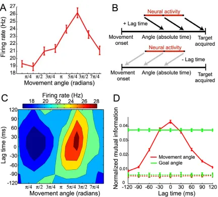
We characterized the encoding properties of each PPC neuron during the movement period by constructing a space-time tuning function (STTF) (Fig 2-2B), which plots a neuron’s instantaneous firing rate as a function of angle (goal or movement) measured at
a particular lag time (L. Paninski et al., 2004). Importantly, lag time, τ, denotes the relative time difference between the instantaneous firing rate and the time that a
particular behavioral angle occurred, and should not be confused with the absolute elapsed time. Therefore, the STTF of a neuron can be thought of as a description of the average temporal dynamics of the angle that can be recovered from the firing rate, for
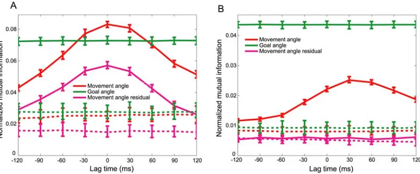
example, by downstream neurons faced with the task of decoding the goal or movement angle at different relative times in the trajectory. We also calculated the mutual
information between firing rate and angle for each lag time in the STTF to generate a temporal encoding function (TEF). Since mutual information is a non-parametric measure of statistical dependency between two random variables, this measure allowed
the angle at different lag times (i.e., from past (τ < 0) to future (τ > 0) angles). The lag time that contained the maximal mutual information was defined as the optimal lag time
(OLT), denoting the relative time at which a neuron’s firing rate contained the most information about the angle.
Fig. 2-2C shows a movement angle STTF for a single neuron. This neuron contained significantly more information about the movement angle than the goal angle at its OLT
of 0 ms (Fig. 2-2D). However, since it is not possible to classify cells as encoding purely goal angle or purely movement angle (due to implicit partial correlation between these
two angles), we instead determined whether tuning for movement angle was significant, independent of tuning for goal angle, and vice versa (Supporting Information). If so, we included that cell in the movement angle population. Similarly, if the cell contained
Figure 2-2. Representative neuron and STTF analysis. (A) Movement angle tuning curve, plotting firing rate as a function of movement angle measured at zero lag time. The tuning curve was well fit by a
cosine model (R2 = 0.92). (B) Diagram describing space-time tuning analysis. Neural activity was
sampled from the middle of the movement period and movement angle was sampled across the entire
movement period, from movement onset to the time the cursor entered the target zone. This sampling
scheme allowed each firing rate sample to be paired with angle samples at all possible lag times
considered. (C) Movement angle space-time tuning function (STTF). Contour plot shows the average firing rate of a cell that occurred for different movement angles measured over a range of lag times
(-120 ms ≤τ≤ 120 ms) relative to the firing rate. (D) Movement angle temporal encoding function (TEF) and corresponding goal angle TEF, where mutual information between firing rate and movement angle
was stationary during each trial (e.g., goal angle did not change during a trajectory), the goal angle
information was approximately constant across lag time. The dashed lines denote surrogate TEFs, for
both movement (red-dashed) and goal (green-dashed) angles, that were derived from surrogate spike
trains and actual angles. Note that there is no temporal tuning structure in the surrogate movement
angle TEF.
During the center-out task, we recorded from 652 neurons from 2 monkeys. 390 neurons were significantly tuned for either the movement angle or goal angle, or both. 220/390
(56%) significantly encoded the movement angle and 292/390 (75%) significantly encoded the goal angle. During the obstacle task, we recorded from 221 neurons from
monkey 1 and 212 of these were significantly tuned for either the movement angle or goal angle. 168/212 (79%) neurons significantly encoded the movement angle and 197/212 (93%) significantly encoded the goal angle. Since our analysis relies upon the
neural tuning properties being stationary in time, the above population counts do not include any cells that exhibited non-stationarity (Supporting Information).
Interestingly, we found an anatomical correlate for the representation of goal angle and movement angle in the medial intraparietal area: the mutual information for goal angle
tended to increase gradually with the depth of the recording electrode, while information for movement angle (peak information, measured at OLT) decreased with depth. A linear
approximately 30% over a 10 mm span (α = 0.038). The average goal angle information increased with depth in the sulcus, by about 60%, over 10 mm (α = 0.002). A stronger encoding of target-related signals deeper in the intraparietal sulcus (IPS) and conversely a
favored representation of arm movement related activity in surface regions of the IPS is consistent with previous PPC studies of reach planning, in which eye-centered target
signals were commonly found in deeper structures such as PRR, and more hand-related activity was reported for surface area 5 neurons (C. A. Buneo et al., 2002).
2.3.2 Static encoding of goal angle
Neurons that were significantly tuned for the goal angle persistently encoded information about the static direction to the target (measured from the starting cursor position, which is also the fixation point), independent of the changing state of the cursor. These cells
were consistent with previous descriptions of target-sensitive tuning in area 5 (J. Ashe and A. P. Georgopoulos, 1994). This target representation is most likely not due simply
to a cue response since the neural activity we analyzed typically occurred more than 240 ms after cue onset. Therefore, the intended goal of the trajectory is maintained in the PPC population during control of the movement. Knowledge of the target direction during the
movement could be used downstream, for example by motor cortices, to adjust upcoming motor commands to more accurately constrain the trajectory toward the target. Similarly,
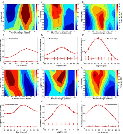
PPC neurons tuned for the movement angle encode dynamic information about the
changing state of the cursor. Fig. 2-3A shows TEFs for the entire movement angle population, normalized on a per cell basis by each cell’s maximal mutual information.
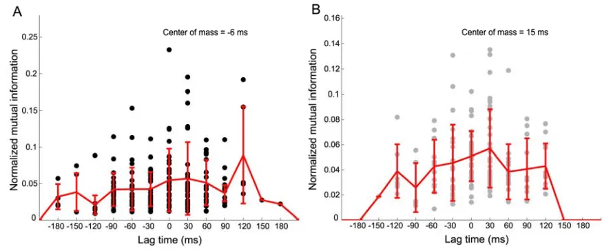
TEFs were typically single-peaked at each cell’s OLT. The histogram in Fig. 2-3B
summarizes the distribution of OLTs for the movement angle population, which was centered at 0 ± 90 ms and 30 ± 90 ms, for the center-out and obstacle tasks respectively
(median ± interquartile range (IQR)). Both of these plots show that movement angle neurons contained a temporal distribution of information about the state of the ongoing
movement; some neurons best represented states in the near future (positive-lag time), some best represented states in the recent past (negative-lag time), and many peaked around the current state (zero-lag time). Passive sensory feedback would require at least
30-90 ms (proprioceptive-visual) to reach PPC; consistent with some of the negative OLTs (≤ -30 ms) observed here (M. Flanders and P. J. Cordo, 1989; R. C. Miall et al.,
1993; N. Petersen et al., 1998; S. E. Raiguel et al., 1999). Conversely, if PPC neurons were encoding an outgoing motor command, subsequent motor processing and execution of the movement would require at least 90 ms to produce the corresponding cursor
motion, resulting in positive OLTs above 90 ms (R. C. Miall et al., 1993). For instance, similar analyses for velocity have been performed in the primary motor cortex and report
average OLTs of approximately 90-100 ms (J. Ashe and A. P. Georgopoulos, 1994; L. Paninski et al., 2004). Therefore, it is unlikely that PPC is driving motor cortex with feedforward commands since it would be expected that PPC would lead the movement
have reported that velocity information is present in area 5 and suggested that those neurons best reflect non-causal, sensory information (J. Ashe and A. P. Georgopoulos, 1994; B. B. Averbeck et al., 2005). We performed an additional temporal encoding
analysis for velocity and obtained very similar results to those reported here for
movement angle (Supporting Information). Neither passive sensory feedback nor efferent
motor explanations best account for many of the OLTs observed in our data. In contrast, the most reasonable description of neurons whose optimal lag times lie between -30 and 90 ms is that they encode a forward estimate, which is used to monitor the current and
upcoming state of the movement angle, prior to the arrival of delayed sensory feedback. We suggest that these forward state estimates most likely reflect the operation of a
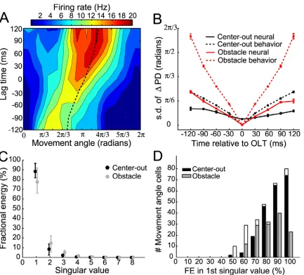
Figure 2-3. Population temporal encoding results. (A) Population TEFs plotted for all movement angle neurons showing cell-normalized mutual information as a function of lag time. TEFs are sorted from
lowest to highest optimal lag time (OLT). The population encoded a distribution of temporal
information, including past, present, and future states of the movement angle. Note that some neurons’
TEFs had more data than others since one monkey made slightly faster movements than the other. (B) Histogram summarizing the OLTs for movement angle neurons for both center-out and obstacle tasks.
Many of these neurons’ OLTs were consistent with a forward estimate of the state of the movement
angle, which did not directly reflect delayed sensory feedback to PPC, nor were they compatible with
outgoing motor commands from PPC. Color-coded horizontal bars beneath the abscissa denote the
approximate lag time ranges for sensory (blue), forward estimate (black), and motor (red)
representations of the state of the movement angle.
We also observed that the peak information (mutual information at the OLT) encoded by
those neurons that were clearly forward-estimating (0 ≤ OLT ≤ 60 ms) was significantly larger than the peak information encoded by the remaining population of movement angle
neurons (OLT ≤ -30 ms, or, OLT ≥ 90 ms) (p < 0.001, Wilcoxon rank sum test, (Supporting Information)). This result shows that, in addition to PPC having a central tendency to encode the current state of the movement angle, these forward-estimating
neurons will also on average encode more information compared to neurons at other OLTs, providing further support that the population best represents forward estimates of
the state of the cursor. In addition, we observed that the median OLT for the obstacle task distribution was significantly larger than the median OLT for the center-out task
distribution, and shifted forward from 0 to 30 ms (p < 1e-4, Wilcoxon rank sum). This
anticipatory estimate of the state of the cursor for online control.
2.3.4 Dynamic tuning and separability of movement angle STTF
We further analyzed the spatio-temporal encoding properties of movement angle neurons by measuring changes in the preferred direction of a neuron, θpd, over a range of lag
times. θpd is the movement angle at which a neuron fired maximally for a particular lag
time. We reasoned that if θpd did not vary significantly as a function of lag time compared
to changes that occurred in the movement angle itself, then that neuron encoded a mostly
straight trajectory. Fig. 2-4A shows an example of a neuron recorded during the obstacle task for which θpd changed smoothly in time. Specifically, θpd changed by 0.87 radians
from -120 ms to 0 ms lag time and by an additional 0.61 radians from 0 ms to 120 ms lag
time, implying that this neuron, on average, encoded a slightly nonlinear trajectory. Across the population, most neurons’ STTFs exhibited small changes in θpd as a function
of lag time. We quantified the tendency for the θpd to vary for the movement angle
population by computing the circular standard deviation of the distribution of all neurons’ angle-changes, for each lag time (N. I. Fisher, 1993). We found that the standard
deviation of θpd changes increased with time away from the OLT and rose to a maximum
difference of 0.36 ± 0.03 and 0.72 ± 0.03 radians over 120 ms, for the center-out and
obstacle tasks respectively (Fig. 2-4B). These deviations, while significantly larger than zero, were significantly smaller than deviations measured in the movement angle itself,
encoded significantly less change in movement direction than was observed in the actual trajectories themselves (< 50%). We also calculated the average curvature of the
monkeys’ trajectories and the curvature of simulated trajectories derived from a neuron’s
STTF, which were considered to be the ‘preferred trajectory’ of a neuron. Consistent with the preferred direction results, the average curvature of preferred trajectories (0.06 ± 0.01
and 0.15 ± 0.04, for center-out and obstacle tasks, respectively) was significantly less than the average curvature of the actual trajectories (0.15 ± 0.04 and 0.26 ± 0.02, for center-out and obstacle tasks respectively). This result further substantiates the claim that
Figure 2-4. Curvature and separability of STTFs. (A) Example STTF containing slight curvature. The
θpd of this cell (dashed line) changed smoothly as a function of lag time. These small changes in θpd over
time do not implicate a non-separable STTF, however; the percentage of fractional energy (FE)
accounted for by the first singular vectors for this cell was 89%. (B) Standard deviation of the
population’s distribution of θpd changes (σdθ), plotted as a function of time relative to the OLT. For both
center-out and obstacle tasks, the population σdθ (neural, solid lines) was less than the σdθ for the
movement angle (behavior, dashed lines) over the same time range. Data points represent mean and
error bars denote 95% confidence intervals derived from bootstrapped distributions of σdθ. (C)
Population summary of FE accounted for by each singular vector (dots denote median FE and error
bars depict interquartile range). The majority of energy in movement angle STTFs was captured by the
distribution of FE of first singular value for all movement angle cells. Unfilled (white) regions of bars
denote fraction of cells that were not significantly separable. Overall, the distributions for the two tasks
were largely separable.
We performed an additional separability analysis to further characterize the relationship
between angle and lag time encoded by a neuron’s STTF. A perfectly separable STTF indicated that lag time and angle were encoded independently of one another. We
determined that the population of movement angle neurons was largely separable in the angle-time plane by using singular value decomposition (SVD) (J. L. Pena and M.
Konishi, 2001; J. A. Mazer et al., 2002). We calculated the fractional energy contained in
the singular values for each cell’s movement angle STTF. 92.0 ± 14.7% and 78.9 ± 25.8% of energy (median + IQR) was contained in the first singular value, for the
center-out and obstacle tasks, respectively (Fig. 2-4C). 209/220 (95%) and 130/168 (77%) of movement angle STTFs were significantly separable when compared to their
corresponding surrogate STTFs, for center-out and obstacle tasks. Fig. 2-4D shows the distribution of fractional energies of the first singular value for both tasks. These SVD results show that movement angle and time were primarily encoded independently by the
PPC population and suggest that they could be combined in a multiplicative fashion to create the observed STTFs.
Together, the above analyses suggest that dynamic sensorimotor control mechanisms in PPC encode mostly straight trajectories, with a less substantial component of neurons’
encoded at a cell’s OLT, with decreasing information encoded away from the OLT.
2.4 Discussion
Our data suggest that neurons in PPC are not only involved in forming plans for
movement (L. H. Snyder et al., 1997; R. A. Andersen and C. A. Buneo, 2002b), but also in monitoring the goal and the dynamic state of the trajectory during movement
execution. This monitoring is likely important for, among other purposes, continuous
control and error correction. Rapid online control of movement cannot rely completely upon slow sensory feedback but instead must make continuous adjustments to motor
commands based upon a best estimate of the current and future states of the effector (R. C. Miall and D. M. Wolpert, 1996; M. Desmurget and S. Grafton, 2000). The temporal encoding properties of many movement angle neurons suggest that PPC computes an
estimate of the state of the cursor forward in time, consistent with the operation of a theoretical forward model. Furthermore, the distribution of OLTs we report in PPC spans a continuum of dynamic state estimates, bridging purely sensory with purely motor
representations of the state of a continuous movement, while having a central tendency to encode a forward estimate of the current state of the cursor. We suggest that PPC
movement angle neurons may provide a sensorimotor linkage necessary for translating desired goals and anticipated errors measured in the sensory domain into appropriate
state of an external object (i.e., the sensory outcome of the cursor motion on the computer screen). However, for our joystick task, the movement of the cursor is strongly correlated with the movement of the hand, and therefore it is also possible that these state estimates
might reflect an intrinsic representation of the hand itself. Further experiments should be carried out to determine to what extent the forward model in PPC encodes a motor,
sensory (visual and/or proprioceptive) or intermediate representation of the expected state of a movement.
A forward model must rely both upon efference copy and a model of the dynamics of the cursor/hand to estimate the next state of a movement from the previous state. Efference
copy signals could be central in origin (fed back from motor and premotor cortices) or may be signals of non-cortical origin that lead the current state of the movement.
Previous psychophysical, clinical and theoretical studies have pointed to both the parietal
lobe and the cerebellum as candidate neural substrates for a forward model (D. M. Wolpert et al., 1998a; D. M. Wolpert et al., 1998b; S. J. Blakemore and A. Sirigu, 2003;
A. J. Bastian, 2006). Since both areas are reciprocally connected (cerebellum projects to parietal cortex via the thalamus (D. M. Clower et al., 2001); parietal cortex projects to cerebellum via the pons (M. Glickstein, 2000)), it is quite possible that the two areas
might comprise a ‘functional loop’ responsible for monitoring and updating the internal state of the limb for online control (S. J. Blakemore and A. Sirigu, 2003). The extent to
The finding that movement angle neurons are largely separable in the angle-time plane implies that online directional tuning is mostly stable over time during the movement.
While PPC neurons do encode some curvature during our tasks, both the average change in θpd (approximately π/6 radians in 120 ms) and the amount of curvature encoded by
PPC neurons were not large and moreover were both significantly smaller than their corresponding values measured for the movement itself over the same time range. Therefore, these ‘preferred trajectories’ are not complex functions of time but for the
most part provide a simple dynamic encoding scheme: state estimation of movement direction at a particular OLT. This explanation is conceptually similar to the claim that
M1 neurons encode an instantaneous estimate of movement direction (or velocity) at a particular lag time (A. B. Schwartz et al., 1988; D. W. Moran and A. B. Schwartz, 1999). Alternatively, Hatsopoulos and colleagues have recently suggested that neurons in M1
encode more complex ‘pathlets’, which are comprised of a broad range of temporally extensive trajectory shapes (N. G. Hatsopoulos et al., 2007). This complex spatio-temporal encoding scheme may in part reflect M1’s involvement in the execution of
actions in coordinate frames appropriate for musculoskeletal control, although a single coordinate frame for M1 has not been identified (W. Wu and N. Hatsopoulos, 2006). In
contrast, we suggest that the encoding of space and time that we observe in PPC may best reflect a visuomotor representation of the trajectory, which seems reasonable given the
strong sensory input to PPC as well as its substantial reciprocal connections with
A representation of the expected state of the cursor may also be useful for reconciling whether the outcome of a movement in space is caused by self-induced or external sensory motion. PPC would be a reasonable brain area to perform such a
reafference-cancelling computation during continuous sensorimotor control given both the presence of a forward model estimate of the sensory consequences of movement and the
convergence of substantial sensory inputs. Unfortunately, since the desired sensory outcome and the actual sensory outcome of the movement coincide closely in our task, we cannot determine from our data whether a comparison between these two signals is
encoded in PPC or not. A future experiment that perturbs the cursor visual feedback so that it is incongruent with the movement of the joystick, dissociating the intended
movement state and the actual visual state, might modulate the activity of cells that did not respond during our task, which would normally encode such a comparison.
Eye-behavior-related signals have also been described previously in PPC (V. B.
Mountcastle et al., 1975). For instance, some smooth pursuit sensitive cells in area MST
appear to reflect the integration of an extraretinal signal related to the continuous movement of the eye, and continue to respond during periods of the pursuit when the stimulus is blinked off (W. T. Newsome et al., 1988). One use of this signal may be for
perceptual stability during pursuit eye movements (D. C. Bradley et al., 1996; T.
Haarmeier et al., 2001). It would be interesting to determine whether MST cells estimate
responses tend to occur before, during or after saccades (R. A. Andersen et al., 1987). These dynamics have led to the suggestion that this saccade activity is important for
perceptual stability and coordinate transformations, but not for the execution of eye movements (R. A. Andersen et al., 1987). (Note that although LIP does not appear to
generate the execution signal for saccades, it does appear to be involved in the
formulation of the plan or intent to saccade (R. A. Andersen and C. A. Buneo, 2002a)). The saccade response dynamics in LIP appear to estimate when a saccade is occurring.
Fixation-related activity commonly found in PPC is sensitive for eye position, and this response characteristic is often multiplicatively combined with the sensory and
saccade-related activity of single neurons (R. A. Andersen et al., 1987). The eye position signal in the posterior parietal cortex could be derived from proprioceptive inputs from the eye muscles (X. L. Wang et al., 2007) and/or the integration of saccade command signals. It
would be interesting to see if a component of the eye position signal might also provide anticipatory information (ahead of passive sensory feedback) about the current state of
the eye position during fixations between saccades.
Finally, the availability of both goal and dynamic arm movement information in PPC
make this brain area an attractive target for a neural prosthesis. For example, a continuous decoder estimating the dynamic state of the cursor could be improved by incorporating
target information to constrain the decoded cursor trajectory toward the goal (L.
becomes available about the target, rapidly jumping the cursor to the correct endpoint. Lastly, the observation that these neurons appear to encode mostly straight lines in visual space may prove to be more straightforward to decode for controlling a variety of end
effectors.
2.5 Methods
In our experiment, 2 monkeys were trained to perform a 2D center-out joystick (ETI Systems) reaction task. Both monkeys performed a center-out task and the first monkey
was also trained to perform an obstacle avoidance task to enforce more curvature in the trajectories and to further de-correlate goal angle and movement angle (center-out task
correlation coefficient:ρT = 0.70 ± 0.06., ρT = 0.57 ± 0.12 for monkeys 1 and 2,
respectively, obstacle task: ρT = 0.16 ± 0.11, (mean ± standard deviation (s.d.)), T-linear
association test (N. I. Fisher, 1993)). Example trajectories are shown for both tasks in
Fig. 2-1 C and D. During joystick trials, we recorded simultaneous single-unit activity from multiple neurons (up to 4-channels) in the medial bank of the intraparietal sulcus of the posterior parietal cortex (PPC), with chamber coordinates centered at 5 mm posterior
and 6 mm lateral.
2.5.1 Behavioral task
The monkeys sat 45 cm in front of an LCD monitor. Eye position was monitored with an infrared oculometer (ISCAN Inc.). The monkeys initiated a trial by moving a white cursor (0.9 degrees (deg)) into a central green target circle (4.4 deg) and then fixated a
into the peripheral target while maintaining central fixation. Once the cursor was held within 2.2 deg of the target center for 350 ms, the monkeys were rewarded with a drop of
juice. In the obstacle task, the monkey initiated the trial as before. After 350 ms, a blue obstacle circle (10.0 deg) appeared and the target was jumped simultaneously to 1 of 8
(or 12) target locations. The obstacle was aligned and equidistant between initial and final target positions. During movement, the monkey maintained central fixation and guided the cursor around the obstacle and into the peripheral target. If the cursor intersected the
obstacle or fixation was broken, the trial was aborted. The duration from movement onset to the time the cursor entered the target was for the center-out task: 259 ± 80 ms and 392
± 173 ms for monkeys 1 and 2 respectively, and for monkey 1 for the obstacle task: 360 ± 99 ms (mean ± s.d.)).
2.5.2 Space-time tuning analysis
Spike trains and raw joystick positional data were smoothed with a Gaussian kernel (s.d. = 20 ms). The standard deviation of the smoothing Gaussian was derived from the standard deviation of the inter-spike interval (ISI) distribution of a typical neuron.
Specifically, a Gaussian curve was fit to every tuned neuron’s ISI distribution. The median ISI standard deviation for all neurons was 23 ms, which we then approximated to
20 ms.