Clinical Interventions in Aging
Video abstract
Point your SmartPhone at the code above. If you have a QR code reader the video abstract will appear. Or use:
http://dvpr.es/1dIuH1f
Dove
press
R e v I e w
open access to scientific and medical research
Open Access Full Text Article
vitamin D and neurocognitive function
Mathias Schlögl
1Michael F Holick
21University Center for Medicine
of Aging Basel, University of Basel, Basel, Switzerland; 2Department of
Medicine, Section of endocrinology, Nutrition, and Diabetes, vitamin D, Skin, and Bone Research Laboratory, Boston University Medical Center, Boston, MA, USA
Correspondence: Michael F Holick Physiology and Biophysics, Boston University School of Medicine, 85 east Newton St, M-1013, Boston, MA, USA 02118 Tel +1 617 638 4546
Fax +1 617 638 8882 email [email protected]
Abstract: In recent years, emerging evidence has linked vitamin D not only to its known effects
on calcium and bone metabolism, but also to many chronic illnesses involving neurocognitive decline. The importance of vitamin D3 in reducing the risk of these diseases continues to increase due to the fact that an increasing portion of the population in developed countries has a significant vitamin D deficiency. The older population is at an especially high risk for vitamin D deficiency due to the decreased cutaneous synthesis and dietary intake of vitamin D. Recent studies have confirmed an association between cognitive impairment, dementia, and vitamin D deficiency. There is a need for well-designed randomized trials to assess the benefits of vitamin D and lifestyle interventions in persons with mild cognitive impairment and dementia.
Keywords: vitamin D, 25(OH)D level, cognition, mild cognitive impairment, Alzheimer’s
disease, vascular dementia
Introduction
Vitamin D is involved in calcium and bone metabolism, as well as in numerous other
metabolic processes that are important for maintaining health. Vitamin D deficiency
is common in the elderly. In this review, we will summarize and discuss the current
knowledge of the association between vitamin D levels and neurocognitive function.
We will begin with overviews of vitamin D metabolism, vitamin D and aging, the
vitamin D receptor (VDR) in the brain, malnutrition in the elderly, and the current
evidence of vitamin D deficiency. Next, we will summarize new clinical data on the
role of vitamin D among patients with mild cognitive impairment (MCI), Alzheimer’s
disease (AD), and vascular dementia (VaD).
Background
vitamin D metabolism
Vitamin D
3is produced in the human skin with the influence of sunlight (ultraviolet B;
290–315 nm) from 7-dehydrocholesterol (7-DHC).
1Even though 7-DHC is the
precur-sor of cholesterol, statins have no influence on the cutaneous synthesis of vitamin D
3.
2Major factors that influence the cutaneous production of vitamin D
3include time of day,
season, latitude, skin pigmentation, sunscreen use, and aging.
3,4Vitamin D (D represents
D
2or D
3) from cutaneous synthesis or dietary/supplemental intake is bound to the
vitamin D binding protein and transported to the liver, where it is hydroxylated on
C-25 by the cytochrome P450 enzyme (CYP2R).
1In addition, 25-hydroxyvitamin D
[25(OH)D] is the main circulating metabolite of vitamin D.
1In the kidneys, a second
Clinical Interventions in Aging downloaded from https://www.dovepress.com/ by 118.70.13.36 on 20-Aug-2020
For personal use only.
Number of times this article has been viewed
This article was published in the following Dove Press journal: Clinical Interventions in Aging
Dovepress
Schlögl and Holick
hydroxylation at the C1-position by the cytochrome P450
[5(OH)D-1
α
-hydroxylase; CYP27B1] occurs.
1This results
in the production of 1,25-dihydroxyvitamin D [1,25(OH)
2D],
the biologically active metabolite of vitamin D.
1The
con-centration of 1,25(OH)
2D in the blood is regulated via a
feedback mechanism by 1,25(OH)
2D itself (via an induction
of the 25(OH)D–24-hydroxylase; CYP24A1), as well as by
parathyroid hormone, calcium, fibroblast growth factor 23,
and various cytokines such as interferon
γ
and tumor necrosis
factor
α
(Figure 1).
1,5For a long time, it was assumed that
only the kidneys were capable of converting 25(OH)D to
1,25(OH)
2D. In vitro experiments and studies in patients
with nephrectomy have shown that numerous extrarenal
cells, including keratinocytes, monocytes, macrophages,
osteoblasts, prostate, and colon cells are capable of expressing
the 1
α
-hydroxylase so as to convert 25(OH)D in these cells to
1,25(OH)
2D.
1,6,7In keratinocytes, both the 1
α
-hydroxylase as
well as the 25-hydroxylase (CYP2R) have been detected.
6,8,9Lehmann et al
10have shown in vitro that keratinocytes are
capable of engaging in the complete enzymatic synthesis
of 1,25(OH)
2D
3from vitamin D
3. Moreover, 1,25(OH)
2D is
produced locally in organs and cells, and it is thought to
func-tion in an autocrine manner to regulate a variety of metabolic
processes that are not related to calcium metabolism. Once
it performs these functions, it induces its own destruction
by increasing the expression and production of CYP24A1,
which hydroxylates and oxidizes the side chain, forming the
inactive water-soluble calcitroic acid.
1vitamin D receptor in the brain
It should be noted that 1,25(OH)
2D signaling is conducted
through the VDR, which shares its structural
characteris-tics with the broader nuclear steroid receptor family.
11In
1992, Sutherland et al
12provided the first evidence that
the VDR is expressed in the human brain. Using
radiola-beled complementary deoxyribonucleic acid probes, the
authors showed that VDR messenger ribonucleic acid is
expressed in the postmortem brains of patients with AD
or Huntington’s disease. In a landmark study, Eyles et al
13described that both the VDR and CYP27B1 are widespread
in important regions of the human brain including the
hippocampus, which is particularly affected by
neurode-generative disorders.
14–17Furthermore, the VDR is also
expressed in the prefrontal cortex, cingulate gyrus, basal
forebrain, caudate/putamen, thalamus, substantia nigra,
lateral geniculate nuclei, hypothalamus, and cerebellum.
18Importantly, VDR gene polymorphisms are associated with
cognitive decline,
19,20AD,
21–24Parkinson’s disease,
25–29and
multiple sclerosis.
30vitamin D and aging
With age, the skin’s ability to synthesize vitamin D
signifi-cantly decreases. MacLaughlin and Holick
31described that
the capacity of the skin’s ability to synthesize vitamin D is
reduced by more than 50% at 70 years of age compared to
20 years of age; however, aging does not affect the intestinal
absorption of vitamin D. While hydroxylation at the C-25
position in the liver is not affected by aging,
32the ability for
the hydroxylation at the C-1 position is reduced by age-related
functional limitations of the kidneys, and is less responsive
to the parathyroid hormone stimulation of CYP27B1.
33,34Decreased thickness of the skin with age,
35in addition to a
reduction in 7-DHC content is considered the reason for the
decrease in vitamin D synthesis with aging.
31In 1989, Holick
et al
36described that a single exposure to simulated solar
radiation (32 mJ/cm²) in younger subjects led to a significant
threefold increase in serum vitamin D
3concentration, as
compared to elderly subjects. Several studies have reported
that 25(OH)D
,
30 ng/mL is common in older persons with
illnesses.
37–39Perry et al
40also described that there is a
longi-tudinal decline in 25(OH)D levels with aging, even in those
taking a vitamin D supplement.
Malnutrition in the elderly
Malnutrition is not a symptom of old age, but it often
accom-panies one or more diseases, and its clinical presentation
is often nonspecific. The type and intensity of symptoms
depend on the patient’s prior nutritional status and on the
nature of the underlying disease and the speed at which it is
progressing.
41Malnutrition can be a causative factor not only
for vitamin D deficiency, but for other fat and water-soluble
vitamins that are important for neurocognitive function.
Alterations in smell
42and taste perception,
43as well as
in chewing and swallowing disorders,
44lead to a decrease in
the enjoyment of food and may contribute to the reduction
of energy consumption. Pain, nausea, and polypharmacy
are among the most common reasons that many hospital
patients do not consume enough nutrients.
45Nutrient loss
can be accelerated by bleeding, diarrhea, abnormally high
sugar levels (glycosuria), kidney disease, and other factors
such as fever, infection, surgery, or benign or malignant
tumors. Furthermore, life events, such as the loss of a spouse,
or social factors, such as the nature and extent of nursing
support,
46have a significant impact on energy consumption.
Patients with depression
47and most patients with dementia
are at a higher risk for malnutrition during the course of their
disease,
48and ensuring adequate oral intake within the group
of patients with dementia is often problematic.
49–51A recent
meta-analysis of 12 articles evaluated the effectiveness of
Clinical Interventions in Aging downloaded from https://www.dovepress.com/ by 118.70.13.36 on 20-Aug-2020
Dovepress vitamin D and neurocognitive function
Supplement
Vitamin D2
(ergocalciferol)
Vitamin D3
(cholecalciferol)
Vitamin D3
(cholecalciferol) 25-hydroxyvitamin D
(25(OH)D) Nutrition and
fortified food Fat cells Sequestered in fat
Dangerous rays DBP UV-C Absorbed by ozone and oxygen UV-B
UV-B irradiation effective at 290–315 nm for previtamin D3 production
UV-A Short
waves wavesLong
100 nm 280 nm 320 nm 440 nm 700 nm
CYP2R1 Liver 25(OH)D 25(OH)D 25(OH)D 25(OH)D 25(OH)D 25(OH)D Endocrine PTH PTH −
25(OH)D 1,25(OH)2D
1,25(OH)2D
1,25(OH)2D
1,25(OH)2D
1,25(OH)2D
1,25(OH)2D
1,25(OH)2D
1,25(OH)2D
CYP27B1 VDR Expression Target genes VDR Nucleus RXR VDR-mediated transcriptional activation Antimicrobial response Chemotaxis Phagocytosis Cathelicidin Defensin β4
Reactive oxygen species IL1
VDR, CYP27B1, CYP24A1 Macrophage differentiation Macrophage proliferation
Cathelicidin
1,25(OH)2D
1,25(OH)2D
VDR-mediated transrepression
Nucleus
Renin gene
1,25(OH)2D
25(OH)D 1,25(OH) 2 D 1,25(OH) 2 D
1,25(OH)2D
1,25(OH)2D
1,25(OH)2D
VDR-mediated transcriptional regulation RXR VDR
RXR VDR transrepressionVDR-mediated
RANKL RANKOsteoclast
precursor
Osteoclast
Release HPO
4 Ca2+
Prostate, colon, breast cells
Autocrine Endocrine
CYP27B1
Differentiation
and fusion Bone
Activated osteoclast Nucleus Thyroid Thyroid Kidney Proximal Tubule cells Pi, Ca, FGF23
other factors − + − − − + + 25(OH)D (25-hydroxylase)
Excreted in bile Calcitroic
acid 24-hydorxylase
(24-OHase, CYP24) 1,25-dihydroxy vitamin D
[1α,25(OH)2D]
1-alpha-hydroxylase (1-OHase, CYP27B1)
Major circulating metabolite Via chylomicrons and lymphatic system Blood circulation Vitamin D Vitamin D Vitamin D3 Vitamin D3
Vitamin D3
Vitamin D3 Previtamin D3
HO HO OH OH OH OH OH OH HO H H O
HO Heat HO
HO
Vitamin D3
Intestine
Vitamin D2
UVB UVB UVB Tachysterol Parathyroid glands Autocrine Endocrine Parathyroid cells Lumisterol Inactive photoproducts 7 dehydrocholesterol HO H PTH gene Target genes CDK2
p21, p27, p53
Ki67 E-cadherin
PSA AR Bcl-2, Bcl-XL
MCL-1 BAG1L XIAP clAP1, clAP2
Apoptosis Cell-cycle arrest Differentiation Penetrates deep into skin Epidermi s Skin 2− HPO4 Ca2+ 2− HPO4 Ca2+ 2− HPO4
Ca2+ 2−
Ultraviolet Visible light Infrared
PT H Calcitonin Calcitonin Absorption Autocrine Monocyte/ macrophage Toll-like receptors (TLRs) Pathogens and lipopolysaccharides (LPS) Mitochondria CYP27B1 CYP27B1 Mitochondria RXR VDR Parafollicular (c) cells ECaC CaBP Intestine
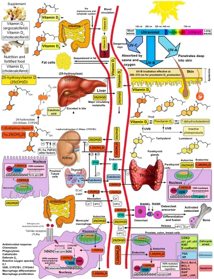
Figure 1 Schematicrepresentation of the synthesis and metabolism of vitamin D for skeletal and nonskeletal function. Note: CopyrightHolick 2013, reproduced with permission.
Abbreviations: DBP, vitamin D-binding protein; Uv, ultraviolet; 25(OH)D, 25-hydroxyvitamin D; 1,25(OH)2D, 1,25-dihydroxyvitamin D; RXR, retinoic acid receptor; vDR,
vitamin D receptor; ecac, epithelial calcium channel; CaBP, calcium-binding protein; RANKL, receptor activator of nuclear factor-kappa B ligand; RANK, receptor activator
of nuclear factor-kappa B; LPS, lipopolysaccharide; TLR, toll-like receptor; PTH, parathyroid hormone; CDK2, cyclin-dependent kinase-2; PSA, prostate-specific antigen; AR,
androgen receptor; Bcl-2, B-cell lymphoma; Bcl-XL, B-cell lymphoma–extra large; 1-OHase, 25-hydroxyvitamin D-la-hydroxylase; IDBP, intracellular D-binding protein; DNA,
deoxyribonucleic acid; RNA, ribonucleic acid; FGF, fibroblast growth factor; 25-OHase, 25-hydroxyvitamin D-24-hydroxylase; NF-κB, nuclear factor-kappa B; IL, interleukin; CYP, cytochrome P450; XIAP, X-linked inhibitor of apoptosis protein; MCL-1, myeloid leukemia cell differentiation protein.
oral nutritional supplements (ONS) in older adults with
and without cognitive impairment.
52The authors showed
that patients exhibited a significant improvement in weight
(P
,
0.0001), body mass index (P
,
0.0001), and cognition at
a 6.5
±
3.9-month follow up (P
=
0.002) when ONS were given,
as compared to the control group.
52Clinical Interventions in Aging downloaded from https://www.dovepress.com/ by 118.70.13.36 on 20-Aug-2020
Dovepress
Schlögl and Holick
However, caution should be applied to the finding
regard-ing the influence of ONS on cognitive performance, as
measured by the Mini-Mental State Examination (MMSE),
since only four studies with a total of 141 patients in the
intervention groups and 130 in the control groups were
included.
52Prevalence of vitamin D deficiency
According to the US Endocrine Society, which addresses
the evaluation and treatment of patients with specific
dis-eases who are at risk for vitamin D deficiency, a cut-off
level
,
20 ng/mL (50 nmol/L) for 25(OH)D defined vitamin D
deficiency.
53The US Institute of Medicine report, which
addresses the dietary reference intake of vitamin D in the
normal, healthy North American population, concluded that
25(OH)D equal to 16 ng/mL (40 nmol/L) should be the
cut-off for vitamin D deficiency, but for maximum bone health,
the team recommended a 25(OH)D level
.
20 ng/mL.
54Recent reviews reported that children, as well as young,
middle-aged, and older adults are at risk for vitamin D
defi-ciency worldwide.
55–57In Europe, vitamin D deficiency in the
elderly is more likely in women than in men, and it is more
common in the south than in the north.
58Based on the
defini-tion of the US Endocrine Society, the prevalence of vitamin D
deficiency was almost one-third of the US population.
59Data
from the Healthy Lifestyle in Europe by Nutrition in
Adoles-cence (HELENA) study, which obtained blood samples from
1,006 adolescents in nine different European countries, also
indicated that vitamin D deficiency is highly prevalent, even
in children.
60Importantly, vitamin D deficiency is associated
with a significantly increased prevalence of hypertension,
obesity, and dyslipidemia, type 2 diabetes, chronic kidney
disease, and endothelial dysfunction.
61,62Vitamin D and neurocognitive
functioning
There is strong evidence that 1,25(OH)
2D contributes to
neuroprotection by modulating the production of nerve
growth,
63–65decreasing L-type calcium channel expression,
66regulating the toxicity of reactive oxygen species,
67–71and
neurotrophic factors such as nerve growth factor,
64,72–76glial
cell-derived neurotrophic factor,
77and nitric oxide synthase.
69Furthermore, vitamin D and its metabolites are involved in
other neuroprotective mechanisms including amyloid
phago-cytosis and clearance,
78,79and vasoprotection.
80Multiple systematic reviews and meta-analyses of
obser-vational studies confirm that cardiovascular risk factors (for
example, hypertension, hypercholesterolemia,
atheroscle-rosis, diabetes mellitus, and smoking) are associated with
low levels of 25(OH)D and predict cardiovascular events
including strokes.
81–86Gunta et al
86recently described
mul-tiple vitamin-D-related pathways that contribute to
cardiovas-cular morbidity and mortality. Vitamin D plays a protective
role in the cardiovascular system through downregulating the
renin–angiotensin–aldosterone system,
87–90cardiac
remodel-ing,
91–93regulating the endothelial response to injury,
94–96and
blood coagulation by increasing thrombus formation and
tissue factor activity (Figure 2).
97Furthermore, 25(OH)D
levels are inversely associated with one’s risk for
develop-ing vascular calcification,
98,99which is known as a marker of
atherosclerotic burden and a risk factor for dementia.
100–103In recent years, the relationship between blood pressure
and cognitive function and dementia has received much
attention from epidemiological research. It is known that
midlife hypertension is an important modifiable risk factor
for late-life cognitive decline,
104,105MCI
106,107and VaD.
108,109Qiu et al
110described that some cross-sectional studies have
shown an inverse association between blood pressure and
the prevalence of dementia and AD, whereas longitudinal
studies yielded mixed results that largely depend on the age
at which blood pressure is measured and the time interval
between blood pressure and outcome assessments.
A recent American Heart Association and American
Stroke Association guidance statement published in 2011
provided an excellent overview of the evidence on vascular
contributions to cognitive impairment and dementia.
111There
is reasonable evidence (class 2a, Level of Evidence B) to
suggest that blood pressure-lowering therapy can be
use-ful for the prevention of late-life dementia among people
who are middle-aged, and for younger elderly individuals.
However, the usefulness of lowering blood pressure in those
over 80 years of age for the prevention of dementia is not well
established (class 2b, Level of Evidence B). Furthermore,
lowering blood pressure in patients who do not have
cogni-tive impairment can reduce the risk of subsequent cognicogni-tive
impairment, whereas lowering blood pressure to preserve
cognition among patients who already have cognitive
impair-ment is not a proven successful strategy.
In 2010, The National Institutes of Health launched a
two-arm, multicenter, randomized clinical trial to
deter-mine whether maintaining blood pressure levels lower than
the current recommendations further reduces one’s risk
of developing cardiovascular and kidney diseases, or
age-related cognitive decline. Called the Systolic Blood
Pres-sure Intervention Trial (SPRINT), this 9-year, $114 million
study will be conducted in more than 80 clinical sites across
Clinical Interventions in Aging downloaded from https://www.dovepress.com/ by 118.70.13.36 on 20-Aug-2020
Dovepress vitamin D and neurocognitive function
the United States. More than 9,000 patients
.
55 years of
age with systolic blood pressure
$
130 mmHg and with
at least one other vascular risk factor will be randomized
to either an “aggressive” treatment arm characterized
by a target systolic blood pressure of
,
120 mmHg, or a
more “routine” arm with a target systolic blood pressure
of
,
140 mmHg. In a substudy (SPRINT-MIND) – which is
funded by the National Institute on Aging and the National
Institute of Neurological Disorders and Stroke –whether
the lower systolic blood pressure goal influences the
occurrence of dementia, change in cognition, and change
in brain structure (on magnetic resonance imaging) will
also be tested.
Vitamin D and mild cognitive
impairment
MCI is a condition that “represents an intermediate state of
cognitive function between the changes seen in aging but does
not fulfill the criteria for dementia.”
112Petersen
112estimated that
between 10% and 20% of people aged 65 years or older suffer
from MCI, and several other studies have shown that patients
with MCI are at a greater risk of developing dementia.
113–116A meta-analysis by Etgen et al
117suggested a more than
dou-bled risk of cognitive impairment in patients with vitamin D
deficiency among 7,688 participants. The authors showed an
increased risk of developing cognitive impairment in those
with low 25(OH)D compared with those with normal 25(OH)D
levels (odds ratio: 2.39; 95% confidence interval: 1.91–3.00;
P
,
0.0001). Only five cross-sectional and two longitudinal
studies were included in the meta-analysis, which underlines
the need for future prospective studies.
One of the studies by Llewellyn et al
118showed an inverse
relationship with serum 25(OH)D and cognitive impairment
in 1,766 adults aged 65 years and older from the Health
Survey for England 2000. There was a 230% increased risk
for cognitive impairment in those with 25(OH)D
,
20 ng/mL
compared to those with a 25(OH)D level
.
20 ng/mL.
Including 2,749 participants from eight studies, Balion
et al
119compared mean MMSE scores between
individu-als with levels of 25(OH)D
,
50 nmol/L and
$
50 nmol/L.
Release ofvitamin D binding protein
Thrombus formation and
tissue factor activity
Migration and proliferation
of VSMCs
Negative inotropic
effects
Osteogenic processes in
VSMCs TNF phosphate
Vascular calcification
Anti-inflammatory cytokine
Inflammation
Macrophage infiltration
Leukocyte and
endothelial adhesion Angiotensin 2
Atherosclerosis
Systolic blood pressure, LVH and cardiovascular
events
Cardiovascular morbidity and mortality
Vitamin D status
Parathyroid hormone
Renin gene promoter activity, renin mRNA expression and plasma
renin
Insulin resistance
Metabolic syndrome or
type 2 diabetes Proinflammatory
cytokines
Figure 2 vitaminD-related pathways, cardiovascular morbidity, and mortality.
Notes: Adecreased serum level of 25-hydroxyvitamin D (vitamin D status) is a risk factor for cardiovascular morbidity and mortality, owing to increases in systolic blood pressure, LvH, and adverse cardiovascular events. These effects may involve various pathways, including increases in endothelial adhesion, which could promote atherosclerosis
causing negative inotropic effects on the heart, vascular calcification through osteogeneic processes in VSMCs, and an increase in thrombogenesis. Furthermore, increases in the inflammatory milieu cause macrophage infiltration, and increased levels of parathyroid hormone could be involved in a complex interaction with the renin–angiotensin
system. Adapted by permission from Macmillan Publishers Ltd: Nature Reviews Nephrology. Gunta SS, Thadhani RI, Mak RH. The effect of vitamin D status on risk factors for
cardiovascular disease. Nat Rev Nephrol. 2013;9(6):337–347.86 Copyright © 2013.
Abbreviations: TNF,tumor necrosis factor; vSMC, vascular smooth muscle cell; LvH, left ventricular hypertrophy; mRNA, messenger RNA.
Clinical Interventions in Aging downloaded from https://www.dovepress.com/ by 118.70.13.36 on 20-Aug-2020
Dovepress
Schlögl and Holick
The authors showed a higher average MMSE score in those
participants with higher 25(OH)D concentrations. There is
also a need for long-term, placebo-controlled, randomized
trials to assess the potential benefits of pharmacologic and
lifestyle interventions in persons with MCI. A very promising
randomized-controlled trial (DO-HEALTH) began
enroll-ing participants in December 2012; it will enroll a total of
2,152 community-dwelling men and women aged 70 years
of age to test the individual and the combined benefits of
2,000 IU of vitamin D/day, 1 g of omega-3 fatty acids/day,
and a simple home exercise program (
http://do-health.eu/
wordpress/
). One of the five primary endpoints is the risk of
functional decline.
Vitamin D, Alzheimer’s disease,
and vascular dementia
AD is the best known and the most common cause of dementia
in older people.
120According to a study by Ferri et al
121that
was conducted in 2005, the global prevalence of dementia
was 24.3 million. The authors hypothesized that this number
will double every 20 years to a total of 42 million individuals
by 2020 and 81 million people by 2040. VaD is the second
most common type of dementia.
122–124According to The
Aging, Demographics, and Memory Study (ADAMS), the
prevalence of VaD in the United States among those aged
71 years and older has been estimated to be approximately
594,000.
125The development of clinical AD and VaD is very
complex,
126,127since several pathophysiological pathways
leading to vascular and neurodegenerative processes are
simi-lar.
128Importantly, macroscopic infarcts are very common in
approximately one-third to one-half of older people,
129–132and
infarcts frequently coexist with AD pathology in the brains
of older people.
130,132–136Several studies showed that
cerebro-vascular lesions lower the threshold of the AD-type changes
that are necessary to cause cognitive decline.
133,135,137It has to be acknowledged that the prevalence and
inci-dence figures from AD and VaD pertain to diagnostic
thresh-olds for these disorders,
111and that there exist multiple sets
of criteria for VaD.
124Most older studies use the construct of
VaD, and more recently, the term “vascular cognitive
impair-ment” has been introduced to capture the entire spectrum of
cognitive disorders that range from MCI to fully developed
dementia.
111Since most of the recent systematic reviews
and meta-analyses that have been published within the last
3–5 years, the old term (VaD) has been used to characterize
cognitive syndromes associated with vascular disease and
cog-nitive decline. A meta-analysis from Balion et al,
119which was
conducted using five different databases including 37 different
studies published in 2012, compared cognition (as measured
by the MMSE) to 25(OH)D levels. The results showed that
individuals with AD had lower 25(OH)D concentrations
compared to those without AD. In addition, MMSE scores
were lower in patients with lower 25(OH)D concentrations.
However, the authors noted that the nature of the
relation-ship between cognition and 25(OH)D concentrations is still
not clear. In contrast to Balion et al,
119who included studies
with and without regression models to answer this question,
Annweiler et al
114restricted their report to studies that used
regression models. The authors concluded that in older adults,
vitamin D deficiency was associated with dementia,
138–141and
that vitamin D supplementation might have a protective effect.
Similar results were reported by Barnard and Colón- Emeric.
142Furthermore, in a systematic review and meta-analysis,
Ann-weiler et al
143critically analyzed the domain-specific cognitive
performance affected in vitamin D deficiency. The authors
demonstrated that vitamin D deficiency “is cross-sectionally
associated in adults with episodic memory disorders and
executive dysfunctions, in particular mental shifting,
infor-mation updating, and processing speed.”
143Recently, van der
Schaft et al
144also conducted a systematic review that included
25 studies with a cross-sectional design and six studies with
a prospective design; three of these studies showed
cross-sectional as well as prospective data.
145–147The main finding
was a statistically significantly worse outcome on one or more
cognitive function tests, or a higher frequency of dementia,
with lower 25(OH)D levels or vitamin D intake in 72% of the
studies. In addition, 67% of the prospective studies showed
a higher risk of cognitive decline after a follow-up period
of 4–7 years in participants with lower 25(OH)D levels at
baseline compared with participants with higher 25(OH)D
levels.
Importantly, several limitations have to be considered
while interpreting the data of the systematic reviews and
meta-analyses. Cross-sectional studies cannot answer the
question of whether vitamin D deficiency leads to cognitive
decline, or whether people with a cognition disorder have
lower exposure to sunlight or lower vitamin D intake, nor do
they reflect seasonal fluctuation of vitamin D status.
144Using
different cut-off points for vitamin D status classification,
and different diagnostic criteria for MCI and VaD, make it
difficult to compare these studies. Finally, the differences in
adjustments for potential confounders such as age, sex, race,
depression, level of education, diabetes, hypertension, kidney
disease, physical activity, and/or season that the sample was
obtained may explain some of the different study results
reported in the systematic reviews and meta-analyses.
Clinical Interventions in Aging downloaded from https://www.dovepress.com/ by 118.70.13.36 on 20-Aug-2020
Dovepress vitamin D and neurocognitive function
Conclusion
Older adults are at a high risk of developing vitamin D
deficiency due to decreased cutaneous synthesis and dietary
intake of vitamin D. Vitamin D deficiency is associated
with substantial increases in the incidence of hypertension,
hyperlipidemia, myocardial infarction, stroke, fractures, and
diabetes. Vitamin D signaling is involved in brain
develop-ment and function. Many studies have shown that AD and
VaD share hypertension as a common risk factor, and there
is reasonable evidence to suggest blood pressure-lowering
therapy can be useful for the prevention of late-life dementia
for middle-aged and younger elderly individuals, whereas
the usefulness of lowering blood pressure among those over
80 years of age for the prevention of dementia is not well
established. The overlap between AD and VaD makes it
difficult to estimate to what extent each disease contributes
to cognitive decline. The majority of the cross-sectional and
prospective studies found that vitamin D deficiency is
associ-ated with a statistically significantly worse outcome on one
or more cognitive function tests, or with a higher frequency
of MCI and dementia. The identification of people who are
at risk for cognitive impairment holds realistic promise for
the prevention or postponement of dementia. There is a need
for long-term, placebo-controlled, randomized trials to assess
the potential benefits of pharmacologic and lifestyle
interven-tions in persons with MCI, VaD, and AD.
Acknowledgments
This work was supported, in part, by the National Institutes
of Health Clinical Translational Science Institute Grant
UL-1-RR-25711.
Disclosure
The authors report no conflicts of interest in this work.
References
1. Holick MF. Vitamin D deficiency. N Engl J Med. 2007;357(3): 266–281. 2. Dobs AS, Levine MA, Margolis S. Effects of pravastatin, a new HMG-CoA reductase inhibitor, on vitamin D synthesis in man. Metabolism. 1991;40(5):524–528.
3. Godar DE, Pope SJ, Grant WB, Holick MF. Solar UV doses of adult Americans and vitamin D(3) production. Dermatoendocrinol. 2011;3(4):243–250.
4. Holick MF. Optimal vitamin D status for the prevention and treatment of osteoporosis. Drugs Aging. 2007;24(12):1017–1029.
5. Holick MF. Evolution and function of vitamin D. Recent Results Cancer Res. 2003;164:3–28.
6. Lehmann B, Tiebel O, Meurer M. Expression of vitamin D3 25-hydroxylase (CYP27) mRNA after induction by vitamin D3 or UVB radiation in keratinocytes of human skin equivalents – a preliminary study. Arch Dermatol Res. 1999;291(9):507–510.
7. Bikle DD, Halloran BP, Riviere JE. Production of 1,25 dihydroxyvitamin D3 by perfused pig skin. J Invest Dermatol. 1994;102(5):796–798.
8. Seifert M, Tilgen W, Reichrath J. Expression of 25-hydroxyvitamin D-1alpha-hydroxylase (1alphaOHase, CYP27B1) splice variants in HaCaT keratinocytes and other skin cells: modulation by culture conditions and UV-B treatment in vitro. Anticancer Res. 2009;29(9): 3659–3667.
9. Fu GK, Lin D, Zhang MY, et al. Cloning of human 25-hydroxyvitamin D-1 alpha-hydroxylase and mutations causing vitamin D-dependent rickets type 1. Mol Endocrinol. 1997;11(13):1961–1970.
10. Lehmann B, Genehr T, Knuschke P, Pietzsch J, Meurer M. UVB-induced conversion of 7-dehydrocholesterol to 1alpha, 25-dihydroxyvitamin D3 in an in vitro human skin equivalent model. J Invest Dermatol. 2001;117(5):1179–1185.
11. Mangelsdorf DJ, Thummel C, Beato M, et al. The nuclear receptor superfamily: the second decade. Cell. 1995;83(6):835–839.
12. Sutherland MK, Somerville MJ, Yoong LK, Bergeron C, Haussler MR, McLachlan DR. Reduction of vitamin D hormone receptor mRNA levels in Alzheimer as compared to Huntington hippocampus: correlation with calbindin-28k mRNA levels. Brain Res Mol Brain Res. 1992;13(3):239–250.
13. Eyles DW, Smith S, Kinobe R, Hewison M, McGrath JJ. Distribution of the vitamin D receptor and 1 alpha-hydroxylase in human brain. J Chem Neuroanat. 2005;29(1):21–30.
14. Dhikav V, Anand K. Potential predictors of hippocampal atrophy in Alzheimer’s disease. Drugs Aging. 2011;28(1):1–11.
15. Mu Y, Gage FH. Adult hippocampal neurogenesis and its role in Alzheimer’s disease. Mol Neurodegener. 2011;6:85.
16. Fotuhi M, Do D, Jack C. Modifiable factors that alter the size of the hippocampus with ageing. Nat Rev Neurol. 2012;8(4):189–202. 17. Calabresi P, Castrioto A, Di Filippo M, Picconi B. New experimental
and clinical links between the hippocampus and the dopaminergic system in Parkinson’s disease. Lancet Neurol. 2013;12(8): 811–821.
18. Nimitphong H, Holick MF. Vitamin D, neurocognitive functioning and immunocompetence. Curr Opin Clin Nutr Metab Care. 2011; 14(1):7–14.
19. Beydoun MA, Ding EL, Beydoun HA, Tanaka T, Ferrucci L, Zonderman AB. Vitamin D receptor and megalin gene polymorphisms and their associations with longitudinal cognitive change in US adults. Am J Clin Nutr. 2012;95(1):163–178.
20. Kuningas M, Mooijaart SP, Jolles J, Slagboom PE, Westendorp RG, van Heemst D. VDR gene variants associate with cognitive function and depressive symptoms in old age. Neurobiol Aging. 2009;30(3): 466–473.
21. Luedecking-Zimmer E, DeKosky ST, Nebes R, Kamboh MI. Association of the 3′ UTR transcription factor LBP-1c/CP2/LSF polymorphism with late-onset Alzheimer’s disease. Am J Med Genet B Neuropsychiatr Genet. 2003;117B(1):114–117.
22. Gezen-Ak D, Dursun E, Ertan T, et al. Association between vitamin D receptor gene polymorphism and Alzheimer’s disease. Tohoku J Exp Med. 2007;212(3):275–282.
23. Lehmann DJ, Refsum H, Warden DR, Medway C, Wilcock GK, Smith AD. The vitamin D receptor gene is associated with Alzheimer’s disease. Neurosci Lett. 2011;504(2):79–82.
24. Gezen-Ak D, Dursun E, Bilgiç B, et al. Vitamin D receptor gene hap-lotype is associated with late-onset Alzheimer’s disease. Tohoku J Exp Med. 2012;228(3):189–196.
25. Kim JS, Kim YI, Song C, et al. Association of vitamin D receptor gene polymorphism and Parkinson’s disease in Koreans. J Korean Med Sci. 2005;20(3):495–498.
26. Butler MW, Burt A, Edwards TL, et al. Vitamin D receptor gene as a candi-date gene for Parkinson disease. Ann Hum Genet. 2011;75(2): 201–210. 27. Han X, Xue L, Li Y, Chen B, Xie A. Vitamin D receptor gene poly-morphism and its association with Parkinson’s disease in Chinese Han population. Neurosci Lett. 2012;525(1):29–33.
28. Lv Z, Tang B, Sun Q, Yan X, Guo J. Association study between vitamin d receptor gene polymorphisms and patients with Parkinson disease in Chinese Han population. Int J Neurosci. 2013;123(1):60–64.
Clinical Interventions in Aging downloaded from https://www.dovepress.com/ by 118.70.13.36 on 20-Aug-2020
Dovepress
Schlögl and Holick
29. Török R, Török N, Szalardy L, et al. Association of vitamin D receptor gene polymorphisms and Parkinson’s disease in Hungarians. Neurosci Lett. 2013;551:70–74.
30. Krizova L, Kollar B, Jezova D, Turcani P. Genetic aspects of vitamin D receptor and metabolism in relation to the risk of multiple sclerosis. Gen Physiol Biophys. Epub September 26, 2013.
31. MacLaughlin J, Holick MF. Aging decreases the capacity of human skin to produce vitamin D3. J Clin Invest. 1985;76(4):1536–1538. 32. Harris SS, Dawson-Hughes B. Plasma vitamin D and 25OHD responses
of young and old men to supplementation with vitamin D3. J Am Coll Nutr. 2002;21(4):357–362.
33. Gallagher JC, Rapuri P, Smith L. Falls are associated with decreased renal function and insufficient calcitriol production by the kidney. J Steroid Biochem Mol Biol. 2007;103(3–5):610–613.
34. Armbrecht HJ, Zenser TV, Davis BB. Effect of age on the conversion of 25-hydroxyvitamin D3 to 1,25-dihydroxyvitamin D3 by kidney of rat. J Clin Invest. 1980;66(5):1118–1123.
35. Need AG, Morris HA, Horowitz M, Nordin C. Effects of skin thickness, age, body fat, and sunlight on serum 25-hydroxyvitamin D. Am J Clin Nutr. 1993;58(6):882–885.
36. Holick MF, Matsuoka LY, Wortsman J. Age, vitamin D, and solar ultraviolet. Lancet. 1989;2(8671):1104–1105.
37. Gau JT. Prevalence of vitamin D deficiency/insufficiency practice patterns in nursing homes. J Am Med Dir Assoc. 2010;11(4):296. 38. Morley JE. Vitamin d redux. J Am Med Dir Assoc. 2009;10(9): 591–592. 39. Braddy KK, Imam SN, Palla KR, Lee TA. Vitamin d deficiency/insuf-ficiency practice patterns in a veterans health administration long-term care population: a retrospective analysis. J Am Med Dir Assoc. 2009;10(9):653–657.
40. Perry HM, Horowitz M, Morley JE, et al. Longitudinal changes in serum 25-hydroxyvitamin D in older people. Metabolism. 1999;48(8): 1028–1032.
41. Clegg A, Young J, Iliffe S, Rikkert MO, Rockwood K. Frailty in elderly people. Lancet. 2013;381(9868):752–762.
42. Welge-Lüssen A. Ageing, neurodegeneration, and olfactory and gusta-tory loss. B-ENT. 2009;5 Suppl 13:129–132.
43. Methven L, Allen VJ, Withers CA, Gosney MA. Ageing and taste. Proc Nutr Soc. 2012;71(4):556–565.
44. Guiglia R, Musciotto A, Compilato D, et al. Aging and oral health: effects in hard and soft tissues. Curr Pharm Des. 2010;16(6):619–630. 45. Zadak Z, Hyspler R, Ticha A, Vlcek J. Polypharmacy and malnutrition.
Curr Opin Clin Nutr Metab Care. 2013;16(1):50–55.
46. Tamura BK, Bell CL, Masaki KH, Amella EJ. Factors associated with weight loss, low BMI, and malnutrition among nursing home patients: a systematic review of the literature. J Am Med Dir Assoc. 2013;14(9): 649–655.
47. van Bokhorst-de van der Schueren MA, Lonterman-Monasch S, de Vries OJ, Danner SA, Kramer MH, Muller M. Prevalence and determinants for malnutrition in geriatric outpatients. Clin Nutr. 2013;32(6):1007–1011.
48. Roqué M, Salvà A, Vellas B. Malnutrition in community-dwelling adults with dementia (NutriAlz Trial). J Nutr Health Aging. 2013;17(4):295–299.
49. Ikeda M, Brown J, Holland AJ, Fukuhara R, Hodges JR. Changes in appetite, food preference, and eating habits in frontotemporal dementia and Alzheimer’s disease. J Neurol Neurosurg Psychiatry. 2002;73(4): 371–376.
50. Manthorpe J, Watson R. Poorly served? Eating and dementia. J Adv Nurs. 2003;41(2):162–169.
51. Greenwood CE, Tam C, Chan M, Young KW, Binns MA, van Reekum R. Behavioral disturbances, not cognitive dete-rioration, are associated with altered food selection in seniors with Alzheimer’s disease. J Gerontol A Biol Sci Med Sci. 2005;60(4): 499–505.
52. Allen VJ, Methven L, Gosney MA. Use of nutritional complete supple-ments in older adults with dementia: Systematic review and meta-analysis of clinical outcomes. Clin Nutr. Epub Mar 28, 2013.
53. Holick MF, Binkley NC, Bischoff-Ferrari HA, et al; Endocrine Society. Evaluation, treatment, and prevention of vitamin D deficiency: an Endocrine Society clinical practice guideline. J Clin Endocrinol Metab. 2011;96(7):1911–1930.
54. Institute of Medicine (US) Committee to Review Dietary Reference Intakes for Vitamin D and Calcium; Ross AC, Taylor CL, Yaktine AL, Del Valle HB, editors. Dietary Reference Intakes for Calcium and Vitamin D. Washington, DC: National Academies Press (US); 2011. 55. Hagenau T, Vest R, Gissel TN, et al. Global vitamin D levels in relation to
age, gender, skin pigmentation and latitude: an ecologic meta-regression analysis. Osteoporos Int. 2009;20(1):133–140.
56. Wahl DA, Cooper C, Ebeling PR, et al. A global representation of vita-min D status in healthy populations. Arch Osteoporos. 2012; 7(1–2): 155–172.
57. Hossein-Nezhad A, Holick MF. Vitamin d for health: a global perspective. Mayo Clin Proc. 2013;88(7):720–755.
58. van der Wielen RP, Löwik MR, van den Berg H, et al. Serum vitamin D concentrations among elderly people in Europe. Lancet. 1995;346(8969):207–210.
59. Ganji V, Zhang X, Tangpricha V. Serum 25-hydroxyvitamin D concentra-tions and prevalence estimates of hypovitaminosis D in the US population based on assay-adjusted data. J Nutr. 2012;142(3): 498–507. 60. Valtueña J, Gracia-Marco L, Huybrechts I, et al; Helena Study Group.
Cardiorespiratory fitness in males, and upper limbs muscular strength in females, are positively related with 25-hydroxyvitamin D plasma concentrations in European adolescents: the HELENA study. QJM. 2013;106(9):809–821.
61. Anderson JL, May HT, Horne BD, et al; Intermountain Heart Collaborative (IHC) Study Group. Relation of vitamin D deficiency to cardiovascular risk factors, disease status, and incident events in a general healthcare population. Am J Cardiol. 2010;106(7): 963–968.
62. Kienreich K, Tomaschitz A, Verheyen N, et al. Vitamin D and cardiovascular disease. Nutrients. 2013;5(8):3005–3021.
63. Chabas JF, Alluin O, Rao G, et al. Vitamin D2 potentiates axon regeneration. J Neurotrauma. 2008;25(10):1247–1256.
64. Brown J, Bianco JI, McGrath JJ, Eyles DW. 1,25-dihydroxyvitamin D3 induces nerve growth factor, promotes neurite outgrowth and inhibits mitosis in embryonic rat hippocampal neurons. Neurosci Lett. 2003;343(2):139–143.
65. Marini F, Bartoccini E, Cascianelli G, et al. Effect of 1alpha, 25-dihydroxyvitamin D3 in embryonic hippocampal cells. Hippocampus. 2010;20(6):696–705.
66. Brewer LD, Thibault V, Chen KC, Langub MC, Landfield PW, Porter NM. Vitamin D hormone confers neuroprotection in parallel with downregulation of L-type calcium channel expression in hippocampal neurons. J Neurosci. 2001;21(1):98–108.
67. Ibi M, Sawada H, Nakanishi M, et al. Protective effects of 1 alpha, 25-(OH)(2)D(3) against the neurotoxicity of glutamate and reactive oxygen species in mesencephalic culture. Neuropharmacology. 2001;40(6):761–771.
68. Kröncke KD, Klotz LO, Suschek CV, Sies H. Comparing nitrosative versus oxidative stress toward zinc finger-dependent transcription. Unique role for NO. J Biol Chem. 2002;277(15):13294–13301. 69. Garcion E, Sindji L, Montero-Menei C, Andre C, Brachet P, Darcy F.
Expression of inducible nitric oxide synthase during rat brain inflammation: regulation by 1,25-dihydroxyvitamin D3. Glia. 1998;22(3):282–294.
70. Chen KB, Lin AM, Chiu TH. Systemic vitamin D3 attenuated oxidative injuries in the locus coeruleus of rat brain. Ann N Y Acad Sci. 2003;993:313–324; discussion 345–349.
71. Lin AM, Fan SF, Yang DM, Hsu LL, Yang CH. Zinc-induced apoptosis in substantia nigra of rat brain: neuroprotection by vitamin D3. Free Radic Biol Med. 2003;34(11):1416–1425.
72. Wion D, MacGrogan D, Neveu I, Jehan F, Houlgatte R, Brachet P. 1,25-Dihydroxyvitamin D3 is a potent inducer of nerve growth factor synthesis. J Neurosci Res. 1991;28(1):110–114.
Clinical Interventions in Aging downloaded from https://www.dovepress.com/ by 118.70.13.36 on 20-Aug-2020
Dovepress vitamin D and neurocognitive function
73. Neveu I, Naveilhan P, Baudet C, Brachet P, Metsis M. 1,25-dihydroxyvitamin D3 regulates NT-3, NT-4 but not BDNF mRNA in astrocytes. Neuroreport. 1994;6(1):124–126.
74. Neveu I, Naveilhan P, Jehan F, et al. 1,25-dihydroxyvitamin D3 regulates the synthesis of nerve growth factor in primary cultures of glial cells. Brain Res Mol Brain Res. 1994;24(1–4):70–76.
75. Musiol IM, Feldman D. 1,25-dihydroxyvitamin D3 induction of nerve growth factor in L929 mouse fibroblasts: effect of vitamin D receptor regulation and potency of vitamin D3 analogs. Endocrinology. 1997;138(1):12–18.
76. Veenstra TD, Fahnestock M, Kumar R. An AP-1 site in the nerve growth factor promoter is essential for 1,25-dihydroxyvitamin D3- mediated nerve growth factor expression in osteoblasts. Biochemistry. 1998;37(17):5988–5994.
77. Naveilhan P, Neveu I, Wion D, Brachet P. 1,25-Dihydroxyvitamin D3, an inducer of glial cell line-derived neurotrophic factor. Neuroreport. 1996;7(13):2171–2175.
78. Masoumi A, Goldenson B, Ghirmai S, et al. 1alpha,25-dihydroxyvitamin D3 interacts with curcuminoids to stimulate amyloid-beta clearance by macrophages of Alzheimer’s disease patients. J Alzheimers Dis. 2009;17(3):703–717.
79. Mizwicki MT, Liu G, Fiala M, et al. 1α,25-dihydroxyvitamin D3 and resolvin D1 retune the balance between amyloid-β phagocytosis and inflammation in Alzheimer’s disease patients. J Alzheimers Dis. 2013;34(1):155–170.
80. Pludowski P, Holick MF, Pilz S, et al. Vitamin D effects on musculoskeletal health, immunity, autoimmunity, cardiovascular dis-ease, cancer, fertility, pregnancy, dementia and mortality-a review of recent evidence. Autoimmun Rev. 2013;12(10):976–989.
81. Witham MD, Nadir MA, Struthers AD. Effect of vitamin D on blood pressure: a systematic review and meta-analysis. J Hypertens. 2009;27(10):1948–1954.
82. Pilz S, Tomaschitz A, März W, et al. Vitamin D, cardiovascular disease and mortality. Clin Endocrinol (Oxf). 2011;75(5):575–584.
83. Muscogiuri G, Sorice GP, Ajjan R, et al. Can vitamin D defi-ciency cause diabetes and cardiovascular diseases? Present evidence and future perspectives. Nutr Metab Cardiovasc Dis. 2012;22(2):81–87.
84. Wang L, Song Y, Manson JE, et al. Circulating 25-hydroxy-vitamin D and risk of cardiovascular disease: a meta-analysis of prospective studies. Circ Cardiovasc Qual Outcomes. 2012;5(6):819–829.
85. Brøndum-Jacobsen P, Nordestgaard BG, Schnohr P, Benn M. 25-hydroxyvitamin D and symptomatic ischemic stroke: an original study and meta-analysis. Ann Neurol. 2013;73(1):38–47.
86. Gunta SS, Thadhani RI, Mak RH. The effect of vitamin D status on risk fac-tors for cardiovascular disease. Nat Rev Nephrol. 2013;9(6):337–347. 87. Li YC, Kong J, Wei M, Chen ZF, Liu SQ, Cao LP. 1,25-Dihydroxyvitamin
D(3) is a negative endocrine regulator of the renin-angiotensin system. J Clin Invest. 2002;110(2):229–238.
88. Li YC. Vitamin D regulation of the renin-angiotensin system. J Cell Biochem. 2003;88(2):327–331.
89. Zhou C, Lu F, Cao K, Xu D, Goltzman D, Miao D. Calcium-independent and 1,25(OH)2D3-dependent regulation of the renin- angiotensin system in 1alpha-hydroxylase knockout mice. Kidney Int. 2008;74(2): 170–179. 90. Forman JP, Williams JS, Fisher ND. Plasma 25-hydroxyvitamin D and
regulation of the renin-angiotensin system in humans. Hypertension. 2010;55(5):1283–1288.
91. Sanna B, Brandt EB, Kaiser RA, et al. Modulatory interacting proteins 1 and 2 function as calcineurin facilitators in vivo. Proc Natl Acad Sci U S A. 2006;103(19):7327–7332.
92. Bodyak N, Ayus JC, Achinger S, et al. Activated vitamin D attenuates left ventricular abnormalities induced by dietary sodium in Dahl salt-sensitive animals. Proc Natl Acad Sci U S A. 2007;104(43): 16810–16815.
93. Chen S, Law CS, Grigsby CL, et al. Cardiomyocyte-specific dele-tion of the vitamin D receptor gene results in cardiac hypertrophy. Circulation. 2011;124(17):1838–1847.
94. Tarcin O, Yavuz DG, Ozben B, et al. Effect of vitamin D deficiency and replacement on endothelial function in asymptomatic subjects. J Clin Endocrinol Metab. 2009;94(10):4023–4030.
95. Caprio M, Mammi C, Rosano GM. Vitamin D: a novel player in endothelial function and dysfunction. Arch Med Sci. 2012;8(1): 4–5.
96. Sypniewska G, Pollak J, Strozecki P, et al. 25-Hydroxyvitamin D, biomarkers of endothelial dysfunction and subclinical organ damage in adults with hypertension. Am J Hypertens. Epub September 16, 2013. 97. Aihara K, Azuma H, Akaike M, et al. Disruption of nuclear vitamin D
receptor gene causes enhanced thrombogenicity in mice. J Biol Chem. 2004;279(34):35798–35802.
98. Watson KE, Abrolat ML, Malone LL, et al. Active serum vitamin D levels are inversely correlated with coronary calcification. Circulation. 1997;96(6):1755–1760.
99. de Boer IH, Kestenbaum B, Shoben AB, Michos ED, Sarnak MJ, Siscovick DS. 25-hydroxyvitamin D levels inversely associate with risk for developing coronary artery calcification. J Am Soc Nephrol. 2009;20(8):1805–1812.
100. Bos D, Vernooij MW, Elias-Smale SE, et al. Atherosclerotic calcification relates to cognitive function and to brain changes on magnetic resonance imaging. Alzheimers Dement. 2012;8(Suppl 5): S104–S111.
101. Yarchoan M, Xie SX, Kling MA, et al. Cerebrovascular atherosclerosis correlates with Alzheimer pathology in neurodegen-erative dementias. Brain. 2012;135(Pt 12):3749–3756.
102. Roher AE, Tyas SL, Maarouf CL, et al. Intracranial atherosclerosis as a contributing factor to Alzheimer’s disease dementia. Alzheimers Dement. 2011;7(4):436–444.
103. Vidal JS, Sigurdsson S, Jonsdottir MK, et al. Coronary artery calcium, brain function and structure: the AGES-Reykjavik Study. Stroke. 2010;41(5):891–897.
104. Knopman D, Boland LL, Mosley T, et al; Atherosclerosis Risk in Com-munities (ARIC) Study Investigators. Cardiovascular risk factors and cognitive decline in middle-aged adults. Neurology. 2001;56(1):42–48. 105. Goldstein FC, Levey AI, Steenland NK. High blood pressure and
cognitive decline in mild cognitive impairment. J Am Geriatr Soc. 2013;61(1):67–73.
106. Kivipelto M, Helkala EL, Hänninen T, et al. Midlife vascular risk factors and late-life mild cognitive impairment: A population-based study. Neurology. 2001;56(12):1683–1689.
107. Reitz C, Tang MX, Manly J, Mayeux R, Luchsinger JA. Hypertension and the risk of mild cognitive impairment. Arch Neurol. 2007;64(12):1734–1740.
108. Launer LJ, Ross GW, Petrovitch H, et al. Midlife blood pressure and dementia: the Honolulu-Asia aging study. Neurobiol Aging. 2000;21(1):49–55.
109. Yamada M, Mimori Y, Kasagi F, Miyachi T, Ohshita T, Sasaki H. Incidence and risks of dementia in Japanese women: Radiation Effects Research Foundation Adult Health Study. J Neurol Sci. 2009;283(1–2):57–61. 110. Qiu C, Winblad B, Fratiglioni L. The age-dependent relation of
blood pressure to cognitive function and dementia. Lancet Neurol. 2005;4(8):487–499.
111. Gorelick PB, Scuteri A, Black SE, et al; American Heart Association Stroke Council, Council on Epidemiology and Prevention, Council on Cardiovascular Nursing, Council on Cardiovascular Radiology and Inter-vention, and Council on Cardiovascular Surgery and Anesthesia. Vascular contributions to cognitive impairment and dementia: a statement for healthcare professionals from the american heart association/ american stroke association. Stroke. 2011;42(9):2672–2713.
112. Petersen RC. Clinical Practice. Mild cognitive impairment. N Engl J Med. 2011;364(23):2227–2234.
113. Ganguli M, Snitz BE, Saxton JA, et al. Outcomes of mild cognitive impairment by def inition: a population study. Arch Neurol. 2011;68(6):761–767.
114. Annweiler C, Schott AM, Berrut G, et al. Vitamin D and ageing: neurological issues. Neuropsychobiology. 2010;62(3):139–150.
Clinical Interventions in Aging downloaded from https://www.dovepress.com/ by 118.70.13.36 on 20-Aug-2020
Clinical Interventions in Aging
Publish your work in this journal
Submit your manuscript here: http://www.dovepress.com/clinical-interventions-in-aging-journal
Clinical Interventions in Aging is an international, peer-reviewed journal focusing on evidence-based reports on the value or lack thereof of treat-ments intended to prevent or delay the onset of maladaptive correlates of aging in human beings. This journal is indexed on PubMed Central, MedLine, the American Chemical Society’s ‘Chemical Abstracts
Service’ (CAS), Scopus and the Elsevier Bibliographic databases. The manuscript management system is completely online and includes a very quick and fair peer-review system, which is all easy to use. Visit http://www.dovepress.com/testimonials.php to read real quotes from published authors.
Dovepress
Dove
press
Schlögl and Holick
115. Annweiler C, Allali G, Allain P, et al. Vitamin D and cognitive per-formance in adults: a systematic review. Eur J Neurol. 2009;16(10): 1083–1089.
116. Grant WB. Does vitamin D reduce the risk of dementia? J Alzheimers Dis. 2009;17(1):151–159.
117. Etgen T, Sander D, Bickel H, Sander K, Förstl H. Vitamin D deficiency, cognitive impairment and dementia: a systematic review and meta-analysis. Dement Geriatr Cogn Disord. 2012;33(5):297–305. 118. Llewellyn DJ, Langa KM, Lang IA. Serum 25-hydroxyvitamin D
concentration and cognitive impairment. J Geriatr Psychiatry Neurol. 2009;22(3):188–195.
119. Balion C, Griffith LE, Strifler L, et al. Vitamin D, cognition, and dementia: a systematic review and meta-analysis. Neurology. 2012;79(13):1397–1405.
120. Defina PA, Moser RS, Glenn M, Lichtenstein JD, Fellus J. Alzheimer’s disease clinical and research update for health care practitioners. J Aging Res. 2013;2013:207178.
121. Ferri CP, Prince M, Brayne C, et al; Alzheimer’s Disease International. Global prevalence of dementia: a Delphi consensus study. Lancet. 2005;366(9503):2112–2117.
122. Chui HC, Victoroff JI, Margolin D, Jagust W, Shankle R, Katzman R. Criteria for the diagnosis of ischemic vascular dementia proposed by the State of California Alzheimer’s Disease Diagnostic and Treatment Centers. Neurology. 1992;42(3 Pt 1):473–480.
123. Román GC, Tatemichi TK, Erkinjuntti T, et al. Vascular dementia: diagnostic criteria for research studies. Report of the NINDS-AIREN International Workshop. Neurology. 1993;43(2):250–260.
124. Hachinski V. Vascular dementia: a radical redefinition. Dementia. 1994;5(3–4):130–132.
125. Plassman BL, Langa KM, Fisher GG, et al. Prevalence of dementia in the United States: the aging, demographics, and memory study. Neuroepidemiology. 2007;29(1–2):125–132.
126. Langbaum JB, Fleisher AS, Chen K, et al. Ushering in the study and treatment of preclinical Alzheimer disease. Nat Rev Neurol. 2013;9(7):371–381.
127. Wiesmann M, Kiliaan AJ, Claassen JA. Vascular aspects of cogni-tive impairment and dementia. J Cereb Blood Flow Metab. Epub September 11, 2013.
128. Iadecola C. The overlap between neurodegenerative and vascular factors in the pathogenesis of dementia. Acta Neuropathol. 2010;120(3):287–296.
129. Neuropathology Group. Medical Research Council Cognitive Func-tion and Aging Study. Pathological correlates of late-onset dementia in a multicentre, community-based population in England and Wales. Neuropathology Group of the Medical Research Council Cognitive Function and Ageing Study (MRC CFAS). Lancet. 2001;357(9251):169–175.
130. White L, Small BJ, Petrovitch H, et al. Recent clinical-pathologic research on the causes of dementia in late life: update from the Honolulu-Asia Aging Study. J Geriatr Psychiatry Neurol. 2005;18(4): 224–227. 131. Sonnen JA, Larson EB, Crane PK, et al. Pathological correlates of
dementia in a longitudinal, population-based sample of aging. Ann Neurol. 2007;62(4):406–413.
132. Schneider JA, Aggarwal NT, Barnes L, Boyle P, Bennett DA. The neu-ropathology of older persons with and without dementia from commu-nity versus clinic cohorts. J Alzheimers Dis. 2009;18(3):691–701. 133. Esiri MM, Nagy Z, Smith MZ, Barnetson L, Smith AD.
Cerebrovascular disease and threshold for dementia in the early stages of Alzheimer’s disease. Lancet. 1999;354(9182):919–920.
134. Snowdon DA, Greiner LH, Mortimer JA, Riley KP, Greiner PA, Markesbery WR. Brain infarction and the clinical expression of Alzheimer disease. The Nun Study. JAMA. 1997;277(10):813–817. 135. Schneider JA, Wilson RS, Bienias JL, Evans DA, Bennett DA. Cerebral
infarctions and the likelihood of dementia from Alzheimer disease pathology. Neurology. 2004;62(7):1148–1155.
136. Schneider JA, Arvanitakis Z, Bang W, Bennett DA. Mixed brain pathologies account for most dementia cases in community-dwelling older persons. Neurology. 2007;69(24):2197–2204.
137. Zekry D, Duyckaerts C, Belmin J, Geoffre C, Moulias R, Hauw JJ. Alzheimer’s disease and brain infarcts in the elderly. Agreement with neuropathology. J Neurol. 2002;249(11):1529–1534.
138. Buell JS, Dawson-Hughes B, Scott TM, et al. 25-Hydroxyvitamin D, dementia, and cerebrovascular pathology in elders receiving home services. Neurology. 2010;74(1):18–26.
139. Annweiler C, Fantino B, Le Gall D, Schott AM, Berrut G, Beauchet O. Severe vitamin D deficiency is associated with advanced-stage dementia in geriatric inpatients. J Am Geriatr Soc. 2011;59(1): 169–171.
140. Annweiler C, Rolland Y, Schott AM, et al. Higher vitamin D dietary intake is associated with lower risk of alzheimer’s disease: a 7-year follow-up. J Gerontol A Biol Sci Med Sci. 2012;67(11): 1205–1211.
141. Annweiler C, Llewellyn DJ, Beauchet O. Low serum vitamin D concentrations in Alzheimer’s disease: a systematic review and meta-analysis. J Alzheimers Dis. 2013;33(3):659–674.
142. Barnard K, Colón-Emeric C. Extraskeletal effects of vitamin D in older adults: cardiovascular disease, mortality, mood, and cognition. Am J Geriatr Pharmacother. 2010;8(1):4–33.
143. Annweiler C, Montero-Odasso M, Llewellyn DJ, Richard-Devantoy S, Duque G, Beauchet O. Meta-analysis of memory and executive dysfunctions in relation to vitamin D. J Alzheimers Dis. 2013;37(1): 147–171.
144. van der Schaft J, Koek HL, Dijkstra E, Verhaar HJ, van der Schouw YT, Emmelot-Vonk MH. The association between vitamin D and cognition: a systematic review. Ageing Res Rev. Epub May 29, 2013. 145. Llewellyn DJ, Lang IA, Langa KM, et al. Vitamin D and risk of
cognitive decline in elderly persons. Arch Intern Med. 2010;170(13): 1135–1141.
146. Slinin Y, Paudel ML, Taylor BC, et al; Osteoporotic Fractures in Men (MrOS) Study Research Group. 25-Hydroxyvitamin D levels and cognitive performance and decline in elderly men. Neurology. 2010;74(1):33–41.
147. Slinin Y, Paudel M, Taylor BC, et al; Study of Osteoporotic Fractures Research Group. Association between serum 25(OH) vitamin D and the risk of cognitive decline in older women. J Gerontol A Biol Sci Med Sci. 2012;67(10):1092–1098.
Clinical Interventions in Aging downloaded from https://www.dovepress.com/ by 118.70.13.36 on 20-Aug-2020