R E V I E W
Open Access
Corticosteroids: way upstream
Therese Riedemann
1,2, Alexandre V Patchev
1, Kwangwook Cho
2, Osborne FX Almeida
1*Abstract
Studies into the mechanisms of corticosteroid action continue to be a rich bed of research, spanning the fields of neuroscience and endocrinology through to immunology and metabolism. However, the vast literature generated, in particular with respect to corticosteroid actions in the brain, tends to be contentious, with some aspects suffer-ing from loose definitions, poorly-defined models, and appropriate dissection kits. Here, rather than presentsuffer-ing a comprehensive review of the subject, we aim to present a critique of key concepts that have emerged over the years so as to stimulate new thoughts in the field by identifying apparent shortcomings. This article will draw on experience and knowledge derived from studies of the neural actions of other steroid hormones, in particular estrogens, not only because there are many parallels but also because‘learning from differences’can be a fruitful approach. The core purpose of this review is to consider the mechanisms through which corticosteroids might act rapidly to alter neural signaling.
The protagonists and their roles
Corticosteroids are the main humoral mediators of stress and their increased secretion in response to adverse stimuli normally results in a cascade of physio-logical and behavioral homeostatic mechanisms that allow survival and the activation of defense mechanisms against future insults. They facilitate arousal and the appropriate channeling of physiological resources; pri-marily, corticosteroids act to conserve essential salts,
sti-mulate gluconeogenesis and lipid metabolism,
cardiovascular and pulmonary function and erythropoei-sis and bone turnover, while inhibiting, among others, reproductive and ingestive behaviors as well as immune responses [1]. Thus, corticosteroids are well suited to serve the fight-or-flight response (first described by Walter B. Cannon in 1915).
Corticosteroids (CS) are primarily produced by the adrenal glands although recent studies suggest that they may also be synthesized in the brain [2,3]. The term ‘corticosteroids’embraces two prototypic steroids with distinct biological functions: glucocorticoids (cortisol in most large mammals, corticosterone in rodents and other taxa), named because of their gluconeogenic prop-erties, and mineralocorticoids (primarily aldosterone), named for their role in the regulation of the salt-water balance. Like other steroid hormones, corticosteroids
are small, lipophilic molecules (ca. 300 Da) that are derived from cholesterol. Their physical properties facili-tate their passage across the blood brain barrier where they act to maintain brain structure (they are implicated in the regulation of neuronal cell birth, differentiation and apoptosis, as well as dendritic arborization and synaptic function), and integrate a variety of behavioral and physiological processes, including their own secre-tion. In this respect, they serve as messengers between the periphery and brain, but also between the external and internal environments and the brain.
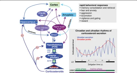
The hypothalamo-pituitary-adrenal axis embraces the feedforward and feedback neuroendocrine mechanisms that regulate CS production and synthesis (Figure 1). Neural inputs trigger the release of adrenocorticotrophic hormone (ACTH) from the pituitary which, in turn, sti-mulates adrenocortical synthesis and secretion of CS. Although CS are not stored in a readily-releasable pool, it is estimated that adequate amounts of CS can be released into the bloodstream within minutes of appro-priate neural stimuli. Noxious (stressful) stimuli are the primary triggers of neural firing that result in increased CS release. On the other hand, CS are secreted accord-ing to strictly-regulated circadian rhythms that are dic-tated by the central nervous system. More recently, CS have been found to have ultradian rhythmic patterns of release. Such patterns are most likely maintained through dynamic cross-talk between the
peripherally-produced CS and centrally-driven regulatory
* Correspondence: [email protected]
1Max-Planck-Institute of Psychiatry, Kraepelin Str. 2-10, 80804 Munich,
Germany
mechanisms; they are also likely important integrators of normo-physiological functions [4].
Since corticosteroids come on stage within 3-7 min-utes of first perception of a stressor [5], they may be considered to be secondary or auxiliary players in com-parison to monoamines (in particular, epinephrine and norepinephrine) whose actions are initiated within milli-seconds to milli-seconds [6] i.e. corticosteroids are secreted during the first stage of the ‘general adaptation syn-drome’, a concept introduced by Hans Selye in 1946. However, since corticosteroids act against the back-ground of increased monoamine secretion, it is thought that they act to fine-tune the organism’s response to stress [7] and to facilitate signal-to-noise discrimination. Moreover, unlike the transient monoamine response, corticosteroids exert sustained actions on cellular activ-ity and behavior, and therefore are essential for ensuring the orchestration of a coordinated adaptive response as
well as ‘preparedness’ of the organism to cope with future challenges.
Although corticosteroids are often thought of in nega-tive terms because of their causanega-tive role in diseases such as diabetes, hypertension, osteoporosis and immune suppression, they are essential for adaptation to stress and for maintaining physiological processes. With respect to brain structure and function, corticosteroids play an important role in maintaining hippocampal cell numbers under basal conditions; this is illustrated by robust observations that removal of corticosteroids by extirpation of the adrenal glands results in massive apoptosis, with parallel increases in neurogenesis, within the granule cell population of the hippocampus [8]. On the other hand, stress and elevated levels of glucocorti-coids inhibit the generation of new granule neurons [9]. Another aspect that suggests an important role of corti-costeroids in normo-physiology is the well-pronounced Figure 1Schematic representation of the hypothalamo-pituitary-adrenal (HPA) axis and its neuronal inputs. Corticotropin-releasing hormone (CRH)- and arginine vasopressin (AVP)-expressing parvocellular neurons in the paraventricular nucleus (PVN) project to pituitary (via the median eminence) where they stimulate adrenocorticotrophic hormone (ACTH) synthesis and secretion, subsequently triggering corticosteroid synthesis and release from the adrenal cortex. Besides acting in the brain to regulate various behaviours, corticosteroids fine-tune the
subsequent pattern (amplitude and duration) of corticosteroid secretion; they activate their cognate receptors in the pituitary, hypothalamus and hippocampus and bed nucleus of the stria terminalis (BNST, a relay between the hippocampus/amygdala and the PVN) to restrain, and in the amygdala to enhance, adrenocortical secretion. Monoaminergic transmitters, namely, norepinephrine, serotonin and dopamine released from midbrain nuclei (the locus coeruleus [LC], raphé and ventral tegmental area [VTA] and substantia nigra [SN], respectively) exert modulatory effects on all brain regions involved in the control of the HPA axis.‘Plus’signs(green)indicate positive drive on the HPA axis;‘minus’signs(red)
circadian pattern of corticosteroid secretion. These rhythms are robust and bi-directionally tightly coupled to the individual’s sleep-activity and feeding cycles, while being entrained and maintained by the daily light-dark cycle.
The magnitude and duration of the humoral response to stress is tightly coupled to the nature (quality, inten-sity and duration) of the stressor, as well as the context in which it occurs. Depending on context (e.g. the pre-vailing physiological or psychological state, as well as history of the individual), stressors may trigger excessive corticosteroid secretion over an extended duration; in such cases, the response switches from being an adap-tive one into a maladapadap-tive one, marked by transient or chronic pathology. Major depression and cognitive impairment are two conditions that represent the so-called stress-induced disorders of the brain. The first of these seems to reflect a sub-optimal stress-coping strat-egy and may largely originate from impairments of the mechanisms contributing to the homeostatic negative feedback processes that act to protect the organism against excessive exposure to corticosteroids; frequently, depressed mood is accompanied by impaired cognition and hyperemotionality, indicating that stress impacts on multiple, inter-related neural circuits. A number of human and animal studies have demonstrated the dis-ruptive effects of excessive corticosteroid secretion on cognition [10-12]. There is now strong evidence that the latter involve structural changes, including severe reduc-tions in the dendritic arborization of hippocampal and prefronto-cortical neurons [13-15]. and synaptic loss [16-18]. In addition, recent studies indicate that stress may initiate neurodegenerative processes that increase the risk for severe cognitive deficits such as those seen in dementia of the Alzheimer type [19]. Lastly, chroni-cally elevated levels of corticosteroids interfere with cen-tral and pituitary integrators and regulators of the hypothalamo-pituitary-adrenal (HPA) axis, resulting in impaired corticosteroid negative feedback and sustained corticosteroid secretion [20].
The soliloquy we’ve come to know and love
Glucocorticoids and mineralocorticoids fulfill their char-acteristic biological functions through the mediation of glucocorticoid receptors (GR) and mineralocorticoid receptors (MR), respectively. Both of these receptors are present in the brain; while GR are expressed ubiqui-tously (most strongly in the hippocampus), MR are more discretely distributed (strongly expressed in certain hippocampal subfields and the septum, and moderately expressed in the amygdala and hypothalamic paraventri-cular nucleus) [21]. The MR has a 7-10-fold greater affi-nity for corticosterone as compared to the GR [22]. It is thus estimated that the MR is some 80% occupied
under basal conditions, and that the GR only becomes activated when corticosterone levels rise during the daily circadian peak of corticosterone secretion or after stress. Although aldosterone may be synthesized in the brain [2,3], it should be noted that brain MR do not normally ‘see’their prototypic endogenous ligand; aldosterone is produced in the periphery at concentrations that are too low to have a direct impact on the brain and in any case, the hormone does not easily cross the blood-brain barrier. On the other hand, it should be mentioned that ligand availability is subject to local regulation through activation/deactivation of cortisol/corticosterone through the actions of 11b-hydroxysteroid dehydrogenase [23].
The MR and GR belong to the phylogenetically ancient superfamily of nuclear receptors, all of which are transcriptional factors. For the sake of clarity, we will herein refer to nuclear MR and GR as nMR and nGR, respectively. Whereas the unliganded nMR is pri-marily localized in the nucleus, the unoccupied nGR resides in the cytoplasm and only translocates to the nucleus upon ligand activation. This process depends on the dissociation of a host of chaperone and co-chaper-one molecules, including heat shock protein 90 (hsp90) as well as on the inclusion of a nuclear translocation signal in the receptor protein [24]. Like other nuclear receptors, nMR and nGR are organized according to canonical modules, including a ligand binding domain (LBD), a DNA binding domain (DBD), and two activa-tion funcactiva-tions (AF-1 and AF-2) at their N- and C-term-inals, respectively. The various domains share considerable homologies (homology between nMR and nGR: ~57% in LBD; ~94% in DBD). Interactions of the DBD with hormone response elements (HRE) in the promoters of specific genes result in the induction or repression of gene transcription and subsequently, changes in the expression of proteins that influence cel-lular functions. Homologies also exist within the HRE sequence of various nuclear receptors, and receptor recruitment and interactions with specific co-regulator proteins (co-activators/-repressors) may endow these structurally similar receptors with differing specificities and potencies.
Stage props
studying events mediated by nGR and nMR include established chaperone inhibitors of hsp90 (e.g. cisplatin and geldanamycin; [25]) and of the FK506-binding pro-teins (e.g. GPI1046; [26]).
Drop sceneb
The mode of action of corticosteroids summarized above, i.e. involving gene transcription and translation, may be generalized to all steroid hormone receptors, including those for estrogens. Since nuclear receptors become transcriptionally active upon ligand activation, their actions are, by definition, slow in onset and poten-tially long-lasting (hours to days, or even months); at best, gene transcription and translation require a mini-mum of 20-30 minutes (translation takes longer than transcription) [27]. However, steroids have been impli-cated in the elicitation of a number of ‘rapid’ or‘fast’ physiological and behavioral responses to external sti-muli; some examples of fast steroid-mediated responses and the mechanisms thought to underlie their actions are presented in Additional File 1. Historically, the idea that steroids can rapidly alter neuronal excitability and conduction stemmed from work on the actions of sex steroids by Kawakami and Sawyer in 1959 [28] and Woolley and Timiras in 1962 [29].
As a rule, fast responses are considered to be those that occur within the first 20 minutes of increased ster-oid secretion, i.e. in a much shorter timeframe than that required for effects on gene transcription and protein synthesis. Somewhat erroneously, these fast actions are referred to as ‘non-genomic’; in fact, rapidly triggered signaling cascades may ultimately converge in the nucleus to regulate gene transcription and protein synthesis. Distinction between the ‘fast’ and ‘slow’ actions of steroid hormones is more of mechanistic than of behavioral or physiological importance, since the lat-ter are the integrated manifestations of sequential events. Viewed from this perspective, the rapid actions of steroids may be considered as‘primers’ of the sub-strates responsible for the manifestation of transcrip-tional events triggered by nuclear receptors; kinase cascades activated during early phases of steroid action and which lead to the phosphorylation of regulatory sites of nuclear receptors [30-32] are a good example of such priming functions.
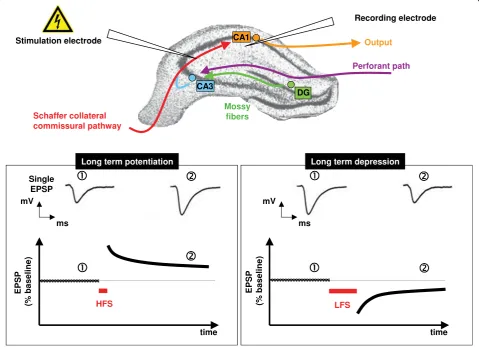
Many of the changes in behavior and brain physiology that are listed in Additional File 1 reflect rapid responses of the hippocampus to steroid hormones. For example, corticosteroids have been consistently shown to influence cognition and their effects are thought to result from their ability to directly or indirectly alter the excitability of hippocampal neurons. The hippocampus has been extensively studied for a number of pragmatic reasons. The input-output connections of the different
hippocampal subfields are well defined, making their electrophysiological study convenient. Of all brain areas, the hippocampus has been best studied in the context of long-term potentiation (LTP) and long-term depres-sion (LTD), the electrophysiological correlates of learn-ing and memory, functions in which the hippocampus is strongly implicated [[33-35]; see Figure 2 and Additional File 2]. The hippocampus also serves as an important homeostatic regulator of the HPA axis upon which it exerts a strong negative drive [36,37] through the med-iation of nMR and nGR [38].
Although the attention paid to the hippocampus is justi-fiable because of its role in the regulation of many beha-vioral and physiological processes, it should be remembered that it constitutes only part of a complex neuronal network that underpins physiology and beha-vior in normal and pathological states. For example, although the hippocampus plays an important role in the regulation of the HPA axis, it should be noted that other brain areas such as the prefrontal cortex [39], amygdala and bed nucleus of the stria terminalis, under the modulatory influence of monoamines from the hind-brain [40], contribute to the control of corticosteroid secretion; all these areas have reciprocal connections with the hippocampus and express nGR.
Several studies have begun to define how corticoster-oids and other stercorticoster-oids act on different brain structures to produce integrated and adaptive behavioral and phy-siological responses, e.g. the prefrontal and orbito-fron-tal cortices (executive functions, including attention, behavioral flexibility, declarative memory, decision mak-ing [41,13,14,42]), thalamus (processmak-ing and gatmak-ing of sensory input [43], amygdala (evaluation of emotional load of sensory input and regulation of fear [44], ventral striatum (motivation and reward [45] and decision-mak-ing [42]), and the cerebellum (learndecision-mak-ing of motor tasks [46]. Of these, the amygdala, involved in the control of fear, aggression and cognition (see Additional File 1), has been the most intensively studied. Interesting work by Roozendaal and colleagues has demonstrated a cross-talk between rapid GC and noradrenergic signaling in contextual memory consolidation [44,47] and suggests that endocannabinoids are key mediators of this cross-talk [48].
Putative membrane receptors - pirates with legs to stand on?
the presence of pharmacological antagonists of nGR (RU 38486 [49] or nMR (spironolactone [50,51]). These find-ings suggest certain homologies between the classical nuclear receptors and the putative receptors mediating the rapid actions of these steroids. Nevertheless, the existence of another class of receptors, with distinct che-mistries and cellular localizations, and that are not sen-sitive to the above-named antagonists, cannot be dismissed.
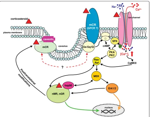
Several mechanisms that may account for membrane-mediated transduction of the rapid actions of estradiol have been proposed (see Figure 3). Substantial evidence
bound ER (mER) that is coupled to a Gaqprotein.
Evi-dence for the latter includes the observation that estra-diol induces activation of the phospholipase C- protein kinase A (PLC-PKC-PKA) pathway in nER knockdown mice [55]. The same investigators demonstrated rapid electrophysiological effects of STX, a diphenylacryla-mide-based selective estrogen receptor modulator, in nER knockout animals; STX, which does not bind to either isoform of the nER, proved to be more potent than estradiol in theirin vitroandin vivotest systems [55,56].
estradiol [60], the latter finding suggesting that GPR 30 may co-exist alongside (an)other mER with unique phar-macological properties. Notably, in an extension of their earlier work, Revankar et al. [61] exploited chemical biol-ogy to explore the subcellular localization of GPR 30 and its signaling potential; on the basis of observations in 4 cancer cell lines, they discarded the notion that sufficient GPR 30 is localized at the plasma membrane and rather suggested that GPR 30 localized in the endoplasmic reti-culum serves as an intracellular transmembrane receptor for estrogen.
Interestingly, Toran-Allerand and colleagues [62,63] described a high affinity (KD for estradiol: 1.6 nM)
caveolin-associated protein in the plasma membranes of neonatal (but not adult) neocortical and uterine tissues. This so-called ER-X seems to come closer to meeting the expectations of a distinct mER insofar that it cannot be blocked by ICI 182,780 [62]; moreover, these authors found that experimentally-induced ischemic stroke in adult animals is accompanied by an upregulation of ER-X in the brain, suggesting that the ER-ER-X mediates the neuroprotective actions ascribed to estrogens.
It is tempting to hypothesize, that the mediators of rapid corticosteroid effects may share similar basic prop-erties and mechanisms with the proposed membrane-associated estrogen receptors. The existence of a mem-brane-bound receptor for corticosteroids (herein referred to as mCR) was postulated by Willmer in 1961 [64]. Willmer’s suggestion that steroid hormones inter-digitate with, and alter the permeability of, lipids in the plasma membrane, lost currency as evidence that ster-oids bind to intracellular proteins (nuclear receptors) and stimulate protein synthesis began to accumulate from 1961 onwards [65,66]. However, in 1974 Satre and Vignais described corticosterone binding to mitochon-drial preparations from the adrenal and kidney [67], a finding that eventually extended to other cell types [68]. A series of authors provided evidence for membrane-bound steroid recognition sites in the brain [69-71]; among these, Towle and Sze demonstrated specific cor-ticosterone binding to plasma membrane preparations from rat brain synapses [72]. These membrane binding sites had a relatively high affinity for corticosterone (KD
10-7Mvs. 10-9M in the case of cytosolic binding sites) and treatment with phospholipase A2 or phospholipase C led to complete dissociation of membrane-bound cor-ticosterone. Similarly, Orchinik et al. described the pre-sence of mCR in brain synaptosomal fractions obtained from the amphibian Taricha granulosa(rough-skinned newt) [69]. These receptors showed pharmacological specificity for corticosterone and cortisol (KD10-9 M),
and lesser affinities for aldosterone and other natural and synthetic steroids (such as dexamethasone and RU 38486). Importantly, Orchinik et al. reported a linear
relationship between the potencies of various com-pounds (corticosterone being the most potent) in inhi-biting male reproductive behavior (inhibition by corticosterone within 8 minutes of application) and their ability to bind the putative mCR [69]. In subse-quent studies, these authors described similar neuronal mCR in mammalian [73] and bird [74] brains and sug-gested a role for guanine nucleotide-binding proteins in the formation of a ternary complex of corticosterone and the putative neuronal mCR, i.e. the mCR appears to be coupled to G proteins [75]. Additional evidence for the existence of a mCR was eventually provided by Orchinik’s colleagues who solubilized and partially puri-fied membrane-bound corticosterone binding sites from the amphibian brain [76]; the assumed mCR had a molecular weight of about 63 kDa, as compared to 97 kDa and 110 kDa in the case of the nGR and nMR, respectively. More recently, studies by Johnson et al. [77] provided anatomical evidence for the existence of nGR within the postsynaptic density of neurons in the rodent amygdala. At present it is unclear as to whether there are any homologies between the mCR and either the nGR or nMR.
Supporting the plausibility of this view, Matthews et al. have shown that nGR interacts with caveolin [81].
Many unliganded nuclear receptors (e.g. nGR), are tethered in the cytoplasm through their association with chaperone proteins such as heat shock protein 90 (hsp90); this complex is dissociated upon arrival of the ligand [24]. Interestingly, hsp90 is known to interact with src kinase [82], a membrane-proximal kinase thought to mediate the rapid activation of the MAPK pathway by corticosteroids. In addition, hsp90 interac-tions with MEK2, another kinase upstream of MAPK, has been shown to mediate MAPK pathway activation by estradiol [83]. In fact, nGR itself reportedly interacts with Raf-1, a downstream effector of Ras, and upstream regulator of the MAPK pathway [84].
Receptors for several neurotransmitters (some of which are ion channels) have been shown to bind CS [76,85,86]. Although it remains unclear as to whether these interactions serve as a conduit of the rapid actions of CS, the latter seems plausible given the evidence that neurosteroids can modulate chloride flux and thereby, neuronal excitability, by binding to an allosteric site on the GABAAreceptor [87].
In summary, there is growing support for the view that CS can initiate signaling at the plasma membrane through one or more of the following mediatory mechanisms: (i) G protein-coupled membrane-bound CS receptors, (ii) steroid modulatory sites on plasma-bound neurotransmitter receptors, (iii) interactions between cytoplasmic CS receptors and kinase family-interacting chaperone molecules, and/or (iv) palmitoyla-tion. Elucidation of the mechanisms underlying the rapid actions of CS will require a stepwise analysis of the contributions of each member of this‘interactome’ -a m-ajor ch-allenge.
From the sightlines - peeping on a rapidly changing stage
This section will focus on the cellular endpoints that can be used to support the view that corticosteroids rapidly influence neuronal activity, focusing on altera-tions in membrane excitability and signaling cascades that originate at or close to the plasma membrane. However, attempts to summarize the existing literature are confronted with the fact that the results derive from disparate protocols and experimental models in different laboratories. For example, a wide range of corticosteroid doses and exposure times have been applied to studying synaptic transmission in either rat or mouse dissociated hippocampal neurons or hippocampal slices. We will, however, first consider early studies on hypothalamic neurons by Kasai and colleagues and Saphier and Feld-man, usingin vitro ionotophoresis. Kasai and colleagues showed that cortisol excited tuberoinfundibular neurons
in the paraventricular nucleus (PVN) which project to the median eminence from where their neurosecretory products reach the anterior pituitary; however, these authors also reported inhibitory effects of cortisol in the PVN, suggesting this to result from inhibition of nora-drenergic inputs [88-90]. Saphier and Feldman, observed a significant reduction in the spontaneous firing rates of similar hypothalamic neurons after the application of corticosterone [91,92]; these changes had a rapid onset and were maintained even after iontophoresis of the hormone was stopped. Further, they reported on a sub-set of neurons whose activity was not altered by corti-costerone; glutamate-induced excitation of these neurons was however suppressed in the presence of corticosterone.
Together, the studies described above represent a hypothalamic electrophysiological correlate of the nega-tive feedback control of adrenocortical secretion, and illustrate that corticosteroids can elicit different responses from different brain areas or neuronal popula-tions within an anatomical region or specific neuronal phenotypes within a given subfield; moreover, the responses depend on neural inputs to the particular set of neurons under investigation [91,93]. Given the sug-gested importance of the hippocampus in mediating glu-cocorticoid negative feedback (see above), it is surprising that Barak [94] failed to observe any changes in the activity of hippocampal neurons upon applying corticos-terone. As will become evident below, despite a large number of studies that focussed on the CA1 subfield of the hippocampus, it is difficult to compile a consensus view of how corticosteroids impact on the activity of this region.
Given that the amplitude of the AHP is determined by Ca2+ and Ca2+-dependent K+ transients [99,100], it is interesting that Landfield and colleagues reported that high doses of the synthetic GR agonist RU28362 (7μM) enhance the amplitudes of voltage-dependent calcium channel (VDCC)- mediated Ca2+ spikes in a protein synthesis-dependent manner [101]. In contrast, Tian et al. suggested that the increase in the slow after-hyperpo-larization amplitude seen after exposure to high doses of corticosterone may involve cAMP-dependent phosphor-ylation and Ca2+-activated K+ channels [102]: dexa-methasone (1 μM), a synthetic glucocorticoid with high selectivity for the nGR, blocked PKA-mediated inhibi-tion of Ca2+-activated K+ channels without influencing VDCC-mediated Ca2+currents in a mouse pituitary cell line (AtT20). It should be noted that Tian et al. treated their cells with dexamethasone for 2 h and that these effects requiredde novoprotein synthesis for their mani-festation [102,103]. Because activation of NMDA recep-tors results in an influx of Ca2+ and, as mentioned above, Ca2+determines the AHP amplitude [99], corti-costeroid-NMDA receptor interactions have been ana-lyzed in a number of studies using electrophysiological recordings as the endpoint. For example, Wiegert et al. showed that exposure of mouse hippocampal slices to corticosterone (100 nM) for 20 min resulted in NMDA receptor-mediated suppression of primed-burst potentia-tion and synaptic potentiapotentia-tion [104] (induced by stimu-lation at 10 Hz, in contrast to the more commonly-used 100 Hz LTP regimen). In contrast, theta-burst potentia-tion (see Addipotentia-tional File 2 for informapotentia-tion on different stimulation protocols), which requires activation of both NMDA receptors and voltage-dependent Ca2+-channels was not affected by corticosterone treatment. The same authors also described a role for L-type Ca2+ channels in the synaptic actions of corticosterone [105]. In the context of the question of whether corticosterone can rapidly alter synaptic function, it is important to note, however, that Wiegert et al. [104] and Chameau et al. [105] made their electrophysiological recordings between 1 and 6 h after initial exposure to the steroid. On the other hand, Chameau et al. [105] found by quantitative PCR that corticosterone did not change the mRNA expression of the pore-forming Cav1 subunit of
the L-type Ca2+channel, and ruled out transcriptional mechanisms in the effects they observed.
Wiegert et al. [104] showed that RU 38486 blocks cor-ticosterone-induced impairments of synaptic plasticity, implying mediation of the effects by nGR. A similar conclusion was drawn from their previous work on GRdim/dim mice, a strain carrying a point mutation of the DNA binding domain of the nGR which precludes transcriptional effects; briefly corticosterone did not influence VDCC-mediated Ca2+currents in hippocampal
slices from GRdim/dim mice [106]. To address the ques-tion of how glucocorticoids enhance Ca2+currents on the one hand, and reduce synaptic efficacy on the other, Joëls’laboratory examined synaptic efficacy 1-4 h after a brief exposure to corticosterone (1 μM CORT for 20 min) [107]. Their investigations revealed that synaptic transmission was potentiated when VDCCs were acti-vated, and impaired only when NMDA receptors were activated; moreover, they found that these effects were RU 38486-sensitive, indicating their mediation by nGR. Together, these observations point to the importance of considering all of the individual components that contri-bute to the overall response in field recordings. In this respect, it is worth recalling that the magnitude of LTP and LTD is a function of the number of AMPA recep-tors that are present at the synaptic surface (see Addi-tional File 2). Miniature excitatory postsynaptic currents (mEPSCs, which represent the spontaneous release of neurotransmitter quanta from presynaptic terminals) are mediated by AMPA receptors and changes in the mEPSC amplitude represent postsynaptic changes in AMPA receptor properties and/or numbers. Indeed, Martin et al. observed that corticosterone increases the amplitude (but not frequency) of miniature excitatory postsynaptic currents and demonstrated that corticoster-one increases trafficking of the GluR1 and GluR2 subu-nits of the AMPA receptor to the synaptic surface, apparently through an nGR-dependent mechanism [108]. This last study is in good agreement with that by Karst and Joëls, who also reported nGR-mediated increases in mEPSC amplitude [109].
receptor-mediated Ca2+ currents in the CA1 subfield of the mouse hippocampus [93], that bath application of corticosterone to hippocampal slices inhibits VDCC-mediated Ca2+ currents within minutes [112], and that corticosterone increases synaptosomal uptake of Ca2+ upon K+-induced depolarization [113].
At this stage, it is important to note that some of the discrepant reports on corticosterone-induced changes in NMDAR-mediated Ca2+currents may reflect the ent durations of exposure to the steroid used by differ-ent groups. In fact, Wiegert et al. defined a narrow time window (10 min before high frequency stimulation) dur-ing which corticosterone facilitates synaptic potentiation; longer bath applications of the hormone were found to impair synaptic potentiation [114].
Most of the evidence reviewed above presumes post-synaptic sites of corticosterone action. New studies of CA1 neurons also report changes in the frequency of mEPSCs, thus implying presynaptic sites of action. Thus, Karst et al. [50] and Olijslagers et al. [51] showed that corticosterone increases the frequency of AMPA receptor-mediated mEPSCs. Both studies show that application of BSA-conjugated corticosterone produced similar effects to those obtained with corticosterone, and interestingly, that de novoprotein synthesis was not essential for their manifestation. Together, these results hint at the involvement of receptors other than nGR and nMR; nevertheless, nMR antagonism by spironolac-tone resulted in a blockade of the corticosterone-induced increases in mEPSC frequency. [50,51] [but see [114]]. On the other hand, since RU 28362, a synthetic nGR agonist, did not reproduce the effects of corticos-terone, and because the effects were not antagonizable with RU 38486, Karst et al. [50] and Olijslagers et al. [51] proposed that the putative mCR might share iden-tity with the nMR. The latter suggestion is supported by experiments in mice with targeted mutations of nGR and nMR [50,106] and work by Groc et al. [115]. Using dissociated hippocampal cells to visualize AMPA recep-tor trafficking, the latter authors observed increased synaptic surface expression of GluR2 subunits of the AMPA receptor within minutes of exposure to corticos-terone, BSA-conjugated corticosterone or aldosterone (the prototypic nMR agonist).
Related to the electrophysiological measures summar-ized in the last few paragraphs, Olijslagers et al. demon-strated that activation of the MAP kinase ERK1/2 is crucial for the corticosterone-induced increase in mEPSC frequency [51]. Interestingly, their experiments showed non-dependence on postsynaptic G protein activity on mEPSC frequency. Rather, by using the H-Ras G12V strain of mouse which displays strong presy-naptic activation of ERK1/2 due to constitutively high expression of the H-Ras transgene, they suggested that
the actions of corticosterone are initiated at presynaptic sites, increasing the probability of presynaptic neuro-transmitter release [50,51]. Moreover, in agreement with other studies [111], Olijslagers et al., reported that intra-cellular Ca2+ stores do not influence mEPSC frequency upon exposure to corticosterone [51]. Lastly, it should be noted that although the involvement of G proteins in corticosterone-induced changes in mEPSC frequency were excluded [51], direct infusion of GDPbS into the postsynaptic cell prevented the decrease of the peak amplitude of IAcurrents (postsynaptic K+ conductance)
by corticosterone [51]; this finding points to mediation through a postsynaptic mCR-dependent mechanism.
A number of studies suggest a role of G proteins in the mediation of the rapid actions of corticosterone. For example, ffrench-Mullen showed that the inhibition of Ca2+ currents by cortisol in guinea pig CA1 neurons depends on pertussis toxin-sensitive G-proteins [112]. The same author also showed that the effects of cortisol are significantly diminished in the presence of PKC inhi-bitors (BIS and PKCI 19-31), and ruled out a role for PKA in the mediation of the actions of cortisol [112]. Similarly, Chen and Qiu showed that corticosterone rapidly inhibits VDCC-mediated Ca2+ currents in a phaeochromocytoma cell line of neural origin (PC12 cells), and that inhibition of G proteins by application of either pertussis toxin or GDPbS significantly attenuates the ability of either corticosterone or BSA-corticosterone to stimulate the influx of Ca2+ [116]. They also demon-strated that activation of PKC with phorbol 12-myristate 13-acetate results in an inhibition of Ca2+ entry though VDCC after depolarization with K+, and that the appli-cation of corticosterone activates PKC within 5-15 min-utes. Lastly, like Qi et al. [117] who obtained similar results in primary hippocampal neurons, Chen and Qiu [116] showed that both, corticosterone and BSA-conju-gated corticosterone trigger the activation of PKC and a series of MAP kinases (ERK1/2, p38MAPK and c-Jun) in PC-12 cells; maximum kinase activation occurred within 15 min of application of the hormone and the effects could not be attenuated by RU 38486.
Reality
‘slow’negative feedback actions of corticosteroids at the level of the pituitary and the brain. Pioneering research by Mary Dallman used ingenious experimental designs which eventually provided evidence for the rapid actions of corticosteroids in reducing their own secretion [118] and, as already mentioned, the search for electrophysio-logical correlates was pursued in the hypothalamus in parallel. Today, predominantly based on work from the laboratories of Stafford Lightman and colleagues [4], it would appear that the ultradian rhythmic secretion of relatively high-amplitude corticosterone may serve to ensure low levels of adrenocortical activity during the organism’s resting phases; these brief pulses presumably act rapidly to suppress brain-pituitary drive of adrenal secretion.
At the behavioral level, Orchinik et al. [69] elegantly demonstrated the potency of corticosterone in inhibiting male reproductive behaviour in newts, within 8 min of application. In mammals, Jozsef Haller and colleagues have shown that corticosterone injections elicit aggres-sive and anxiety-related behavior (latency of 7 min) in rats whose endogenous adrenocortical activity is sup-pressed by inhibition of 11b-hydroxylase activity with metyrapone [119-121]. Several authors have also described the ability of corticosterone to rapidly alter locomotor behavior in rodents; for example, acute sys-temic injections of corticosterone to rats (placed in a novel environment) were shown to stimulate locomotion within 7.5 minutes of administration [122].
Rhythms in the secretion of corticosteroids and other neuromodulatory molecules can influence experimental outcomes, even in in vitrosettings. For instance, Ca2+ currents into hippocampal CA3 neurons inin vitro pre-parations are highest during the subjective night, when corticosterone levels are highest [123]. Similarly, Brunel and de Montigny [124] reported that the firing rate and pharmacological responsiveness of CA3 neurons is high-est during the nocturnal peak in corticosterone secretion in vivo. Importantly, using hippocampal slice cultures, Chaudhury et al. demonstrated that the amplitude of LTP is greatest during the subjective night [125]. Addi-tionally, Eckel-Mahan and colleagues reported circadian dependency in the efficiency of consolidation of long term memory [126].
Many studies support the idea that stress, a large part of whose actions are mediated by corticosteroids, influ-ences learning and memory. Besides the quality and intensity of the stressor, the context in which the stress-ful stimulus is perceived, is an important determinant of the behavioral outcome. The latter is more easily explained in terms of ‘intrinsic’ and ‘extrinsic stress [127];‘intrinsic stress’refers to situations in which stress is either elicited by, or directly associated with, the cog-nitive experience (e.g. spatial learning), whereas
‘extrinsic stress’describes situations in which the stress occurs outside the context of the momentary stress situation (e.g. foot shock stressbefore spatial learning). According to a model developed by Sandi and Pinelo-Nava [127], learning and memory will be facilitated by stressors that activate the same (or similar) neural cir-cuitries that are required for interpreting and respond-ing to a particular cognitive challenge. Supportrespond-ing this view, Cahill and McGaugh [128] and Sandi [129] reported that emotionally arousing experiences are bet-ter remembered than neutral ones. In fear conditioning experiments, Cordero et al. noted that post-training cor-ticosterone levels correlate with the strength of stimulus required to encode memories [130,131]. Moreover, the importance of corticosterone in information acquisition and consolidation of memory is well known, even if still poorly understood [132-135]. The relative importance of nMR and nGR in these processes are elegantly discussed by Schwabe et al. [136], and Revest et al. [134] have demonstrated a mediatory role of the MAPK pathway in the facilitation of hippocampus-dependent contextual fear conditioning by corticosteroids. In the previously-cited work on long-term contextual fear memory by Eckel-Mahan and colleagues [126], rhythms of MAPK (ERK1/2) activation were shown to coincide temporally with the degree of persistence of memory. Given that corticosterone acutely increases ERK1/2 phosphorylation [51,116,117,134], the results presented by Eckel-Mahan and colleagues [126] should be considered in the con-text of the hypothesis proposed by Sandi and Pinelo-Nava [127] and the pioneering work by Oitzl and de Kloet [137]; in addition, since the amygdala plays a major part in the regulation of fear and has reciprocal interactions with the hippocampus and other cognition-regulating brain areas, future interpretations of the work by Eckel-Mahan and colleagues [126] should embrace the idea that corticosteroids can exert actions on a net-work of interconnected brain structures, whose indivi-dual responses will determine the ultimate behavioral output.
adrenocortical response to stress primarily serves an adaptive purpose, in certain circumstances, it may switch to being maladaptive, marked by transient or chronic pathology, as discussed earlier in this article.
The physiological and behavioral responses to stress depend on myriad molecules and processes, with an important contribution by corticosteroids; effects of the latter are often studied in isolation at the cost of other contributory factors and the neural networks which reg-ulate, or may be regulated by, corticosteroids. This can be exemplified by considering our earlier discussion of corticosteroid interactions with glutamatergic transmis-sion and reports that the direction and/or magnitude of LTP and LTD are influenced by the intensity and emo-tional value of a given stressor; for example, LTP is only reduced in animals exposed to uncontrollable stress [138], but not in animals that can escape from the stres-sor [139]. Using the paradigm of foot-shock stress, Wang et al. reported that stress induces a shift in synap-tic plassynap-ticity; thus, whereas stress facilitates LTD induc-tion, it impairs LTP induction [140]. Besides showing that these effects of stress can be blocked by RU 38486, these last authors showed that blockade of the NMDA receptor restores LTP inducibility in stressed animals; further they demonstrated that stress-induced changes in synaptic efficacy can be abolished by prior adminis-tration of Ro25-6981, a specific antagonist of the NR2B subunit of the NMDA receptor. A role for the NR2B subunit in the synaptic plasticity thought to be essential for the orchestration of the behavioral response to stress was also suggested by Wong et al. who showed that Ro25-6981 reverses elevated platform stress-induced deficits in spatial learning and memory, as tested in the Morris water maze (MWM) [141].
The NR2B subunit is predominantly associated with extrasynaptic NMDA receptors whose activation depends on glutamate“spill-over”, a phenomenon that can be mimicked with threo-b-benzyloxyaspartate (TBOA), a blocker of glutamate re-uptake. Wong et al. [141] found that TBOA application to animals 5 min before low frequency stimulation resulted in the suc-cessful induction of LTD, indicating that stress leads to glutamate“spill-over”. Linking LTD with stress-induced memory impairment, the authors showed that prevent-ing LTD induction by infusion of a GluR2 peptide ana-logue that cannot be internalized abolished the ability of stress to cause memory deficits in the MWM test; these findings add to the evidence that acute stress results in the internalization of AMPA receptors, followed by synaptic depression and learning and memory deficits.
We previously discussed how the MAPK signaling pathways may be linked with LTP and LTD (and learn-ing and memory). In this respect, it is interestlearn-ing to note that this pathway is concomitantly activated by
stress, presumably due to activation of nGR [142,143], believed to be essential for the phosphorylation of ERK1/2 [134]. Moreover, the observation that tail shock and restraint stress robustly activate ERK1/2 and impair synaptic potentiation in the CA1 subfield suggests a major role for the MAPK pathway in mediating the actions of stress [144]. In addition to inducing the phos-phorylation of ERK1/2, stress activates other kinases (e. g. p38 MAPK, CaMKII) and pCREB within 2 min of swim stress [145]. Surprisingly, however, the latter responses are accompanied by a reinforcement (rather than impairment) of LTP in the dentate gyrus of the hippocampus. This finding indicates that different stres-sors may elicit quite different electrophysiological responses and/or, that the synaptic effects of stress differ from one hippocampal subfield to another. Since the effects of stress on biochemical and electrophysiological signalling in the dentate gyrus were found to be subject to modulation by serotonin [145], it is plausible that dif-ferential monoaminergic innervation of the different hippocampal subfields defines the ultimate cellular response.
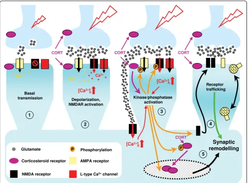
We summarize some potential mechanisms that may account for the rapid and slower effects of corticoster-oids on neuronal physiology, with a focus on synaptic events, in Figure 4. An attempt is made to show how signals originating at the neuronal surface are integrated both at the synaptic and transcriptional levels.
Critique
From the preceding, it appears safe to assume that, irrespective of the behavioural or physiological out-comes, acute and chronic elevations of corticosteroid secretion initiate common mechanisms and biochem-ical processes; convergence of these events will depend on parameters such as exposure dosage and time, as well as the context in which they occur. Given the potential for convergence (as well as potentiation), improved knowledge of the initial stages of corticoster-oid signalling, whether membrane- or nuclear recep-tor-mediated, is clearly desirable. Studies on the rapid neural actions of corticosteroids are likely to gain further interest, especially as newer analytical tools become available and knowledge about the fast actions of other steroid hormones grows. It therefore seems appropriate to list some critical issues and needs, the consideration of which may foster progress through cautious reflection:
• standardized test protocols (steroid dose, animal or cellular models, and sexcof animals); inin vitro studies, drug diffusion times and active concentra-tions achieved at target cells should be controlled; similarly, in in vivoresearch, pharmacokinetic fac-tors, including solvent and route of administration,
should be considered;age of animals, but also of material used for in vitro testing, is important because of dynamic age-related changes in the expression of key partners such as glutamate recep-tor subunits [146]; since corticosteroids are secreted according to a strict circadian rhythm, both the Figure 4Working model of sequential corticosteroid influences on synaptic physiology. Corticosterone-mediated changes in synaptic transmission occur at different levels and in different sequential steps.①depicts synaptic transmission under basal conditions. Neuronal excitation results in glutamate secretion from synaptic vesicles at presynaptic sites into the synaptic cleft. Glutamate binds to postsynaptic glutamate-gated ion channels (in particular, AMPA receptors), which open to permit ion fluxes (Na+influx, K+efflux) across the AMPA receptor,
resulting in a depolarization of the postsynaptic cell. Due to a voltage-dependent Mg2+block in its membrane domain, the NMDA receptor
remains inactive under basal conditions, and is activated when a certain transmission threshold is reached.②Exposure to corticosteroids (e.g. during stress) may lead to activation of ERK1/2 in the presynaptic terminal (possibly through membrane corticosteroid receptors [51]); increased glutamatergic stimulation of postsynaptic AMPA receptors results in an increase in the frequency of AMPA receptor-mediated miniature postsynaptic currents (mEPSCs).③Enhanced activation of AMPA receptors in the previous step further depolarizes the postsynaptic membrane and activates NMDA receptors. Activated NMDA receptors (Na+and Ca2+influx, K+efflux) lead to further depolarization of the postsynaptic cell, resulting in the opening of voltage-dependent Ca2+channels (VDCC) and high postsynaptic concentrations of Ca2+. Corticosteroids may stimulate glutamate secretion so strongly, causing glutamate“spill-over”which activates not only synaptic, but also extrasynaptic, glutamate receptors [141]; the latter are mainly NMDA receptors of the NR2B subtype. The increased intracellular levels of Ca2+trigger a cascade of Ca2
+
-dependent signaling pathways in the postsynaptic cell, which may, in turn, induce the phosphorylation and de-phosphorylation of postsynaptic glutamatergic receptors and of nuclear corticosteroid receptors (nMR and nGR). Activation of extrasynaptic NMDA receptors is thought to trigger NR2B-dependent kinases, which might initiate trafficking of extrasynaptic NR2B receptors into the postsynaptic surface. Furthermore, Ca2+-dependent signaling pathways in the postsynaptic cell participate in the regulation of AMPA receptor trafficking to and from
availability of endogenous corticosteroids as well as of primary and secondary downstream effectors will vary over the day - this demands testing at a given circadian timeto ensure comparable measurements [123-125].
•while surgical adrenalectomyis a useful approach to ensure that only the actions of exogenously-admi-nistered steroids are being recorded, the operation requires anaesthesia and may involve potentially confounding post-operative pain;chemical adrena-lectomy is a good alternative (e.g. blockade of corti-costeroid synthesis with metyrapone), but it may have (indirect) non-selective effects on the produc-tion of other steroids; adrenalectomy, in general, induces massive apoptosis and stimulates neurogen-esis in the dentate gyrus within just a few hours, changes that probably result inreorganized neuro-nal circuits and measurable outputs [147].
•attention to the fact that acute and chronic corti-costeroid exposures differ significantly, and that administration of corticosteroids only mimics an intermediate phase of the organism’s response to stress;
• clear exclusion of transcriptional and transla-tional events initiated by activation of cognate nuclear receptors;
The show must (will) go on
While the nuclear receptor-mediated actions of corticos-teroids are well established, those that appear to be mediated through non-classical, possibly membrane-bound receptors, have perhaps not received sufficient appreciation. The lack of consistent results (see need for standardization in previous section), compounded by the relatively fruitless hunt for putative membrane receptors, accounts for the scepticism that haunts this area of research. Increased respectability might be gained by initially seeking answers to some of the following ques-tions:
• How can the neural actions ascribed to peripher-ally-produced corticosteroids be distinguished from those that result from those elicited by corticoster-oids thought to be produced in neural tissue? • Can the rapid actions of corticosteroids observed predominantly in the CA1 subfield of the hippocam-pus be generalized to other hippocampal subfields, or indeed other brain regions?
•Do the endpoints assessed after application of cor-ticosteroids reflect actions exclusively at the hippo-campus? In vitro, do we get only a partial (or perhaps, false) picture? In vivo, are we monitoring responses from a network of corticosteroid-sensitive
brain regions? How are the outputs modulated by other neurochemical states and inputs?
•Do corticosteroids directly interact with membrane proteins? What is the chemical identity of these molecules? Are they distinct from the known nuclear receptors and if not,
◦Do they represent post-translational modifica-tions (e.g. palmitoylated versions of the nuclear receptors, as suggested for the mER)?
◦ Is there biochemical evidence for interactions with other known membrane receptors (e.g. glu-tamate receptors); do these receptors have allos-teric binding sites for corticosteroids as well as for pharmacological antagonists of nMR and nGR? (cf. estrogens, progestins)
•How do events that are triggered by corticosteroids at the membrane funnel into long-term cellular and organismic adaptations (e.g. by positive or negative priming of the gene machinery regulated by nMR and nGR)?
•How do the rapid actions of corticosteroids contri-bute to their longer-lasting actions (e.g.‘priming’of nuclear receptor-mediated events?)
•Is it possible to define corticosteroid actions - fast and slow - in terms of spatio-temporal maps, keep-ing in mind that damage induced in a relatively short time in one area may take longer to spread to other interconnected areas [cf. [13]]?
•Is it feasible to generate genetic or pharmacological tools that will facilitate acceptance and further study of mCR?
Appendix
a) Corticosteroids: way upstream - the title of this article is adapted from Alan Ayckbourne’s stage play
Way Upstreamin which two couples on a boating
holiday run into some strange happenings.
b) A painted cloth in front of which a short scene is played while the main stage set is changed.
c) Research on the rapid actions of corticosteroids has mainly exploited male rodents or tissues derived from them. Corticosteroid secretion is strongly influ-enced by sex, as are physiology and behaviour. Many of the physiological and behavioural readouts moni-tored in such studies reflect the prevailing sex ster-oidmilieu; in females, sex steroids are secreted in a cyclical fashion.
Additional file 1: Summary of rapid effects of corticosteroids and estrogens on the central nervous system[148-181].
Click here for file
Additional file 2: Synaptic plasticity and learning and memory [182-221].
Click here for file
[ http://www.biomedcentral.com/content/supplementary/1756-6606-3-2-S2.PDF ]
Acknowledgements
The authors thank Silei Yang and members of the Munich and Bristol laboratories for their critique and encouragement. The article was written within the framework of the European Union’s CRESCENDO Consortium (FP6 Contract LSHM-CT-2005-018652). TR was supported by LINE and a fellowship from the Max Planck Society.
Author details
1
Max-Planck-Institute of Psychiatry, Kraepelin Str. 2-10, 80804 Munich, Germany.2Henry Wellcome Laboratories for Integrative Neuroscience and
Endocrinology, Faculty of Medicine and Dentistry, University of Bristol, Bristol, UK.
Authors’contributions
TR, AP and OFX wrote the manuscript; KC critically reviewed the manuscript and suggested improvements. All authors read and approved the final form of the manuscript.
Competing interests
The authors declare that they have no competing interests.
Received: 18 September 2009
Accepted: 11 January 2010 Published: 11 January 2010
References
1. Chrousos GP, Gold PW:The concepts of stress and stress system disorders. Overview of physical and behavioral homeostasis.JAMA1992, 267:1244-1252.
2. Ye P, Kenyon CJ, Mackenzie SM, Nichol K, Seckl JR, Fraser R, Connell JM, Davies E:Effects of ACTH, dexamethasone, and adrenalectomy on 11beta-hydroxylase (CYP11B1) and aldosterone synthase (CYP11B2) gene expression in the rat central nervous system.J Endocrinol2008, 196:305-311.
3. Gomez-Sanchez EP, Ahmad N, Romero DG, Gomez-Sanchez CE:Is aldosterone synthesized within the rat brain?.Am J Physiol Endocrinol Metab2005,288:E342-346.
4. Lightman SL, Wiles CC, Atkinson HC, Henley DE, Russell GM, Leendertz JA, McKenna MA, Spiga F, Wood SA, Conway-Campbell BL:The significance of glucocorticoid pulsatility.Eur J Pharmacol2008,583:255-262.
5. Bassett JR, Cairncross KD:Time course for plasma 11-hydroxycortico-steroid elevation in rats during stress.Pharmacol Biochem Behav1975, 3:139-142.
6. Morilak DA, Barrera G, Echevarria DJ, Garcia AS, Hernandez A, Ma S, Petre CO:Role of brain norepinephrine in the behavioral response to stress.Prog Neuropsychopharmacol Biol Psychiatry2005,29:1214-1224. 7. Radley JJ, Williams B, Sawchenko PE:Noradrenergic innervation of the
dorsal medial prefrontal cortex modulates hypothalamo-pituitary-adrenal responses to acute emotional stress.J Neurosci2008, 28:5806-5816.
8. Yu S, Holsboer F, Almeida OF:Neuronal actions of glucocorticoids: focus on depression.J Steroid Biochem Mol Biol2008,108:300-309.
9. Fuchs E, Gould E:Mini-review: in vivo neurogenesis in the adult brain: regulation and functional implications.Eur J Neurosci2000,12:2211-2214. 10. Starkman MN, Giordani B, Gebarski SS, Schteingart DE:Improvement in
learning associated with increase in hippocampal formation volume.Biol Psychiatry2003,53:233-238.
11. Lupien SJ, McEwen BS, Gunnar MR, Heim C:Effects of stress throughout thelifespan on the brain, behaviour and cognition.Nat Rev Neurosci2009, 10:434-445.
12. Gilpin H, Whitcomb D, Cho K:Atypical evening cortisol profile induces visual recognition memory deficit in healthy human subjects.Mol Brain
2008,1:4.
13. Cerqueira JJ, Mailliet F, Almeida OF, Jay TM, Sousa N:The prefrontal cortex as a key target of the maladaptive response to stress.J Neurosci2007, 27:2781-2787.
14. Holmes A, Wellman CL:Stress-induced prefrontal reorganization and executive dysfunction in rodents.Neurosci Biobehav Rev2009,33:773-783. 15. Radley JJ, Rocher AB, Rodriguez A, Ehlenberger DB, Dammann M,
McEwen BS, Morrison JH, Wearne SL, Hof PR:Repeated stress alters dendritic spine morphology in the rat medial prefrontal cortex.J Comp Neurol2008,507:1141-1150.
16. Sousa N, Almeida OFX:Corticosteroids: sculptors of the hippocampal formation.Rev Neurosci2002,13:59-84.
17. Grillo CA, Piroli GG, Wood GE, Reznikov LR, McEwen BS, Reagan LP: Immunocytochemical analysis of synaptic proteins provides new insights into diabetes-mediated plasticity in the rat hippocampus.Neuroscience
2005,136:477-486.
18. Bessa JM, Ferreira D, Melo I, Marques F, Cerqueira JJ, Palha JA, Almeida OFX, Sousa N:Hippocampal neurogenesis induced by antidepressant drugs: an epiphenomenon in their mood-improving actions.Mol Psychiatry
2009,14:739.
19. Sotiropoulos I, Catania C, Riedemann T, Fry JP, Breen KC, Michaelidis TM, Almeida OFX:Glucocorticoids trigger Alzheimer disease-like pathobio-chemistry in rat neuronal cells expressing human tau.J Neurochem2008, 107:385-397.
20. de Kloet ER, Joëls M, Holsboer F:Stress and the brain: from adaptation to disease.Nat Rev Neurosci2005,6:463-475.
21. Reul JM, de Kloet ER:Anatomical resolution of two types of
corticosterone receptor sites in rat brain with in vitro autoradiography and computer-ized image analysis.J Steroid Biochem1986,24:269-272. 22. Reul JM, Gesing A, Droste S, Stec IS, Weber A, Bachmann C, Bilang-Bleuel A,
Holsboer F, Linthorst AC:The brain mineralocorticoid receptor: greedy for ligand, mysterious in function.Eur J Pharmacol2000,405:235-249. 23. Seckl JR, Holmes MC:Mechanisms of disease: glucocorticoids, their
placental metabolism and fetal‘programming’of adult pathophysiology.
Nat Clin Pract Endocrinol Metab2007,3:479-488.
24. Gronemeyer H, Gustafsson JA, Laudet V:Principles for modulation of the nuclear receptor superfamily.Nat Rev Drug Discov2004,3:950-964. 25. Rosenhagen MC, Sōti C, Schmidt U, Wochnik GM, Hartl FU, Holsboer F,
Young JC, Rein T:The heat shock protein 90-targeting drug cisplatin selectively inhibits steroid receptor activation.Mol Endocrinol2003, 17:1991-2001.
26. Edlich F, Weiwad M, Wildemann D, Jarczowski F, Kilka S, Moutty MC, Jahreis G, Lücke C, Schmidt W, Striggow F, Fischer G:The specific FKBP38 inhibitor N-(N’, N’dimethylcarboxamidomethyl) cycloheximide has potent neuroprotective and neurotrophic properties in brain ischemia.J Biol Chem2006,281:14961-14970.
27. Tata JR:Hormonal regulation of growth and protein synthesis.Nature
1968,219:331-337.
28. Kawakami M, Sawyer CH:Neuroendocrine correlates of changes in brain activity thresholds by sex steroids and pituitary hormones.Endocrinology
1959,65:652-668.
29. Woolley DE, Timiras PS:The gonad-brain relationship: effects of female sex hormones on electroshock convulsions in the rat.Endocrinology1962, 70:196-209.
30. Chen D, Washbrook E, Sarwar N, Bates GJ, Pace PE, Thirunuvakkarasu V, Taylor J, Epstein RJ, Fuller-Pace FV, Egly JM, Coombes RC, Ali S:
Phosphorylation of human estrogen receptor alpha at serine 118 by two distinct signal transduction pathways revealed by phosphorylation-specific antisera.Oncogene2002,21:4921-4931.
31. Bruck N, Vitoux D, Ferry C, Duong V, Bauer A, de Thé H, Rochette-Egly C:A coordinated phosphorylation cascade initiated by p38MAPK/MSK1 directs RARalpha to target promoters.EMBO J2009,28:34-47. 32. Kino T, Ichijo T, Amin ND, Kesavapany S, Wang Y, Kim N, Rao S, Player A,
Zheng YL, Garabedian MJ, Kawasaki E, Pant HC, Chrousos GP: Cyclin-dependent kinase 5 differentially regulates the transcriptional activity of the glucocorticoid receptor through phosphorylation: clinical
implications for the nervous system response to glucocorticoids and stress.Mol Endocrinol2007,21:1552-1568.
34. Bliss TV, Collingridge GL:A synaptic model of memory: long-term potentiation in the hippocampus.Nature1993,361:31-39. 35. Stanton PK, Sejnowski TJ:Associative long-term depression in the
hippocampus induced by hebbian covariance.Nature1989,339:215-218. 36. Wilson M, Critchlow V:Effect of fornix transection or hippocampectomy
on rhythmic pituitary-adrenal function in the rat.Neuroendocrinology
1974,13:1973-29.
37. Sapolsky RM, Plotsky PM:Hypercortisolism and its possible neural bases.
Biol Psychiatry1990,27:937-952.
38. Wintermantel TM, Berger S, Greiner EF, Schütz G:Evaluation of steroid receptor function by gene targeting in mice.J Steroid Biochem Mol Biol
2005,93:107-112.
39. Mizoguchi K, Ishige A, Takeda S, Aburada M, Tabira T:Endogenous glucocorticoids are essential for maintaining prefrontal cortical cognitive function.J Neurosci2004,24:5492-5499.
40. Ulrich-Lai YM, Herman JP:Neural regulation of endocrine and autonomic stress responses.Nat Rev Neurosci2009,10:397-409.
41. Radley JJ, Gosselink KL, Sawchenko PE:A discrete GABAergic relay mediates medial prefrontal cortical inhibition of the neuroendocrine stress response.J Neurosci2009,29:7330-7340.
42. Dias-Ferreira E, Sousa JC, Melo I, Morgado P, Mesquita AR, Cerqueira JJ, Costa RM, Sousa N:Chronic stress causes frontostriatal reorganization and affects decision-making.Science2009,325:621-625.
43. Jaferi A, Bhatnagar S:Corticosterone can act at the posterior para-ventricular thalamus to inhibit hypothalamic-pituitary-adrenal activity in animals that habituate to repeated stress.Endocrinology2006, 147:4917-4930.
44. Roozendaal B, McEwen BS, Chattarji S:Stress, memory and the amygdala.
Nat Rev Neurosci2009,10:423-433.
45. Piazza PV, Le Moal ML:Pathophysiological basis of vulnerability to drug abuse: role of an interaction between stress, glucocorticoids, and dopa-minergic neurons.Annu Rev Pharmacol Toxicol1996,36:359-378. 46. Katz DB, Steinmetz JE:Psychological functions of the cerebellum.Behav
Cogn Neurosci Rev2002,1:229-241.
47. Roozendaal B, Quirarte GL, McGaugh JL:Glucocorticoids interact with the basolateral amygdala beta-adrenoceptor–cAMP/cAMP/PKA system in influencing memory consolidation.Eur J Neurosci2002,15:553-560. 48. Campolongo P, Roozendaal B, Trezza V, Hauer D, Schelling G, McGaugh JL,
Cuomo V:Endocannabinoids in the rat basolateral amygdala enhance memory consolidation and enable glucocorticoid modulation of memory.Proc Natl Acad Sci USA2009,106:4888-4893.
49. Cho K, Little HJ:Effects of corticosterone on excitatory amino acid responses in dopamine-sensitive neurons in the ventral tegmental area.
Neuroscience1999,88:837-845.
50. Karst H, Berger S, Turiault M, Tronche F, Schütz G, Joëls M: Mineralocort-icoid receptors are indispensable for nongenomic modulation of hippo-campal glutamate transmission by corticosterone.Proc Natl Acad Sci USA
2005,102:19204-19207.
51. Olijslagers JE, de Kloet ER, Elgersma Y, van Woerden GM, Joëls M, Karst H: Rapid changes in hippocampal CA1 pyramidal cell function via pre- as well as postsynaptic membrane mineralocorticoid receptors.Eur J Neurosci2008,27:2542-2550.
52. Hart SA, Snyder MA, Smejkalova T, Woolley CS:Estrogen mobilizes a subset of estrogen receptor-alpha-immunoreactive vesicles in inhibitory presynaptic boutons in hippocampal CA1.J Neurosci2007,27:2102-2111. 53. Kalita K, Szymczak S, Kaczmarek L:Non-nuclear estrogen receptor beta
and alpha in the hippocampus of male and female rats.Hippocampus
2005,15:404-412.
54. Milner TA, Ayoola K, Drake CT, Herrick SP, Tabori NE, McEwen BS, Warrier S, Alves SE:Ultrastructural localization of estrogen receptor beta immunoreactivity in the rat hippocampal formation.J Comp Neurol2005, 491:81-95.
55. Qiu J, Bosch MA, Tobias SC, Krust A, Graham SM, Murphy SJ, Korach KS, Chambon P, Scanlan TS, Rønnekleiv OK, Kelly MJ:A G-protein-coupled estrogen receptor is involved in hypothalamic control of energy homeo-stasis.J Neurosci2006,26:5649-5655.
56. Qiu J, Bosch MA, Tobias SC, Grandy DK, Scanlan TS, Ronnekleiv OK, Kelly MJ: Rapid signaling of estrogen in hypothalamic neurons involves a novel G-protein-coupled estrogen receptor that activates protein kinase C.J Neurosci2003,23:9529-9540.
57. Revankar CM, Cimino DF, Sklar LA, Arterburn JB, Prossnitz ER:A trans-membrane intracellular estrogen receptor mediates rapid cell signaling.
Science2005,307:1625-1630.
58. Thomas P, Pang Y, Filardo EJ, Dong J:Identity of an estrogen membrane receptor coupled to a G protein in human breast cancer cells.
Endocrinology2005,146:624-632.
59. Filardo EJ, Quinn JA, Frackelton AR Jr, Bland KI:Estrogen action via the G protein-coupled receptor, GPR30: stimulation of adenylyl cyclase and cAMP-mediated attenuation of the epidermal growth factor receptor-to-MAPK signaling axis.Mol Endocrinol2002,16:70-84.
60. Qiu J, Rønnekleiv OK, Kelly MJ:Modulation of hypothalamic neuronal activity through a novel G-protein-coupled estrogen membrane receptor.Steroids2008,73:985-991.
61. Revankar CM, Mitchell HD, Field AS, Burai R, Corona C, Ramesh C, Sklar LA, Arterburn JB, Prossnitz ER:Synthetic estrogen derivatives demo-nstrate the functionality of intracellular GPR30.ACS Chem Biol2007,2:536-544. 62. Toran-Allerand CD, Guan X, MacLusky NJ, Horvath TL, Diano S, Singh M,
Connolly ES Jr, Nethrapalli IS, Tinnikov AA:ER-X: a novel, plasma membrane-associated, putative estrogen receptor that is regulated during development and after ischemic brain injury.J Neurosci2002, 22:8391-8401.
63. Singh M, Sétáló G Jr, Guan X, Frail DE, Toran-Allerand CD: Estrogen-induced activation of the mitogen-activated protein kinase cascade in the cerebral cortex of estrogen receptor-alpha knock-out mice.J Neurosci2000,20:1694-1700.
64. Willmer EN:Steroids and cell surfaces.Biol Rev Camb Philos Soc1961, 36:368-398.
65. Jensen EV:From chemical warfare to breast cancer management.Nat Med2004,10:1018-1021.
66. Tata JR:Signalling through nuclear receptors.Nat Rev Mol Cell Biol2002, 3:702-710.
67. Satre M, Vignais PV:Steroid 11beta-hydroxylation in beef adrenal cortex mitochondria. Binding affinity and capacity of specific (14C)steroids and for (3H)metyrapol, an inhibitor of the 11beta-hydroxylation reaction.
Biochemistry1974,13:2201-2209.
68. Gametchu B:Glucocorticoid receptor-like antigen in lymphoma cell membranes: correlation to cell lysis.Science1987,236:456-461. 69. Orchinik M, Murray TF, Moore FL:A corticosteroid receptor in neuronal
membranes.Science1991,252:1848-1851.
70. Ke FC, Ramirez VD:Binding of progesterone to nerve cell membranes of rat brain using progesterone conjugated to 125I-bovine serum albumin as a ligand.J Neurochem1990,54:467-472.
71. Kelly MJ, Moss RL, Dudley CA:The effect of ovariectomy on the respons-iveness of preoptic-septal neurons to microelectrophoresed estrogen.
Neuroendocrinology1978,25:204-211.
72. Towle AC, Sze PY:Steroid binding to synaptic plasma membrane: differ-ential binding of glucocorticoids and gonadal steroids.J Steroid Biochem
1983,18:135-143.
73. Orchinik M, Hastings N, Witt D, McEwen BS:High-affinity binding of corticosterone to mammalian neuronal membranes: possible role of corticosteroid binding globulin.J Steroid Biochem Mol Biol1997, 60:229-236.
74. Breuner CW, Orchinik M:Pharmacological characterization of intra-cellular, membrane, and plasma binding sites for corticosterone in house sparrows.Gen Comp Endocrinol2009,163:214-224.
75. Orchinik M, Murray TF, Franklin PH, Moore FL:Guanyl nucleotides modu-late binding to steroid receptors in neuronal membranes.Proc Natl Acad Sci USA1992,89:3830-3834.
76. Evans SJ, Murray TF, Moore FL:Partial purification and biochemical characterization of a membrane glucocorticoid receptor from an amphibian brain.J Steroid Biochem Mol Biol2000,72:209-221. 77. Johnson LR, Farb C, Morrison JH, McEwen BS, LeDoux JE:Localization of
glucocorticoid receptors at postsynaptic membranes in the lateral amygdala.Neuroscience2005,136:189-299.
78. Liposits Z, Bohn MC:Association of glucocorticoid receptor immuno-reactivity with cell membrane and transport vesicles in hippocampal and hypothalamic neurons of the rat.J Neurosci Res1993,35:14-19. 79. Razandi M, Alton G, Pedram A, Ghonshani S, Webb P, Levin ER: