OncoTargets and Therapy
Dove
press
R e v i e w
open access to scientific and medical research
Open Access Full Text Article
Primary effusion lymphoma: current perspectives
Mayur Narkhede1
Shagun Arora2
Chaitra Ujjani1
1Lombardi Comprehensive Cancer Center, Georgetown University Hospital, washington, DC, USA; 2Division of Hematology and Oncology, University of California, San Francisco, CA, USA
Abstract: Primary effusion lymphoma (PEL) is a rare and aggressive disease, affecting a unique population of patients who are often elderly or immunocompromised. PEL is associated with human herpesvirus type-8 infection and most commonly presents as malignant effusions of the body cavities. Patients diagnosed with PEL often have a compromised immune system from secondary conditions such as HIV. Chemotherapy has traditionally been the cornerstone of treatment for patients with a good performance status and no significant comorbidities. However, an optimal regimen does not exist. Most patients with PEL experience a relapse after frontline therapy within 6–8 months and subsequently require further treatment. In recent years, our understanding of the molecular drivers and environmental factors affecting the pathogenesis of PEL has expanded. This review will discuss the pathogenesis of PEL and various management approaches available in the frontline and relapsed setting as well as targeted agents that have shown promise in this disease.
Keywords: HIV-associated lymphomas, primary effusion lymphoma, HHV8-associated lymphomas
Introduction
Originally referred to as body cavity lymphoma, primary effusion lymphoma (PEL) is a distinct B-cell non-Hodgkin lymphoma (NHL) with an aggressive phenotype. It is caused by human herpesvirus type 8 (HHV8), also referred to as Kaposi sarcoma-associated herpesvirus (KSHV).1 This virus was initially described in association with
AIDS-associated Kaposi sarcoma (KS) in 1994.2 Subsequently, PEL was found to be
associated with HHV8 in 1995 and was first reported as a unique neoplasm by the World Health Organization (WHO) classification in 2001.3
Epidemiology
PEL is rare and accounts for ~4% of HIV-associated NHL and ,1% of non-HIV-related lymphomas.4 There is a male predominance of 6:1.5 PEL typically presents
in middle-aged patients infected with HIV or harboring other immunocompromised states, such as recipients of solid-organ transplants, patients with cirrhosis, and in the elderly, often in HHV8 endemic areas.6–10 Epstein Bar virus (EBV) co-infection is
commonly found (60%–90% of cases) although its role in the pathogenesis of PEL is not clear.11 EBV-negative PELs are typically found in elderly HIV-negative patients
from HHV8-endemic areas.12
Pathogenesis
The gamma-herpesvirus HHV8 is found in association with a variety of malignancies including PEL, KS, a variant of Multicentric Castleman Disease, HHV8-positive
Correspondence: Chaitra Ujjani Lombardi Comprehensive Cancer Center, Georgetown University Hospital, 3800 Reservoir Road Nw, washington, DC, 200057, USA
Tel +1 202 444 2000 email [email protected]
Journal name: OncoTargets and Therapy Article Designation: Review
Year: 2018 Volume: 11
Running head verso: Narkhede et al
Running head recto: Primary effusion lymphoma DOI: 167392
OncoTargets and Therapy downloaded from https://www.dovepress.com/ by 118.70.13.36 on 25-Aug-2020
For personal use only.
Dovepress Narkhede et al
diffuse large B-cell lymphoma (DLBCL), and germinotropic lymphoproliferative disease.4,13 It is universally implicated
with the oncogenesis of PEL, infecting the B-cell during its latent phase and replicating during the lytic phase.6,14
In the latent phase of infection by HHV8, many viral tran-scripts are expressed promoting oncogenesis. These include latency-associated nuclear antigen (LANA), viral FLICE inhibitory protein (v-FLIP) and viral cyclin (vCyclin) and are implicated in the progression of HHV8-associated malignancies.3 LANA maintains the latent phase of the
virus, and additionally represses the tumor suppressor protein p53 and retinoblastoma protein, leading to cell growth and survival.15,16 It also may contribute to NOTCH
dysregulation and tumor progression.17 vCyclin and v-FLIP
contribute to tumor growth via constitutively activating cyclin-dependent kinase 6 and the transcription factor nuclear factor kappa B (NF-κB) pathway, respectively, leading to tumor proliferation and inhibition of apoptosis while maintaining viral latency.18 HHV8 additionally
pro-duces interleukin IL-6 (vIL-6) which is found in a high concentration in PEL-related effusions and induces VEGF increasing vascular permeability and augmenting the forma-tion of PEL-related effusions.19 Additionally, vIL-6 prevents
apoptosis by suppressing proapoptotic cathepsin D. Other HHV8 genes expressed during the latent phase of the viral life cycle affect oncogenesis via cell binding, prolifera-tion, apoptosis, angiogenesis, cytokine producprolifera-tion, B-cell proliferation all leading to tumor growth.4,13 The lytic and
reproductive phase of HHV8 leads to lysis and death of the infected cell therefore it behooves the disease and virus to remain in the latent phase to promote tumor growth and cell mortality.
Clinical presentation
As its name describes, PEL presents with malignant lymphomatous effusions in body cavities (pleural space, peritoneal cavity, pericardium). The clinical presentation in PEL is based upon disease location and quantity of the effusion. A common presentation is an immunocompromised man who complains of shortness of breath, leading to an imaging finding of a pleural effusion. Increased abdominal girth, lower extremity edema, and increases in abdominal pressures may lead one to discover accumulation of ascitic fluid from PEL. Pericardial involvement may present with cardiac tamponade with symptoms of dizziness, low blood pressure, and electrocardiogram changes. Some patients may also present with typical B symptoms of fever, weight loss, and night sweats. More rarely, presentations such as an extracavitary mass have also been described. Areas of involvement include organs adjacent to a cavitary space, regional lymph nodes, bone marrow, skin, central nervous system, and the gastrointestinal tract.20–29
Diagnostic testing
Histopathological assessment
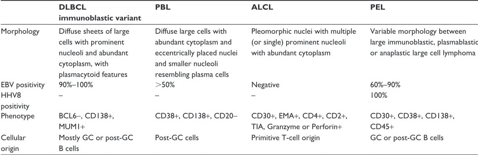
Initial diagnosis is made by hematopathologic assessment of the effusion at presentation. As morphologically described by the WHO, PEL cells bridge those of large-cell immunoblastic lymphoma, plasmablastic lymphoma, and anaplastic large-cell lymphoma30 (Table 1). The cells are large, with moderate
to abundant deeply basophilic cytoplasm, a large round to irregular nuclei, and prominent nucleoli. They express CD45, proving lymphoid origins, however, are a “null” lymphocyte phenotype as they do not exhibit typical B-cell or T-cell immunophenotype characteristics and are indeterminate
Table 1 Pathologic features differentiating PeL from other aggressive lymphomas
DLBCL
immunoblastic variant
PBL ALCL PEL
Morphology Diffuse sheets of large cells with prominent nucleoli and abundant cytoplasm, with plasmacytoid features
Diffuse large cells with abundant cytoplasm and eccentrically placed nuclei and smaller nucleoli resembling plasma cells
Pleomorphic nuclei with multiple (or single) prominent nucleoli with abundant cytoplasm
variable morphology between large immunoblastic, plasmablastic or anaplastic large cell lymphoma
eBv positivity 90%–100% .50% Negative 60%–90%
HHv8 positivity
– – – 100%
Phenotype BCL6−, CD138+, MUM1+
CD38+, CD138+, CD20− CD30+, eMA+, CD4+, CD2+, TiA, Granzyme or Perforin+
CD30+, CD38+, CD138+, CD45+
Cellular origin
Mostly GC or post-GC B cells
Post-GC cells Primitive T-cell origin GC or post-GC B cells
Abbreviations: PeL, primary effusion lymphoma; eBv, epstein Bar virus; HHv8, human herpes virus 8; GC, germinal center; DLBCL, diffuse large B cell lymphoma; PBL, plasmablastic lymphoma; ALCL, anaplastic large cell lymphoma.
OncoTargets and Therapy downloaded from https://www.dovepress.com/ by 118.70.13.36 on 25-Aug-2020
Dovepress Primary effusion lymphoma
by immunohistochemistry (IHC). CD30, a marker found in 70% of classical Hodgkin lymphoma. Reed-Sternberg cells are commonly observed in PEL, whereas CD15 is typi-cally not expressed. Markers of plasma cell differentiation, including CD38 and CD138, are present; of note plasma-cell myeloma is typically CD45 negative.31 human leukocyte
antigen – antigen D related (HLA-DR), activation antigen, and epithelial membrane antigen are variably expressed.
Most importantly, the confirmation in diagnosis of PEL is dependent on identifying HHV8 viral infection in the nuclei of the malignant cells. This is evidenced via expression of LANA-1 by IHC stain or via DNA extraction and polymerase chain reaction amplification of HHV8.31 This verification by
itself can differentiate the null lymphocyte phenotype PEL from other lymphomas demonstrating strikingly similar clinical presentations and morphologic features.
Cell of origin and genetic alterations in PeL
Gene expression profiling reveals the PEL cell of origin to be closely related to postgerminal center late-differentiating B-cells and likely of plasmablastic derivation.32 While
clonal rearrangement of the heavy immunoglobulin gene is detected, demonstrating B-cell derivation, a recurrent cytogenetic abnormality or PEL-specific driver mutation has not been identified.31 Typical NHL-related gross
rear-rangements or mutations of BCL2, c-Myc, and TP53 are not identified in PEL.
Gaidano et al found a high frequency of BCL6 5′
noncoding region nucleotide substitution mutations in PEL.33
Bcl6 5′ mutations are markers of B-cell transition through the germinal center. Therefore, these mutations suggest the origin as postgerminal-center B-cells.31 In addition, a frequent
occurrence of complete or partial trisomy 12, trisomy 7, and abnormalities of bands 1q21-25 have been noted.33
Interleukin 1 receptor-associated kinase 1 (IRAK1), together with its binding partner MYD88, mediate toll-like receptor signaling and reactivate HHV8 leading to prolonged survival of PEL in culture. Via X chromosome targeted sequencing of PEL exudate cells, Yang et al found IRAK1 constitutively phosphorylated (mutated) in PEL and required for survival of these tumor cells.34 Of note, the IRAK1 mutation is a
common, essential driver for Kaposi sarcoma.
Staging
As per the Lugano classification for NHL, all patients who present an effusion have stage IV disease at diagnosis.35 Radiographic evaluation is akin to other
aggressive NHL diagnoses including positron emission
tomography-computed tomography. Additionally, a bone marrow biopsy and/or lumbar puncture may be considered if clinically indicated.
Prognosis
Given its resistance to cytotoxic therapies, treatment with PEL has generally been associated with a poor prognosis. Several factors have been evaluated as potential markers of prognosis. In a retrospective study of 28 HIV-positive patients with PEL, two independent predictors of decreased survival were identified by multivariate analysis: poor perfor-mance status and absence of combined antiretroviral therapy (c-ART) prior to PEL diagnosis.36 In a different analysis of
104 patients with PEL, the number and location of affected cavities appeared to play a role in prognosis.5 Specifically,
the involvement of more than one body cavity was associated with an overall survival (OS) of 4 months in comparison to 18 months in patients with only one cavity involved.
Management
Given the low incidence of PEL, there are no large-scale randomized studies to guide treatment and management decisions. Most of the evidence is based on retrospective studies, case reports, and preclinical data.
Frontline treatment
Chemotherapy has traditionally been the cornerstone of treatment for patients with a good performance status and no significant comorbidities. However, an optimal regimen does not exist. As the majority of the PEL are diagnosed in the setting of HIV infection, the approach to their manage-ment is also based on HIV status.
Chemotherapy
Although there is no one standard regimen that is universally accepted for the frontline treatment of PEL, an aggressive lymphoma regimen is typically used. Examples include dose-adjusted (DA) EPOCH (etoposide, prednisolone, vincristine, cyclophosphamide, doxorubicin) or CHOP (cyclophosph-amide, doxorubicin, vincristine, and prednisone).
The use of DA-EPOCH for PEL can be extrapolated from a study conducted at the National Cancer Institute in 39 patients with newly diagnosed AIDS-associated aggressive B-cell lymphoma.37 The regimen produced an overall response
rate (ORR) of 87%, 74% of which were complete remissions (CRs). At 52 months, the disease-free survival and OS were 92% and 60%, respectively. The treatment was well tolerated with grade 3 or 4 neutropenia, anemia, and thrombocytopenia
OncoTargets and Therapy downloaded from https://www.dovepress.com/ by 118.70.13.36 on 25-Aug-2020
Dovepress Narkhede et al
occurring in 30%, 17%, and 21% of the cycles, respectively. Serious constipation or stomatitis occurred in less than 3% of cycles, and grade 3 peripheral neuropathies occurred in two patients. Though no randomized controlled trials exist for PEL, there are case reports with the use of DA-EPOCH in combination with ART. In one case report, an HIV patient with extracavitary PEL received four cycles of EPOCH which led to a complete response after four cycles that lasted for 14 months after completing treatment.38
Simonelli et al conducted a retrospective analysis evaluat-ing the efficacy of a CHOP-like regimen in which prednisone was omitted in the majority of the eight patients included in order to prevent the emergence or exacerbation of KS. Three patients achieved a CR (42%), and the median OS was 6 months.39 Boulanger et al explored the efficacy of another
multi-agent regimen, CHVp plus methotrexate, in another retrospective study of seven patients with AIDS-associated PEL. Patients received cyclophosphamide 650–700 mg/m2,
doxorubicin 35–40 mg/m2, vincristine 0.8 mg/m2, or
etopo-side 80 mg/m2 on day 1 followed by 4-hour intravenous
infu-sion of methotrexate 2.5–3 g/m2 on day 2 for 6–8 cycles every
21 days.40 Three achieved a CR and remained in remission at
18, 26, and 78 months. Most of the patients had hematologi-cal toxicity with delayed clearance of methotrexate due to the presence of effusions. The use of methotrexate should be avoided as majority of the patients have an effusion that leads to decreased clearance and prolonged toxicity with methotrexate.
Rarely, PEL are CD20 positive. As with other B-cell lymphomas, treatment should incorporate rituximab-based chemoimmunotherapy. Two single-patient case reports have demonstrated the efficacy of RCHOP in this population, resulting in durable CR lasting 22 and 30 months.41,42 The
use of R-EPOCH in an HIV-patient resulted in CR lasting 12 months.43
Anti-retroviral therapy
Anti-retroviral treatment (ART) is an important component of the management in patients with lymphoproliferative malignancies in the setting of HIV. In a retrospective study of HIV-infected patients diagnosed with PEL, Boulanger et al demonstrated a shorter OS for patients with a poor perfor-mance status and untreated HIV infection prior to diagnosis.36
Similarly, patients treated with a CHOP-like regimen without ART were unable to achieve a CR and had a shorter OS of 3 months.39 ART given by itself can also lead to a CR, as
evident by one case report in which a patient remained in CR at 14 months.44
Treatment with ART should therefore be the mainstay of HIV-positive patients with PEL. In terms of choice of antiretroviral agents, some agents have more activity in PEL than others based on preclinical studies. Azidothymidine has been shown to sensitize primary effusion lymphoma cells to Kaposi sarcoma associated herpesvirus-specific CD4+
T cells and inhibits proliferation in PEL cell lines.45 Similarly,
preclinical studies with the protease inhibitor Lopinavir has shown induction of apoptosis of PEL cells via suppression of the NF-κB pathway. Interestingly, this effect, though present, was not as significant with other protease inhibitors like Ritonavir and Darunavir.46 Hence, involving the infectious
disease specialist in managing these patients is crucial.
Relapsed and refractory disease
Most patients with PEL experience a relapse after frontline therapy within 6–8 months and subsequently require further treatment.11 Treatment options will depend upon the
perfor-mance status, comorbidities, and goals of care based on the individual. Therapies that have been evaluated in patients include stem cell transplant, radiation, and targeted agents such as bortezomib.
Stem cell transplant
Autologous stem cell transplant (ASCT) has been attempted in a handful of patients. Waddington et al published a case report of an HIV-positive patient with PEL who received high-dose chemotherapy followed by autologous stem cell transplant in PEL.47 The patient had persistent disease prior
to ASCT. The transplant was unsuccessful as he developed recurrent accumulation of pleural fluid consistent with persistent disease indicating failure of ASCT. Won et al demonstrated the effectiveness of ASCT in an HIV-negative patient who proceeded to ASCT after achieving a CR with salvage chemotherapy (ifosfamide, carboplatin, and etoposide).48 The patient remained in CR 12 months after
completion of ASCT.
As the majority of patients with PEL have concurrent HIV infection, there has been great apprehension regarding the use of allogenic stem cell transplant (allo-SCT) in this population. Nevertheless, Bryant et al successfully performed a reduced intensity conditioning regimen followed by allo-SCT in one HIV-positive patient with PEL.49 The patient
achieved a CR and remained in CR for a period of 31 months after transplantation. There have been no other reports of the use of allo-SCT in PEL, therefore, the role for this therapy remains uncharted.
OncoTargets and Therapy downloaded from https://www.dovepress.com/ by 118.70.13.36 on 25-Aug-2020
Dovepress Primary effusion lymphoma
Radiation
When the goal of treatment shifts toward comfort in patients with refractory or relapsed disease, an important compo-nent is to identify if the site of disease is causing physical distress. Radiation by itself is inadequate in most instances for achieving a complete response as with other aggressive lymphomas; however, it can be useful to control the disease burden. In a case report by Cassoni et al, a multiply-refractory patient with PEL with a pleural-based mass was treated with localized radiation resulting in a sustained remission for 12 months.70 Radiation can be considered for PEL with a
solid component involved within one radiation field.
Antiviral therapies
As the malignant cells in PEL are infected with HHV8 virus, they theoretically can be targeted through antiviral therapy. Unfortunately, since the HHV8 virus is in a latent state, it is resistant to most antiviral agents. Klass and Offermann proposed a potential solution whereby induction of the HHV8 virus into the lytic phase via valproate followed by treatment with a lytic-phase effective anti-herpetic drug may lead to apoptosis of the infected cell.50 Using an HHV-8 positive
PEL cell line, they found lytic replication to be sensitive to ganciclovir, foscarnet and cidofovir, and resistant to acyclovir.50,51
The effectiveness of antiviral therapy in PEL has been demonstrated in a handful of case reports. An HIV patient who was refractory to frontline chemotherapy regimen of bortezomib, cyclophosphamide, Adriamycin, and predni-sone experienced a CR with valganciclovir while continu-ing to receive c-ART.52 The patient received treatment for
12 months but had achieved a CR by 6 months with eradi-cation of the HHV8 virus. Seven months after completion of therapy, the patient continued on c-ART and remained disease free. Cidofovir has also been used in combination with ART and interferon. Hocqueloux et al published a case report of a patient with HIV-associated PEL treated with cido-fovir and interferon (IFN)-α: cidofovir 5 mg/kg every 15 days in combination, IFN-α 3 million units 3 times a week.53
A CR was achieved after 2 months of treatment; cidofovir was stopped at 3 months, whereas IFN-α was continued for 7 months. The patient remained in a complete remission for 24 months. The inferior activity of cidofovir is postulated to be due to poor penetration of the drug into the effusion.54,55
It has been shown to be highly active as a single agent when administered directly into the affected cavity. Luppi et al suc-cessfully treated three patients with HIV-negative, HHV8+
PEL with intracavitary cidofovir 2.5–5 mg/kg every week.55
The first patient achieved a CR with two doses of intrapleural cidofovir and maintained a remission for 10 months. The second patient achieved a CR after 3 doses of intraperito-neal cidofovir, maintaining a remission for 5 months. The third patient achieved a CR after three doses of intrapleural cidofovir and remained treatment free for 15 months. This approach is limited to disease involving only one cavity but may be extrapolated to HIV-positive patients as well. There are no data for the use of intracavitary cidofovir injection in multiple cavities.
Talc pleurodesis
When PEL presents solely as an effusion, palliative approaches to decrease accumulation of fluid can be performed. One commonly utilized approach in other malignancies including malignant mesothelioma is pleurodesis. In this procedure, the visceral and parietal pleural layers are fused together so that there is no accumulation of fluid. Talc is a sclerosant which has been effectively used in this process. In addition to causing a fusion of the visceral and parietal pleura, it also has an apoptotic effect on mesothelioma cells by regulating the surface expression of the proto-oncogene c-myc and enhanc-ing apoptosis.56 A case series has shown that it is possible to
achieve and maintain long-term remissions in patients with HIV-negative HHV8-associated PEL with video-assisted talc pleurodesis.57 Remissions were achieved instantaneously
after the procedure and lasting 50–60 months in the three patients evaluable. This is a reasonable option for elderly, frail individuals who have limited treatment options and are unable to tolerate aggressive treatments.
Targeted therapies
Proteasome inhibitors
Proteasomal activity is required for the survival of PEL cells and viral replication of KSHV cells. Preclinical data have shown that proteasome inhibitors reduce cell proliferation and induce apoptosis in KSHV-positive, EBV-positive and KSHV-positive, EBV-negative PEL cell lines.54 As described
previously, HHV8 viral latent transcripts also constitutively activate the NF-κB pathway, leading to tumor cell prolif-eration and survival. Bortezomib is a proteasome inhibitor currently approved in multiple myeloma and mantle cell lymphoma.58 While single-agent bortezomib was not shown
to be active in a small case series of three HIV-infected patients with relapsed/refractory PEL, it has promise in combination with other agents.8 Siddiqi et al administered a
combination of bortezomib, PEGylated liposomal doxoru-bicin, and rituximab to an HIV-negative patient with CD20
OncoTargets and Therapy downloaded from https://www.dovepress.com/ by 118.70.13.36 on 25-Aug-2020
Dovepress Narkhede et al
positive PEL, which resulted in a durable CR lasting at least 2 years.59 Bortezomib has also been studied with the histone
deacetylase inhibitor, vorinostat, in a murine PEL xenograft model. The combination of bortezomib and vorinostat led to KSHV lytic replication and cell death, thus demonstrating the synergy of vorinostat and bortezomib.60
Brentuximab vedotin (Bv)
BV is an antibody drug conjugate against CD30, currently approved in previously treated classical Hodgkin lymphoma and anaplastic large cell lymphoma.61 As PEL is typically
CD30 positive, Bhatt et al evaluated the role of BV in CD30 positive UM-PEL-1 and UM-PEL-3 cell lines and xenograft models.62 BV induced G2 phase cell cycle arrest with
intracel-lular delivery of the drug monomethyl auristatin E, inducing apoptosis of both the CD30 expressing cells and intratumoral cells lacking the target antigen. This effect was also observed in xenograft models inoculated with PEL-1 and UM-PEL-3 cells. While there have been no clinical trials of BV in PEL, it has shown promise in other CD30+ lymphomas. Jacobsen et al administered BV to patients with CD30+
non-Hodgkin lymphoma patients, resulting in notable activ-ity among the different histologies.63 The ORR in DLBCL
was 44%, including 17% complete remissions and a median duration of response of 16.6 months (range 2.7–22.7 months). Responses were also seen in patients with gray zone and posttransplant lymphoproliferative disorders. While the true efficacy in PEL is unclear, BV may be considered in select patients.
Preclinical studies
Preclinical data have provided insight into targeted thera-pies that may have efficacy in PEL. The mammalian target of rapamycin (mTOR), its activator AKT, and the target p70S6 kinases are frequently phosphorylated in PEL. Rapamycin, an inhibitor of mTOR, has been tested in a variety of PEL cell lines (BC-1, BC-3, JSC-1, BCBL-1, BCP-1, and VG-1), proving its ability to inhibit prolifera-tion and induce apoptosis.64 It has also been studied in
xenograft models, confirming its ability to inhibit PEL tumor growth. PEL cell lines also secrete high levels of VEGF and L-6.65 Given these findings, the VEGF
inhibi-tor, bevacizumab, and the IL-6 inhibiinhibi-tor, tocilizumab, have also been studied in PEL cell lines (BCBL-1, BC-1, BC-3, TY-1) and xenograft mouse models.66 Neither
beva-cizumab or tocilizumab were able to inhibit proliferation in the cell line studies, however, both agents were able to inhibit the development of malignant pleural effusion and
provide a survival benefit in the xenograft models. These data support the exploration of the agents’ utility in the clinical setting.
The immunomodulatory agent, lenalidomide, was ini-tially approved in multiple myeloma but has also shown activity in other lymphoid malignancies including mantle cell lymphoma.67 It has been evaluated with arsenic trioxide,
a potent agent in acute promyelocytic leukemia.68 The
combination resulted in inhibition of growth in PEL cell lines (BC-1 and BC-3) by upregulating p53 and inducing apoptosis.69 Reduction of the expression of latent viral
transcripts (LANA-1, LANA-2, v-FLIP, and v-Cyclin) and proteins (LANA-1/LANA-2) was also noted. In PEL xenograft models, lenalidomide and arsenic trioxide induced greater responses and OS than lenalidomide alone. These preclinical studies provide a rational for the use of these agents in the clinical setting. Unfortunately, given the rarity of this disease and the comorbidities associated with it, it is difficult to develop readily accessible trials for patients.
Conclusion
PEL is an uncommon and aggressive disease affecting a unique population of patients who are often elderly or immu-nocompromised. Most of the patients diagnosed with PEL have significant other comorbidities, such as HIV, along with a poor performance status which limit their enrollment in clinical trials. For the fit and healthy patients, intensive che-motherapy remains the preferred choice, however, the avail-ability of various targeted agents may alter this approach. For the unfit individuals, palliative treatments or newer targeted agents should be considered. With the improvement in our understanding of PEL, the treatment landscape is evolving and promising. Despite our advances, for now the use of ART for HIV-positive patients will continue to remain the backbone of treatment.
Disclosure
The authors report no conflicts of interest in this work.
References
1. Jarrett RF. Viruses and lymphoma/leukaemia. J Pathol. 2006;208(2): 176–186.
2. Chang Y, Cesarman E, Pessin MS, et al. Identification of herpesvirus-like DNA sequences in AIDS-associated Kaposi’s sarcoma. Science. 1994;266(5192):1865–1869.
3. Cesarman E, Chang Y, Moore PS, Said JW, Knowles DM. Kaposi’s sarcoma-associated herpesvirus-like DNA sequences in AIDS-related body-cavity-based lymphomas. N Engl J Med. 1995;332(18):1186–1191. 4. Kaplan LD. Human herpesvirus-8: Kaposi sarcoma, multicentric Castle-man disease, and primary effusion lymphoma. Hematol Am Soc Hematol
Educ Program. 2013;2013:103–108.
OncoTargets and Therapy downloaded from https://www.dovepress.com/ by 118.70.13.36 on 25-Aug-2020
Dovepress Primary effusion lymphoma
5. Castillo JJ, Shum H, Lahijani M, Winer ES, Butera JN. Prognosis in primary effusion lymphoma is associated with the number of body cavities involved. Leuk Lymphoma. 2012;53(12):2378–2382. 6. Chen Y-B, Rahemtullah A, Hochberg E. Primary effusion lymphoma.
Oncologist. 2007;12(5):569–576.
7. Boulanger E, Hermine O, Fermand J-P, et al. Human herpesvirus 8 (HHV-8)-associated peritoneal primary effusion lymphoma (PEL) in two HIV-negative elderly patients. Am J Hematol. 2004;76(1):88–91. 8. Boulanger E, Afonso PV, Yahiaoui Y, Adle-Biassette H, Gabarre J, Agbalika F. Human herpesvirus-8 (HHV-8)-associated primary effu-sion lymphoma in two renal transplant recipients receiving rapamycin.
Am J Transplant. 2008;8(3):707–710.
9. Said JW, Tasaka T, Takeuchi S, et al. Primary effusion lymphoma in women: report of two cases of Kaposi’s sarcoma herpes virus-associated effusion-based lymphoma in human immunodeficiency virus-negative women. Blood. 1996;88(8):3124–3128.
10. Sasaki Y, Isegawa T, Shimabukuro A, Yonaha T, Yonaha H. Primary effusion lymphoma in an elderly HIV-negative patient with hemodialy-sis: importance of evaluation for pleural effusion in patients receiving hemodialysis. Case Rep Nephrol Urol. 2014;4(2):95–102.
11. Dunleavy K, Wilson WH. How I treat HIV-associated lymphoma. Blood. 2012;119(14):3245–3255.
12. Swerdlow SH, Campo E, Harris NL, et al. WHO Classification of
Tumours of Haematopoietic and Lymphoid Tissues. Revised 4th ed,
Volume 2. Lyon: International Agency for Research on Cancer; 2016.
13. Carbone A, Gloghini A. KSHV/HHV8-associated lymphomas. Br J
Haematol. 2008;140(1):13–24.
14. Said J. Kaposi’s sarcoma-associated herpesvirus (KSHV): a new viral pathogen associated with Kaposi’s sarcoma, primary effusion lym-phoma, and multicentric Castleman’s disease. West J Med. 1997;167(1): 37–38.
15. Friborg J Jr, Kong W, Hottiger MO, Nabel GJ. p53 inhibition by the LANA protein of KSHV protects against cell death. Nature. 1999; 402(6764):889–894.
16. Radkov SA, Kellam P, Boshoff C. The latent nuclear antigen of Kaposi sarcoma-associated herpesvirus targets the retinoblastoma-E2F pathway and with the oncogene Hras transforms primary rat cells. Nat Med. 2000;6(10):1121–1127.
17. DeCotiis JL, Lukac DM. KSHV and the role of NOTCH receptor dysregulation in disease progression. Pathogens. 2017;6(3):E34. 18. Direkze S, Laman H. Regulation of growth signalling and cell cycle
by Kaposi’s sarcoma-associated herpesvirus genes. Int J Exp Pathol. 2004;85(6):305–319.
19. Sakakibara S, Tosato G. Viral interleukin-6: role in Kaposi’s sarcoma-associated herpesvirus–sarcoma-associated malignancies. J Interferon Cytokine
Res. 2011;31(11):791–801.
20. Ely SA, Powers J, Lewis D, et al. Kaposi’s sarcoma-associated herpesvirus-positive primary effusion lymphoma arising in the suba-rachnoid space. Hum Pathol. 1999;30(8):981–984.
21. Pielasinski U, Santonja C, Rodríguez-Pinilla SM, Requena L. Extracavi-tary primary effusion lymphoma presenting as a cutaneous tumor: a case report and literature review. J Cutan Pathol. 2014;41(9):745–753. 22. Inoue S, Miyamoto T, Yoshino T, Yamadori I, Hagari Y, Yamamoto O.
Primary effusion lymphoma with skin involvement. J Clin Pathol. 2006; 59(11):1221–1222.
23. Ibrahim U, Saqib A, Mohammad F, Ding J, Hussein S, Atallah JP. KSHV-associated extracavitary primary effusion lymphoma in an HIV seronegative patient: a case report and review of the literature. Postgrad
Med. 2017;129(3):402–407.
24. Kabiawu-Ajise OE, Harris J, Ismaili N, Amorosi E, Ibrahim S. Primary effusion lymphoma with central nervous system involvement in an HIV-negative homosexual male. Acta Haematol. 2012;128(2):77–82. 25. Chadburn A, Hyjek E, Mathew S, Cesarman E, Said J, Knowles DM.
KSHV-positive solid lymphomas represent an extra-cavitary vari-ant of primary effusion lymphoma. Am J Surg Pathol. 2004;28(11): 1401–1416.
26. Medeiros BC, Maness LJ, Bauer FA, Ross JW, Kapur D. Unusual pre-sentation of “extracavitary” primary effusion lymphoma in previously unknown HIV disease. Conn Med. 2000;64(10):591–594.
27. Pantanowitz L, Wu Z, Dezube BJ, Pihan G. Extracavitary primary effusion lymphoma of the anorectum. Clin Lymphoma Myeloma. 2005; 6(2):149–152.
28. Kim Y, Leventaki V, Bhaijee F, Jackson CC, Medeiros LJ, Vega F. Extracavitary/solid variant of primary effusion lymphoma. Ann Diagn
Pathol. 2012;16(6):441–446.
29. Santonja C, Medina-Puente C, Serrano Del Castillo C, Cabello Úbeda A, Rodríguez-Pinilla SM. Primary effusion lymphoma involving cerebro-spinal fluid, deep cervical lymph nodes and adenoids. Report of a case supporting the lymphatic connection between brain and lymph nodes.
Neuropathology. 2017;37(3):249–258.
30. Nador RG, Cesarman E, Chadburn A, et al. Primary effusion lymphoma: a distinct clinicopathologic entity associated with the Kaposi’s sarcoma-associated herpes virus. Blood. 1996;88(2):645–656.
31. Brimo F, Michel RP, Khetani K, Auger M. Primary effusion lymphoma: a series of 4 cases and review of the literature with emphasis on cyto-morphologic and immunocytochemical differential diagnosis. Cancer. 2007;111(4):224–233.
32. Klein U, Gloghini A, Gaidano G, et al. Gene expression profile analysis of AIDS-related primary effusion lymphoma (PEL) suggests a plas-mablastic derivation and identifies PEL-specific transcripts. Blood. 2003;101(10):4115–4121.
33. Gaidano G, Capello D, Cilia AM, et al. Genetic characterization of HHV-8/KSHV-positive primary effusion lymphoma reveals frequent mutations of BCL6: implications for disease pathogenesis and histo-genesis. Genes Chromosomes Cancer. 1999;24(1):16–23.
34. Yang D, Chen W, Xiong J, Sherrod CJ, Henry DH, Dittmer DP. Inter-leukin 1 receptor-associated kinase 1 (IRAK1) mutation is a common, essential driver for Kaposi sarcoma herpesvirus lymphoma. Proc Natl
Acad Sci U S A. 2014;111(44):E4762–E4768.
35. Cheson BD, Fisher RI, Barrington SF, et al. Recommendations for initial evaluation, staging, and response assessment of Hodgkin and non-Hodgkin lymphoma: the Lugano classification. J Clin Oncol. 2014; 32(27):3059–3068.
36. Boulanger E, Gérard L, Gabarre J, et al. Prognostic factors and outcome of human herpesvirus 8-associated primary effusion lymphoma in patients with AIDS. J Clin Oncol. 2005;23(19):4372–4380. 37. Little RF, Pittaluga S, Grant N, et al. Highly effective treatment of
acquired immunodeficiency syndrome-related lymphoma with dose-adjusted EPOCH: impact of antiretroviral therapy suspension and tumor biology. Blood. 2003;101(12):4653–4659.
38. El-Ayass W, Yu E-M, Karcher DS, Aragon-Ching JB. Complete response to EPOCH in a patient with HIV and extracavitary primary effusion lymphoma involving the colon: a case report and review of literature. Clin Lymphoma Myeloma Leuk. 2012;12(2):144–147. 39. Simonelli C, Spina M, Cinelli R, et al. Clinical features and outcome of
primary effusion lymphoma in HIV-infected patients: a single-institution study. J Clin Oncol. 2003;21(21):3948–3954.
40. Boulanger E, Daniel M-T, Agbalika F, Oksenhendler E. Combined che-motherapy including high-dose methotrexate in KSHV/HHV8-associated primary effusion lymphoma. Am J Hematol. 2003;73(3):143–148. 41. Terasaki Y, Okumura H, Saito K, et al. HHV-8/KSHV-negative and
CD20-positive primary effusion lymphoma successfully treated by pleural drainage followed by chemotherapy containing rituximab. Intern
Med. 2008;47(24):2175–2178.
42. Suzuki K, Ino K, Sugawara Y, Mizutani M, Sekine T, Katayama N. [Pro-longed survival in a patient with human herpesvirus-8-negative primary effusion lymphoma after combination chemotherapy with rituximab].
Gan To Kagaku Ryoho. 2008;35(4):691–694. [In Japanese].
43. Foster WR, Bischin A, Dorer R, Aboulafia DM. Human herpesvirus type 8-associated large B-cell lymphoma: a nonserous extracavitary variant of primary effusion lymphoma in an HIV-infected man: a case report and review of the literature. Clin Lymphoma Myeloma Leuk. 2016; 16(6):311–321.
OncoTargets and Therapy downloaded from https://www.dovepress.com/ by 118.70.13.36 on 25-Aug-2020
OncoTargets and Therapy
Publish your work in this journal
Submit your manuscript here: http://www.dovepress.com/oncotargets-and-therapy-journal
OncoTargets and Therapy is an international, peer-reviewed, open access journal focusing on the pathological basis of all cancers, potential targets for therapy and treatment protocols employed to improve the management of cancer patients. The journal also focuses on the impact of management programs and new therapeutic agents and protocols on
patient perspectives such as quality of life, adherence and satisfaction. The manuscript management system is completely online and includes a very quick and fair peer-review system, which is all easy to use. Visit http://www.dovepress.com/testimonials.php to read real quotes from published authors.
Dovepress
Dove
press
Narkhede et al
44. Oksenhendler E, Clauvel JP, Jouveshomme S, Davi F, Mansour G. Complete remission of a primary effusion lymphoma with antiretroviral therapy. Am J Hematol. 1998;57(3):266.
45. Williamson SJ, Nicol SM, Stürzl M, Sabbah S, Hislop AD. Azidothy-midine sensitizes primary effusion lymphoma cells to Kaposi sarcoma-associated herpesvirus-specific CD4+ T cell control and inhibits vIRF3 function. PLoS Pathog. 2016;12(11):e1006042.
46. Kariya R, Taura M, Suzu S, Kai H, Katano H, Okada S. HIV protease inhibitor Lopinavir induces apoptosis of primary effusion lymphoma cells via suppression of NF-κB pathway. Cancer Lett. 2014;342(1): 52–59.
47. Waddington TW, Aboulafia DM. Failure to eradicate AIDS-associated primary effusion lymphoma with high-dose chemotherapy and autolo-gous stem cell reinfusion: case report and literature review. AIDS Patient
Care STDs. 2004;18(2):67–73.
48. Won J-H, Han S-H, Bae S-B, et al. Successful eradication of relapsed primary effusion lymphoma with high-dose chemotherapy and autolo-gous stem cell transplantation in a patient seronegative for human immunodeficiency virus. Int J Hematol. 2006;83(4):328–330. 49. Bryant A, Milliken S. Successful reduced-intensity conditioning
allo-geneic HSCT for HIV-related primary effusion lymphoma. Biol Blood
Marrow Transplant. 2008;14(5):601–602.
50. Klass CM, Offermann MK. Targeting human herpesvirus-8 for treat-ment of Kaposi’s sarcoma and primary effusion lymphoma. Curr Opin
Oncol. 2005;17(5):447–455.
51. Kedes DH, Ganem D. Sensitivity of Kaposi’s sarcoma-associated herpesvirus replication to antiviral drugs. Implications for potential therapy. J Clin Invest. 1997;99(9):2082–2086.
52. Marquet J, Velazquez-Kennedy K, López S, Benito A, Blanchard M-J, Garcia-Vela JA. Case report of a primary effusion lymphoma suc-cessfully treated with oral valganciclovir after failing chemotherapy.
Hematol Oncol. 2018;36(1):316–319.
53. Hocqueloux L, Agbalika F, Oksenhendler E, Molina JM. Long-term remission of an AIDS-related primary effusion lymphoma with antiviral therapy. AIDS. 2001;15(2):280–282.
54. Halfdanarson TR, Markovic SN, Kalokhe U, Luppi M. A non-chemotherapy treatment of a primary effusion lymphoma: durable remission after intracavitary cidofovir in HIV negative PEL refractory to chemotherapy. Ann Oncol. 2006;17(12):1849–1850.
55. Luppi M, Trovato R, Barozzi P, et al. Treatment of herpesvirus associ-ated primary effusion lymphoma with intracavity cidofovir. Leukemia. 2005;19(3):473–476.
56. Nasreen N, Mohammed KA, Dowling PA, Ward MJ, Galffy G, Antony VB. Talc induces apoptosis in human malignant mesothelioma cells in vitro. Am J Respir Crit Care Med. 2000;161(2 Pt 1):595–600.
57. Birsen R, Boutboul D, Crestani B, et al. Talc pleurodesis allows long-term remission in HIV-unrelated Human Herpesvirus 8-associated pri-mary effusion lymphoma. Leuk Lymphoma. 2017;58(8):1993–1998. 58. Raedler L. Velcade (Bortezomib) receives 2 new FDA indications: for
retreatment of patients with multiple myeloma and for first-line treat-ment of patients with mantle-cell lymphoma. Am Health Drug Benefits. 2015;8(Spec Feature):135–140.
59. Siddiqi T, Joyce RM. A case of HIV-negative primary effusion lym-phoma treated with bortezomib, pegylated liposomal doxorubicin, and rituximab. Clin Lymphoma Myeloma. 2008;8(5):300–304.
60. Bhatt S, Ashlock BM, Toomey NL, et al. Efficacious proteasome/HDAC inhibitor combination therapy for primary effusion lymphoma. J Clin
Invest. 2013;123(6):2616–2628.
61. ADCETRIS® (brentuximab vedotin) | U.S. | Seattle Genetics. http://
www.seattlegenetics.com/products/adcetris-us. Accessed February 24, 2018.
62. Bhatt S, Ashlock BM, Natkunam Y, et al. CD30 targeting with bren-tuximab vedotin: a novel therapeutic approach to primary effusion lymphoma. Blood. 2013;122(7):1233–1242.
63. Jacobsen ED, Sharman JP, Oki Y, et al. Brentuximab vedotin demonstrates objective responses in a phase 2 study of relapsed/refractory DLBCL with variable CD30 expression. Blood. 2015;125(9):1394–1402. 64. Sin S-H, Roy D, Wang L, et al. Rapamycin is efficacious against primary
effusion lymphoma (PEL) cell lines in vivo by inhibiting autocrine signaling. Blood. 2007;109(5):2165–2173.
65. Aoki Y, Tosato G. Role of vascular endothelial growth factor/ vascular permeability factor in the pathogenesis of Kaposi’s sarcoma-associated herpesvirus-infected primary effusion lymphomas. Blood. 1999;94(12):4247–4254.
66. Goto H, Kudo E, Kariya R, Taura M, Katano H, Okada S. Targeting VEGF and interleukin-6 for controlling malignant effusion of primary effusion lymphoma. J Cancer Res Clin Oncol. 2015;141(3):465–474. 67. Dimopoulos MA, Kastritis E, Rajkumar SV. Treatment of plasma cell
dyscrasias with lenalidomide. Leukemia. 2008;22(7):1343–1353. 68. Trisenox (Arsenic trioxide) [package insert]. Teva Pharmaceuticals
USA, Inc. North Wales, PA 19454.
69. Hleihel R, Tawil N, Karam M, Merhi RA, Bazarbachi A, Hajj HE. Arsenic trioxide and lenalidomide: a promising combination therapy for primary effusion lymphoma. Blood. 2017;130(Suppl 1):2817. 70. Cassoni A, Ali U, Cave J, et al. Remission after radiotherapy for a
patient with chemotherapy-refractory HIV-associated primary effusion lymphoma. J Clin Oncol. 2008;26(32):5297–5299.
OncoTargets and Therapy downloaded from https://www.dovepress.com/ by 118.70.13.36 on 25-Aug-2020
![Bis(2,2′ bipyridine){ethyl 4′ [N (4 carbamoylphenyl)carbamoyl] 2,2′ bipyridine 4 carboxylate}ruthenium(II) bis[hexafluoridophosphate(V)]](data:image/gif;base64,R0lGODlhAQABAIAAAP///wAAACH5BAEAAAAALAAAAAABAAEAAAICRAEAOw==)