s Z
RESEARCH ARTICLE
CONE-BEAM COMPUTED TOMOGRAPHY STUDY OF MORPHOMETRIC EVALUATION OF
PTERYGOIDHAMULUS
*Dr. Ashwini Nerkar, Dr. Rajeev Gadgil, Dr. Ajay Bhoosreddy, Dr. Karan Shah, Dr. Shweta Varma
Department of Oral Medicine and Radiology, MGV’S KBH Dental College and Hospital,
Nashik, Maharashtra, India
ARTICLE INFO ABSTRACT
aim: The aim of the study was morphometricevaluation of pterygoidhamulus using CBCT.
Material and Methods: CBCT images of 40males and 40females with age >20years were evaluated.The length, width, inclinationandincidence of shape of PH was evaluatedwith respect to side and gender.Independent t-test was used for statistical analysis.
Results: The mean length of PH of both sides in males and females was found to be 11.5 +0.75 and 10.09 + 0.45 mm, inclinations in sagittal plane was 12.74° + 30.45 and 15.22° + 30.65 in anterior direction,inclinations in coronal plane was 11.5° + 31.05, 19.7° + 39.15 in medial direction whereas, it was 79.13°+30.4 and 71.5°+38.7 in lateral direction.Incidence of shapes of PH was 2.6,2.4 for triangle 1.7 and 2.2 for slenderrespectively with statistically significant difference. Mean width values in males & females was found to be (left) 3.04 + 0.6 , 3.06 + 0.5 and (right) 2.01 + 0.4 and 2.02 + 0.3mm,inclination in sagittal plane (left) was 47.2°+ 20.1 and (right) 47.28°+ 22.1 ,47° + 20.1 and 47.1° + 22.1 in posterior direction respectively with statistically insignificant difference. No statistically significant difference was found between left and right side except the inclination in coronal plane was more in left as compared to right side.Inclination in coronal and sagittal plane was more on lateral and posterior side respectively.
Conclusion: Appraisal of the morphology of PH is helpful as it provides insightfor differential diagnosis of the imperceptible pains inoral cavity and pharynx.
INTRODUCTION
Tracing an unusual pain in the soft palate and pharyngeal region has been a diagnostic dilemma since years. Symptoms in this area forms a complex, which can include hearing disorders, TMD’s., uncontrolled movements of facial muscles, stylo-hyoid ligament calcification or stylo-mandibular ligament inflammation (Eyrich et al., 1997; Shankland, 1996; Ramirez
et al., 2006). Also any of the following factors may be
responsible such as bursitis or an osteophyte in the tensor velipalatini, elongation of the Pterygoidhamulus (PH), consistent repetition of minimal trauma to the overlying soft tissue and of PH, hyper awareness of the PH, muscular discoordination, or fracture of the PH after extensive and repeated manipulation (Eyrich et al., 1997). PH is a structurebeneaththe skull base, which has been scarcely described till now. It is biomechanically unique in its position (Putz Kroyer, 1999). The position, length, and inclination of PH are of great importance for the function of several muscles: tensor velipalatini, palatopharyngeus, and upper part of the upper pharyngeal constrictor. These muscles contribute to the separation of the oral from the nasal cavity during sucking and
*Corresponding author: Dr. Ashwini Nerkar,
Department of Oral Medicine and Radiology, MGV’S KBH Dental College and Hospital, Nashik, Maharashtra, India.
swallowing during growth and development and into adulthood (Hjørting-Hansen and Lous, 1987). Anatomically, the PH and the edge of the medial pterygoid plate give rise to the origin of the superior constrictor muscle of the pharynx. The palate pharyngeus muscle originates in layers from the PH as well as from the border of the hard palate and from the fibers of the levatorvelipalatini (LVP) muscle. It is generally considered that the lowest and most anterior fibers of the LVP arise from the base of the pterygoid process and reach up to and a little beyond the base of the PH (Orhan et al., 2011; Krmpoti et al.,
2006). According to Krmpoti c-Nemani et al., 2006 if the PH remains short, as it is in newborns, thancephalopharyngeus does not have firm support and its contraction will lead to uncontrolled narrowing of the upper pharynx, causing problems such as snoring or sleep apnea (Krmpoti et al., 2006; Jo, 2006). Variations in the distance between the left and right PH influence the volume of the epipharynx (Krmpoti et al.,
2006; Jo, 2006). Ulas et al. (2016) has shown in his study that the PH length is inversely associated with sleep apnea severity.Heconcluded, that the size of PH seems to play an important function in affecting the level of muscle activity and thereby the airway collapse.(10)Hjørting- Hansen and Lous (Hjørting-Hansen and Lous, 1987) were the first to coin the term pterygoidhamulussyndrome which described pain in the palate and pharyngeal area as a result of an abnormal growth of a pterygoidhamulus.
Article History: Received 19th April, 2017
Received in revised form 24th May, 2017
Accepted 06th June, 2017
Published online 30th July, 2017
www.ijramr.com
International Journal of Recent Advances in Multidisciplinary Research
Vol. 04, Issue 07, pp.2678-2682, July, 2017
Keywords:
Also the present study states the clinical importance of pterygoidhamulus which can help in
• In locating hamular notch (between tuberosity & pterygoidhamulus), required for fabrication of maxillary denture (Posterior, 2014).
• In knowing the anatomy with exact dimensions which are required prior to lefort 1 osteotomy surgery (EdwardBuchanan, 2013).
• For placement of pterygoid implants (
for rehabilitation of a patient with a bilateral maxillectomydefect).
• Length of PH influences the procedure selection and objective treatment in patients with Obstructive sleep apnea (Ulas et al., 2016).
• Excessive length and improper inclination inc chances of pterygoidhamulus syndrome
features of pterygoidhamulus and its clinical significance).
Recently cone beam computed tomography (CBCT) is introduced in dental imaging which provides three dimensional evaluations of dental and maxillofacial structures.CBCT explains the internal structure of object as a cone shaped beam of radiations, which acquires data in a single 360degree rotation.WhenCBCT compared with conventional periapical radiograph it has combination of axial, cor
sections & as a result it minimizes distortion and overlapping of anatomical structures. When CBCT is compared with conventional computed tomography (CT),
radiation dose, ower scan time and it also incre
and resolution (Scarfe, 2005). The aim of the present study was morphometric evaluation of pterygoidhamulus using CBCT. The objectives of the study was to evaluate the length, width, inclination, incidence of shape with respect to side and ge
MATERIALS AND METHODS
The study was conducted in Oral medicine and radiology department in MGV’S Dental college and Hospital,
India. Protocol of the study was approved from authorized ethical committee. The CBCT dental imaging system (SIRONA, Germany) operating at 85 kVp and 6mA and used was 8 × 8 cm with 14 second of exposure time. Total 80 patient’s scan of age > 20years were evaluated by selecting them randomly. (As the ossification age of PH is estimated to be 18-20years).Both sides (Right and left) and gender (40 males&40 females) correlation was evaluated with respect to each parameter. Following parameters were noted:
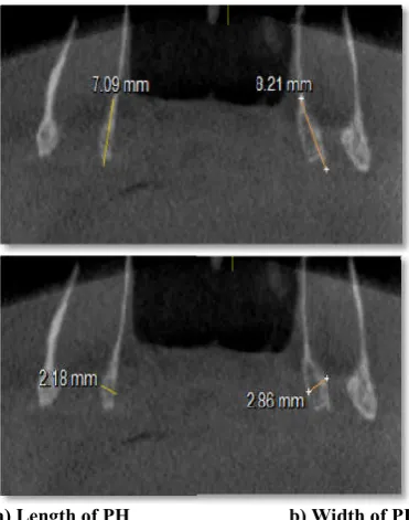
Length of PH: junction of medial pterygoid
through the tip. First a line was drawn from the junction of medial pterygoid plate and pH parallel to the horizontall plane. Following the identification of the midpoint of this line, the length was measured starting from this point to the tip of the pH
Width of PH: the distance between the most thickest
portion of the PH in the coronal plane
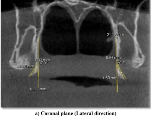
Inclination of PH: Inclination of PH along its long axis in
sagittal and coronal plane
Types of inclination:
i ) Sagittal section
International Journal of Recent Advances in Multidisciplinary Research
Also the present study states the clinical importance of
In locating hamular notch (between tuberosity & pterygoidhamulus), required for fabrication of maxillary
In knowing the anatomy with exact dimensions which are required prior to lefort 1 osteotomy surgery
(Pterygoidimpalnts for rehabilitation of a patient with a bilateral
Length of PH influences the procedure selection and objective treatment in patients with Obstructive sleep
Excessive length and improper inclination increases the chances of pterygoidhamulus syndrome (Morphological features of pterygoidhamulus and its clinical
Recently cone beam computed tomography (CBCT) is introduced in dental imaging which provides three dimensional l and maxillofacial structures.CBCT explains the internal structure of object as a cone shaped beam which acquires data in a single 360degree rotation.WhenCBCT compared with conventional periapical coronal and sagittal as a result it minimizes distortion and overlapping When CBCT is compared with conventional computed tomography (CT), it provides less ower scan time and it also increases accuracy The aim of the present study was evaluation of pterygoidhamulus using CBCT. The objectives of the study was to evaluate the length, width, incidence of shape with respect to side and gender.
The study was conducted in Oral medicine and radiology department in MGV’S Dental college and Hospital, Nasik, Protocol of the study was approved from authorized The CBCT dental imaging system 85 kVp and 6mA and FOV used was 8 × 8 cm with 14 second of exposure time. Total 80 scan of age > 20years were evaluated by selecting (As the ossification age of PH is estimated to Right and left) and gender (40 males&40 females) correlation was evaluated with respect to
Following parameters were noted:
junction of medial pterygoid plate and PH through the tip. First a line was drawn from the junction of medial pterygoid plate and pH parallel to the horizontall plane. Following the identification of the midpoint of this line, the length was measured starting from this point to the
the distance between the most thickest
Inclination of PH along its long axis in
a) Anterior, b) Posterior
ii) Coronal section a) Medial, b) Lateral,
Shapes of PH a) Triangle type b) Slender type.
Independent t-test was used for statistical analysis.p value <0.05 was considered as significant
RESULTS
The CBCT images of 40males and 40femaleswere
mean lengthof PH(left side) in males and females was found to be 7.72 +0.5,6.78+0.3mm. Inclination
found to be53.01°+20.3°,
7.72°+20.7°, 13.91°+26.1°(medial) and that was found to be 8.46°+20.3°,10.17°
incidence of shape in left side of PH was 1.8,1.5 (Triangle) and 1.2, 1.5(slender) respectively. Independent t
significant difference of this paramters
mean width value in males and females were found to be 2.04+0.4, 2.05+0.4mm and inclination in sagittal plane 47.22°+20.1°, 47.28°+22.1°(posterior) respectively with statistically insignificant (Table 1)
(right side) in males and females wa
6.62+0.3. Inclination in coronal plane was found to be 52.24°+20.3°, 47.23°+25.18 (lateral) and 7.6° 13.03°+26.1°(medial) and that
8.56°+20.3°, 10.01°+20.1° (anterior)
in right side of PH 1.7, 1.4(triangle) and 1.1,
respectively. Independent t-test stated significant difference in this paramters with p value > 0.05.
males and females were found to 2.01 inclination in sagittal plane 47°
respectively with statistically insignificant
a) Length of PH
Figure 1. Linear dimensions of PH
International Journal of Recent Advances in Multidisciplinary Research
test was used for statistical analysis.p value <0.05 was considered as significant
40males and 40femaleswere studied. The mean lengthof PH(left side) in males and females was found to Inclination in coronal plane was 47.95°+26.18 (lateral) and 26.1°(medial) and that in sagittal plane 20.3°,10.17°+21.1°(anterior).The incidence of shape in left side of PH was 1.8,1.5 (Triangle) and 1.5(slender) respectively. Independent t-test stated significant difference of this paramters with p value > 0.05.The idth value in males and females were found to be 0.4mm and inclination in sagittal plane 22.1°(posterior) respectively with (Table 1). The mean lengthof PH (right side) in males and females was found to be 7.56+0.5, in coronal plane was found to be 25.18 (lateral) and 7.6°+20.7°, in sagittal plane was found to be 20.1° (anterior). The incidence of shape 1.4(triangle) and 1.1, 1.5 (slender) test stated significant difference in p value > 0.05. The mean width value in males and females were found to 2.01+0.4, 2.02+0.3 and in sagittal plane 47°+20.1°, 47.12°+22.1°(posterior) respectively with statistically insignificant (Table 2).
b) Width of PH
Linear dimensions of PH
No significant difference was found between right and left side except the coronal plane inclination (posterior) was greater of left side as compared to right side. Also, in coronal and sagittal plane inclination, lateral and posterior inclination was seen more and among shapes more incidence of triangular shape PH were found.
a) Coronal plane (Lateral direction)
b) Sagittal plane (Anterior direction)
Figure 2. Inclination of PH
Table 1. Mean measurements with standard deviations (SD) of Pterygoidhamulus( PH ) of different parameters of left s
L W
M 7.72 +0.5 2.04+0.4
F 6.78+0.3 2.05+0.4
p VALUE < 0.01** >0.05
Table 2. Mean measurements with standard deviations (SD) of Pterygoidhamulus (PH) of different parameters of right side
L W
M 7.56+0.5 2.01+0.4
F 6.62+0.3 2.02+0.3
p VALUE < 0.01** >0.05
International Journal of Recent Advances in Multidisciplinary Research
No significant difference was found between right and left side except the coronal plane inclination (posterior) was greater of in coronal and sagittal lateral and posterior inclination was seen more and among shapes more incidence of triangular shape PH
Coronal plane (Lateral direction)
Sagittal plane (Anterior direction)
a) Triangle shape
Figure 3. Shape of PH
Figure 4. An elongated PH case of length 10.53mm of a young female.
DISCUSSION
IfPH plays the etiological role,
is caused. Common symptoms include sharp or burning pain in the palatal and pharyngeal region that may remain localized or refer to the ipsilaleral ear or temporomandibular joint. This may occur spontaneously or elicited by touch or eating and drinking (Naidoo et al., 2014).
sometimes ulceration of the palatal mucosa over the hamu are also common signs (Sasaki
Occasional soreness upon swallowing and while manipulating the area with the tongue or finger are seen.After several months of elapsation, their may be history of occasional exacerbations and remissions of swelling and discomfort
Table 1. Mean measurements with standard deviations (SD) of Pterygoidhamulus( PH ) of different parameters of left s
Left pH Measurements ( Mean +SD )
CPI SPI
CPI-L CPI-M SPI-A SPI-P
53.01°+20.3° 7.72°+20.7° 8.46°+20.3° 47.22°+20.1°
47.95°+26.18 13.91°+ 26.1° 10.17°+21.1° 47.28°+22.1°
< 0.05* < 0.05* < 0.05* >0.05
Mean measurements with standard deviations (SD) of Pterygoidhamulus (PH) of different parameters of right side
Rightph Measurements ( Mean +SD )
CPI SPI
CPI-L CPI-M SPI-A SPI-P
52.24°+20.3° 7.6°+20.7° 8.56°+20.3° 47°+20.1°
47.23°+25.18 13.03°+26.1° 10.01°+20.1° 47.12°+22.1°
< 0.05* < 0.05* < 0.05* >0.05
International Journal of Recent Advances in Multidisciplinary Research
b) Slender shape
Figure 3. Shape of PH
elongated PH case of length 10.53mm of a young female.
IfPH plays the etiological role, deviant or uncharacteristic pain Common symptoms include sharp or burning pain in the palatal and pharyngeal region that may remain localized or refer to the ipsilaleral ear or temporomandibular joint. This may occur spontaneously or elicited by touch or eating and 2014). Firm swelling, erythema and of the palatal mucosa over the hamulus Sasaki et al., 2001; Shankland, 1996). Occasional soreness upon swallowing and while manipulating the area with the tongue or finger are seen.After several months their may be history of occasional exacerbations elling and discomfort (Charbeneau and Table 1. Mean measurements with standard deviations (SD) of Pterygoidhamulus( PH ) of different parameters of left side
SH
T SL
1.8 1.2
1.5 1.5
< 0.01** < 0.01**
Mean measurements with standard deviations (SD) of Pterygoidhamulus (PH) of different parameters of right side
SH
T SL
1.7 1.1
1.4 1.5
< 0.01** < 0.01**
Blanton, 1981). The following symptoms present are not always associated with elevation in the soft palate.Although an excessively long hamulus could have been present in such cases,it is conceivable that one of the three other realtionships could have existed ,the medial pterygoid plate (and the PH) may have been situated in more inferior position than normal or the soft palate mucosa may have been situated more closer than normal or soft palate would have been thinner than usual in thickness (Charbeneau and Blanton, 1981). Several studies describe the morphology of PH in different populations. Eyrich et al. 1997 found the mean length of the left hamulus to be 5 mm and the right to be 4.9 mm.But,the present study showed mean length in left and right side in males and females as 7.726, 6.78,7.56 and 6.62 mm respectively. Putz and Kroyer (1999) reported the average length to be 7.2 mm and the sagittal and transverse diameter to be 1.4 mm and 2.3 mm, respectively.Wheareas in present study the mean width values found were 2.04, 2.05, 2.01 and 2.02mm respectively.Sasaki et al. 2001 reported an elongated PH case of 13 mm. Also they found the mean length of PH to be 6.8 mm.In the present study, an elongated PH of 10.53mm in a young female patient (Figure 4) and shortest PH found of 6.63mmwas reported. The inclination of PH in Putz and Kroyer’s (1999) study wasfound to be 75° in the sagittal plane and 58° in the coronal plane.Wheareas, in the present study, it was found to be 47° and 53° in sagittal and coronal plane respectively. Also as per their findings of Putz and Kroyer’sstudy, all the hamuluifound were inclined dorsolaterally. Similar to this study, the present study findings showed more number of scans with dorsolateral inclination but few scans with ventromedial inclination were also noted. The incidence of shape of PH of triangle shape in present study was found to be 30.43%, which was not in accordance with the percentage found to be 12% in NIOsang et al (2005). Incidcence of 28.12 % of slender shape PH was found in present study which was in accordance to Nio sang et al study, which found it to be 29.33%.
Orhan et al. in 2011 showed the mean length of PHs for left and right sides were 5.48 (SD 1.94) and 5.40 (SD 2.0) mm, respectively with no significant difference according to gender and location. They reported an elongated PH(10.9 mm) in a young female.Wheareas in present study, comparing gender wise, males had greater degree of inclination in both the planes than females But with respect to location ,all the parameters were almost of same value for both the sides (left and right) except the coronal inclination,which was found to be more in left side as compared to right side. The strength of the present study is use of CBCT which provides high-resolution images of high diagnostic quality with significantly reduced acquisition time and radiation burden.Reformatted from CBCT imaging data have been shown to have measurement accuracy equivalent to MDCT imaging.data (2014). However,limitation of this study is that this is a retrospective study which was conducted in limited geographic areas.
Conclusion
The length, width, inclination in coronal and sagittal plane of PH is greater in males as compared to females.The inclination in coronal plane is more of lateral than medial and that of sagittal plane is more of posterior than anterior.There was no difference in the metric measurements of left side when compared with right side, except the inclination in coronal
plane. (Left side>Right side) The triangular shape of PH shows higher incidence than slender shape in both the genders. CBCT is an excellent imaging modality for the identification of PH. The morphometric evaluation of PH helps us to trace and manage obscure and conflicting symptoms related to its morphology. So henceforth; consideration of the PH as a pain-inducing factor should be included in the diagnosis list.
REFERENCES
Charbeneau, T.D., Blanton, P.L. 1981. The Pterygoid Hamulus. Aconsiderationin the diagnosis of posterior palatal lesions. Oral SurgOral Med Oral Pathol., 52: 574-6. EdwardBuchanan, Charles, H. Hyman. Lefort I osteotomy.
Semin PlastSurg 2013;27:149-154
Eyrich, G. K., Locher, M.C., Warnke, T., Sailer, H.F. 1997. The pterygoidhamulus as a pain inducing factor. A report of a case and a radiographic study. Int J Oral Maxillofac
Surg., 26:275-77.
Hjørting-Hansen, E,. Lous, I. 1987. The Pterygoid Hamulussyndrome. Ugeskr Laeger., 6:979-82.
Hjørting-Hansen, E., Lous, I. 1987. Hamuluspterygoids yndrome. Tandlaegebladet, 91:833-7.
Hjørting-Hansen, E., Lous, I. 1987. The pterygoidhamulus syndrome. UgeskrLaeger., 6:979-82.
Jo, A. 1980. Kraniometrischeanalyse der knöchernenumrah mung des epipharynx. VerhAnatGes. 74:803-806.
Krmpoti Nemani, J., Vinter, I., Marusi, A. 2006. Relations of the pterygoidhamulus and hard palate in children and adults: Anatomical implications for the function of the soft palate. Ann Anat., 188:69-74.
Krmpoti Nemani, J., Vinter, I., Marusi, A. 2006. Relations of the pterygoidhamulus and hard palate in children and adults: anatomical implications for the function of the soft palate. Ann Anat., 188:69-74.
Morphological features of pterygoidhamulus and its clinical significance. Clinical Journal of Anatomy, 2004-2005.NIU Song-Qing, ZHANG Zhen-You, CHEN Chas, SUNYong, ZHANG Xi.
Naidoo, S., Roode, G.J., Bütow, K.W. 2014. Palatal pain due to exostosis of the posterior palatal spine in a cleft patient. J
Cleft Lip Palate Craniofacial Anatomy, 2001. A case of
elongated pterygoidhamulus syndrome. Oral Dis., 2001; 7:131 7. Hjørting-Hansen E, Lous I. (1987).
Orhan, K., Sakul, B.U., Oz, U., Bilecenoglu, B. 2011. Evaluation of the pterygoidhamulus morphology using cone beam computed tomography. Oral Surg Oral Med Oral
Pathol Oral Radiol Endod., 112:48-55.
Posterior palatal seal (PPS): A brief review. AliMariyam, Verma, A.K., ChaturvediSaurabh, AhmadNaeem, ShuklaAnuj. 2014. Journal of scientific and innovative
fresearch, 3(6):602-605.
Pterygoidimpalnts for rehabilitation of a patient with a bilateral maxillectomydefect.AvinashBidra, George. W. May, Greggory E. Tharp and Mark. Chambers. Journal of oral
ilmpalntology, Vol.39,issue 1,91-97
Putz, R., Kroyer, A. 1999. Functional morphology in the pterygoidhamulus. Ann Anat., 181:85-8.
Ramirez, L.M., Ballesteros, L.E., Sandoval, G.P. 2006. Hamular bursitis and its possible craniofacial referred symptomatology: two case reports. Med Oral Patol Oral
Cir Bucal., 11:E329-33.
Scarfe, W.C. 2005. Imaging of maxillofacial trauma : Evolutions and emerging revolutions. OralSurg Oral Med
Oral Pathol Oral Radiol Endod., 100:75-96
Shankland, W.E 2 nd. Pterigoidhamulusbursitis:One cause of craniofacial pain. J Prosthet dent., 1996; 75:205-10. Shankland, W.E. 1996. 2nd. Bursitis of the hamular process.
Part II: diagnosis, treatment and report of three case studies.
Cranio., 14:306-11.
UlasOz, KaanOrhan, SecilAksoy, FatmaCiftci, TunisÖzdo gano glu, and Finn Rasmussen, 2016. Association between pterygoidhamulus length and apnea hypopnea index in patients with obstructive sleep apnea: a combined three-dimensional cone beam computed tomography and polysomnographic study. Oral Surg Oral MedOralPathol
Oral Radiol., 1-10.
White, S.C., Pharoah, M.J. 2014. Oral Rdiology, Principles and Interpretation. 7th ed. St. Louis : Mosby Elsevier.