© 2017 IJSRST | Volume 3 | Issue 8 | Print ISSN: 2395-6011 | Online ISSN: 2395-602X Themed Section: Science and Technology
Biological activity, Spectroscopic and DNA Cleavage of Binuclear Schiff Base
Complexes
N. Mahalakshmi, M. R. Kuppusamy, C. Vanitha
Department of Chemistry, Rajeswari Vedachalam Government Arts College, Chengalpattu, Tamilnadu, India
ABSTRACT
A new series of transition metal complexes of Cu(II), Ni(II) and VO(II) have been synthesized from the Schiff base (L) derived from 5- iodosalicylaldehyde and 3,3’,4,4’-tetraminobiphenyl. Structural features were obtained from their spectral and analytical data. The data show that these complexes have composition of [M2(L)]X type. (where M = (Cu(II), Ni(II), and VO(II) X=ClO4-, SO42- L = binucleating tetradendate ligand). The spectral data of the complexes suggest a square–planar geometry around the central metal ion except VO(II) complex which has square–pyramidal geometry. The redox behavior of copper and vanadyl complexes was studied by cyclic voltammetry. The pUC18 DNA cleavage study was monitored by gel electrophoresis method. The results suggest that binuclear Cu(II), Ni(II) and VO(II) complexes cleaves pUC18 DNA in presence of the oxidant H2O2. The invitro antimicrobial activities of the synthesized compounds have been tested against the gram negative and gram positive bacteria’s. The binuclear Schiff base complexes were found to be higher antibacterial activity than the free ligand.
Keywords : 3,3’,4,4’-tetraminobiphenyl,Schiff base, binuclear, Antimicrobial, CT-DNA.
I.
INTRODUCTION
Schiff base complexes have been extensively investigated for more than a century and have been employed in areas that include magnetochemistry [1], non-linear optics [2] photophysical studies [3], catalysis [4] and materials chemistry [5]. Schiff bases derived from an amine and any aldehyde are a class of compounds which co-ordinate to metal ions via the azomethine nitrogen [6]. Metal complexes of Schiff bases derived from substituted salicylaldehydes and various amines have been widely investigated because of their wide applicability [7-10]. The oxovanadium(IV) ion, VO2+ is considered to be the most stable oxocation of the first-row transition metal ion [11]. It forms stable anionic, cationic and neutral complexes with various types of ligands. Monomeric and five coordinate oxovanadium(IV) complexes are formed with several bidentate Schiff bases. Chelating ligands containing O and N donor atoms show broad biological activity and are of special interest because of the variety of ways in which they are bonded to metal ions [12]. It is well known that several Schiff base complexes have inflammatory, antipyretic, analgesic, diabetic,
anti-bacterial, anti-cancer and anti-HIV activity [13, 14]. The interaction of transition metal complexes with DNA has been extensively studied in the development of new tools for nanotechnology [15, 16]. In the present investigation we report here the synthesis, spectroscopic, redox, DNA cleavage studies and antimicrobial studies of new tetradentate N2O2 donor type Schiff base and their metal complexes. In this paper the synthesis of new tetradentate N2O2 donor type Schiff base and its metal complexes (Cu(II) Ni(II) and VO(II)) derived by the condensation of 3,3’,4,4’-tetraminobiphenyl and 5- iodosalicylaldehyde is described.
II.
EXPERIMENTAL
2.1 Materials and methods
Synthesis: Caution! Perchlorate salts are potentially explosive and were handled only in small quantities with care.
2.2 Physical measurements
The elemental analysis were carried out with a Carlo-Erba 1106-model 240 Perkin Elmer analyzer. The solution conductivity measurements were performed to establish the charge type of the complexes. The complexes were dissolved in MeCN/DMF/DMSO and molar conductivities of 10-3M of their solutions at 28 0C were measured. Infrared spectra were recorded on the Perkin Elmer FT-IR-8300 model spectrometer using KBr disc and Nujol mull techniques in the range of 4000-400 cm-1. Electronic absorption spectra in the UVVisible range were recorded on Perkin Elmer Lambda -25 between 200-800 nm by using DMF as the solvent. Cyclic voltammetry studies were performed on a CHI760C electrochemical analyzer in single compartmental cells at 29 0C with H
2O/DMSO (95:5) solution using tetrabutylammonium perchlorate (TBAP) as a supporting electrolyte.
2.3 Preparation of binucleating tetradentate Schiff base ligand
The binucleating tetradentate Schiff base was prepared by condensation of tetramine with appropriate aldehydes (Scheme 1). 3,3’,4,4’-tetraminobiphenyl (1 mmol) in 20 ml of methanol was stirred with 5- iodosalicylaldehyde (4 mmol) in 20 ml of methanol for 4 h. The resulting yellow orange solid was separated and dried in vacuum. Yield: 85%.
Scheme 1 Structure of binucleating tetradendate Schiff base ligand
2.4 Synthesis of binuclear Schiff base complexes
[Cu2(L)]4ClO4 (1) [Ni2(L)]4ClO4 (2) [VO2(L)]2SO4 (3)
Metal(II) perchlorates of [Cu(II), Ni(II)] and [VO(II)] sulphate (0.2 mmol) and the potential binucleating Schiff base ligand (0.1 mmol) were dissolved in DMF (20 ml) and the mixture was heated to reflux for 3 h and the reactions were monitored by TLC. After partial evaporation of the solvent, solid (60–70%) metal(II) Schiff base complexes (Scheme 2) were separated and dried in vacuum.
Scheme 2 Structure of binuclear Cu(II), Ni(II) and VO(II) Schiff base complexes
Where, M = Cu(II), Ni(II) and VO(II) X = 4ClO4-, 2SO42
2.5 Cyclic voltammetry
All voltammetric experiments were performed with a CHI760C electrochemical analyzer, in single compartmental cells using Tetrabutylammonium perchlorate as a supporting electrolyte. The redox behavior of the complexes have been examined in absence and in presence of CT-DNA at a scan rate 0.2 Vs−1 in the potential range +1.2 to -2.0 V. A three-electrode configuration was used, comprised of a glassy carbon electrode as the working electrode, a Pt-wire as the auxiliary electrode, and an Ag/AgCl electrode as the reference electrode. The electrochemical data such as cathodic peak potential (Epc) and anodic peak potential (Epa) were measured.
2.6 Gel electrophoresis
experiments were performed by incubation of the samples containing 40 μM pUC18 DNA, 50 μM metal complexes and 50 μM H2O2 in Tris-HCl buffer (pH 7.3) at 39oC for 2 h. After incubation, the samples were electrophoresed for 3 h at 55 V on 1% agarose gel using Tris-acetic acid-EDTA buffer (pH 7.3). The gel was then stained using 1 μg cm-3 ethidium bromide (EB) and photographed under ultraviolet light at 355nm. All the experiments were performed at room temperature unless otherwise mentioned.
2.7 Antimicrobial activity
The in vitro antibacterial activity of the ligand and the complexes were tested against the bacterias Bacillus subtilis, Klebsiella pneumoniae, Escherichia coli and
Staphylococcus aureus by well diffusion method using nutrient agar as the medium. Streptomycin was used as standard for bacteria. The stock solution (10-2 mol L-1) was prepared bydissolving the compound in DMF and the solution was serially diluted in order to find minimum inhibitory concentration (MIC) values. In a typical procedure, a well was made on the agar medium inoculated with microorganisms in a Petri plate. The well was filled with the test solution and the plate was incubated for 24 h for bacteria at 40 oC. During the period, the test solution diffused and the growth of the inoculated microorganisms was affected. The inhibition zone was developed, at which the concentration was noted.
III.
RESULTS AND DISCUSSION
The binuclear Schiff base ligand prepared and reacts with transition metals Cu(II), Ni(II), and VO(II) . The Schiff base ligand has been synthesized from 5- iodosalicylaldehyde and 3,3’,4,4’-tetraminobiphenyl (scheme 1) in 4:1 mole ratio. The results of elemental analyses were in good agreement with those required by the proposed formulae. The data in consistent with the earlier reports support the proposed formulation of the binuclear complexes (scheme 2). The higher conductance values (Table 3) of chelates support the electrolytic (1:2) nature of metal complexes.
3.1 IR spectra
The important IR absorption frequencies of the synthesized complexes are shown in Table 1. The
azomethine nitrogen υC=N stretching frequency of the free ligand appears around 1625 cm-1, which is shifted to lower frequencies in the spectra of all the complexes (1600-1610 cm-1). These bands are shifted to lower wave numbers indicating the involvement of azomethine nitrogen in coordination to the metal ion [19]. This is further supported by the disappearance of the υOH in the range of 3400-3440 cm-1 in all the complexes . Accordingly, the ligand acts as a tetradendate chelating agent bonded to the metal ion via two –C-O groups & two –C=N azomethine nitrogen atoms of the Schiff base (scheme 2). Assignment of the proposed coordination sites is further supported by the appearance of medium bands at 480-510cm-1 and 430-470 cm-1 which could be attributed to M-O, M-N respectively [20]. In addition, the Oxovanadium complexes shows a band at 970 cm-1 - 980 cm-1 attributed to V=O stretching frequency [21]. A further examination of Infrared spectra of complexes shows the presence of a band in the 1070-1115 cm-1 region. The strong band is ascribable to ClO4- & SO4 2-ions [22].
Table 1. Infrared spectral data for the ligand and binuclear Schiff base complexes
Complexes υ(-C=N)
(cm -1)
υ(-OH) (cm
-1)
υ(V=O) (cm-1) υ(M-O)
(cm -1)
υ(M-N) (cm -1)
ClO4 -/SO4 2-(cm-1)
(Ligand) 1625 3400 - - - - [Cu2(L)]4ClO4- 1600 3410 - 10 460 1080 [Ni2(L)]4ClO4- 1610 3440 - 490 470 1115 [VO2(L)]2SO4- 1602 3405 980 480 430 1070
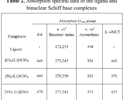
3.2 Electronic spectra
centered at 440 nm for Cu(II) complex. This is due to 2B
1g → 2A1g transition [23]. The spectra of Ni(II) complex in the visible region at about 370 and 380 nm is assigned to 1A
1g → 1A2g, 1A1g → 1B1g, transitions, suggesting an approximate square planar geometry of the ligand around the metal ions [24]. The intense charge transfer band at 410-425 nm in Oxovanadium(IV) complex assigned to 2B
2 → 2A1, 2B2 → 2E transitions. This is due to electron delocalization over whole molecule on complexation. Based on these data, a square planar geometry has been assigned to the complexes except VO(II) complex which has square pyramidal geometry. These values are comparable with other reported complexes [25].
Table 2. Absorption spectral data of the ligand and binuclear Schiff base complexes
3.3 Molar Conductance studies
The complexes are insoluble in most common polar and non-polar organic solvent. They are soluble in DMSO, DMF and CH2Cl2. The molar conductivities of the complexes were measured in DMSO for freshly prepared solutions and after standing for two weeks. The conductivity increased very slightly with time in DMSO for all the complexes. The present coordination moieties cannot be replaced by the solvent molecules. The value of these conductance’s are in the range 25-30 ohm-1 cm2 mol-1 in DMSO and are in accordance with those reported for electrolyte in this solvent. This is indicative that these complexes dissociate very slightly in this solvent. As observed in the IR spectra, where the compounds were formulated as electrolytes the molar conductivities show that all the anions are present
outside the coordination sphere in solution [26-31]. The data are presented in Table 3.
Table 3. Molar conductance data of the binuclear Schiff base the complexes
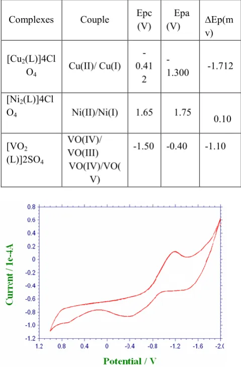
3.4 Cyclic voltammetry studies
the shift in E0value and increase in peak heights potentials suggest that both Cu(II) and Cu(I) form of complex 1 bind to CT-DNA [32].
Table 4. Cyclic voltammetric data of the binuclear Schiff base Complexes in DMSO solution.
Complexes Couple Epc (V) Epa (V)
∆Ep(m
v)
[Cu2(L)]4Cl
O4 Cu(II)/ Cu(I) -0.41
2 -
1.300 -1.712
[Ni2(L)]4Cl
O4 Ni(II)/Ni(I) 1.65 1.75 0.10
[VO2 (L)]2SO4
VO(IV)/ VO(III)
VO(IV)/VO( V)
-1.50 -0.40 -1.10
Figure 1a. Cyclic voltammogram of complex 1 alone
Figure 1b. Complex 1 in presence of CT-DNA [Complex 1]
3.5 Cleavage of Plasmid pUC18 DNA
Figure 2. Changes in the agarose gel electrophoretic pattern of pUC18DNA induced by H2O2 and metal complexes: Lane 1, DNA alone; Lane 2, DNA alone + H2O2; Lane 3, DNA + Cu binuclear complex + H2O2; Lane 4, DNA + Ni binuclear complex + H2O2; Lane5, DNA + VO binuclear complex + H2O2.
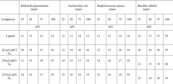
3.6 Antimicrobial activity
The ligand and their complexes have been tested for
invitro growth inhibitory activity against gram-positive
microbes Bacillus subtilis, staphylococcus aureus and
gram-negative microbes Klebsiella pneumonia,
Escherichia coli by using well-diffusion method. As the
test solution concentration increases, the biological activity also increases. It is found that the activity increases upon co-ordination. The increased activity of the metal chelates can be explained on the basis of chelation theory [35]. The orbital of each metal ion is made so as to overlap with the ligand orbital. Increased activity enhances the lipophilicity of complexes due to delocalization of pi-electrons in the chelate ring [36]. In some cases increased lipophilicity leads to breakdown of the permeability barrier of the cell [37]. The results revealed that the metal complexes Cu(II), Ni(II) and VO(II) have higher antimicrobial activity than the ligand are shown in Table 5.
Table 5. Antibacterial activity of the ligand and binuclear Schiff base complexes
IV.
CONCLUSION
The N2O2 type Schiff base ligand is synthesized from 5- iodosalicylaldehyde and 3,3’,4,4’-tetraminobiphenyl. It acts as a tetradentate ligand and forms stable complexes with transition metal ions such as Copper(II), Nickel(II), and Oxovanadium(IV). The ligand and its complexes are characterized using spectral and analytical data. The interaction of these complexes with CT-DNA was
investigated by gel electrophoresis. All the transition metal complexes have higher activity than the control CT-DNA. The Cu(II), Ni(II) complexes have more activity than VO(IV) complex and the control CT-DNA. . The metal complexes have higher antimicrobial activity than the free ligand.
Klebsiella pneumoniae
(mm) Escherichia coli (mm) Staphylococcus aureus (mm) Bacillus subtilis (mm)
Complexes 25 50 75 100 25 50 75 100 25 50 75 100 25 50 75 100
(μl) (μl) (μl) (μl)
Ligand 11 13 12 14 12 13 14 15 11 13 14 16 12 13 15 18
[Cu2(L)]4Cl
O4 10 14 13 16 13 14 16 16 12 15 16 18 10 16 18 19
[Ni2(L)]4Cl
O4 11 15 18 19 14 15 17 18 14 16 17 19 13 15 19 20
[VO2(L)]2S
V.
REFERENCES
[1]. J.-W. Lu, Y.-H. Huang, S.-I. Lo, H.-H. Wei, Inorg. Chem. Commun. 10 (2007)1210.
[2]. S. Di Bella, I. Fragala, New J. Chem. 26 (2002) 285.
[3]. P.G. Cozzi, L.S. Dolci, A. Garelli, M. Montalti, L. Prodi, N. Zaccheroni, New J.Chem. 27 (2003) 692.
[4]. N.C. Gianneschi, S.T. Nguyen, C.A. Mirkin, J. Am. Chem. Soc. 127 (2005)1644.
[5]. M. Bandini, P.G. Cozzi, A. Umani-Ronchi, Chem. Commun. 919 (2002).
[6]. A. Anora, K.P. Sharma, Synth. React. Inorg. Met.-Org. Chem. 32 (2000)913.
[7]. E. Canpolat, M. Kaya, J. Coord. Chem. 57 (2004) 127.
[8]. A.P. Mishra, M. Khare, S.K. Gautam, Synth. React. Inorg. Met.-Org. Chem. 32 (2002) 1485. [9]. Y. Fan, C. Bi, J. Li, Synth. React. Inorg.
Met.-Org. Chem. 33 (2003) 137.
[10]. 0E. Canpolat, M. kaya, A. Yazici, Spectrosc. Lett. 38 (2005) 35.
[11]. N. D. Chasteen, Biological Magnetic Resonance, Vol. 3. Plenum Press, New York, 1981, p. 54. [12]. R.C. Maurya, P. Patel, S. Rajput, Synth. React.
Inorg. Met.-Org. Chem. 33(2003) 817.
[13]. A.P. Mishra, S.K. Gavtarm, J. Ind. Chem. Soc. 81 (2004) 324.
[14]. N.A. Venkariya, M.D. Khunt, A.P. Parikh, Ind. J. Chem. 42B (2003) 421.
[15]. A.R. Banerjee, J.A. Jaeger, D.H. Turner, Biochemistry, 1993, Vol 32, pp153-163.
[16]. N.Y. Sardesai, K. Zimmerman, JK Barton, J. Am. Chem. Soc, 1994, Vol 116, pp7502-7508.
[17]. Vogel AI (1989) Text Book of Practical Organic Chemistry, 5th ed., Longman, London.
[18]. N. Ramana, R. Jeyamurugana, A. Sakthivel, L. Mitub, Spectrochim. Acta PartA, 2010, Vol 75, pp88–97.
[19]. Pal S, Pal S (2002) J Chem Soc Dalton Trans 2102-2108.
[20]. Yamada S, Takeuchi A (1982) Coord Chem Rev 43:187-204.
[21]. Kaitner B., and Parlovic G., Croatica Chemica Acta., 72, 607(1999).
[22]. Nakamoto K (1978) Infrared and Raman spectra, of inorganic and coordination compounds, III edition, John Wiley.
[23]. Sharadha LN, Ganorkar MC (1988) Indian J Chem 27A:617-621.
[24]. Lever ABP (1968) Crystal Field Spectra, Inorganic Electronic Specroscopy, first ed., Elsevier, Amsterdam 249-360.
[25]. Sivasankaran Nair M, Kalalakshmi G, Sankaranarayana Pillai M (1999) J Indian Chem Soc 76:310.
[26]. A. Muller, M. Peng, E. Krichmeyer and H.Walberg, J.Angew chem. Int. Ed. Engl.,27,1719 (1988).
[27]. A.Muller, R. Rohlfing, J. Doring and M. Penk. J. Angew chem., Int. Ed. Engl., 30, 588 (1991). [28]. Q. Chen, D.P. Gosham, C.P.Scholes, X.Tan and
J.Zubieta, J.Am.chem. Soc., 114, 4667 (1992). [29]. M.I. Khan, Q.Chen, D.P. Gosham and J.Zubieta,
J.Inorg. Chem., 32, 672 (1991).
[30]. M.J. Pelezar, E.C.S. Chan and N.R. Krieg ,“Microbiology”, 5th edition, Wiley Interscience, New York, (1983).
[31]. Willson and Gisvold, Text Book of Organic Medicinal and Pharamaceutical Chemistry. 9th Edn., J.B. Lippin Colt Co., 1 (1991).
[32]. Z. S. Yang, Y. L. Wang and G. C. Zhao., Anal Sci., 2004, 20, 1127.
[33]. N. Raman, T. Baskaran, A. Selvan, J. Iran. Chem. Res, 2008, Vol 1, pp29–139.
[34]. Thomas AM, Naik AD, Nethaji M, Chakravarty AR (2004) Indian J Chem A43:691-700.
[35]. N. Raman, A. Kulandaisamy, A. Shanmugasundaram, K. Jeyasubramanian,Trans. Met. Chem. 26 (2001) 131.
[36]. R.S. Srivastava, Inorg. Chim. Acta 56 (1981) 65. [37]. A. Cukurovali, I. Yilmaz, H. Ozmen, M.