1
SECOND PRIMARY CANCERS IN NON-HODGKIN LYMPHOMA:
BI-DIRECTIONAL ANALYSES SUGGESTING ROLE FOR IMMUNE
DYSFUNCTION
Subhayan Chattopadhyay1,2, Amit Sud1,3, Guoqiao Zheng 1,2, Hongyao Yu 1,2, Kristina Sundquist4,5,6, Jan Sundquist4,5,6 Asta Försti1,4 , Richard Houlston3,7, Akseli Hemminki, 7,8 and Kari Hemminki, 1,4
1
Division of Molecular Genetic Epidemiology, German Cancer Research Center (DKFZ), Im Neuenheimer Feld 580, D-69120, Heidelberg, Germany
2
Faculty of Medicine, University of Heidelberg, Heidelberg, Germany
3
Division of Genetics and Epidemiology, The Institute of Cancer Research, London, UK.
4
Center for Primary Health Care Research, Lund University, 205 02 Malmö, Sweden
5
Department of Family Medicine and Community Health, Department of Population Health Science and Policy, Icahn School of Medicine at Mount Sinai, New York, USA
6
Center for Community-based Healthcare Research and Education (CoHRE), Department of Functional Pathology, School of Medicine, Shimane University, Japan
7
Division of Molecular Pathology, The Institute of Cancer Research, London, UK.
8
Cancer Gene Therapy Group, Faculty of Medicine, University of Helsinki, Finland
9
Comprehensive Cancer Center, Helsinki University Hospital, Helsinki, Finland
Correspondence: Kari Hemminki, Division of Molecular Genetic Epidemiology, German Cancer Research Center (DKFZ), Im Neuenheimer Feld 580, Heidelberg 69120, Germany.
Telephone: +496221421800 Fax: +496221421810
Email: [email protected]
Words: 245 (abstract), 2998(text)
Key words: second cancers, immune suppression, bi-directional risk, therapeutic implication.
2
NOVELTY AND IMPACT
The nationwide study found non-Hodgkin lymphoma and 7 cancers, including squamous cell skin cancer and melanoma, were bi-directionally associated with each other. Bi-directional associations may help to resolve the role for therapeutic side effects because treatment for two cancers is rarely similar,
particularly considering primary surgical treatment for skin cancers. The data suggest that an immune suppressed state is a key contributing mechanism for second primary cancers and immune therapy in patient management will have a clinical impact.
ABSTRACT
Second primary cancers (SPCs) account for an increasing proportion of all cancer diagnoses. It is
unlikely that prior therapy is solely responsible for SPC risk. To investigate risk of SPC after diagnosis of non-Hodgkin lymphoma (NHL) and 10 of its subtypes we conducted a novel bi-directional analysis, SPCs after NHL and NHL as SPC. Using the Swedish Family-Cancer Database, we identified 19,833 individuals with primary NHL diagnosed between 1993 and 2015. We calculated relative risks (RRs) of SPCs in NHL survivors and, for bi-directional analysis, risk of NHL as SPC. The overall RRs were significantly bi-directionally increased for NHL and 7 cancers. After diagnosis of NHL risks were increased for upper aerodigestive tract (RR=1.96), colorectal (1.35), kidney (3.10), bladder (1.54) and squamous cell skin cancer (SCC) (4.12), melanoma (1.98) and Hodgkin lymphoma (9.38). The
concordance between RRs for each bi-directional association between NHL and 31 different cancers was highly significant (r= 0.86, P<0.0001). Melanoma was bi-directionally associated with all 10 subtypes of NHL. The observed bi-directional associations between NHL and cancer suggest that therapy-related carcinogenic mechanisms cannot solely explain the findings. Considering that skin SCC and melanoma are usually treated by surgery and that these cancers and NHL are most responsive of any cancer to immune suppression, the consistent bi-directional results provide population-level evidence that immune suppressed state is a key underlying mechanism in the context of SPCs. Furthermore, the quantified risks for NHL subtypes have direct clinical application in the management of NHL patients.
3
INTRODUCTION
Non-Hodgkin lymphoma (NHL), the most common hematological malignancy, is a cancer of the lymphatic system caused by either B- or T-cell clonal expansion 1, 2. Established risk factors for NHL include
immunosuppression and other types of immunodeficiency or autoimmunity, in addition to chronic inflammation induced by viral or other microbial causes 2.
There are multiple subtypes of NHL, including, and not restricted, to benign forms of follicular lymphoma, small lymphocytic lymphoma and mantle cell lymphoma and aggressive forms of diffuse large B-cell lymphoma and Burkitt lymphoma1. Perhaps not surprisingly, the epidemiology of NHL has been hampered by the changes in NHL classification and studies have been confined to the major subtypes, diffuse large B-cell lymphoma and follicular lymphoma 3.
Until the advent of multidrug chemotherapy to complement radiotherapy in the 1970s, including the CHOP regimen (cyclophosphamide, doxorubicin, vincristine, prednisone), and variations thereof, patient outcome of NHL was typically poor 1. In the late 1990s the monoclonal antibody directed against tumor antigens was licensed to be used in combination with chemotherapy and radiotherapy 1, 2. Subsequently, other monoclonal antibodies, some radioactive labeled, have been developed for treatment of specific NHL subtypes,
including most recently immune checkpoint inhibitors 2, 4.
While the advances in the management of NHL over the past 40 years have undoubtedly led to a markedly improved survival, this has come at the cost of an increased number of second primary cancers (SPCs) and other treatment-related complications. A number of studies have estimated risks of SPCs after NHL but have confined their analyses to the most common NHL subtypes 5-10.
Here we report analysis of the Swedish Cancer Registry to assess risks of SPCs following the diagnosis of NHL, and also the risk of NHL after the diagnosis of another cancer. The rationale for the bi-directional analysis was to understand mechanisms for SPC susceptibility beyond therapeutic side effects and shared risk factors 11, 12. Indeed, the novel bi-directional analysis of 19,833 NHL patients (including 10 NHL subtypes) is a powerful approach in search of evidence for reciprocal relationships between cancer risks which may suggest that SPCs may be a model of tumor biology.
4 The Swedish Family-Cancer Database includes the whole Swedish population organized in families and linked to the national Cancer Registry with more than 2 million cancers registered since 1958 13. The registry is based on compulsory cancer notifications from clinicians and pathologists/cytologists 14. All registered NHL cases were histologically verified. While the cancer registry does not publish statistics on histological verification of SPCs they are included with primary cancers for which histological verification has been around 98% from the 1970s 15. An ad hoc study on the diagnostic accuracy of second neoplasms found 98% to be correctly classified. 16. NHL subtypes were identified through reference to the 10th revision of International Classification of Diseases (ICD) in combination with SNOMED (ICD-O2) codes that were introduced in 1993. The Swedish Cancer Registry orders tumors by diagnostic date into first, second, third etc. primary cancers. This ordering was used either to select NHL as first primary cancer or as SPC after another primary cancer. We did not study patients diagnosed with NHL after NHL. NHL patients were followed from diagnosis until death, detection of a SPC, emigration or December 31, 2015, whichever came first. Person-years and SPCs were categorized according to age (5-year bands), sex, socioeconomic index (six groups), region (four groups), calendar year (1993-99, 2000-09, 2010-15), time since NHL diagnosis and age at NHL diagnosis. Category-specific incidence rates among NHL patients were multiplied by the corresponding person-years at risk to estimate the expected number of malignancies in respective strata. In stratified analyses over calendar periods case accrual was stopped at the termination of the period to allow uniform follow-up times.
Relative risks (RRs) were assessed by means of incidence rate ratios, regressed over a fixed-effects
generalized Poisson model. RRs for SPCs were obtained by comparing incidence rates for each SPC in NHL patients with respective population background rates for the primary cancer. In the reverse analysis, RRs for specific NHLs were considered as SPCs following any primary cancer. Sex, age group, calendar-period, socio-economic status and residential area were treated as potential confounders and were adjusted for in the regression model. Confidence intervals (CIs) were calculated for 5% level of significance and p-values associated with RRs were obtained with two-tailed tests against Chi-square distribution with one degree of freedom 17. In referring to differences between RRs we call risks only when they are significant (i.e., 95%CIs are non-overlapping). The concordance between RRs was assessed by Pearsonian correlation. All analyses are performed in SAS (v9.4) or R (v3.3.4).
The study was approved by the Ethical Committee of Lund University.
RESULTS
5 Risks for SPCs following a diagnosis of NHL and risk of NHL following the diagnosis of 31 non-NHL cancers are shown in Table 2. Overall, the risk of SPC was increased 1.53-fold. RRs were significantly increased for 12 cancers with the largest RRs being shown for Hodgkin lymphoma (RR: 9.38,), squamous cell skin (SCC, 4.12,) and kidney cancers (3.10). Statistically significant RRs of more than 2.00 were also documented for leukemia, anal and thyroid tumors. In the reverse analysis, for all non-NHL cancers the risk of being diagnosed subsequently with NHL was significantly increased 1.40-fold (Table 2). RRs were significantly increased for 14 cancers with the largest RRs being shown for Hodgkin lymphoma (7.29), followed by skin SCC (2.44) and testicular cancer (2.29). The concordance between RR for each non-NHL cancer following a diagnosis of NHL and RR for NHL following diagnosis of a non-NHL cancer was highly statistically significant (r=0.86, P<0.0001).
Sensitivity analysis was performed on data shown in Table 2 by deleting in both of the reciprocal analyses the first year of follow-up after first diagnosis of cancer (Supplementary Table 1). No essential differences in RRs to Table 2 were noted even though case numbers were reduced. The overall risks for SPC increased to 1.62 from 1.53 and the overall risks of NHL as SPC remained at 1.40.
6 Table 3 shows the RRs of all non-NHL cancers following diagnosis of each of the 5 of the major NHL subtypes. Statistically significant RRs were shown after mantle cell lymphoma (1.78), marginal zone lymphoma (1.70), follicular lymphoma (1.69), lymphoplasmacytic lymphoma (1.64, Waldenstrom macroglobulinemia) and diffuse large B-cell lymphoma (1.33). Skin SCC and melanoma risks were increased after all 5 NHL subtypes; the RR for skin SCC was 6.20 after mantle cell lymphoma. Lung and kidney cancer risks were increased after four NHLs. Risks for upper aerodigestive tract and bladder cancers, Hodgkin lymphoma and leukemia were increased after 3 NHLs; the RR for Hodgkin lymphoma was 9.64 after follicular lymphoma. Rare anal cancer risk was increased to 6.34 after follicular lymphoma.
The results of the reverse analysis, risk of the 5 NHL subtypes as SPC, are shown in Table 4. Melanoma and skin SCC were first primaries for five and four subtypes, respectively; RRs were highest for marginal zone lymphoma (after melanoma 2.81), for follicular lymphoma (after skin SCC 2.56) and for mantle cell lymphoma (after melanoma and skin SCC both 2.46). Testicular cancer was associated with high risks of lymphoplasmacytic lymphoma (4.59) and mantle cell lymphoma (5.27). Diffuse large B-cell lymphoma risk was increased to 13.76 after Hodgkin lymphoma.
Risks for SPC after 5 rare NHL subtypes are shown in Supplementary Table 6. The overall risks were increased and statistically significant for all of them, most for small lymphocytic lymphoma (1.85), followed by and anaplastic T-cell lymphoma (1.62). Skin SCC was increased after three of these subtypes, most after small lymphocytic lymphoma (7.41). Kidney cancer was increased after two NHL subtypes, cutaneous T-cell lymphoma and anaplastic T-T-cell lymphoma both with RRs exceeding 6.00. Melanoma (4.77) and leukemia (7.82) were increased after Burkitt lymphoma
Reverse analyses for 5 rare NHL subtypes as SPC are shown in Supplementary Table 7. The overall RRs were increased for small lymphocytic lymphoma (2.28) and cutaneous T-cell lymphoma (1.59). Skin SCC and cancer of connective tissue were first primaries for three NHL subtypes whereas kidney cancer and melanoma were primaries for two subtypes. Small lymphocytic lymphoma was increased to an RR of 13.51 after testicular cancer. Melanoma, skin SCC and connective tissue cancer each associated with increased risks of cutaneous T-cell lymphoma with RRs of 5.93, 4.11 and 9.22, respectively.
Figure 1 shows plots of the RRs over follow-up time since the diagnoses of NHL and non-NHL cancer. Following diagnosis of NHL, risks of SPCs were persistent and somewhat increasing for all non-NHL cancers, skin SCC and melanoma. For NHL following the diagnosis of a non-NHL cancers, risks were moderately decreasing.
7 Novel findings of the present study were the demonstration of an increase in overall SPC risk after each of the 10 different subtypes of NHL and, conversely, an increase of six of these NHL subtypes as SPC. Skin SCC risk was most systematically increased and was found in excess after 8 NHL subtypes, followed by melanoma after 7 NHL subtypes and kidney cancer after 6 NHL subtypes. In the reverse analysis on NHL as SPC, melanoma and skin SCC were the most common primary cancers, both of which were followed by an excess risk of 7 NHL subtypes as SPC. The correlation of the bidirectional associations was highly
significant P<0.0001. Sensitivity analyses on data for which the first year of follow-up were excluded did not change results; this would be expected on cancer cases with practically complete histological
confirmation. Nor showed age- and period-stratified data unexpected findings on individual cancers, including melanoma and skin SCC.
A major strength of this study is that we have avoided ascertainment bias in patient selection because our cohort analysis was based on the Swedish population, for which there is near complete case registration with long-term follow-up. We do acknowledge, however, that as a limitation of our study we did not have the opportunity to incorporate information on treatment.
Therapy-related side effects are generally considered to be the cause of many SPCs. Since chemotherapy and radiotherapy regimes are generally used to treat NHL the finding of an increased risk of cancers such as those of the lung and colorectum reported herein and documented by others 5-10, has a plausible etiology. Such a mechanism is not however likely to be solely responsible for any SPC risk as evidenced by the bi-directional associations we observed. Indeed, given the diversity of cancer treatment across tumor types the prominent inter-relationships shown between NHL and kidney, skin, bladder and melanoma invite
alternative hypotheses.
Aside from some form of shared environmental/lifestyle factors common to cancers, the association of NHL with skin SCC and melanoma for which surgery is the primary mode of treatment raises the possibility of immune dysfunction playing a role. In keeping with such a postulate is the fact that immunosuppressed organ transplantion patients have an increased risk of not only skin SCC and NHL (20-fold) but also kidney cancer (15-fold), melanoma, leukemia and anogenital cancers (5-fold) and other cancers 18. High risks were reported also on lip, oral and pharyngeal cancers, which in the present study were included as minor
components under ‘upper aerodigestive tract’ cancers 19, 20
. The bi-directional spectrum of cancer risk seen in the present analysis is reminiscent such observations. Even though observational results may be
8 As only a small proportion of NHL patients will have been therapeutically immune suppressed because of bone marrow transplantation, for immune dysfuntion to play any role requires an alternative explanation. Increasingly it is being recognized that tumors can influence immune function, specifically T-cell function, which again mirrors the impact of any iatrogenic immune suppression 21. NF-kappaB signaling is a master regulator of cancer-associated chronic inflammation which contributes to immunosuppression through induction of proinflammatory mediators and activation of immune suppressor cells 22-24. Myeloid-derived suppressor cells are the main type of tumor-associated macrophages that produce chemokines, cytokines, growth factors and proteases which are involved in extracellular matrix remodeling 25.
Although there were similarities in the bi-directional risks between NHL subtypes and cancers, there were also differences. The highest overall RRs for SPC after NHL were associated with small lymphocytic, mantle cell and marginal zone lymphoma with the smallest effect being shown in respect of Burkitt lymphoma; these risks were approximately correlated with known survival for these subtypes, and hence good survival increases the life-time chance for a SPC. Myeloma risk was high only after
lymphoplasmacytic lymphoma (Waldenstrom) which could be predicted from the known familial
association between the two diseases 26; consistently, lymphoplasmacytic lymphoma was also increased as SPC after myeloma. Other types of unique findings included anal cancer with a high risk after follicular lymphoma, high risk of leukemia after mantle cell and Burkitt lymphoma and high risk of kidney cancer after cutaneous and anaplastic T-cell lymphoma. In the reverse analysis high risks were noted for diffuse large B-cell lymphoma after Hodgkin lymphoma and cervical cancer, the latter even influencing the risk of mantle cell lymphoma. Finally, testis cancer was associated with a high risk of small lymphocytic
lymphoma, mantle cell lymphoma and lymphoplasmacytic lymphoma.
Immune suppression would be a timely explanation to the findings concerning cancers that are known to be increased in immunosuppressed patients, in view of the current successes in immune therapy. Immune-checkpoint inhibitors, which can promote cytotoxic activity of T cells, are effective in some individuals with NHL and are gaining wider clinical acceptance 4, 27, 28. However, can we exclude other alternative
explanations, such as shared familial risk, microbial agents or other environmental factors playing a role? Given that many cancer susceptibility genes have pleotropic effects it is plausible that a small part of the excess risk is enshrined in inherited genetic factors, either through high penetrance alleles or co-inheritance of multiple common risk variants. Neither would infection appear as a plausible candidate mechanism because of the multiple cancers involved. However, reactivation of endogenous viruses may be important, as for example Epstein-Barr virus activation is an essential mechanism in post-transplantation carcinogenesis
18
9 melanoma, has been suggested to be both a risk and protective factors for NHL but large prospective studies show no association 29, 30.
Generalizability of the results is likely to encompass the cancers with bi-directional significance (NHL, upper aerodigestive tract, kidney, bladder, melanoma, skin SCC and Hodgkin) which account for 28% of cancers in Sweden 15; skin SCC is the most common of these, accounting for 10% of cancers in Sweden. Adding also colorectal cancer with modest bi-directional risks would increase the total to 39% of all cancers. The main limitation of our study in terms of understanding etiology is our reliance on purely cancer registry information. Another point of concern may be that we included all SPCs even though we know that upon diagnosis of first cancer a large number of SPCs are synchronously diagnosed. However the level of histological verification of all relevant cancers for this study is high and there is no reason to believe that diagnostic accuracy would have been compromised. Moreover, the data show that the risk for many SPC remains elevated after 15 years. Surveillance for these cancers with elevated risks should be considered for integration into ongoing cancer survivorship programs.
In conclusion, we have provided a comprehensive analysis of cancer risks associated with NHL.
Additionally through analysis of NHL as SPC we propose that immune suppression is a key mechanisms responsible for the development of SPCs. Our findings further substantiate the significant cancer risks associated with survivorship from NHL and are informative in defining the long-term management of patients successfully treated for NHL in terms of surveillance for SPCs. When immune therapy will become widely used it will be possible to test the present hypothesis; if correct, the suggested immune-responsive SPCs should be suppressed.
ACKNOWLEDGEMENT
A.S. is the recipient of Guest Scientist Fellowship of DKFZ. Supported by the Harald Huppert Foundation, Deutsche Krebshilfe, Jane and Aatos Erkko Foundation, Sigrid Juselius Foundation, Finnish Cancer
Organizations, University of Helsinki and Helsinki University Central Hospital
AUTHOR CONTRIBUTIONS
Design: KH, AS, SC Acquisition of data: JS, KS
Statistical analysis and interpretation: SC, GZ, HY, KH, AH. Manuscript writing: KH, RSH, SC, AS, AH, AF.
Approval of the final text: All authors
10 A.H. is shareholder in Targovax ASA. A.H. is employee and shareholder in TILT Biotherapeutics Ltd. Other authors declared no conflict of interest.
REFERENCES
1. Evans LS, Hancock BW. Non-Hodgkin lymphoma. Lancet 2003;362: 139-46.
2. Shankland KR, Armitage JO, Hancock BW. Non-Hodgkin lymphoma. Lancet 2012;380: 848-57. 3. Jaffe ES. The 2008 WHO classification of lymphomas: implications for clinical practice and translational research.
4. Pianko MJ, Moskowitz AJ, Lesokhin AM. Immunotherapy of Lymphoma and Myeloma: Facts and Hopes. Clin Cancer Res 2018;24: 1002-10.
5. Brennan P, Scelo G, Hemminki K, Mellemkjaer L, Tracey E, Andersen A, Brewster DH, Pukkala E, McBride ML, Kliewer EV, Tonita JM, Seow A, et al. Second primary cancers among 109 000 cases of non-Hodgkin's lymphoma. Br J Cancer 2005;93: 159-66.
6. Tward JD, Wendland MM, Shrieve DC, Szabo A, Gaffney DK. The risk of secondary malignancies over 30 years after the treatment of non-Hodgkin lymphoma. Cancer 2006;107: 108-15.
7. Mudie NY, Swerdlow AJ, Higgins CD, Smith P, Qiao Z, Hancock BW, Hoskin PJ, Linch DC. Risk of second malignancy after non-Hodgkin's lymphoma: a British Cohort Study. J Clin Oncol 2006;24: 1568-74.
8. Hemminki K, Lenner P, Sundquist J, Bermejo JL. Risk of subsequent solid tumors after non-Hodgkin's lymphoma: effect of diagnostic age and time since diagnosis. J Clin Oncol 2008;26: 1850-7.
9. Morton LM, Curtis RE, Linet MS, Bluhm EC, Tucker MA, Caporaso N, Ries LA, Fraumeni JF, Jr. Second malignancy risks after non-Hodgkin's lymphoma and chronic lymphocytic leukemia: differences by lymphoma subtype. J Clin Oncol 2010;28: 4935-44.
10. Lorenzo Bermejo J, Pukkala E, Johannesen TB, Sundquist J, Hemminki K. Age-time risk patterns of solid cancers in 60 901 non-Hodgkin lymphoma survivors from Finland, Norway and Sweden. Br J Haematol 2014;164: 675-83.
11. Travis LB, Demark Wahnefried W, Allan JM, Wood ME, Ng AK. Aetiology, genetics and prevention of secondary neoplasms in adult cancer survivors. Nature reviews Clinical oncology 2013;10: 289-301.
12. Vogt A, Schmid S, Heinimann K, Frick H, Herrmann C, Cerny T, Omlin A. Multiple primary tumours: challenges and approaches, a review. ESMO open 2017;2: e000172.
13. Hemminki K, Ji J, Brandt A, Mousavi SM, Sundquist J. The Swedish Family-Cancer Database 2009: Prospects for histology-specific and immigrant studies. Int J Cancer 2010;126: 2259-67.
14. Pukkala E, Engholm G, Hojsgaard Schmidt LK, Storm H, Khan S, Lambe M, Pettersson D, Olafsdottir E, Tryggvadottir L, Hakanen T, Malila N, Virtanen A, et al. Nordic Cancer Registries - an overview of their procedures and data comparability. Acta Oncol 2017: 1-16.
15. CentreforEpidemiology. Cancer incidence in Sweden 2012ed. Stockholm: The National Board of Health and Welfare, 2013.
16. Frödin J-E, Ericsson J, Barlow L. Multiple primary malignant tumors in a national cancer registry. Reliability of reporting. Acta Oncol 1997;36: 465-9.
17. Tibshirani R. Estimating Transformations for Regression via Additivity and Variance Stabilization. J Am Stat Associat 1988;83: 394-405.
18. Rama I, Grinyo JM. Malignancy after renal transplantation: the role of immunosuppression. Nature reviews Nephrology 2010;6: 511-9.
11 20. Wimmer CD, Rentsch M, Crispin A, Illner WD, Arbogast H, Graeb C, Jauch KW, Guba M. The janus face of immunosuppression - de novo malignancy after renal transplantation: the experience of the Transplantation Center Munich. Kidney Int 2007;71: 1271-8.
21. Friman V, Winqvist O, Blimark C, Langerbeins P, Chapel H, Dhalla F. Secondary immunodeficiency in lymphoproliferative malignancies. Hematol Oncol 2016;34: 121-32.
22. Wang D, DuBois RN. Immunosuppression associated with chronic inflammation in the tumor microenvironment. Carcinogenesis 2015;36: 1085-93.
23. Taniguchi K, Karin M. NF-kappaB, inflammation, immunity and cancer: coming of age. Nat Rev Immunol 2018.
24. Schreiber S, Rosenstiel P, Albrecht M, Hampe J, Krawczak M. Genetics of Crohn disease, an archetypal inflammatory barrier disease. Nat Rev Genet 2005;6: 376-88.
25. Shalapour S, Karin M. Immunity, inflammation, and cancer: an eternal fight between good and evil. J Clin Invest 2015;125: 3347-55.
26. Frank C, Fallah M, T. C, Mai EK, Sundquist J, Forsti A, Hemminki K. Search for familial clustering of multiple myeloma with any cancer Leukemia 2016;30: 627-32.
27. Hude I, Sasse S, Engert A, Brockelmann PJ. The emerging role of immune checkpoint inhibition in malignant lymphoma. Haematologica 2017;102: 30-42.
28. Wang Y, Wu L, Tian C, Zhang Y. PD-1-PD-L1 immune-checkpoint blockade in malignant lymphomas. Ann Hematol 2018;97: 229-37.
29. Freedman DM, Kimlin MG, Hoffbeck RW, Alexander BH, Linet MS. Multiple indicators of ambient and personal ultraviolet radiation exposure and risk of non-Hodgkin lymphoma (United States). Journal of photochemistry and photobiology B, Biology 2010;101: 321-5.
12
FIGURE LEGEND
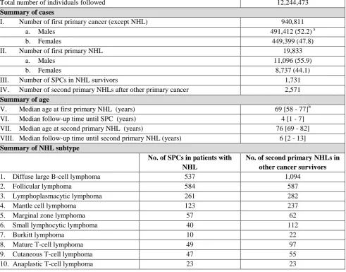
13 Table 1. Summary of the cohort
Abbreviation:
SCC, squamous cell carcinoma; SPC, second primary cancer; a Percentages in parentheses
b Inter-quartile distance shown in square brackets
Total number of individuals followed 12,244,473
Summary of cases
I. Number of first primary cancer (except NHL) 940,811
a. Males 491,412 (52.2) a
b. Females 449,399 (47.8)
II. Number of first primary NHL 19,833
a. Males 11,096 (55.9)
b. Females 8,737 (44.1)
III. Number of SPCs in NHL survivors 1,731
IV. Number of second primary NHLs after other primary cancer 2,571
Summary of age
V. Median age at first primary NHL (years) 69 [58 - 77]b
VI. Median follow-up time until SPC (years) 4 [1 - 7]
VII. Median age at second primary NHL (years) 76 [69 - 82]
VIII. Median follow-up time until second primary NHL (years) 6 [2 - 13]
Summary of NHL subtype
No. of SPCs in patients with NHL
No. of second primary NHLs in other cancer survivors
1. Diffuse large B-cell lymphoma 537 1,094
2. Follicular lymphoma 584 587
3. Lymphoplasmacytic lymphoma 261 282
4. Mantle cell lymphoma 123 237
5. Marginal zone lymphoma 57 62
6. Small lymphocytic lymphoma 40 112
7. Burkitt lymphoma 10 22
8. Mature T-cell lymphoma 49 97
9. Cutaneous T-cell lymphoma 47 55
14 Table 2. Risk of second primary cancer after diagnosis of non-Hodgkin lymphoma (A) and risk of non-Hodgkin lymphoma after diagnosis of non-NHL cancer (B).
Abbreviations:
N, frequency; RR, relative risk; CI, 95% confidence interval; P, probability; UAT, upper aerodigestive tract; SCC, squamous cell carcinoma; CUP, cancer of unknown primary;
Bolding indicates statistical significance at 0.05 level Cancer
A. Risk of non-NHL cancer after diagnosis of NHL B. Risk of NHL after diagnosis of non-NHL cancer
N RR CI lower CI upper P N RR CI lower CI upper P
UAT 40 1.96 1.44 2.68 <.0001 66 1.71 1.34 2.17 <.0001
Esophagus 9 0.94 0.51 1.75 0.85 3 0.73 0.28 1.96 0.5363
Stomach 34 1.52 1.08 2.13 0.015 21 1.01 0.66 1.56 0.947
Small intestine 3 0.60 0.19 1.87 0.382 6 0.88 0.39 1.95 0.7451
Colorectum 182 1.35 1.16 1.56 <.0001 269 1.28 1.13 1.44 <.0001
Anus 7 2.65 1.26 5.58 0.01 3 0.68 0.22 2.12 0.511
Liver 30 1.29 0.91 1.83 0.159 10 0.89 0.48 1.66 0.7204
Pancreas 25 0.95 0.64 1.40 0.779 5 0.53 0.22 1.29 0.1617
Nose 3 2.25 0.72 7.00 0.161 3 1.24 0.40 3.84 0.7099
Lung 126 1.48 1.25 1.77 <.0001 42 0.88 0.65 1.20 0.4216
Breast 93 0.88 0.72 1.08 0.221 377 1.28 1.16 1.42 <.0001
Cervix 1 0.19 0.03 1.36 0.099 53 1.81 1.38 2.37 <.0001
Endometrium 20 0.74 0.48 1.14 0.175 111 1.36 1.13 1.65 0.0012
Ovary 9 0.65 0.34 1.25 0.193 37 1.14 0.83 1.58 0.4225
Other female genitals 4 0.91 0.34 2.42 0.844 9 1.34 0.70 2.57 0.3835
Prostate 265 0.89 0.79 1.01 0.068 576 1.14 1.05 1.24 0.0027
Testis 0 28 2.29 1.58 3.32 <.0001
Other male genitals 4 1.71 0.64 4.56 0.286 2 0.41 0.10 1.65 0.2097
Kidney 70 3.10 2.45 3.92 <.0001 61 1.57 1.22 2.02 0.0004
Urinary bladder 83 1.54 1.24 1.91 <.0001 128 1.29 1.09 1.54 0.0037
Melanoma 83 1.98 1.60 2.44 <.0001 185 1.80 1.56 2.08 <.0001
Skin (SCC) 314 4.12 3.69 4.60 <.0001 237 2.44 2.14 2.77 <.0001
Eye 2 0.85 0.21 3.41 0.822 9 1.70 0.92 3.17 0.0918
Nervous system 24 1.14 0.76 1.70 0.525 46 0.96 0.72 1.29 0.8004
Thyroid gland 10 2.14 1.15 3.98 0.017 18 0.92 0.58 1.46 0.7152
Endocrine glands 14 1.16 0.70 1.92 0.576 70 1.37 1.08 1.73 0.009
Bone 1 1.30 0.18 9.25 0.793 6 2.01 0.90 4.47 0.0881
Connective tissue 9 1.67 0.87 3.21 0.125 18 1.58 0.99 2.50 0.0535
Hodgkin lymphoma 15 9.38 5.81 15.15 <.0001 46 7.29 5.46 9.73 <.0001
Multiple myeloma 14 0.89 0.52 1.50 0.648 24 1.54 1.03 2.30 0.034
Leukemia 94 2.90 2.36 3.56 <.0001 2 1.75 0.44 7.01 0.4269
CUP 48 1.31 0.99 1.74 0.061 12 0.67 0.38 1.19 0.1719
15 Table 3. Risk of second primary cancers among survivors of five frequent subtypes of non-Hodgkin lymphoma.
Abbreviations:
N, frequency; RR, relative risk; CI, confidence interval; P, probability; UAT, upper aerodigestive tract; SCC, squamous cell carcinoma; CUP, cancer of unknown primary; Bolding indicates statistical significance at 5% level
Second cancers
Diffuse large B-cell lymphoma Follicular lymphoma Lymphoplasmacytic lymphoma Mantle cell lymphoma Marginal zone lymphoma
N RR 95% CI P N RR 95% CI P N RR 95% CI P N RR 95% CI P N RR 95% CI P
UAT 14 1.89 1.12 - 3.20 0.017 13 2.06 1.19 -3.54 0.0094 7 2.53 1.21 - 5.31 0.0141 3 2.33 0.75 - 7.22 0.1435 1 1.66 0.23 - 11.77 0.613
Stomach 10 0.98 0.52 - 1.81 0.9382 11 1.31 0.72 - 2.36 0.3748 5 1.21 0.50 - 2.91 0.6666 3 1.76 0.57 - 5.45 0.3292 3 3.91 1.26 - 12.14 0.0181 Colorectum 65 1.31 1.03 - 1.67 0.03 52 1.27 0.97 - 1.67 0.0806 32 1.65 1.16 - 2.33 0.0049 11 1.31 0.72 - 2.36 0.3766 8 1.99 1.00 - 3.99 0.049
Anus 1 1.09 0.15 - 7.73 0.9326 5 6.34 2.63 - 15.25 <0.0001 1 2.90 0.41 - 20.62 0.2867
Liver 15 1.56 0.94 - 2.59 0.085 6 0.75 0.34 - 1.67 0.4833 3 0.77 0.25 - 2.39 0.6511 3 3.91 1.26 - 12.14 0.0181
Nose 2 3.89 0.97 - 15.59 0.0549
Lung 28 0.94 0.65 - 1.36 0.7511 51 1.93 1.46 - 2.53 <0.0001 19 1.68 1.07 - 2.63 0.0241 10 1.89 1.02 --3.52 0.0436 6 2.41 1.08 - 5.37 0.0311 Breast 32 0.86 0.61 - 1.22 0.4051 39 1.01 0.74 - 1.38 0.9593 12 0.96 0.55 - 1.70 0.8981 3 0.79 0.25 - 2.44 0.6803 2 0.54 0.14 - 2.18 0.3893
Cervix 1 0.50 0.07 - 3.53 0.4853
Endometrium 11 1.27 0.70 - 2.30 0.4245 6 0.65 0.29 - 1.45 0.2927 2 0.68 0.17 - 2.74 0.5912
Ovary 1 0.20 0.03 - 1.43 0.1098 5 0.92 0.38 - 2.21 0.8504 3 1.79 0.58 - 5.54 0.315
Prostate 77 0.72 0.58 - 0.90 0.0044 82 1.10 0.89 - 1.37 0.3781 52 1.13 0.86 - 1.48 0.3796 18 0.72 0.45 - 1.14 0.1608 1 0.15 0.02 - 1.05 0.056 Kidney 20 2.33 1.51 - 3.62 0.0002 25 3.36 2.27 - 4.97 <0.0001 6 1.90 0.85 - 4.23 0.1163 6 3.97 1.79 - 8.85 0.0007 4 5.99 2.25 - 15.96 0.0003 Urinary bladder 30 1.53 1.07 - 2.19 0.0198 29 1.79 1.24 - 2.58 0.0017 8 1.06 0.53 - 2.12 0.8681 9 2.64 1.38 - 5.08 0.0035 1 0.63 0.09 - 4.49 0.6462 Melanoma 23 1.58 1.05 - 2.37 0.029 28 2.28 1.57 - 3.30 <0.0001 10 2.04 1.10 - 3.79 0.0245 7 2.84 1.36 - 5.97 0.0057 7 5.85 2.79 - 12.28 <0.0001 Skin (SCC) 108 3.90 3.23 - 4.72 <0.0001 87 4.30 3.48 - 5.31 <0.0001 51 4.78 3.64 - 6.30 <0.0001 27 6.20 4.25 - 9.04 <0.0001 11 4.75 2.63 - 8.58 <0.0001
Eye 1 1.20 0.17 - 8.54 0.8542 1 1.38 0.19 - 9.80 0.7479
Nervous system 9 1.18 0.62 - 2.28 0.6114 6 0.87 0.39 - 1.94 0.7378 3 1.15 0.37 - 3.55 0.8143 1 0.76 0.11 - 5.41 0.7855 1 1.70 0.24 - 12.08 0.5949
Thyroid gland 2 1.08 0.27 - 4.31 0.9154 1 0.65 0.09 - 4.61 0.6651 1 1.57 0.22 - 11.14 0.6526 1 3.43 0.48 - 24.37 0.2175
Endocrine glands 4 0.90 0.34 - 2.40 0.8351 6 1.50 0.67 - 3.33 0.3248 2 1.29 0.32 - 5.16 0.7189 1 2.80 0.39 - 19.90 0.3029
Bone 1 3.91 0.55 - 27.83 0.1726
Connective tissue 2 1.00 0.25 - 3.99 0.9967 3 1.78 0.57 - 5.53 0.3176 2 2.72 0.68 - 10.87 0.1577 1 2.84 0.40 - 20.17 0.2966 1 6.59 0.93 - 46.83 0.0593 Hodgkin lymphoma 4 4.64 1.74 - 12.37 0.0022 7 9.64 4.59 - 20.25 <0.0001 1 3.33 0.47 - 23.63 0.2294 1 7.21 1.01 - 51.20 0.0483
Multiple myeloma 2 0.35 0.09 - 1.39 0.1343 4 0.82 0.31 - 2.19 0.6927 6 2.69 1.21 - 5.98 0.0156 2 1.97 0.49 - 7.87 0.3385
Leukemia 30 2.68 1.87 - 3.83 <0.0001 34 3.66 2.61 - 5.12 <0.0001 8 1.91 0.96 - 3.82 0.0668 12 6.20 3.52 - 10.91 <0.0001 1 1.13 0.16 - 8.00 0.9049
CUP 13 0.99 0.58 - 1.71 0.9837 20 1.79 1.16 - 2.78 0.0092 7 1.30 0.62 - 2.73 0.4866 4 1.79 0.67 - 4.76 0.2453 1 1.00 0.14 - 7.06 0.996
16 Table 4. Risk of five frequent subtypes of non-Hodgkin lymphoma as second primary cancer among survivors of other cancers.
Abbreviations:
N, frequency; RR, relative risk; CI, confidence interval; P, probability; UAT, upper aerodigestive tract; SCC, squamous cell carcinoma; CUP, cancer of unknown primary; Bolding indicates statistical significance at 5% level
First cancer
Diffuse large B-cell lymphoma Follicular lymphoma Lymphoplasmacytic lymphoma Mantle cell lymphoma Marginal zone lymphoma
N RR 95% CI P N RR 95% CI P N RR 95% CI P N RR 95% CI P N RR 95% CI P
UAT 27 1.59 1.09 - 2.32 0.0164 15 1.67 1.01 - 2.78 0.0464 10 2.20 1.18 - 4.10 0.0129 5 1.56 0.65 - 3.77 0.3181 3 3.10 0.99 - 9.66 0.0512
Stomach 11 1.19 0.66 - 2.15 0.5685 3 0.64 0.21 - 1.98 0.4373 1 0.38 0.05 - 2.69 0.3307 2 1.21 0.30 - 4.85 0.7862 1 2.16 0.30 - 15.36 0.4435
Colorectum 102 1.06 0.87 - 1.29 0.5534 72 1.58 1.25 - 2.00 0.0001 34 1.36 0.97 - 1.91 0.0767 26 1.55 1.05 - 2.29 0.0275 7 1.35 0.64 - 2.85 0.4385
Anus 1 0.52 0.07 - 3.66 0.5073 1 0.99 0.14 - 7.04 0.993 1 2.07 0.29 - 14.71 0.467
Liver 7 1.43 0.68 - 3.00 0.3463 2 0.77 0.19 - 3.08 0.7114 1 1.11 0.16 - 7.89 0.9173
Nose 1 0.93 0.13 - 6.61 0.9426 2 10.09 2.52 - 40.40 0.0011
Lung 17 0.82 0.51 - 1.32 0.41 9 0.81 0.42 - 1.56 0.5346 7 1.31 0.62 - 2.74 0.4821 4 1.03 0.39 - 2.75 0.9557 1 0.78 0.11 - 5.53 0.8008
Breast 144 1.10 0.93 - 1.30 0.2754 113 1.40 1.15 - 1.69 0.0006 35 1.20 0.85 - 1.69 0.2956 25 1.65 1.10 - 2.49 0.0162 12 1.24 0.69 - 2.22 0.4704
Cervix 25 1.97 1.33 - 2.92 0.0007 14 1.64 0.97 - 2.78 0.0646 4 1.34 0.50 - 3.59 0.5569 4 2.81 1.05 - 7.53 0.0402 1 1.08 0.15 - 7.69 0.9402
Endometrium 47 1.26 0.94 - 1.68 0.1234 29 1.37 0.95 - 1.98 0.0921 15 1.78 1.06 - 2.97 0.0279 6 1.43 0.64 - 3.22 0.3848 3 1.16 0.37 - 3.62 0.8037
Ovary 17 1.19 0.74 - 1.92 0.4663 9 0.98 0.51 - 1.88 0.9451 3 0.90 0.29 - 2.80 0.8564 5 3.01 1.25 - 7.30 0.0144 1 0.97 0.14 - 6.91 0.9743
Prostate 237 1.06 0.92 - 1.21 0.4227 112 1.23 1.01 - 1.49 0.0404 68 1.04 0.81 - 1.34 0.7313 79 1.47 1.16 - 1.86 0.0015 13 1.17 0.66 - 2.08 0.5975
Testis 6 1.15 0.52 - 2.57 0.7284 5 1.78 0.74 - 4.28 0.1988 5 4.59 1.90 - 11.06 0.0007 6 5.27 2.36 - 11.77 <0.0001
Kidney 21 1.22 0.79 - 1.87 0.3628 19 2.13 1.36 - 3.35 0.001 7 1.54 0.73 - 3.23 0.2554 4 1.25 0.47 - 3.34 0.6533
Urinary bladder 57 1.27 0.98 - 1.65 0.0724 26 1.21 0.82 - 1.78 0.3406 17 1.43 0.88 - 2.30 0.1459 10 1.19 0.64 - 2.22 0.585 2 0.84 0.21 - 3.37 0.8027
Melanoma 68 1.50 1.18 - 1.91 0.0009 41 1.71 1.26 - 2.33 0.0006 18 1.64 1.03 - 2.62 0.0361 20 2.46 1.58 - 3.83 <0.0001 8 2.81 1.39 - 5.67 0.0039 Skin (SCC) 110 2.39 1.97 - 2.89 <0.0001 50 2.56 1.93 - 3.39 <0.0001 26 2.26 1.53 - 3.33 <0.0001 19 2.46 1.56 - 3.88 0.0001 3 1.26 0.40 - 3.94 0.6913
Eye 7 2.69 1.28 - 5.65 0.0088 1 1.53 0.22 - 10.89 0.6695
Nervous system 21 1.01 0.66 - 1.55 0.9557 9 0.77 0.40 - 1.49 0.4417 6 1.21 0.54 - 2.69 0.6439 2 0.55 0.14 - 2.22 0.4042 2 1.56 0.39 - 6.25 0.5317 Thyroid gland 9 1.06 0.55 - 2.04 0.8591 2 0.42 0.10 - 1.66 0.2148 2 0.97 0.24 - 3.87 0.9614 2 1.43 0.36 - 5.74 0.6115
Endocrine glands 34 1.51 1.07 - 2.11 0.0174 20 1.68 1.08 - 2.60 0.0213 4 0.70 0.26 - 1.86 0.468 7 1.80 0.86 - 3.79 0.1206 2 1.47 0.37 - 5.89 0.5896
Bone 4 3.09 1.16 - 8.22 0.0243 1 1.36 0.19 - 9.62 0.7613 1 4.34 0.61 - 30.82 0.1425
Connective tissue 3 0.60 0.19 - 1.85 0.3688 6 2.24 1.01 - 4.99 0.0482 2 1.57 0.39 - 6.28 0.5246 1 1.11 0.16 - 7.88 0.9178
Hodgkin lymphoma 37 13.76 9.96 - 19.02 <0.0001 6 3.72 1.67 - 8.30 0.0013 1 6.02 0.84 - 42.82 0.0732
Multiple myeloma 14 2.05 1.22 - 3.47 0.0072 3 0.84 0.27 - 2.59 0.7559 5 2.69 1.12 - 6.48 0.027
Leukemia 2 3.91 0.98 - 15.65 0.0538
CUP 6 0.77 0.34 - 1.71 0.5136 2 0.48 0.12 - 1.93 0.3033 1 0.47 0.07 - 3.36 0.4545 1 0.72 0.10 - 5.14 0.746