Circuitry Comprised of Ole1, Rsp5, and Hsf1 in
Candida albicans
Michelle D. Leach,a,bLeah E. Cowenb
Aberdeen Fungal Group, School of Medical Sciences, University of Aberdeen, Institute of Medical Sciences, Foresterhill, Aberdeen, United Kingdoma; Department of Molecular Genetics, University of Toronto, Toronto, Ontario, Canadab
Temperature is a ubiquitous environmental variable which can profoundly influence the physiology of living cells as it changes over time and space. When yeast cells are exposed to a sublethal heat shock, normal metabolic functions become repressed and the heat shock transcription factor Hsf1 is activated, inducing heat shock proteins (HSPs).Candida albicans, the most prevalent human fun-gal pathogen, is an opportunistic pathogen that has evolved as a relatively harmless commensal of healthy individuals. Even thoughC. albicansoccupies thermally buffered niches, it has retained the classic heat shock response, activating Hsf1 during slow thermal transitions such as the increases in temperature suffered by febrile patients. However, the mechanism of temperature sensing in fungal pathogens remains enigmatic. A few studies withSaccharomyces cerevisiaesuggest that thermal stress is transduced into a cellular signal at the level of the membrane. In this study, we manipulated the fluidity ofC. albicansmembrane to dissect mecha-nisms of temperature sensing. We determined that in response to elevated temperature, levels ofOLE1, encoding a fatty acid de-saturase, decrease. Subsequently, loss ofOLE1triggers expression ofFAS2, encoding a fatty acid synthase. Furthermore, deple-tion ofOLE1prevents full activation of Hsf1, thereby reducingHSPexpression in response to heat shock. This reduction in Hsf1 activation is attributable to the E3 ubiquitin ligase Rsp5, which regulatesOLE1expression. To our knowledge, this is the first study to define a molecular link between fatty acid synthesis and the heat shock response in the fungal kingdom.
M
icroorganisms inhabit dynamic environments in which they are continually exposed to environmental stimuli and stresses. Survival depends upon effective environmental response strategies that have been uniquely tuned over evolutionary time. By reacting to environmental changes via a sense and respond logic, cells continuously monitor their environment and coordi-nate appropriate cellular responses to specific stimuli (1). These cellular response strategies have been intensively studied for a va-riety of model organisms (2–5). Fundamentally, organisms utilize a myriad of signaling pathways that drive physiological adaptation to diverse environmental stresses, including temperature fluctua-tions and osmotic, oxidative, and weak acid stresses, as well as nutrient limitation (6,7).Temperature is a ubiquitous environmental variable, which can profoundly influence the physiology of living cells as it changes over time and space. When yeast cells are exposed to a sublethal heat shock, normal metabolic functions become re-pressed, and genes encoding heat shock proteins (HSPs) are in-duced (8). This induction occurs through the evolutionarily con-served heat shock transcription factor Hsf1. How the cell senses changes in ambient temperature, thus triggering Hsf1 activation, was thought to be driven by the accumulation of denatured pro-teins (9). However, a sustained heat shock would give rise to on-going protein denaturation, leading to continual activation of HSPs. Instead, the transient nature of the heat shock response suggests that the thermal sensor becomes desensitized, allowing cells to adapt to a new basal level of activity (10,11).
The cellular membrane serves as a direct sensor of its orga-nism’s environment. Its lipid structure is key to determining the physicochemical environment of the membrane, with the molec-ular packing of lipids acting as a direct determinant of membrane fluidity. At physiological temperatures, the fluidity of the mem-brane is increased through disorganized, unpacked unsaturated fatty acids, whereas saturated fatty acids remain tightly packed and
retain a higher melting temperature (12,13). Consequently, lipid saturation is regulated in response to changes in temperature. The fluidity of the membrane is governed by an intricate balance be-tween saturated and unsaturated fatty acids. In the model yeast
Saccharomyces cerevisiae, after the initial reaction of fatty acid syn-thesis, elongation of the carbon chain is catalyzed by the fatty acid synthases Fas1 and Fas2 to produce the saturated fatty acids palmitic acid (16:0) and stearic acid (18:0) (14). These are then converted to the monounsaturated fatty acids palmitoleic acid (16:1) and oleic acid (18:1) by the fatty acid⌬9 desaturase (Ole1), located at the surface of the endoplasmic reticulum (ER), by in-troducing double bonds at carbon 9 in the carbon chains (15).
The degree of unsaturation in theS. cerevisiaecellular mem-brane is highest at 15°C and lowest at 37°C, with fatty acid chains becoming longer with increasing temperature (16). But how does this affect the heat shock response? Studies on the fungal pathogen
Histoplasma capsulatumestablished that addition of increasing concentrations of palmitic acid, a saturated fatty acid, paired with a temperature upshift upregulated transcription of the heat shock protein HSP82; conversely, addition of oleic acid, an unsaturated fatty acid, decreased heat shock gene transcription upon a rise in temperature (17). Furthermore, anS. cerevisiaemutant that lacks the fatty acid desaturase geneOLE1was complemented using
con-Received27 May 2014Accepted11 June 2014
Published ahead of print20 June 2014
Address correspondence to Leah E. Cowen, [email protected]. Supplemental material for this article may be found athttp://dx.doi.org/10.1128 /EC.00138-14.
Copyright © 2014 Leach and Cowen. This is an open-access article distributed under the terms of theCreative Commons Attribution 3.0 Unported license.
doi:10.1128/EC.00138-14
on September 8, 2020 by guest
http://ec.asm.org/
structs with the native (S. cerevisiae)OLE1promoter or theOLE1
promoter from a temperature-tolerant or temperature-sensitive
H. capsulatumstrain, with the resulting strains each displaying differential expression ofOLE1(18). Depending on the promoter used, complemented strains adjusted the physiology of the mem-brane by modifying the ratio of saturated to unsaturated fatty acids. Subsequently, each mutant displayed a different threshold temperature of heat shock gene expression. For instance, the tem-perature-sensitive strain complemented with theOLE1promoter that upregulatesOLE1expression displayed a dramatic decrease in palmitic acid, as well asHSPexpression (17).
Candida albicans, one of the most prevalent human fungal pathogens, is an opportunistic pathogen that has evolved as a rel-atively harmless commensal of the mucous membranes and diges-tive tracts of healthy individuals (19,20).C. albicansfrequently causes infections of mucosal membranes (thrush), and in immu-nocompromised patients this yeast can cause life-threatening sys-temic infections (19,21). Even thoughC. albicans is obligately associated with warm-blooded mammals and occupies thermally buffered niches, it has retained the classic heat shock response (22). Indeed, Hsf1 is essential for viability inC. albicans, reflecting the fundamental importance of heat shock adaptation in all or-ganisms (22–24). Our recent exploration of the dynamic regula-tion of Hsf1 during thermal adaptaregula-tion has suggested that Hsf1 is activated even during slow thermal transitions such as the in-creases in temperature suffered by febrile patients (11). However, the mechanisms by whichC. albicanssenses changes in ambient temperature and activates Hsf1 remain an enigma.
In this study, we explore whetherC. albicans utilizes mem-brane fluidity as a direct sensor of temperature. We determined that cells alter the levels of the fatty acid desaturase Ole1 in re-sponse to increased temperature and that loss ofOLE1induces expression ofFAS2, encoding a fatty acid synthase. We show that depletion ofOLE1prevents full activation of Hsf1, contributing to a reduction inHSPexpression in response to heat shock, and that this reduction in Hsf1 activation is attributable to the E3 ubiquitin ligase Rsp5, which regulatesOLE1through the transcription fac-tor Spt23. Therefore, we have established for the first time in the fungal kingdom a molecular link between membrane fluidity and the heat shock response.
MATERIALS AND METHODS
Strains and growth conditions.All strains used are listed inTable 1. Strains were grown in YPD (1% yeast extract, 2% Bacto peptone, 2% glucose) (25). To impose an instant heat shock of 30°C to 42°C, cells were grown in YPD at 30°C to exponential phase and mixed with an equal volume of medium that had been prewarmed to 54°C in flasks that had been prewarmed at 42°C. Cells were grown at 42°C for the times indicated below. Doxycycline was added to YPD medium at a concentration of 1
g/ml or 20g/ml.
Strain construction.To regulate oleic acid levels, one copy of the
OLE1gene was deleted, and the other was placed under the control of the
tetOpromoter. Briefly, the NAT (nourseothricin) flipper cassette (pLC49) was amplified with oLC2798/oLC2799 containing regions of homology upstream and downstream ofOLE1. The PCR product was transformed into the wild-type strain SN95 (CaLC239), and NAT-resistant transfor-mants were PCR tested with oLC275/oLC2802 and oLC274/oLC2803 to verify integration of the cassette. The NAT cassette was then excised to create CaLC2851. The tetracycline-repressible transactivator, thetetO
promoter, and the NAT flipper cassette were PCR amplified from pLC605 using oligonucleotides oLC2801 and oLC2798. The PCR product was transformed into CaLC2851. Correct upstream and downstream integra-tion was verified by amplifying across both juncintegra-tions by colony PCR using primer pairs oLC2802/oLC275 and oLC2804/oLC300, respectively. Loss of the wild-type band was verified using primer pair oLC2802/oLC2804. Hsf1 was then tagged in this strain as described below to create CaLC3032. To regulateRSP5, one copy of theRSP5gene was deleted, and the other was placed under the control of thetetOpromoter. Briefy, the NAT flipper cassette (pLC49) was amplified with oLC3359/oLC3360 contain-ing regions of homology upstream and downstream ofRSP5. The PCR product was transformed into the wild-type strain SN95 (CaLC239), and NAT-resistant transformants were PCR tested with oLC275/oLC3361 and oLC274/oLC3362 to verify integration of the cassette. The NAT cassette was then excised to create CaLC3032. The tetracycline-repressible trans-activator, thetetOpromoter, and the NAT flipper cassette were PCR am-plified from pLC605 using oligonucleotides oLC3357/oLC3388. The PCR product was transformed into CaLC3302, and NAT-resistant transfor-mants were PCR tested with oLC534/oLC3361 (upstream), oLC300/ oLC3389 (downstream), and oLC3361/oLC3389 (wild type) to verify in-tegration of the cassette, the NAT cassette was then excised. Hsf1 was then tagged in this strain as described below to create CaLC3366.
To determine Hsf1 phosphorylation status, Hsf1 was tagged with the TAP (tandem affinity purification) tag at its C terminus in the wild-type strain SN95 (creating CaLC2993) (Table 1) and in thetetO-HSF1/hsf1⌬
strain to confirm functionality of the tagged allele, using a PCR-based strategy as described previously (26). Briefly, the tag and a selectable marker (ARG4) were PCR amplified from pLC573 (pFA-TAP-ARG4
[27]) using oligonucleotides oLC2950/2922 (see Table S1 in the supple-mental material). Fifty microliters of PCR product was transformed into
C. albicans. Correct genomic integration was verified using appropriate primer pairs that anneal⬃500 bp upstream (oLC1597) or downstream (oLC1598) from both insertion junctions together with oLC1593 (TAP-R) and oLC1594 (ARG4-F), which target the TAP and the selectable marker (see Table S1).
qRT-PCR.To monitor gene expression changes in response toOLE1
orRSP5depletion, strains SN95 (CaLC2993), CaLC3032 (tetO-OLE1/ ole1⌬), and CaLC3366 (tetO-RSP5/rsp5⌬) were grown overnight at 30°C in YPD, with shaking at 200 rpm. Stationary-phase cultures were split and adjusted to an optical density at 600 nm (OD600) of 0.1; one culture was
treated with doxycycline (BD Biosciences), while the other was left un-treated. Cells were grown for 4 h at 30°C. To monitor gene expression changes in response to heat shock, wild-type (CaLC2993) andtetO-OLE1/ ole1⌬(CaLC3032) cells were grown to mid-log phase and subjected to a 30°C to 42°C heat shock, and 50 ml was harvested from each culture at the
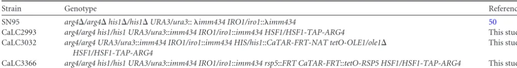
TABLE 1C. albicansstrains used in this study
Strain Genotype Reference
SN95 arg4⌬/arg4⌬his1⌬/his1⌬URA3/ura3::imm434 IRO1/iro1::imm434 50
CaLC2993 arg4/arg4 his1/his1 URA3/ura3::imm434 IRO1/iro1::imm434 HSF1/HSF1-TAP-ARG4 This study CaLC3032 arg4/arg4 URA3/ura3::imm434 IRO1/iro1::imm434 HIS/his1::CaTAR-FRT-NAT tetO-OLE1/ole1⌬
HSF1/HSF1-TAP-ARG4
This study
CaLC3366 arg4/arg4 his1/his1 URA3/ura3::imm434 IRO1/iro1::imm434 rsp5::FRT CaTAR-FRT::tetO-RSP5 HSF1/HSF1-TAP-ARG4 This study
on September 8, 2020 by guest
http://ec.asm.org/
specified time, centrifuged at 3,000 rpm for 2 min at 4°C, and washed once with distilled water (dH2O) before being frozen at⫺80°C. RNA was then
isolated using the Qiagen RNeasy kit, and cDNA synthesis was performed using the AffinityScript cDNA synthesis kit (Stratagene). PCR was carried out using the SYBR green JumpStartTaqReadyMix (Sigma-Aldrich) un-der the following cycle conditions: 95°C for 3 min, 95°C for 10 s, and 60°C for 30 s for 39 rounds, 95°C for 10 s, and 65°C for 5 s. All reactions were done in triplicate using the following primer pairs: forHSP104, oLC1620/ oLC1621; forHSP21, oLC3217/oLC3218; forOLE1, oLC2805/oLC2806; forRSP5, oLC3408/oLC3409; and forFAS2, oLC3168/oLC3169. Tran-script levels were normalized toACT1(oLC2285/oLC2286) (see Table S1 in the supplemental material). Data were analyzed using Bio-Rad CFX Manager software, version 3.1 (Bio-Rad).
Western blotting.Total soluble protein was extracted and subjected to Western blotting using published protocols (28,29). Briefly, mid-log-phase cells were pelleted by centrifugation, washed with sterile water, and resuspended in lysis buffer (0.1 M Tris-HCl [pH 8.0], 10% glycerol, 1 mM dithiothreitol [DTT], phenylmethylsulfonyl fluoride [PMSF], and pro-tease inhibitor cocktail). An equal volume of 0.5-mm acid-washed beads was added to each tube. Cells were mechanically disrupted on a BioSpec (Bartlesville, OK) Mini-Beadbeater for six 30-s periods, with 1 min on ice between each cycle. The lysate was pelleted by high-speed centrifugation and the supernatant removed for analysis. Protein concentration was de-termined using a Bradford reagent (Sigma-Aldrich) assay. Protein sam-ples were mixed with one-sixth volume of 6⫻sample buffer (0.35 M Tris-HCl, 10% [wt/wt] SDS, 36% glycerol, 5%-mercaptoethanol, and 0.012% bromophenol blue). Between 2g and 30g of protein was loaded in wells of a 6% SDS-PAGE gel. Separated proteins were trans-ferred to a polyvinylidene difluoride (PVDF) membrane for 1 h at 100 V at 4°C. Membranes were blocked in 5% milk in phosphate-buffered saline (PBS) containing 0.1% Tween 20 (PBS-T) at room temperature for 1 h and subsequently incubated in primary antibody as follows. All primary antibodies were left on the membrane for 1 h at room temperature. Mem-branes were washed with 1⫻PBS-T and probed for 1 h with secondary antibody dissolved in 1⫻PBS-T and 5% milk. Membranes were washed in PBS-T and signals detected using an ECL Western blotting kit as per the manufacturer’s instructions (Pierce).
TAP-tagged Hsf1 was detected using a 1:5,000 dilution of anti-TAP tag rabbit polyclonal antibody (Thermo Scientific; CAB1001) in PBS-T plus 5% milk. To detect Act1, an anti-Act1 antibody was used (Santa Cruz Biotechnology; sc47778) at a 1:1,000 dilution in PBS-T plus 5% milk. To detect Hsp90, a 1:10,000 dilution of anti-Hsp90 antibody was used (cour-tesy of Bryan Larson) in PBS-T plus 5% milk.
RESULTS
OLE1is regulated by temperature.Mechanisms of temperature sensing in fungal pathogens remain an enigma, but evidence ob-tained with the benign yeastS. cerevisiaeimplicates membrane fluidity as a primary sensor of temperature (17). To establish whether temperature sensing and membrane fluidity are linked in the fungal pathogenC. albicans, which is obligately associated with warm-blooded mammals, we aimed to determine ifOLE1 expres-sion is affected by temperature. As oleic acid is an unsaturated fatty acid, we hypothesized that during high-temperature growth,
OLE1would be downregulated, promoting a less fluid membrane. Wild-type cells (CaLC2993) were grown at 30°C, 37°C, or 42°C for 4 h or subjected to a 30°C to 42°C heat shock before being har-vested and snap-frozen. RNA was extracted, and expression of
OLE1was determined relative to that of the housekeeping gene
ACT1(Fig. 1A). As expected, levels ofOLE1 were significantly depleted when cells were heat shocked or grown at 42°C versus 30°C and 37°C (Fig. 1A). These data reinforce the notion that temperature affects fluidity of the membrane.
To elucidate the mechanisms of temperature sensing through
membrane fluidity further, we created a conditional mutant with doxycycline-repressible expression of the fatty acid desaturase geneOLE1, which is known for its essentiality (30). One allele of
OLE1was deleted, and the other was placed under the control of a tetracycline-repressible promoter. The addition of 1g/ml of the tetracycline analog doxycycline halted growth of thetetO-OLE1/ ole1⌬strain after 6 h but had no effect on wild-type cells (data not shown). Expression ofOLE1was measured by quantitative reverse transcription-PCR (qRT-PCR) after 4 h of growth in the absence
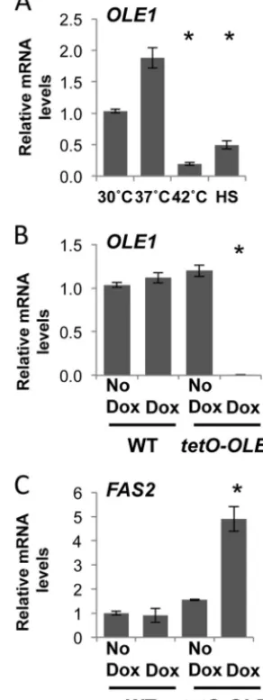
FIG 1Membrane fluidity is tightly regulated in response to temperature and fatty acid ratios. (A)OLE1transcript levels decrease upon exposure to high temperatures.C. albicanswild-type (WT) cells were grown to exponential phase at 30°C, 37°C, or 42°C or subjected to a 15-min 30°C to 42°C heat shock (HS), andOLE1transcript levels were measured and normalized to theACT1
loading control. *,P⬍0.05 compared to 30°C growth (Studentttest). (B)
OLE1transcript levels decrease following depletion intetO-OLE1/ole1⌬cells. WT (CaLC2993 [Table 1]) and tetO-OLE1/ole1⌬ cells (CaLC3032) were treated or not with 1g/ml of doxycycline (Dox) for 4 h, andOLE1transcript levels were measured by qRT-PCR and normalized to theACT1loading con-trol. *,P⬍0.05 compared to the value for the wild type (Studentttest). (C) Depleting Ole1 leads to an increase inFAS2transcript levels. WT and tetO-OLE1/ole1⌬cells were treated or not with 1g/ml of doxycycline for 4 h, and
FAS2transcript levels were measured and normalized to theACT1loading control. *,P⬍0.05 compared to the value for the wild type (Studentttest).
on September 8, 2020 by guest
http://ec.asm.org/
or presence of 1g/ml of doxycycline, when cells were in mid-log phase (OD600⫽0.6) (Fig. 1B). The addition of doxycycline had no
effect onOLE1expression in wild-type cells but caused a signifi-cant decrease inOLE1expression in thetetO-OLE1/ole1⌬strain (Fig. 1B).
Ole1 is responsible for introducing the double bond into satu-rated fatty acetyl coenzyme A (acetyl-CoA) substrates to produce monounsaturated fatty acids. After the initial reaction of fatty acid synthesis, which involves the incorporation of acetyl-CoA with CO2 to generate malonyl-CoA (31), elongation of the carbon
chain is catalyzed by the fatty acid synthases Fas1 and Fas2 to produce long-chain saturated fatty acids such as palmitic and ste-aric acids (14); these act as the precursors for the subsequent de-saturation reactions to produce monounsaturated fatty acids. We postulated that depletion of Ole1 would act as a signal for the production of saturated fatty acid precursors, essentially leading to an increase in the levels of the fatty acid synthases. To test this, we depletedOLE1as previously described and determined expres-sion of the fatty acid synthase geneFAS2by qRT-PCR (Fig. 1C). As expected, loss ofOLE1triggered a 5-fold increase inFAS2levels. Therefore, the cell responds to temperature by intricately regulat-ing the levels of saturated and unsaturated fatty acids.
Ole1 regulates components of the heat shock response.To determine whether a link exists between membrane fluidity and temperature sensing inC. albicans, we looked to components of the heat shock response. The heat shock transcription factor Hsf1 is rapidly activated by phosphorylation following a heat shock, leading to the upregulation of heat shock proteins (22). We hy-pothesized that if the membrane does indeed sense temperature, depletion of oleic acid would misregulate Hsf1 activation. Wild-type andtetO-OLE1/ole1⌬cells were grown in the absence or pres-ence of 1g/ml of doxycycline for 4 h to depleteOLE1. Cells were either left untreated (30°C) or treated with a 15-min 30°C to 42°C heat shock. Protein was extracted, and Hsf1-TAP was visualized using an anti-TAP antibody (Fig. 2A). Wild-type cells in the ab-sence and preab-sence of doxycycline respond similarly, activating Hsf1 by phosphorylation, as seen by an upshift and broadening of the band corresponding to Hsf1 upon the thermal shock. The
tetO-OLE1/ole1⌬strain also activates Hsf1 similarly to wild-type cells in the absence of doxycycline. However, in the presence of doxycycline, we observed a dramatic decrease not only in the phosphorylation status of Hsf1 but also in its protein levels. Based on this, we hypothesized that levels of the heat shock protein Hsp90, which is regulated by Hsf1, would be reduced. However, when we probed the blot with an anti-Hsp90 antibody, Hsp90 levels remained constant. This is most likely due to the stability of Hsp90 (M. D. Leach, unpublished data).
Next, we aimed to determine if the impact ofOLE1depletion on Hsf1 levels manifests exclusively at the translational level, or if it is also evident at the transcriptional level. Expression ofHSF1
was measured in wild-type cells and thetetO-OLE1/ole1⌬cells in the presence of 1g/ml of doxycycline after growth at 30°C, 37°C, or 42°C or upon a 15-min 30°C to 42°C heat shock (Fig. 2B). In wild-type cells,HSF1levels increased up to 8-fold with increasing temperature and rapidly increased upon heat shock, equivalent to levels of cells grown at 42°C. However, loss ofOLE1hampers
HSF1expression during high-temperature growth, with levels in-creasing only 3-fold at 42°C. Additionally, when cells are exposed to a heat shock,HSF1expression is only half that of wild-type cells. Doxycycline has no effect onHSF1expression (see Fig. S1 in the
supplemental material). These data provided the first molecular link between membrane fluidity and temperature sensing in fungi. Our next goal was to ascertain if misregulation of Hsf1 upon
OLE1depletion affects expression of key heat shock proteins in-volved in the heat shock response. Under the same conditions as used to determine HSF1 levels, we monitored expression of
HSP104andHSP21, which encode two heat shock proteins essen-tial for thermal adaptation inC. albicans(22,32). We made three observations. First, there was an increase in bothHSP104 and
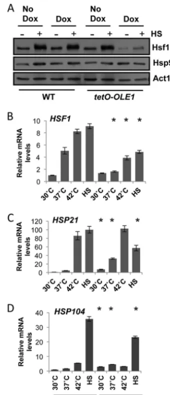
FIG 2Depletion of Ole1 inhibits full activation of Hsf1, leading to a loss in
HSPexpression during high-temperature growth and heat shock. (A) Phos-phorylation ofC. albicansHsf1 during a 30°C to 42°C heat shock, revealed by Western analysis. Exponentially growing WT (wild type, CaLC2993 [Table 1]) andtetO-OLE1/ole1⌬cells (CaLC3032) were treated or not with1g/ml of doxycycline for 4 h and subjected to a 15-min 30°C to 42°C heat shock. Protein was extracted immediately, and Hsf1 phosphorylation was monitored using an anti-TAP antibody recognizingHSF1-TAP. Membranes were stripped and reprobed for Hsp90, and actin served as an internal loading control. (B to D) Expression ofHSF1(B),HSP21(C), andHSP104(D) during growth at 30°C, 37°C, or 42°C or after a 15-min 30°C to 42°C heat shock (HS), as determined by qRT-PCR of the corresponding transcripts relative to the internalACT1
mRNA control. *,P⬍0.05 compared to the value for the wild type at the corresponding temperature (Studentttest).
on September 8, 2020 by guest
http://ec.asm.org/
HSP21under basal conditions uponOLE1depletion (Fig. 2Cand
D). This increase remained at 37°C, with both genes presenting significantly higher expression in thetetO-OLE1/ole1⌬strain than in its wild-type counterpart. Therefore, depletion ofOLE1 pro-motes the induction ofHSPexpression in the absence of any heat stress. Second, depletion ofOLE1had little impact on expression of theHSPs at 42°C. This could be due to a balance between the initial upregulation of theseHSPgenes in response to exposure to 42°C, before full depletion of Ole1, and stability of theHSPgenes during high-temperature growth. Third, upon treatment with a 30°C to 42°C heat shock,HSPexpression increased 100-fold for
HSP21(Fig. 2C) and 35-fold forHSP104(Fig. 2D) in wild-type cells but reached only half of wild-type levels whenOLE1 was depleted. Again, addition of doxycycline to wild-type cells had no effect onHSP expression (Fig. S1). In summary, depletion of
OLE1reduces Hsf1 protein levels and Hsf1 activation upon heat shock. In addition,HSF1andHSPlevels are misregulated at the transcriptional level.
Rsp5 regulates expression ofOLE1and activation of Hsf1. The promoter ofS. cerevisiae OLE1contains multiple transcrip-tional regulatory elements, allowing for the elaborate control of the levels of unsaturated fatty acids. Transcription can be re-pressed through the addition of unsaturated fatty acids and acti-vated in response to low temperature and hypoxia via the ER-bound transcription factors ScSpt23p and ScMga2p (33). Both factors are, in turn, activated by ubiquitin/proteasome-dependent ER-associated degradation, through the E3 ubiqutin ligase ScRsp5 (34).C. albicansretains only one homolog of theS. cerevisiae func-tionally redundant gene,CaSPT23. Repression ofSPT23inC. al-bicansblocks expression ofOLE1(35), suggesting that Rsp5 could be the upstream element that transduces signals pertaining to temperature changes sensed at the membrane.
We hypothesized that Rsp5 regulates OLE1 expression and hence Hsf1 activation inC. albicans. To test this, we constructed a tetracycline-repressible RSP5 conditional expression strain, in whichRSP5expression is repressed upon the addition of doxycy-cline. Addition of 20g/ml of doxycycline for prolonged periods did not affect the viability of thetetO-RSP5/rsp5⌬strain, suggest-ing thatRSP5is not an essential gene (data not shown). To ensure full depletion ofRSP5, tetO-RSP5/rsp5⌬cells were subcultured into YPD in the presence or absence of 20g/ml of doxycycline for 18 h and then subcultured again the following morning under the same conditions and grown to mid-log phase (approximately 5 h). RNA was extracted for qRT-PCR analysis ofRSP5transcript levels (Fig. 3A). As expected,RSP5was significantly decreased upon ad-dition of 20g/ml of doxycycline. We also noted that in the ab-sence of doxycycline,RSP5levels in thetetO-RSP5/rsp5⌬strain were significantly higher than in the wild-type counterpart, sug-gesting that thetetOpromoter is stronger than the native pro-moter. To determine if Rsp5 regulatesOLE1expression, we de-pleted RSP5 at 30°C overnight and until mid-log phase and examinedOLE1 expression by qRT-PCR. Notably,OLE1levels were 4-fold lower uponRSP5depletion than in wild-type cells (Fig. 3B), indicating that Rsp5 regulatesOLE1, likely through the transcription factor Spt23 (35). Even thoughRSP5expression was significantly increased in the absence of doxycycline (Fig. 3A),
OLE1expression remained the same as in wild-type cells (see Fig. S2A in the supplemental material).
Given that we can manipulateRSP5expression and thereby reduce levels ofOLE1, we wanted to examine the effect ofRSP5
depletion on Hsf1 activity in response to heat shock. Wild-type cells andtetO-RSP5/rsp5⌬cells were grown in the absence or pres-ence of 20g/ml of doxycycline as previously described and either kept at 30°C or exposed to a 15-min 30°C to 42°C heat shock. Proteins were extracted and subjected to Western analysis to visu-alize Hsf1 activation (Fig. 3C). As predicted, doxycycline-medi-ated depletion ofRSP5led to reduced levels of Hsf1 activation, as indicated by a decrease in phosphorylation with a narrower band and loss of a band shift. We also noted that in the absence of doxycycline, although a visible shift is seen in the presence of a heat shock in thetetO-RSP5/rsp5⌬strain, the levels of Hsf1 are significantly lower than in the wild-type counterpart, suggesting that the misregulation ofRSP5affects Hsf1 protein levels.
In order to confirm that Hsf1 activation upon depletion of
RSP5is not equivalent to that seen in wild-type cells upon a heat shock, we monitored expression ofHSP104andHSP21under the same conditions (Fig. 3D). As expected, expression of bothHSPs increased significantly upon heat shock; however, this increase was reduced whenRSP5was depleted. Expression ofHSPgenes was not affected by doxycycline in the wild-type strain (see Fig. S2B and C in the supplemental material). Although expression of
HSP104 andHSP21 was significantly lower upon depletion of
RSP5, it was not reduced to the same extent as that observed upon depletion ofOLE1(Fig. 3CandD). In part, this may be because depletion ofRSP5does not fully depleteOLE1(Fig. 3A), even in the presence of heat shock (Fig. S2D). Therefore, depletion of
RSP5phenocopies loss ofOLE1.
DISCUSSION
In this paper, we have addressed one of the most fundamental ques-tions in biology: how do cells sense temperature? The ability to sense the surrounding temperature is key for virulence. Indeed, many fun-gal pathogens inhabit a remarkable diversity of environments; for example,Cryptococcus neoformans,Histoplasma capsulatum, and
Aspergillus fumigatusare found in diverse environments such as pigeon excreta and soil, but each retains the ability to grow at 37°C. The loss of genes necessary for high-temperature growth in these pathogens results in attenuated virulence and at times even death (36–38), suggesting the importance of temperature sensing for pathogenesis. Besides governing virulence, temperature regu-lates numerous cellular processes that often contribute to patho-genesis. For example, morphological transitions inC. albicansare controlled by temperature. At ambient temperature, this organ-ism favors the yeast form, while elevated temperatures induce fil-amentous growth, enabling penetration of the epithelium (39).C. albicanscan also switch from a white to opaque cellular state in host niches with lower temperatures, such as skin, facilitating mat-ing (40).
By manipulatingC. albicansmembrane composition, we have begun to understand the ways in which fungal pathogens sense temperature. Using a tetracycline-repressible promoter, we were able to deplete levels of the fatty acid desaturase geneOLE1, which regulates synthesis of the monounsaturated fatty acid oleic acid. In doing so, we triggered a 5-fold increase in the fatty acid synthase geneFAS2(Fig. 1C). These data suggest that the cell compensates for the loss of unsaturated fatty acids by increasing the levels of
FAS2in an attempt to increase levels of saturated fatty acids, which are the precursors for unsaturated fatty acids. Furthermore, heat shock or high-temperature growth caused a significant decrease in
OLE1expression (Fig. 1A). This is consistent with previous studies
on September 8, 2020 by guest
http://ec.asm.org/
withS. cerevisiaeillustrating a 4- to 6-fold increase inOLE1mRNA when cells were rapidly shifted from 30°C to 10°C (41). These data reinforce the notions that temperature affects fluidity of the mem-brane and that the cell continually monitors the ratio of saturated to unsaturated fatty acids.
Previous studies with the fungal pathogenH. capsulatumand the model yeastS. cerevisiaehave suggested a role for the mem-brane in sensing the surrounding temperature. Indeed, Carratù and colleagues observed a strong increase inHSPexpression inH. capsulatumwhen cells were heat shocked at 37°C upon the addi-tion of external saturated fatty acids into the media. Addiaddi-tion of unsaturated fatty acids, however, reducedHSPexpression during the same heat shock (17). Furthermore, Chatterjee and colleagues demonstrated a 9°C increase in the optimal activation of the heat shock response upon supplementation ofS. cerevisiaewith unsat-urated fatty acids (42). Therefore, it would appear that the ratio of saturated to unsaturated fatty acids of the membrane is a key de-terminant in the perception of rapid temperature changes. Con-sistent with these data, we discovered that loss ofOLE1caused upregulated expression of key components of the heat shock
re-sponse during growth at 30°C and 37°C, but this upregulation was lost upon high-temperature growth or exposure to a short heat shock (Fig. 2BtoD). Indeed, based on our findings and those of others, it would appear that upon loss ofOLE1, levels of unsatu-rated fatty acids are severely depleted within the membrane, prompting the cell to assume that the temperature has increased. In turn,HSPexpression is increased at 30°C and 37°C. However, if the ratio of fatty acids is acting as the primary sensor of tempera-ture, upon subjecting the cell to a drastic heat shock, the temper-ature change in a membrane that is depleted of unsaturated fatty acids is not properly recognized, thereby preventing full activation of Hsf1 and reducing upregulation ofHSPgenes (Fig. 2).
One of the conundrums surrounding temperature sensing is the remarkable speed at which cells are able to sense and respond to the surrounding environment.C. albicansis able to activate Hsf1 within 1 min of exposure to a heat shock (11), suggesting that the sensor itself may reside in the plasma membrane, becoming activated upon slight changes in membrane fluidity. This would then be transduced into a signal that could further regulate fatty acid expression. Based on this notion, we examined regulators of
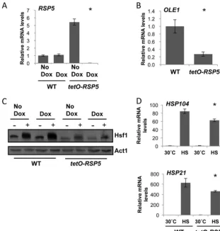
FIG 3Rsp5 regulates Hsf1 in part throughOLE1. (A)RSP5transcript levels are upregulated intetO-RSP5/rsp5⌬strain in the absence of doxycycline and depleted in the presence of doxycycline. WT (wild type, CaLC2993 [Table 1]) andtetO-RSP5/rsp5⌬(CaLC3366) cells were treated or not with 20g/ml of doxycycline overnight for 18 h and then subcultured the following morning and grown for a further 5 h under the same conditions.RSP5transcript levels were normalized to theACT1loading control. (B) Depleting Rsp5 causes a decrease inOLE1transcript levels. WT andtetO-RSP5/rsp5⌬cells were treated or not with 20g/ml of doxycycline as stated above andOLE1transcript levels were measured and normalized to theACT1loading control. (C) Rsp5 is required for Hsf1 expression and full activation. WT andtetO-RSP5/rsp5⌬cells were treated or not with 20g/ml of doxycycline as stated above, exposed to a 15-min 30°C to 42°C heat shock, and subjected to Western analysis. Decreased Hsf1-TAP levels were observed in thetetO-RSP5/rsp5⌬strain. Membranes were stripped and reprobed for actin, which served as the internal loading control. (D) Expression ofHSP21andHSP104during growth at 30°C or after a 15-min 30°C to 42°C heat shock (HS), as determined by qRT-PCR relative to the internalACT1mRNA control. *,P⬍0.05 intetO-RSP5/rsp5⌬cells compared to wild-type cells (Studentttest).
on September 8, 2020 by guest
http://ec.asm.org/
fatty acid synthesis. InS. cerevisiae,OLE1expression is regulated via the ER-bound transcription factors Spt23 and Mga2 (33), which are activated by the E3 ubiquitin ligase Rsp5 (34).C. albi-canshas retained only one homolog of theS. cerevisiae function-ally redundant genes,CaSPT23. Oh and colleagues found that upon repression ofSPT23inC. albicans, expression ofOLE1was blocked (35). Therefore, we postulated that Rsp5 regulates expres-sion ofOLE1, through Spt23, thereby acting as a downstream element that is activated upon changes that occur in the plasma membrane and subsequently regulating activation of the heat shock response. By regulating the levels of Rsp5 using the tetracycline-repressible pro-moter (Fig. 3A), we noted a significant decrease inOLE1expression upon loss ofRSP5(Fig. 3B). This translated to a reduction in Hsf1 activation during a short heat shock (Fig. 3C), similar to that seen upon loss ofOLE1(Fig. 2A). Furthermore, this decrease in Hsf1 ac-tivation contributed to a decrease inHSP21andHSP104expression upon a heat shock (Fig. 3D).
We also observed that in the absence of doxycycline in the
tetO-RSP5/rsp5⌬strain,RSP5levels are significantly higher than in wild-type cells (Fig. 3A; see also Fig. S2E in the supplemental material). When we looked at Hsf1 activation under these condi-tions, although Hsf1 is activated andHSPexpression remains sim-ilar to that observed in wild-type cells (see Fig. S2B and C), Hsf1 levels are much lower than in the wild-type counterpart and sim-ilar to those observed upon Rsp5 depletion. ForS. cerevisiae, two recent studies by Haitani and colleagues show that an Rsp5 mu-tant,rsp5A410E, exhibits a decrease in Hsf1 levels compared to that
for wild-type cells but relatively similarHSF1mRNA levels. Fur-ther investigation showed accumulation ofHSF1mRNA in the nucleus, suggesting that Rsp5 is required forHSF1nuclear export (43,44). Our data suggest that a similar mechanism operates inC. albicans, whereby misregulation of Rsp5 prevents proper export of
HSF1mRNA. However, depletion ofOLE1does reduceHSF1 ex-pression, suggesting more complex circuitry connecting Rsp5-Ole1-Hsf1.
These data provide evidence for the first inhibitor of Hsf1 ac-tivation in response to a heat shock. The loss of the unsaturated fatty acid desaturase geneOLE1drastically increases the fatty acid synthase geneFAS2(Fig. 1C), leads to an upregulation of HSP
genes in the absence of heat shock but inhibits fullHSPexpression upon heat shock (Fig. 2CandD). This inhibition is likely due to a reduction in Hsf1 activation (Fig. 2A). This suggests that mem-brane fluidity is a key sensor of temperature; however, before changes can occur in levels of fatty acids, a signal must be trans-duced.OLE1is regulated by the transcription factor Spt23, which is regulated by the E3 ubiquitin ligase Rsp5. We discovered that loss ofRSP5phenocopies loss ofOLE1, suggesting that Rsp5 could be one of the early sensors of temperature, which coordinately regulates fatty acid synthesis and activation of the heat shock re-sponse.
Understanding circuitry governing temperature sensing offers therapeutic opportunities for crippling diverse microbial patho-gens. In the context ofCandidaspecies, many are human com-mensals and able to disseminate into the bloodstream, causing systemic infections. Inhibiting fatty acid biosynthesis would be an ideal way to eradicateCandidaspecies from the mycobiome prior to immunosuppressive treatments that render patients vulnerable to infection. Importantly, fungal Ole1 is comprised of two do-mains: the N and C domains, containing the catalytic motif for the desaturase activity and the cytochromeb5activity, respectively. In
contrast, humans have two separate enzymes to carry out these activities, making fungal Ole1 an ideal antifungal target (33). Fur-thermore, recent studies illustrated that components of fatty acid biosynthesis from differentCandidaspecies are essential for estab-lishing and maintaining infection in a mouse model of candidiasis (45–47). Xu and colleagues demonstrated thatC. albicansOle1 is necessary for virulence, and Nguyen and colleagues obtained sim-ilar results withC. parapsilosis. This is consistent with our findings that loss of Ole1 leads to a significant decrease in Hsf1 activation (Fig. 2A) and a previous study showing that activation of Hsf1 is necessary for virulence inC. albicans(48). Finally, Krishnamurthy and colleagues discovered that depletion ofOLE1blocks one of the key virulence traits inC. albicans, hyphal development, when cells are grown under aerobic conditions (30). This study provides us with the first mechanistic link between fatty acid synthesis and the heat shock response in the fungal kingdom, highlighting Ole1 as an attractive antifungal. Beyond fungal pathogens, fatty acid biosynthesis is emerging as a powerful target for the development of antibacterial agents, with small-molecule inhibitors currently in clinical use to treat tuberculosis and as ubiquitous consumer antimicrobials (49).
ACKNOWLEDGMENTS
We thank Grant Hatch and Fred Xu for help with data analysis. We also thank Joe Heitman and Al Brown for inspiring scientific discussions.
M.D.L. is supported by a Sir Henry Wellcome Postdoctoral Fellowship (Wellcome Trust grant 096072), and L.E.C. is supported by a Canada Research Chair in Microbial Genomics and Infectious Disease and by Canadian Institutes of Health Research Grant MOP-119520.
REFERENCES
1.Kussell E, Leibler S.2005. Phenotypic diversity, population growth, and information in fluctuating environments. Science309:2075–2078.http: //dx.doi.org/10.1126/science.1114383.
2.Tirosh I, Weinberger A, Carmi M, Barkai N.2006. A genetic signature of interspecies variations in gene expression. Nat. Genet.38:830 – 834.http: //dx.doi.org/10.1038/ng1819.
3.Gasch AP, Spellman PT, Kao CM, Carmel-Harel O, Eisen MB, Storz G, Botstein D, Brown PO.2000. Genomic expression programs in the re-sponse of yeast cells to environmental changes. Mol. Biol. Cell11:4241– 4257.http://dx.doi.org/10.1091/mbc.11.12.4241.
4.Enjalbert B, Nantel A, Whiteway M.2003. Stress-induced gene expres-sion inCandida albicans: absence of a general stress response. Mol. Biol. Cell14:1460 –1467.http://dx.doi.org/10.1091/mbc.E02-08-0546. 5.Causton HC, Ren B, Koh SS, Harbison CT, Kanin E, Jennings EG, Lee
TI, True HL, Lander ES, Young RA.2001. Remodeling of yeast genome expression in response to environmental changes. Mol. Biol. Cell12:323– 337.http://dx.doi.org/10.1091/mbc.12.2.323.
6.Estruch F.2000. Stress-controlled transcription factors, stress-induced genes and stress tolerance in budding yeast. FEMS Microbiol. Rev.24:
469 – 486.http://dx.doi.org/10.1111/j.1574-6976.2000.tb00551.x. 7.de Dios CH, Roman E, Monge RA, Pla J.2010. The role of MAPK signal
transduction pathways in the response to oxidative stress in the fungal pathogenCandida albicans: implications in virulence. Curr. Protein Pept. Sci.11:693–703.http://dx.doi.org/10.2174/138920310794557655. 8.Mager WH, De Kruijff AJ.1995. Stress-induced transcriptional
activa-tion. Microbiol. Rev.59:506 –531.
9.Parsell DA, Lindquist S.1993. The function of heat-shock proteins in stress tolerance: degradation and reactivation of damaged proteins. Annu. Rev. Genet.27:437– 496.http://dx.doi.org/10.1146/annurev.ge.27.120193 .002253.
10. Sorger PK.1991. Heat shock factor and the heat shock response. Cell
65:363–366.http://dx.doi.org/10.1016/0092-8674(91)90452-5. 11. Leach MD, Tyc KM, Brown AJP, Klipp E.2012. Modelling the regulation
of thermal adaptation inCandida albicans, a major fungal pathogen of humans. PLoS One 7:e32467. http://dx.doi.org/10.1371/journal.pone .0032467.
on September 8, 2020 by guest
http://ec.asm.org/
12. Leach MD, Cowen LE.2013. Surviving the heat of the moment: a fungal pathogens perspective. PLoS Pathog. 9:e1003163. http://dx.doi.org/10 .1371/journal.ppat.1003163.
13. Leach MD, Cowen LE.2014. To sense or die: mechanisms of temperature sensing in fungal pathogens. Current Fungal Infect. Rep.8:185–191.http: //dx.doi.org/10.1007/s12281-014-0182-1.
14. Schweizer E, Hofmann J. 2004. Microbial type I fatty acid synthases (FAS): major players in a network of cellular FAS systems. Microbiol. Mol. Biol. Rev. 68:501–517. http://dx.doi.org/10.1128/MMBR.68.3.501-517 .2004.
15. Stukey JE, McDonough VM, Martin CE.1989. Isolation and character-ization ofOLE1, a gene affecting fatty acid desaturation from Saccharomy-ces cerevisiae. J. Biol. Chem.264:16537–16544.
16. Klose C, Surma MA, Gerl MJ, Meyenhofer F, Shevchenko A, Simons K.
2012. Flexibility of a eukaryotic lipidome—insights from yeast lipidomics. PLoS One7:e35063.http://dx.doi.org/10.1371/journal.pone.0035063. 17. Carratù L, Franceschelli S, Pardini CL, Kobayashi GS, Horvath I, Vigh
L, Maresca B.1996. Membrane lipid perturbation modifies the set point of the temperature of heat shock response in yeast. Proc. Natl. Acad. Sci. U. S. A.93:3870 –3875.http://dx.doi.org/10.1073/pnas.93.9.3870. 18. Gargano S, Di Lallo G, Kobayashi GS, Maresca B.1995. A
temper-ature-sensitive strain ofHistoplasma capsulatumhas an altered⌬9-fatty acid desaturase gene. Lipids 30:899 –906. http://dx.doi.org/10.1007 /BF02537480.
19. Odds FC.1988. Candida and candidosis, 2nd ed. Bailliere Tindall, Lon-don, United Kingdom.
20. Piispanen AE, Hogan DA.2008. PEPped up: induction ofCandida albi-cansvirulence by bacterial cell wall fragments. Cell Host Microbe4:1–2. http://dx.doi.org/10.1016/j.chom.2008.06.005.
21. Calderone RA.2002. Candida and candidiasis.. ASM Press, Washington, DC.
22. Nicholls S, Leach MD, Priest CL, Brown AJ.2009. Role of the heat shock transcription factor, Hsf1, in a major fungal pathogen that is obligately associated with warm-blooded animals. Mol. Microbiol. 74:844 – 861. http://dx.doi.org/10.1111/j.1365-2958.2009.06883.x.
23. Leach MD, Budge S, Walker L, Munro C, Cowen LE, Brown AJP.2012. Hsp90 orchestrates transcriptional regulation by Hsf1 and cell wall re-modelling by MAPK signalling during thermal adaptation in a pathogenic yeast. PLoS Pathog.8:e1003069.http://dx.doi.org/10.1371/journal.ppat .1003069.
24. Leach MD, Klipp E, Cowen LE, Brown AJ. 2012. Fungal Hsp90: a biological transistor that tunes cellular outputs to thermal inputs. Nat. Rev. Microbiol.10:693–704.http://dx.doi.org/10.1038/nrmicro2875. 25. Sherman F.1991. Getting started with yeast. Methods Enzymol.194:3–21.
http://dx.doi.org/10.1016/0076-6879(91)94004-V.
26. Diezmann S, Michaut M, Shapiro RS, Bader GD, Cowen LE. 2012. Mapping the Hsp90 genetic interaction network inCandida albicans re-veals environmental contingency and rewired circuitry. PLoS Genet.
8:e1002562.http://dx.doi.org/10.1371/journal.pgen.1002562.
27. Lavoie H, Sellam A, Askew C, Nantel A, Whiteway M.2008. A toolbox for epitope-tagging and genome-wide location analysis inCandida albi-cans. BMC Genomics9:578.http://dx.doi.org/10.1186/1471-2164-9-578. 28. Leach MD, Stead DA, Argo E, MacCallum DM, Brown AJ. 2011. Molecular and proteomic analyses highlight the importance of ubiquiti-nation for the stress resistance, metabolic adaptation, morphogenetic reg-ulation and virulence ofCandida albicans. Mol. Microbiol.79:1574 –1593. http://dx.doi.org/10.1111/j.1365-2958.2011.07542.x.
29. Leach MD, Stead DA, Argo E, Brown AJ.2011. Identification of sumoy-lation targets, combined with inactivation ofSMT3, reveals the impact of sumoylation upon growth, morphology, and stress resistance in the pathogenCandida albicans. Mol. Biol. Cell22:687–702.http://dx.doi.org /10.1091/mbc.E10-07-0632.
30. Krishnamurthy S, Plaine A, Albert J, Prasad T, Prasad R, Ernst JF.2004. Dosage-dependent functions of fatty acid desaturase Ole1p in growth and morphogenesis ofCandida albicans. Microbiology150:1991–2003.http: //dx.doi.org/10.1099/mic.0.27029-0.
31. Schneiter R, Guerra CE, Lampl M, Gogg G, Kohlwein SD, Klein HL.
1999. TheSaccharomyces cerevisiaehyperrecombination mutant hpr1delta is synthetically lethal with two conditional alleles of the acetyl coenzyme A carboxylase gene and causes a defect in nuclear export of polyadenylated RNA. Mol. Cell. Biol.19:3415–3422.
32. Mayer FL, Wilson D, Jacobsen ID, Miramon P, Slesiona S, Bohovych IM, Brown AJ, Hube B.2012. Small but crucial: the novel small heat
shock protein Hsp21 mediates stress adaptation and virulence inCandida albicans. PLoS One 7:e38584. http://dx.doi.org/10.1371/journal.pone .0038584.
33. Martin CE, Oh CS, Jiang Y.2007. Regulation of long chain unsaturated fatty acid synthesis in yeast. Biochim. Biophys. Acta1771:271–285.http: //dx.doi.org/10.1016/j.bbalip.2006.06.010.
34. Hoppe T, Matuschewski K, Rape M, Schlenker S, Ulrich HD, Jentsch S.
2000. Activation of a membrane-bound transcription factor by regulated ubiquitin/proteasome-dependent processing. Cell102:577–586.http://dx .doi.org/10.1016/S0092-8674(00)00080-5.
35. Oh CS, Martin CE.2006.Candida albicansSpt23p controls the expression of the Ole1p⌬9 fatty acid desaturase and regulates unsaturated fatty acid biosynthesis. J. Biol. Chem.281:7030 –7039.http://dx.doi.org/10.1074/jbc .M510746200.
36. Bhabhra R, Miley MD, Mylonakis E, Boettner D, Fortwendel J, Pan-epinto JC, Postow M, Rhodes JC, Askew DS.2004. Disruption of the
Aspergillus fumigatusgene encoding nucleolar protein CgrA impairs ther-motolerant growth and reduces virulence. Infect. Immun.72:4731– 4740. http://dx.doi.org/10.1128/IAI.72.8.4731-4740.2004.
37. Lamoth F, Juvvadi PR, Fortwendel JR, Steinbach WJ.2012. Heat shock protein 90 (Hsp90) is required for conidiation and cell wall integrity in
Aspergillus fumigatus. Eukaryot. Cell11:1324 –1332.http://dx.doi.org/10 .1128/EC.00032-12.
38. Odom A, Muir S, Lim E, Toffaletti DL, Perfect J, Heitman J.1997. Calcineurin is required for virulence ofCryptococcus neoformans. EMBO J.
16:2576 –2589.http://dx.doi.org/10.1093/emboj/16.10.2576.
39. Gow NA, Brown AJ, Odds FC.2002. Fungal morphogenesis and host invasion. Curr. Opin. Microbiol.5:366 –371.http://dx.doi.org/10.1016 /S1369-5274(02)00338-7.
40. Lachke SA, Lockhart SR, Daniels KJ, Soll DR.2003. Skin facilitates
Candida albicansmating. Infect. Immun.71:4970 – 4976.http://dx.doi .org/10.1128/IAI.71.9.4970-4976.2003.
41. Nakagawa Y, Sakumoto N, Kaneko Y, Harashima S.2002. Mga2p is a putative sensor for low temperature and oxygen to induceOLE1 transcrip-tion inSaccharomyces cerevisiae. Biochem. Biophys. Res. Commun.291:
707–713.http://dx.doi.org/10.1006/bbrc.2002.6507.
42. Chatterjee MT, Khalawan SA, Curran BPG.1997. Alterations in cellular lipids may be responsible for the transient nature of the yeast heat shock response. Microbiology 143:3063–3068. http://dx.doi.org/10.1099 /00221287-143-9-3063.
43. Haitani Y, Shimoi H, Takagi H.2006. Rsp5 regulates expression of stress proteins via post-translational modification of Hsf1 and Msn4 in Saccha-romyces cerevisiae. FEBS Lett.580:3433–3438.http://dx.doi.org/10.1016/j .febslet.2006.05.016.
44. Haitani Y, Takagi H.2008. Rsp5 is required for the nuclear export of mRNA ofHSF1andMSN2/4under stress conditions inSaccharomyces cerevisiae. Genes Cells13:105–116.http://dx.doi.org/10.1111/j.1365-2443 .2007.01154.x.
45. Xu D, Sillaots S, Davison J, Hu W, Jiang B, Kauffman S, Martel N, Ocampo P, Oh C, Trosok S, Veillette K, Wang H, Yang M, Zhang L, Becker J, Martin CE, Roemer T.2009. Chemical genetic profiling and characterization of small-molecule compounds that affect the biosynthe-sis of unsaturated fatty acids inCandida albicans. J. Biol. Chem.284:
19754 –19764.http://dx.doi.org/10.1074/jbc.M109.019877.
46. Nguyen LN, Trofa D, Nosanchuk JD.2009. Fatty acid synthase impacts the pathobiology ofCandida parapsilosisin vitro and during mammalian infection. PLoS One 4:e8421. http://dx.doi.org/10.1371/journal.pone .0008421.
47. Nguyen LN, Gacser A, Nosanchuk JD.2011. The stearoyl-coenzyme A desaturase 1 is essential for virulence and membrane stress inCandida parapsilosisthrough unsaturated fatty acid production. Infect. Immun.
79:136 –145.http://dx.doi.org/10.1128/IAI.00753-10.
48. Nicholls S, MacCallum DM, Kaffarnik FA, Selway L, Peck SC, Brown AJ.2011. Activation of the heat shock transcription factor Hsf1 is essential for the full virulence of the fungal pathogenCandida albicans. Fungal Genet. Biol.48:297–305.http://dx.doi.org/10.1016/j.fgb.2010.08.010. 49. Heath RJ, White SW, Rock CO.2002. Inhibitors of fatty acid synthesis as
antimicrobial chemotherapeutics. Appl. Microbiol. Biotechnol.58:695– 703.http://dx.doi.org/10.1007/s00253-001-0918-z.
50. Noble SM, Johnson AD.2005. Strains and strategies for large-scale gene deletion studies of the diploid human fungal pathogenCandida albicans. Eukaryot. Cell 4:298 –309. http://dx.doi.org/10.1128/EC.4.2.298-309 .2005.