STATE-OF-THE-ART REVIEW ARTICLE
Editor’s Note
The Journal is interested in receiving for review short articles (1000 words) summarizing recent advances which have been made in the past 2 or 3 years in specialized areas of research and patient care.
Pediatric Autonomic Disorders
Felicia B. Axelrod, MDa, Gisela G. Chelimsky, MDb, Debra E. Weese-Mayer, MDc
aDepartment of Pediatrics and Neurology, New York University School of Medicine, New York, New York;bDepartment of Pediatrics, Case Western Reserve School of Medicine, Cleveland, Ohio;cDepartment of Pediatrics, Rush University School of Medicine, Chicago, Illinois
The authors have indicated they have no financial relationships relevant to this article to disclose.
ABSTRACT
The scope of pediatric autonomic disorders is not well recognized. The goal of this review is to increase awareness of the expanding spectrum of pediatric autonomic disorders by providing an overview of the autonomic nervous system, including the roles of its various components and its pervasive influence, as well as its intimate relationship with sensory function. To illustrate further the breadth and complexities of autonomic dysfunction, some pediatric disorders are described, concentrating on those that present at birth or appear in early childhood.
www.pediatrics.org/cgi/doi/10.1542/ peds.2005-3032
doi:10.1542/peds.2005-3032
Key Words
autonomic nervous system, cardiovascular, sympathetic nervous system,
parasympathetic nervous system, viscerosensory
Abbreviations FD—familial dysautonomia ANS—autonomic nervous system CAN— central autonomic network PHOX2B—paired-like homeobox 2B NGF—nerve growth factor CFS— chronic fatigue syndrome HSAN— hereditary sensory and autonomic neuropathy
CIPA— congenital insensitivity to pain with anhidrosis
CCHS— congenital central hypoventilation syndrome
CVS— cyclic vomiting syndrome POTS—postural orthostatic tachycardia Accepted for publication Feb 13, 2006
Address correspondence to Felicia B. Axelrod, MD, Dysautonomia Treatment and Evaluation Center, New York University School of Medicine, 530 First Ave, Suite 9Q, New York, NY 10016. E-mail: [email protected]. edu
A
PPRECIATION OF THEbreadth of autonomic disorders has increased since Langley1 originally proposedthe generic term “autonomic nervous system” (ANS) and designated its division into the sympathetic, para-sympathetic, and enteric nervous systems. Although a number of texts dedicated to various aspects of auto-nomic function now are available,2–4 they tend to
con-centrate on adult disorders, with pediatric autonomic disorders poorly represented. Even within the first text dedicated to describing various clinical disorders by Dancis,5 the only pediatric disorder included was
fa-milial dysautonomia (FD). Now, more than 20 years later, investigators are beginning to appreciate the value of genetic autonomic disorders as models to advance the understanding of pathophysiologic mechanisms in-volved in autonomic dysfunction.6,7In fact, the original
description of FD in 19498preceded the description by
Shy and Drager9 of the adult neurodegenerative
syn-drome characterized by central autonomic dysfunction by 11 years. Although the report by Shy and Drager initiated expansion of autonomic research and eventual founding of an autonomic subsection within neurology and autonomic societies in the United States and Eu-rope, the same level of interest has not been seen within the pediatric community. Perhaps this disparity has evolved through lack of awareness of the myriad of pediatric autonomic disorders or inadequate residency education regarding evaluation of this particular system. The ANS is pervasive and integrates multiple second-ary functions so that symptoms can be widespread and confounding. In addition, there are often associated sen-sory perturbations, because the development and main-tenance of the autonomic and sensory systems are closely linked. The goal of this article is to increase awareness of the expanding spectrum of pediatric auto-nomic disorders so that this population can benefit from the advances being made in evaluation and treatment.2,3
We provide an overview of the ANS and stress the extent of its influence, discuss the protean symptoms and man-ifestations caused by autonomic perturbations, and em-phasize the expanding number of pediatric disorders that feature autonomic dysfunction.
THE ANATOMY AND PHYSIOLOGY OF THE ANS
General Description
The ANS is a visceral and largely involuntary motor/ effector system that is traditionally divided into sympa-thetic (thoracolumbar) and parasympasympa-thetic (craniosa-cral) divisions, each with a central and a peripheral component.10In addition, there is an important enteric
division. Outflow can occur independently, but to some extent it is regulated and integrated by the central au-tonomic network (CAN).11 The CAN maintains integral
relationships with visceral sensory neurons via afferent input from the vagus nerve and relays transmission
through the nucleus tractus solitarius to the hypothala-mus, amygdala, and forebrain.11
Embryologic Development
Development of the ANS is intimately related to the development of the sensory nervous system; both have their embryonic origins in the multipotential neural crest cells. These cells migrate and eventually evolve into sensory and autonomic ganglia as well as the adrenal chromaffin cells. Their differentiation and commitment to function in the mature nervous system is incumbent on exposure to growth factors released by structures along the migratory route and then within the target tissue. Eventually, specificity will be determined by their ability to produce specific neurotransmitters. Therefore, one could postulate that an early genetic error affecting initial migration would cause profound decreases in both sensory and autonomic populations, whereas a later ge-netic error might only affect cell survival to one or both populations, causing more erratic and varied clinical ex-pression.
Growth Factors and Neurotransmitters
Various factors promote normal progression from the embryonic to the mature autonomic and sensory ner-vous systems.12,13Several key transcription factors have
been identified that play critical roles in the develop-ment of the ANS, such as the MASH1 (mammalian achaete-scute homologue) and PHOX (paired-like ho-meobox) 2B genes, which are necessary for differentia-tion of uncommitted neural crest cells to the developing ANS.14,15 Another important regulator of development
and survival is nerve growth factor (NGF).16,17 In the
embryonic neuron, NGF binding promotes migration from the neural crest and enhances maturation through neurite outgrowth. In the mature neuron, dependence on NGF decreases, but it continues to enhance neuro-transmitter synthesis.17
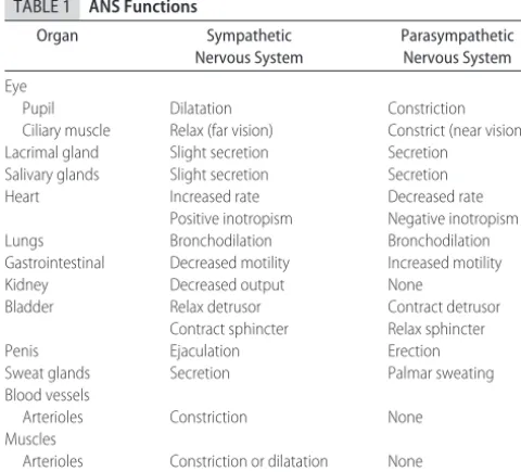
systems are antagonistic, that is not always the case (as indicated in Table 1). Thus, when the sympathetic sys-tem is stimulated, a host of receptor syssys-tems are acti-vated, including dilation of the pupil, increase in glan-dular secretions, bronchodilation, increase in heart rate and force of contraction, decrease in gastrointestinal tract motility, decrease in function of the reproductive organs, and mobilization of energy substrates. The para-sympathetic system tends to have more focal responses, but some effects may be quite broad, particularly with the wide-ranging innervation of the vagus nerve. How-ever, the parasympathetic system seems to have less influence on exocrine and endocrine function.
INTEGRATION OF THE PERIPHERAL AND CENTRAL AUTONOMIC NERVOUS SYSTEMS
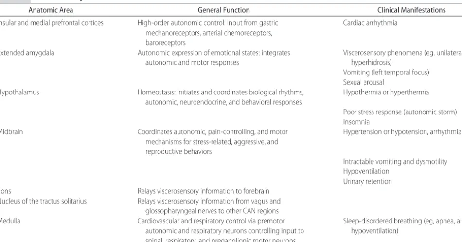
The varied functions of the peripheral ANS are inte-grated and regulated by the CAN, the extensive circuitry of which ranges from the forebrain to the brainstem (Table 2).11 Disorders in the forebrain circuits, such as
ischemia secondary to blood-flow disturbance or sei-zures, can cause cardiac arrhythmia.18 Within this
cir-cuitry, the nucleus tractus solitarius in the medulla ob-longata, which receives input from the vagus and glossopharyngeal nerves, functions as a major relay sta-tion, allowing continuous feedback and integration. The hypothalamic area seems to have major influences on thermoregulation and sleep/wake cycling. Thus, the CAN serves many critical functions and affects viscero-motor and neuroendocrine function as well as viscero-motor and pain modulation. It aids in reflex adjustments of autonomic responses and integrates autonomic, neu-roendocrine, and behavioral responses that, in turn, maintain homeostasis, emotional expression, and re-sponse to stress.19
SYMPTOMS OF AUTONOMIC DYSFUNCTION IN THE PEDIATRIC PATIENT
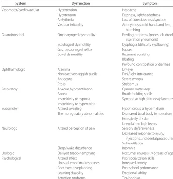
Because the ANS and its CAN component have perva-sive effects that affect multiple other systems second-arily, clinical manifestations can be extremely varied. Rather than use an anatomic approach, one can use a functional or system approach, as listed in Table 3. For this review, only those autonomic disorders with multi-system involvement are considered. Although children with gastroesophageal reflux or asthma have obvious autonomic dysfunction, their care is best relegated to the appropriate subspecialist. However, when more than one system is perturbed, then one might consider that the patient is affected with a more global autonomic disorder. At that point, the differential diagnosis expands and starts to include a number of autonomic disorders that can be considered on the basis of age at presenta-tion.
PEDIATRIC AUTONOMIC DISORDERS
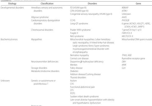
Many pediatric autonomic disorders are apparent at birth or within the first year of life. Some of these disorders occur as a result of developmental abnormali-ties caused by specific genetic mutations required for neural crest cell migration and maturation; others occur as a result of prematurity or generalized central dysfunc-tion (Table 4). Those disorders that occur as a result of biochemical errors causing neurotransmitter deficiencies or inefficient mitochondrial metabolism can be more insidious and later in their presentation. In addition, autonomic dysfunction has been noted with various dis-orders for which mechanisms remain obscure, such as autism and chronic fatigue syndrome (CFS), and also may be associated with various chronic diseases. Table 4 lists some of these disorders, but the list continues to expand. A few representative disorders will be described.
Autonomic Disorders Associated With Developmental Arrest or Aberrant Development of Function
Hereditary Sensory and Autonomic Neuropathies
General Description
The complexities of the ANS and its intimate relation-ship with sensory function is especially well illustrated in the group of genetic disorders known as hereditary sen-sory and autonomic neuropathies (HSANs).6,7,20 Each
HSAN disorder is probably caused by different genetic errors affecting a specific aspect of small fiber neuro-development and resulting in variable phenotypic ex-pression.6,7,20 With the exception of hereditary sensory
radicular neuropathy (HSAN type I), which is a domi-nant disorder presenting in the second decade of life, the other HSANs are autosomal recessive disorders that present at birth. Two HSANs with specific genetic mu-tations are FD (HSAN type III) and congenital
insensi-TABLE 1 ANS Functions
Organ Sympathetic
Nervous System
Parasympathetic Nervous System
Eye
Pupil Dilatation Constriction
Ciliary muscle Relax (far vision) Constrict (near vision)
Lacrimal gland Slight secretion Secretion
Salivary glands Slight secretion Secretion
Heart Increased rate Decreased rate
Positive inotropism Negative inotropism
Lungs Bronchodilation Bronchodilation
Gastrointestinal Decreased motility Increased motility
Kidney Decreased output None
Bladder Relax detrusor Contract detrusor
Contract sphincter Relax sphincter
Penis Ejaculation Erection
Sweat glands Secretion Palmar sweating
Blood vessels
Arterioles Constriction None
Muscles
Arterioles Constriction or dilatation None
tivity to pain with anhidrosis (CIPA or HSAN type IV). For each HSAN type, penetrance is complete, but there can be marked variability in expression. Characteristic to all HSANs is that intradermal injection of histamine phosphate fails to elicit a normal axon-flare response.7
However, FD is the only HSAN for which there is com-mercially available genetic testing.
FD
In FD, the gene is IKBKAP (IB kinase–associated protein gene), and ⬎99% of individuals with FD are homozygous for a mutation in intron 20 that causes a drastic reduction in correctly spliced messenger RNA in neuronal tissue and, therefore, a lack of expression of the normal protein product IKAP (IB kinase–associated protein).21,22 It has been postulated that IKAP aids in
expression of various neurotransmitters and that pro-duction of the abnormal gene product impedes this ability.23Although FD is almost exclusive to individuals
of Eastern European Jewish extraction,21,22,24 it is the
most prevalent HSAN type and often used as the proto-type with which to compare other HSAN disorders.7
In FD there is inadequate development, as well as lim-ited survival, of sensory and autonomic neurons, with the sympathetic population more widely affected than the parasympathetic population. Pathologic studies have demonstrated decreased unmyelinated and small my-elinated neuronal populations in the peripheral sensory nervous system and the ANS. Although central auto-nomic symptoms are present, no consistent central neu-ropathology has yet been described.
Although patients with FD have decreased pain and
temperature perception, the sensory perturbations are not as profound as in the other HSANs.25Bone and skin
pain are diminished but not absent; sensitivity to visceral pain is intact. Corneal and tendon reflexes are hypo-active, and taste appreciation is diminished, consistent with absence of lingual fungiform papillae. With age, vibratory sensory loss and impaired coordination ap-pear.26
The autonomic disturbances, however, are very prom-inent, involve peripheral and central tracts, and im-pose the greatest impediments to function, especially in the neonatal period.7,27 In addition to absence of tears
(alacrima) with emotional crying, a cardinal feature of the disorder, feeding difficulties resulting from poor oral coordination and hypotonia are frequent. Recurrent misdirection, especially of liquids, and frequent gastro-esophageal reflux put the patient at risk for aspiration and chronic lung disease. Protracted episodes of nausea and vomiting can be triggered by emotional or physical stress or even arousal from sleep. These episodes, also termed the dysautonomic crisis, are usually associated with a constellation of signs including agitation, tachy-cardia, and hypertension. Vasomotor and cardiovascular perturbations manifest as erythematous skin blotching and hyperhidrosis with excitation or even eating. Pa-tients can exhibit both extreme hypertension and pro-found and rapid postural hypotension without compen-satory tachycardia.7Supersensitivity to cholinergic and
adrenergic agents has been demonstrated.28,29 Patients
have relative insensitivity to hypoxemia,30–33which
lim-its their ability to cope with pneumonia or travel to high
TABLE 2 CAN: Anatomy and Function
Anatomic Area General Function Clinical Manifestations
Insular and medial prefrontal cortices High-order autonomic control: input from gastric mechanoreceptors, arterial chemoreceptors, baroreceptors
Cardiac arrhythmia
Extended amygdala Autonomic expression of emotional states: integrates
autonomic and motor responses
Viscerosensory phenomena (eg, unilateral hyperhidrosis)
Vomiting (left temporal focus) Sexual arousal
Hypothalamus Homeostasis: initiates and coordinates biological rhythms,
autonomic, neuroendocrine, and behavioral responses
Hypothermia or hyperthermia
Poor stress response (autonomic storm) Insomnia
Midbrain Coordinates autonomic, pain-controlling, and motor
mechanisms for stress-related, aggressive, and reproductive behaviors
Hypertension or hypotension, arrhythmias
Intractable vomiting and dysmotility Hypoventilation
Urinary retention
Pons Relays viscerosensory information to forebrain
Nucleus of the tractus solitarius Relays viscerosensory information from vagus and glossopharyngeal nerves to other CAN regions
Medulla Cardiovascular and respiratory control via premotor
autonomic and respiratory neurons controlling input to spinal, respiratory, and preganglionic motor neurons
altitudes. Ensuing hypoxemia may lead to hypotension, bradyarrhythmia, and even syncope. Developmental milestones are commonly delayed, but intelligence is usually within normal ranges.34
Although the gene has been identified, the mainstay of treatment remains preventative and supportive. These treatments have included measures to maintain eye moisture, fundoplication with gastrostomy to pro-vide nutrition and avoid risk of aspiration, use of central agents such as benzodiazepines and clonidine to con-trol vomiting and the dysautonomic crisis, and fludro-cortisone and midodrine to combat cardiovascular la-bility.7,27 As a result of improved supportive measures,
approximately half of these patients now reach adult-hood.35
CIPA
CIPA is caused by mutations in the neurotrophic ty-rosine kinase receptor type 1 (NTRK1) gene located on chromosome 1 (1q21-q22).36,37 As a result of
loss-of-function mutations, signal transduction at the NGF re-ceptor is impeded and NGF-dependent neurons, the small sensory and sympathetic neurons, fail to survive.
There is no particular ethnic distribution for this disor-der, but one half of the reported cases have occurred in consanguineous marriages.6,7
CIPA/HSAN type IV is characterized by anhidrosis (absent or markedly decreased sweating),6 which is
probably secondary to impaired thoracolumbar sympa-thetic outflow. It is the anhidrosis that causes episodic fevers and extreme hyperpyrexia that is usually the ear-liest sign of the disorder. Anhidrosis also contributes to the thick and calloused appearance of the skin with lichenification of palms, dystrophic nails, and areas of hypotrichosis on the scalp.38As evidence of
parasympa-thetic dysfunction, patients exhibit miosis with dilute intraocular mecholyl and have mild postural hypoten-sion. In contrast to patients with FD, emotional tearing is normal, there is no acrocyanosis, and cardiovascular responses are normal in the early years. Gastrointestinal dysmotility is infrequent; vomiting is not a feature of the disease, and cyclical crises do not occur. Insensitivity to hypoxia and hypercapnia has not been noted.
The insensitivity to pain is profound and can result in self-mutilation, autoamputation, and corneal scarring.
TABLE 3 Functional Organization of Symptoms Associated With ANS Disorders
System Dysfunction Symptom
Vasomotor/cardiovascular Hypertension Headache
Hypotension Dizziness, lightheadedness
Arrhythmia Loss of consciousness/syncope
Vascular irritability Acrocyanosis, cold hands and feet,
blotching
Gastrointestinal Oropharyngeal dysmotility Feeding problems (poor suck, drooling,
aspiration pneumonia)
Esophageal dysmotility Dysphagia (difficulty swallowing)
Gastroesophageal reflux Nausea
Bowel dysmotility Recurrent vomiting
Bloating
Profound constipation or diarrhea
Ophthalmologic Alacrima Dry eye
Nonreactive/sluggish pupils Dark/light intolerance
Anisocoria Severe myopia
Ptosis Strabismus
Respiratory Alveolar hypoventilation Cyanosis with sleep
Apnea Breath-holding spells
Insensitivity to hypoxia Syncope at high altitudes/plane travel
Insensitivity to hypercarbia
Sudomotor Altered sweating Hypohidrosis or hyperhidrosis
Thermoregulatory abnormalities Decreased basal body temperature
Excessively dry skin Unexplained high fevers
Neurologic Altered perception of pain Sensory defensiveness
Decreased response to injury, injections, and dental procedures Self-mutilation
Sleep/wake disturbance Insomnia
Urologic Delayed bladder emptying Nocturnal enuresis (⬎5 years of age)
Psychological Altered affect Poor socialization skills
Unusual emotional responses Increased anxiety
Poor executive planning Poor school performance
Learning disability Emotional lability
Although there is no immunologic problem, ectodermal structures, skin and bone, heal poorly. Fractures are slow to heal, and large weight-bearing joints seem par-ticularly susceptible to repeated trauma and infection. Temperature sensation is also decreased or absent, but deep-tendon reflexes are usually intact. Hypotonia and delayed developmental milestones are frequent in the early years, and there can be severe learning problems, often associated with hyperactivity. For patients with CIPA/HSAN type IV, the prognosis for independent function depends on the ability to manage secondary clinical problems, especially the orthopedic issues.
Allgrove Syndrome
Allgrove syndrome is a rare autosomal recessive syn-drome that was first described in 1978.39Initially, it was
also termed the “triple-A syndrome” because it was characterized by the triad of adrenocorticotropic hormo-ne–resistant adrenal insufficiency, achalasia, and alac-rima. However, because it is now appreciated that auto-nomic dysfunction is also a feature, the term “4-A syndrome” has been considered more appropriate.40,41
All components are not present in every patient, and age at onset is variable. The syndrome can present in the first decade of life with severe hypoglycemic episodes, which
can cause seizures or death, or dysphagia secondary to achalasia and decreased oral secretions. However, recog-nition of both achalasia and adrenocorticotropic hor-mone insensitivity may not be appreciated until adoles-cence or even adulthood.42,43 Many patients have
progressive neurologic findings that consist of sensori-motor degeneration, optic neuropathy, and cerebellar features, as well as predominant abnormalities in the parasympathetic ANS.40,41The autonomic ocular findings
include alacrima, keratoconjunctivitis sicca, lacrimal gland atrophy, pupillary abnormalities with hypersensi-tivity to dilute pilocarpine, and inappropriate accommo-dation.43–45Autonomic dysfunction also results in
ortho-static hypotension with preservation of compensatory tachycardia and affects secretions so that sweating and oral secretions are diminished and males suffer sexual impotence.46
The Allgrove locus is on chromosome 12q13.45–47
Mu-tations have been found in theAAASgene, which codes for the WD-repeat– containing ALADIN (alacrima-acha-lasia-adrenal insufficiency-neurologic disorder) pro-tein.48 It is interesting to note that there is significant
clinical variability between patients with the sameAAAS
mutation, suggesting genetic heterogeneity.
TABLE 4 Pediatric Autonomic Disorders
Etiology Classification Disorders Gene
Developmental disorders Hereditary sensory and autonomic disorders
FD (HSAN type III) CIPA (HSAN type IV)
Congenital sensory neuropathy (HSAN type II)
IKBKAP NTRK1 Unknown
Allgrove syndrome AAAS
Cardiorespiratory dysregulation disorders
CCHS
Long-QT syndrome
PHOX2B
6 genes (KCNQ1, KVLQT1, HERG, SCN5A,KCNE1,MiRP1)
Chromosomal disorders Prader-Willi syndrome ? gene/Ch15q11-q13
Fragile X FMR1/Ch X
Rett syndrome MECP2/Ch X
Biochemical errors Myopathies Mitochondrial myopathies: Leber hereditary
optic neuropathy; X-linked kinky-hair disease; Leigh syndrome; Kerns-Sayre syndrome; myoneurogastrointestinal disorder with encephalopathy
Mitochondrial DNA point mutations
Nemaline myopathy TPM3,NEB
Central core disease Ryanodine receptor gene
Neurotransmitter deficiencies Dopamine-hydroxylase deficiency DBH
Menkes MNK
Storage disorders Fabry disease GLA
Metabolic/endocrine disorders Diabetes
Addison disease/Cushing disease Thyroid disorders
Unknown Genetic or autoimmune or
postinfectious ?
Autism CVS
Functional abdominal pain CFS
POTS
Sudden infant death syndrome
Late-onset alveolar hypoventilation with obesity and hypothalamic dysfunction
Disorders With Cardiorespiratory Dysregulation
Disorders with cardiorespiratory dysregulation as their prominent feature affect breathing control. Thus, their consequences can be fatal. One of these disorders, con-genital central hypoventilation syndrome (CCHS), is de-scribed below.
CCHS was first described in 1970 and soon thereafter referred to by the literary misnomer “Ondine’s curse.”49,50
It typically presents in the newborn period with cyanosis during sleep, although those who are more severely af-fected hypoventilate awake and asleep with resultant hypercarbia and hypoxemia.50With compromised
ventila-tory and arousal responses to hypercarbia and hypoxemia, patients do not increase their minute ventilation nor per-ceive the physiologic compromise from breath-holding or exercise.
The mainstay of management to optimize neurode-velopmental outcome is tracheostomy with mechanical ventilation.50Diaphragm pacing is a daytime alternative
for the child who requires ventilation 24 hours/day or potentially a nighttime alternative for the older adoles-cent or young adult who requires ventilation 12 hours/ day. Mask ventilation and negative-pressure ventilation are other options for the child who requires ventilation only during sleep, although the transition to mask ven-tilation is better delayed until the child is old enough to understand the need to wear the mask for life support.50
In addition to its prominent effect on cardiorespira-tory regulation,children with CCHS often have symp-toms of diffuse ANS dysfunction that affect heart rate and blood pressure responses, gastrointestinal motility, and other homeostatic functions including sweating and body-temperature regulation.51–53Altered perceptions of
pain and anxiety and ophthalmologic abnormalities in-cluding strabismus, altered pupillary responses, and ac-commodation have been described also.51,54–56Although
children with CCHS can experience an overall good quality of life, neurodevelopmental outcome can vary as a result of ANS dysregulation specific to CCHS or chron-ic/intermittent hypoxemia.50
CCHS is considered a unique genetic entity with dif-fuse autonomic dysregulation, Hirschsprung disease in
⬃20% of cases, various tumors of neural crest origin in ⬃5% of cases, and characteristic facies.54,57–59
Indi-viduals with the CCHS phenotype are heterozygous for
a PHOX2B gene mutation located on chromosome
4p12.60–64In 90% to 95% of CCHS cases there is a
poly-alanine expansion mutation in exon 3, and in 5% to 10% of cases there is a unique mutation. A relationship between polyalanine-repeat length and the severity of autonomic dysfunction, as indicated by the number of associated autonomic symptoms, has been noted.62,64
Subjects with unique mutations in PHOX2B have a higher rate of Hirschsprung disease, higher frequency of ventilation requirement for 24 hours/day, and more
fre-quent neural crest tumors than in the polyalanine ex-pansion-mutation group.61,62,64–66
A polymerase chain reaction– based DNA test is clin-ically available for diagnosis of CCHS. This test can be used to identify probands and the presence of mosaicism in parents and for prenatal diagnosis. The assay also has applicability in diagnosing CCHS in adults with unex-plained hypercarbia or control of breathing deficits.67
Chromosomal Disorders
Chromosomal disorders usually have multisystem per-turbations, and it is increasingly appreciated that auto-nomic dysfunction can be a feature of many of these disorders because of either expansions or deletions of particular genes. One illustrative disorder is Rett syn-drome.
Rett syndrome is a neurodevelopmental disorder that predominantly affects females. Mutations in the gene encoding methyl-CpG-binding protein 2 (MECP2) on the X chromosome have been identified in 95% of girls with the Rett syndrome phenotype.68,69The diagnosis is based
on clinical criteria.70 The phenotype typically includes
normal development until 6 to 18 months of age, and then there is regression with slowing of head circumfer-ence growth, loss of language, development of stereo-typical hand movements, and gait and truncal apraxia. Some girls also develop electroencephalogram abnor-malities, seizures, spasticity, and scoliosis.
The autonomic features include cardiorespiratory dysregulation and abnormal blood pressure responses. Respiratory dysregulation includes hyperventilation, ap-nea, breath-holding, and rapid shallow-breathing.71–82
During wakefulness, breathing dysrhythmias are associ-ated with agitation or excitement as well as other motor functions. During sleep, polysomnography has docu-mented increased frequency of desaturation events and periodic breathing. Girls with Rett syndrome who demonstrate hypoxemia without hypercarbia, awake or asleep, should be treated with supplemental oxygen. Likewise, girls who demonstrate evidence for obstruc-tive sleep apnea (without a treatable cause) should be treated with mask bilevel positive airway pressure dur-ing sleep. In so dodur-ing, the girls will be protected from the sequelae of acute and chronic intermittent hypoxemia and hypercarbia.
An imbalance of sympathovagal input has been re-ported.77,81Decreased cardiac vagal tone and cardiac
sen-sitivity to baroreflex have been identified and result in unopposed sympathetic activity with extreme hyperten-sion and tachycardia. Additional support for autonomic dysregulation comes from observations of decreased heart rate variability, prolongation of corrected QT in-tervals, and sinus bradycardia.83–86
because of autonomic dysregulation.82,86,87Additional
as-sessment of heart rate variability and control of breath-ing is needed to elucidate the mechanisms involved in sudden death.
Autonomic Disorders Associated With Biochemical Errors
Mitochondrial encephalomyopathies are heterogene-ous multisystem disorders characterized by structural or biochemical defects in the mitochondria that impair normal oxidative phosphorylation.88 Common
symp-toms include hypotonia, ophthalmoplegia, seizures, py-ramidal and extrapypy-ramidal signs, psychomotor regres-sion, ataxia, stroke-like episodes, lactic acidosis, and sometimes endocrinopathies.88,89 In general,
involve-ment of the ANS has not been stressed; however, on occasion, the autonomic abnormalities can be so severe that they can overshadow the myopathic features and may delay definitive diagnosis.89 Autonomic features
including vomiting, impaired respiratory control, and cardiac arrhythmia have been observed with Leigh syn-drome and Kerns-Sayre synsyn-drome and in myoneuro-gastrointestinal disorder with encephalopathy.90–92 In
addition, autonomic or visceral features such as cardiac conduction defects or hypothermia and feeding prob-lems may occasionally occur in other mitochondrial diseases including Leber hereditary optic neuropathy93
and X-linked recessive kinky-hair disease.94In addition,
decreased lacrimation, vasomotor disturbances charac-terized by blotchy erythema and skin mottling, altered sweating, and postural hypotension have also been noted in individuals in whom muscle biopsy has verified abnormal respiratory-chain enzymes.89
Because mitochondrial disorders are multisystem dis-eases, dysfunction of the ANS may be a result of struc-tural abnormalities of mitochondria within the central or peripheral nervous system.95Diagnosis is verified by
biochemical assays for mitochondrial enzyme activities.
Unknown
Autism
Autism is a complex neurodevelopmental disorder that produces social, behavioral, and language impairment. Approximately three quarters of the children with au-tism have mental retardation, and one third have sei-zures. It affects more males than females.96However, in
addition to traditional neurodevelopmental symptoms, it is now appreciated that autism also produces symptoms attributable to other organ systems. Some of these man-ifestations, including unexplained constipation or diar-rhea, urinary retention, cold and clammy extremities, and sleep disturbances, suggest underlying autonomic dysfunction.97 These children seem to have impaired
parasympathetic activity resulting in unrestrained sym-pathetic activity.97 Autonomic tests have demonstrated
blunted autonomic arousal responses to visual and
au-ditory social stimuli.98,99In addition, there is low baseline
cardiac vagal tone and low cardiac baroreceptor sensi-tivity, resulting in hyperactive heart rate and blood pres-sure responses.97However, paradoxically, children with
autistic behavior are less flexible in their autonomic adaptation to attention-demanding tasks and demon-strate less decrease in heart rate variability than normal controls during periods of task performance.97
In support of central autonomic dysfunction, patho-logic examinations have revealed abnormalities in cen-tral structures often associated with autonomic con-trol, such as the brainstem, the amygdala, the limbic system, the cerebellum, and the prefrontal lobes.100 In
addition, abnormal levels of monoaminergic and cholin-ergic neurotransmitters, including norepinephrine, do-pamine, acetylcholine, serotonin, and various neuro-peptides, have been reported.97In addition, secretin and
oxytocin, both polypeptide neurotransmitters that cross the blood-brain barrier, have each been reported to im-prove some of the symptoms in different autistic sub-groups.101,102 The reversal of symptoms through agents
that seem to alter central autonomic function further supports direct involvement of autonomic centers in autism.
Functional Gastrointestinal Disorders
By definition, in functional gastrointestinal disorders, there are no anatomic, inflammatory, or biochemical abnormalities to explain the symptoms.103 Although
their pathophysiology is generally unknown, it is hy-pothesized that the interaction between specific psycho-social factors and gut innervation through the brain-gut axis, including both neuroendocrine and ANS, may pro-duce an abnormality of gut function. The gut dysfunc-tion may be expressed by abnormal motility, visceral hyperalgesia, or both.
The functional gastrointestinal disorders of childhood are classified according to the ROME II criteria in 4 groups and several subgroups.104 The groups include
(1) vomiting, (2) abdominal pain, (3) functional diar-rhea, and (4) disorders of defecation. Although all of these may be associated with autonomic dysfunction, the evidence is clearest for cyclic vomiting syndrome (CVS) and functional abdominal pain.
CVS
CVS is characterized by severe, discrete episodes of nausea, vomiting, and lethargy of unclear etiology, with baseline return to health between episodes.105–107 It is
predominantly a disease of childhood, affecting⬃1.9% of school-aged children and frequently evolves into mi-graine headaches in adulthood.105 Episodes often are
prodrome of headaches, photophobia, or vertigo often precedes the period of vomiting.105–108Autonomic testing
has demonstrated abnormalities characterized by in-creased sympathetic modulation as reflected in heart rate variability and postural intolerance.109,110 Although
some consider CVS to be a migraine variant, these stud-ies suggest an autonomic basis.110The cause of CVS is
unknown, but genetic factors have been suggested, be-cause a subset of children with CVS seems to have maternal inheritance and an associated mitochondrial DNA variation.111
Functional Abdominal Pain
The association of functional abdominal pain and autonomic dysfunction in children is still poorly un-derstood. It is not uncommon for children with func-tional gastrointestinal disorders to report various auto-nomic symptoms including dizziness, headaches, flushing, sweating, Raynaud’s phenomena, and severe fatigue.112
In addition, in a subset of children with functional ab-dominal pain, postural orthostatic tachycardia syn-drome (POTS) and mild peripheral neuropathy have been noted.113 An operational definition of POTS
in-cludes symptoms of orthostatic intolerance, such as fatigue, light-headedness, nausea, vomiting, headache, palpitations, and tremulousness, associated with in-creased heart rate exceeding 30 beats per minute or to a heart rate⬎120 beats per minute within 10 minutes of head-up tilt.114 Thus, patients with POTS also report a
variety of gastrointestinal symptoms such as nausea, bloating, early satiety, abdominal pain, and other gas-trointestinal manifestations.115 Furthermore, patients
with functional abdominal pain often respond favorably to treatments directed toward ANS dysfunction such as relaxation and guided imagery and medical treatment such as increasing dietary salt, fludrocortisone, and  blockers.115
CFS
CFS is now recognized as a distinct disorder with specific diagnostic criteria.116 It is characterized by chronic or
relapsing fatigue, lasting for at least 6 months, causing impaired overall physical and mental functioning. Often there is a paucity of physical findings resulting in CFS being a diagnosis of exclusion. Self-reported symptoms can include cognitive difficulties, muscle pain, joint pain, headache, sleep disturbance, poor sleep, and postexer-cise malaise, as well as a variety of gastrointestinal symp-toms.116–118
Onset of symptoms often follows an infectious dis-ease and may be related to inflammatory mediators.119,120
According to a report generated from a Centers for Dis-ease Control and Prevention workshop,116pediatric CFS
patients are mostly teenage females who report a pre-ceding inflammatory condition. Similar to the adult ex-perience, orthostatic intolerance in adolescents with CFS
is consistent with POTS.118,121CFS may represent a severe
form of POTS in adolescents, and the autonomic findings may be related to circulatory abnormalities at rest and during orthostasis. Stewart et al121 have demonstrated
loss of heart rate variability consistent with vagal with-drawal, increased blood pressure variability consistent with enhanced modulation of sympathetic tone, and impaired baroreflex.
Because no cause for CFS has been identified and the pathophysiology remains unknown, treatment pro-grams are directed at relief of symptoms, with the goal of the patient regaining some level of preexisting function and well-being. Nonpharmacologic therapies include light exercise and patient education. Pharma-cologic therapy is directed toward the relief of specific symptoms experienced by the individual patient. Fludrocortisone has been prescribed for patients with CFS who have had a positive tilt-table test, but it may need to be combined with other treatments such as midodrine, an agent that directly increases blood pres-sure, as well as increased salt and water intake.
EVALUATION AND THERAPEUTIC INTERVENTIONS
It is beyond the scope of this article to give extensive descriptions of the various diagnostic techniques that can be used to differentiate and characterize the various autonomic disorders. In addition, there is still a need to reach consensus among investigators as to which tech-niques provide the most accurate means of assessment in the pediatric population. To this end, the American Au-tonomic Society created a task force for the specific purpose of providing a consensus statement regarding assessment guidelines.
In the interim, it is recommended that evaluation of the child suspected of having autonomic dysfunction start with a comprehensive history and be accompanied by a clinical examination that focuses on neurologic features. Questions and examination should attempt to discern if the problem is static or progressive, if there are peripheral and/or central autonomic disturbances, if there are associated sensory problems, and if there is muscle weakness. Because decreased response to pain can be caused by emotional indifference, as well as true insensitivity resulting from neuronal dysfunction, the response to particular injuries should be documented. Although the indifferent patient might not respond to a fall or laceration, the response to a fracture or a burn is expected to be appropriate, because deep pain fibers are intact. Objective tests also can be performed to verify neurologic dysfunction, such as the histamine test and the sympathetic skin response. The intradermal hista-mine test still remains a good screening test for sensory dysfunction caused by small-fiber neuropathy, and an abnormal response (ie, absence of the axon flare) can be seen in all the HSAN types.
sympathetic or parasympathetic deficits, a few relatively simple “bedside” tests can be performed. Active standing or a passive head-up tilt evaluates orthostatic cardiovas-cular control. For a detailed description of other auto-nomic tests that are used in the adult population, such as metronomic breathing (which challenges parasympa-thetic cardiovascular modulation) and the Valsalva ma-neuver (which tests baroreflex buffer capacity and reflex bradycardia), the reader is referred to standard text-books.2–4However, many of these tests cannot be
admin-istered to the pediatric patient, for whom we desire noninvasive quantitative tests that require minimal par-ticipation and cooperation.
Although gene identification holds the promise of eventually yielding more specific treatments for some of the autonomic disorders, at present most treatments are supportive. If the autonomic perturbations seem to in-volve only one organ system, then it is reasonable that evaluation and therapy decisions will be system specific. In some instances, the treatments that have been found effective in the genetic autonomic disorders such as the HSANs (in particular, FD) have been tried in the other autonomic disorders. For episodic signs of central sym-pathetic storm that include symptoms such as tachycar-dia, hypertension, diaphoresis, and hyperpyrexia, cen-tral ␣ agonists such as clonidine have been tried. However, when there is multisystem involvement, then a more comprehensive assessment with careful evalua-tion of the autonomic and sensory systems is warranted, because choices for therapeutic interventions can be complicated and treatment for one system may provoke perturbations in another. In such cases, a comprehensive approach is needed for optimal management.
CONCLUSIONS
The ANS innervates every organ in the body, and thus its effects are pervasive and perturbations can cause a broad spectrum of symptoms. The list of pediatric disorders with autonomic dysfunction, primary or secondary, is continuing to expand. Therefore, to increase our diag-nostic acumen in this area and develop better treat-ments, it is essential that we better understand the ANS, its normal functioning, and the role of its various com-ponents.
The goal of this review is to promote awareness of ANS disorders that affect the pediatric population. We are not describing new disorders; rather, we are provid-ing a new perspective on a number of well-recognized pediatric disorders, which may lead to innovative treat-ment approaches such as correcting neurotransmitter imbalances. We are continuing to learn about the vari-ous signs and symptoms of autonomic dysfunction in the young child, and there still remains a need for ongoing research into the genetics and pathophysiology of the various disorders and consensus regarding techniques
for objective assessment of autonomic and sensory func-tion in the pediatric populafunc-tion.
REFERENCES
1. Langley JN.The Autonomic Nervous System: Part I. Cambridge, United Kingdom: Heffer; 1921
2. Low PA, ed.Clinical Autonomic Disorders: Evaluation and Man-agement. 2nd ed. Philadelphia, PA: Lippencott Raven; 1997 3. Robertson D, Biaffioni I, Burnstock G, Low PA, eds.Primer of
the Autonomic Nervous System. San Diego, CA: Elsevier Aca-demic Press; 2004
4. Appenzeller O. The Autonomic Nervous System. Amsterdam, Netherlands: Elsevier Science; 1990
5. Dancis J. Familial dysautonomia (Riley-Day syndrome). In: Bannister R, ed.Autonomic Failure: A Textbook of Clinical Disor-ders of the Autonomic Nervous System. Oxford, United Kingdom: Oxford University Press; 1983:615– 639
6. Axelrod FB. Genetic disorders as models to understand auto-nomic dysfunction.Clin Auton Res.2002;12(suppl 1):1 7. Axelrod FB. Hereditary sensory and autonomic neuropathies:
familial dysautonomia and other HSANs.Clin Auton Res.2002; 12(suppl 1):2–14
8. Riley CM, Day RL, McL Greeley D, Langford WS. Central autonomic dysfunction with defective lacrimation: report of 5 cases.Pediatrics.1949;3:468 – 477
9. Shy GM, Drager GA. A neurological syndrome associated with orthostatic hypotension.Arch Neurol.1960;2:511–527 10. Pick J. The Autonomic Nervous System. Philadelphia, PA:
Lippencott; 1970
11. Loewy AS. Central autonomic pathways. In: Loewy AS, Spyer KM, eds.Central Regulation of Autonomic Functions. New York, NY: Oxford University Press; 1990:88 –103
12. Edlund T, Jessell TM. Progression from extrinsic to intrinsic signaling in cell fate specification: a view from the nervous system.Cell.1999;96:211–224
13. Goridis C, Brunet JF. Transcriptional control of neurotrans-mitter phenotype.Curr Opin Neurobiol.1999;9:47–53 14. Sommer L, Shah N, Rao M, Anderson D. The cellular function
of MASH1 in autonomic neurogenesis. Neuron. 1995;15: 1245–1258
15. Tiveron M, Hirsch M, Brunet J. The expression pattern of the transcription factor Phox2 delineates synaptic pathways of the autonomic nervous system.J Neurosci.1996;16:7649 –7690 16. Levi-Montalcini R. The morphological effects of
immunosym-pathectomy. In: Steiner G, Schonbam E, eds. Immunosympa-thectomy. Amsterdam, Netherlands: Elsevier; 1972:55–78 17. Thoenen H, Barde YA. Physiology of nerve growth factor.
Physiol Rev.1980;60:1284 –1335
18. Hilz MJ, Devinsky O, Doyle W, Mauerer A, Dutsch M. De-crease of sympathetic cardiovascular modulation after temporal lobe epilepsy surgery.Brain.2002;25:985–995 19. Benarroch EE, Chang FL. Central autonomic disorders.J Clin
Neurophysiol.1993;10:39 –50
20. Dyck P, Ohta M. Neuronal atrophy and degeneration pre-dominantly affecting peripheral sensory neurons. In: Dyck PJ, Thomas PK, Lambert EH, eds.Peripheral Neuropathy. Vol II. Philadelphia, PA: WB Saunders; 1975:791
21. Anderson SL, Coli R, Daly IW, et al. Familial dysautonomia is caused by mutations of the IKAP gene. Am J Hum Genet. 2001;68:753–758
22. Slaugenhaupt SA, Blumenfeld A, Gill SP, et al. Tissue-specific expression of a splicing mutation in theIKBKAPgene causes familial dysautonomia.Am J Hum Genet.2001;68:598 – 605 23. Cuajungco MP, Leyne M, Mull J, et al. Cloning,
24. Leyne M, Mull J, Gill SP, et al. Identification of the first non-Jewish mutation in familial dysautonomia. Am J Med Genet A.2003;118:305–308
25. Hilz MJ, Axelrod FB. Quantitative sensory testing of thermal and vibratory perception in familial dysautonomia.Clin Auton Res.2000;10:177–183
26. Axelrod FB, Iyer K, Fish I, Pearson J, Sein ME, Spielholz N. Progressive sensory loss in familial dysautonomia.Pediatrics. 1981;65:517–522
27. Axelrod FB. Autonomic and sensory disorders. In: Emory AEH, Rimoin DL, eds.Principles and Practice of Medical Genetics. 3rd ed. Edinburgh, Scotland: Churchill Livingstone; 1996: 397– 411
28. Smith AA, Hirsch JI, Dancis J. Responses to infused metha-choline in familial dysautonomia.Pediatrics.1965;36:225–230 29. Bickel A, Axelrod FB, Schmetz M, Marthal H, Hilz MJ. Dermal microdialysis provides evidence for hypersensitivity to nor-adrenaline in patients with familial dysautonomia.J Neurol Neurosurg Psychiatry.2002;73:299 –302
30. Filler J, Smith AA, Stone S, Dancis J. Respiratory control in familial dysautonomia.J Pediatr.1965;81:509 –516
31. Edelman NH, Cherniack NS, Lahiri S, et al. The effects of abnormal sympathetic nervous function upon the ventilatory response to hypoxia.J Clin Invest.1970;41:1153–1165 32. Maayan C, Carley DW, Axelrod FB, Grimes J, Shannon DC.
Respiratory system stability and abnormal carbon dioxide ho-meostasis.J Appl Physiol.1992;72:1186 –1193
33. Bernardi L, Hilz M, Stemper B, Passino C, Welsch G, Axelrod FB. Respiratory and cerebrovascular responses to hypoxia and hypercapnia in familial dysautonomia.Am J Respir Crit Care Med.2002;167:141–149
34. Welton W, Clayson D, Axelrod FB, Levine DB. Intellectual development and familial dysautonomia.Pediatrics.1979;63: 708 –712
35. Axelrod FB, Goldberg JD, Ye XY, Maayan C. Survival in familial dysautonomia: impact of early intervention.J Pediatr. 2002;141:518 –523
36. Indo Y, Tsuruta M, Hayashida Y, et al. Mutations in the NTRKA/NGF receptor gene in patients with congenital insen-sitivity to pain with anhidrosis.Nat Genet.1996;13:485– 488 37. Oddoux C, Wang J, Clayton CM, et al. Genetic heterogeneity
in hereditary and autonomic sensory neuropathy (HSAN4) [abstract].Society of Human Genetics.October 1999
38. Pinsky L, DiGeorge AM. Congenital familial sensory neurop-athy with anhidrosis.J Pediatr.1966;68:1–13
39. Allgrove J, Clayden GS, Grant DB, Macaulay JC. Familial glucocorticoid deficiency with achalasia of the cardia and deficient tear production.Lancet.1978;1(8077):1284 –1286 40. Chu ML, Berlin D, Axelrod FB. Allgrove syndrome:
docu-menting cholinergic dysfunction by autonomic tests.J Pediatr. 1996;129:156 –159
41. Kimber J, McLean BN, Prevett M, Hammans SR. Allgrove or 4 “A” syndrome: an autosomal recessive syndrome causing multisystem neurological disease.J Neurol Neurosurg Psychia-try.2003;74:654 – 657
42. Moore PS, Couch RM, Perry YS, Shuckett EP, Winter JS. Allgrove syndrome: an autosomal recessive syndrome of ACTH insensitivity, achalasia and alacrima. Clin Endocrinol (Oxf).1991;34:107–114
43. Mullaney PB, Weatherhead R, Millar L, et al. Keratocon-junctivitis sicca associated with achalasia of the cardia, adre-nocortical insufficiency, and lacrimal gland degeneration: keratoconjunctivitis sicca secondary to lacrimal gland degen-eration may parallel degenerative changes in esophageal and adrenocortical function.Ophthalmology.1998;105:643– 650 44. Tsilou E, Stratakis CA, Rubin BI, Hay BN, Patronas N,
Kaiser-Kupfer MI. Ophthalmic manifestations of Allgrove syndrome: report of a case.Clin Dysmorphol.2001;10:231–233
45. Brooks BP, Kleta R, Caruso RC, Stuart C, Ludlow J, Stratakis CA. Triple-A syndrome with prominent ophthalmic features and a novel mutation in the AAAS gene: a case report.BMC Ophthalmol.2004;4:7
46. Houlden H, Smith S, De Carvalho M, et al. Clinical and genetic characterization of families with triple A (Allgrove) syndrome.Brain.2002;125:2681–2690
47. Weber A, Wienker TF, Jung M, et al. Linkage of the gene for the triple A syndrome to chromosome 12q13 near the type II keratin gene cluster.Hum Mol Genet.1996;5:2061–2066 48. Sandrini F, Farmakidis C, Kirschner LS, et al. Spectrum of
mutations of theAAASgene in Allgrove syndrome: lack of mutations in six kindreds with isolated resistance to cortico-tropin.J Clin Endocrinol Metab.2001;86:5433–5437
49. Mellins RB, Balfour HH Jr, Turino GM, Winters RW. Failure of automatic control of ventilation (Ondine’s curse): report of an infant born with this syndrome and review of the litera-ture.Medicine (Baltimore).1970;49:487–504
50. Weese-Mayer DE, Shannon DC, Keens TG, Silvestri JM. American Thoracic Society statement on the diagnosis and management of idiopathic congenital central hypoventilation syndrome.Am J Respir Crit Care Med.1999;160:368 –373 51. Weese-Mayer DE, Silvestri JM, Huffman AD, et al. Case/
control family study of autonomic nervous system dysfunc-tion in idiopathic congenital central hypoventiladysfunc-tion syn-drome.Am J Med Genet.2001;100:237–245
52. Trang H, Girard A, Laude D, Elghozi JL. Short-term blood pressure and heart rate variability in congenital central hy-poventilation syndrome.Clin Sci (Lond).2005;108:225–230 53. Faure C, Viarme F, Cargill G, et al. Abnormal esophageal
motility in children with congenital central hypoventilation syndrome.Gastroenterology.2002;122:1258 –1263
54. Weese-Mayer DE, Silvestri JM, Menzies LJ, Morrow-Kenny AS, Hunt CE, Hauptman SA. Congenital central hypoventila-tion syndrome: diagnosis, management, and long-term out-come in thirty-two children.J Pediatr.1992;120:381–387 55. Goldberg DS, Ludwig IH. Congenital central hypoventilation
syndrome: ocular findings in 37 children.J Pediatr Ophthalmol Strabismus.1996;33:175–180
56. Pine DS, Weese-Mayer DE, Silvestri JM, Davies M, Whitaker AH, Klein DF. Anxiety and congenital central hypoventilation syndrome.Am J Psychiatry.1994;151:864 – 870
57. Haddad GG, Mazza NM, Defendini R, et al. Congenital failure of automatic control of ventilation, gastrointestinal motility and heart rate.Medicine (Baltimore).1978;57:517–526 58. Bower RJ, Adkins JC. Ondine’s curse and neurocristopathy.
Clin Pediatr (Phila).1980;19:665– 668
59. Todd ES, Weinberg SM, Berry-Kravis EM, et al. Facial phe-notype in children and young adults with PHOX2B deter-mined congenital central hypoventilation syndrome: quanti-tative pattern of dysmorphology.Pediatr Res.2006;59:39 – 45 60. Weese-Mayer DE, Berry-Kravis EM, Marazita ML. In pursuit (and discovery) of a genetic basis for congenital central hy-poventilation syndrome. Respir Physiol Neurobiol.2005;149: 73– 82
61. Amiel J, Laudier B, Attie-Bitach T, et al. Polyalanine expan-sion and frameshift mutations of the paired-like homeobox gene PHOX2B in congenital central hypoventilation syn-drome.Nat Genet.2003;33:459 – 461
62. Weese-Mayer DE, Berry-Kravis EM, Zhou L, et al. Idiopathic congenital central hypoventilation syndrome: analysis of genes pertinent to early autonomic nervous system embryo-logic development and identification of mutations inPHOX2B. Am J Med Genet A.2003;123:267–278
congenital central hypoventilation syndrome. Hum Genet. 2003;114:22–26
64. Matera I, Bachetti T, Puppo F, et al.PHOX2Bmutations and polyalanine expansions correlate with the severity of the respiratory phenotype and associated symptoms in both con-genital and late onset central hypoventilation syndrome. J Med Genet.2004;41:373–380
65. Trochet D, O’Brien LM, Gozal D, et al. PHOX2B genotype allows for prediction of tumor risk in congenital central hy-poventilation syndrome.Am J Hum Genet.2005;76:421– 426 66. Berry-Kravis EM, Zhou L, Rand CM, Weese-Mayer DE.
UniquePHOX2Bmutations in children with congenital cen-tral hypoventilation syndrome (CCHS) [abstract].Pediatr Res. 2005;57:2289
67. Weese-Mayer DE, Berry-Kravis EM, Zhou L. Adult identified with CCHS-mutation inPHOX2Bgene and late onset CHS. Am J Respir Crit Care Med.2005;171:88
68. Fang P, Jin W, Glaze DG, Percy A, Zoghbi HY, Roa BB.MECP2 gene deletions account for ⬃10% of Rett syndrome cases [abstract].Am Soc Hum Genet.2004;476:2652
69. Amir RE, Van den Veyver IB, Wan M, Tran CQ, Francke U, Zoghbi HY. Rett syndrome is caused by mutations in X-linked MECP2, encoding methyl-CpG-binding protein 2.Nat Genet. 1999;23:185–188
70. Kerr AM, Nomura Y, Armstrong D, et al. Guidelines for re-porting clinical features in cases withMECP2mutations.Brain Dev.2001;23:208 –211
71. Cirignotta F, Lugaresi E, Montagna P. Breathing impairment in Rett syndrome.Am J Med Genet.1986;24(suppl 1):167–173 72. Southall DP, Kerr AM, Tirosh E, Amos P, Lang MH, Stephen-son JBP. Hyperventilation in the awake state: potentially treatable component of Rett syndrome.Arch Dis Child.1988; 63:1039 –1048
73. Kerr AM. A review of the respiratory disorder in the Rett syndrome.Brain Dev.1992;14(suppl):S43–S45
74. Elian M, Rudolf NM. EEG and respiration in Rett syndrome. Acta Neurol Scand.1991;83:123–128
75. Julu POO, Kerr AM, Hansen S, Apartopoulos F, Jamal GA. Functional evidence of brain stem immaturity in Rett syn-drome.Eur Child Adolesc Psychiatry.1997;6(suppl 1):47–54 76. Kerr AM, Julu POO. Recent insights into hyperventilation
from the study of Rett syndrome.Arch Dis Child. 1999;80: 384 –387
77. Julu POO, Kerr AM, Apartopoulos F, et al. Characterization of breathing and associated central autonomic dysfunction in the Rett disorder.Arch Dis Child.2001;85:29 –37
78. Glaze DG, Frost JD Jr, Zoghbi HY, Percy AK. Rett’s syndrome: characterization of respiratory patterns and sleep.Ann Neurol. 1987;21:377–382
79. Schlu¨ter B, Aguigah G, Buschatz D, Trowitzsch E, Aksu F. Polysomnographic recordings of respiratory disturbances in Rett syndrome.J Sleep Res.1995;4(suppl 1):203–207 80. Marcus CL, Carroll JL, McColley SA, et al. Polysomnographic
characteristics of patients with Rett syndrome.J Pediatr.1994; 125:218 –224
81. Nomura Y, Kimura K, Arai H, Segawa M. Involvement of the autonomic nervous system in the pathophysiology of Rett syndrome.Eur Child Adolesc Psychiatry.1997;6(suppl 1):42– 45 82. Weese-Mayer DE, Boothby CM, Lieske SP, et al. Autonomic nervous system dysregulation in breathing and heart rate in girls with Rett syndrome [abstract].Pediatr Res.2005;57:1210 83. Guideri F, Acampa M, DiPerri T, Zappella M, Hayek Y. Pro-gressive cardiac dysautonomia observed in patients affected by classic Rett syndrome and not in the preserved speech variant.J Child Neurol.2001;16:370 –373
84. Sekul EA, Moak JP, Schultz RJ, Glaze DG, Dunn JK, Percy
AK. Electrocardiographic findings in Rett syndrome: an ex-planation for sudden death?J Pediatr.1994;125:80 – 82 85. Ellaway CJ, Sholler G, Leonard H, Christodoulou J. Prolonged
QT interval in Rett syndrome. Arch Dis Child. 1999;80: 470 – 472
86. Guideri F, Acampa M, Hayek G, Zappella M, DiPerri T. Re-duced heart rate variability in patients affected with Rett syndrome: a possible explanation for sudden death. Neuro-pediatrics.1999;30:146 –148
87. Kerr AM, Armstrong DD, Prescott RJ, Doyle D, Kearney DL. Rett syndrome: analysis of deaths in the British survey.Eur Child Adolesc Psychiatry.1997;6(suppl 1):71–74
88. DiMauro S. Mitochondrial myopathies. In: Rosenberg RN, Pruisner SB, DiMauro S, Barachi RL, Kunkel LM, eds.The Molecular and Genetic Basis of Neurological Diseases. Boston, MA: Butterworth-Henemann; 1993:665– 694
89. Zelnik N, Axelrod FB, Leshinsky E, Griebel ML, Kolodny E. Mitochondrial encephalopathies presenting with features of autonomic and visceral dysfunction.Pediatr Neurol.1996;14: 251–254
90. Berenberg RA, Pellock JM, DiMauro S, et al. Lumping or splitting? “Ophthalmoplegia plus” or Kearns-Sayre syn-drome? Ann Neurol. 1977;1:37– 43
91. Pincus JH. Subacute necrotizing encephalomyopathy (Leigh’s disease): a consideration of clinical features and etiology. Dev Med Neurol.1972;14:87–101
92. Bardosi A, Creutzfeld W, DiMauro S, et al. Myo-neurogas-trointestinal encephalopathy (MNGIE syndrome) due to par-tial deficiency of cytochrome c-oxidase: a new mitochondrial multisystem disorder. Acta Neuropathol (Berl). 1987;74: 248 –258
93. Newman NJ, Wallace DC. Mitochondria and Leber’s heredi-tary optic neuropathy.Am J Ophthalmol.1990;109:726 –730 94. Kodama H. Recent developments in Menkes disease.J Inherit
Metab Dis.1993;16:791–799
95. Schroeder MJ. Neuropathy associated with mitochondrial dis-orders.Brain Pathol.1993;3:177–190
96. Nelson KB, Grether JK, Croen LA, et al. Neuropeptides and neurotrophins in neonatal blood of children with autism or mental retardation.Ann Neurol.2001;49:597– 606
97. Ming X, Julu P, Brimacombe M, Connor S, Daniels M. Re-duced cardiac parasympathetic activity in children with au-tism.Brain Dev.2005;27:509 –516
98. Palkovitz RJ, Wiesenfeld AR. Differential autonomic re-sponses of autistic and normal children.J Autism Dev Disord. 1980;10:347–360
99. Hirstein W, Iversen P, Ramachandran VS. Autonomic re-sponses of autistic children to people and objects.Proc Biol Sci. 2001;268:1883–1888
100. Courchesne E. Brainstem, cerebellar and limbic neuroana-tomical abnormalities in autism [published correction appears inCurr Opin Neurobiol. 1997;7:568].Curr Opin Neurobiol.1997; 7:269 –278
101. Lamson DW, Plaza SM. Transdermal secretin for autism: a case report.Altern Med Rev.2001;6:311–313
102. Hollander E, Novotny S, Hanratty M, et al. Oxytocin infusion reduces repetitive behaviors in adults with autistic and As-perger’s disorders.Neuropsychopharmacology.2003;28:193–198 103. Drossman DA. The functional gastrointestinal disorders and
the Rome II process.Gut.1999;45(suppl 2):II1–II115 104. Li BU, Issenman RM, Sarna SK. Consensus statement: 2nd
International Scientific Symposium on CVS. The Faculty of the 2nd International Scientific Symposium on Cyclic Vom-iting Syndrome.Dig Dis Sci.1999;44(8 suppl):9S–11S 105. Stickler GB. Relationship between cyclic vomiting syndrome
106. Stein MT, Katz RM, Jellinek MS, Olness K. Cyclic vomiting. J Dev Behav Pediatr.2001;22:S139 –S142
107. Haan J, Kors EE, Ferrari MD. Familial cyclic vomiting syn-drome.Cephalalgia.2002;22:552–554
108. Pfau BT, Li BU, Murray RD, Heitlinger LA, McClung HJ, Hayes JR. Differentiating cyclic from chronic vomiting pat-terns in children: quantitative criteria and diagnostic implica-tions.Pediatrics.1996;97:364 –368
109. To J, Issenman RM, Kamath MV. Evaluation of neuro-cardiac signals in pediatric patients with cyclic vomiting syn-drome through power spectral analysis of heart rate variabil-ity.J Pediatr.1999;135:363–366
110. Rashed H, Abell TL, Familoni BO, Cardoso S. Autonomic function in cyclic vomiting syndrome and classic migraine. Dig Dis Sci.1999;44(8 suppl):74S–78S
111. Wang Q, Ito M, Adams K, et al. Mitochondrial DNA control region sequence variation in migraine headache and cyclic vomiting syndrome.Am J Med Genet A.2004;131:50 –58 112. Chelimsky G, Chelimsky T. The role of autonomic testing in
pediatric gastrointestinal disorders [abstract].J Pediatr Gastro-enterol Nutr.2005;41:521
113. Chelimsky G, Boyle JT, Tusing L, Chelimsky TC. Autonomic abnormalities in children with functional abdominal pain: coincidence or etiology?J Pediatr Gastroenterol Nutr.2001;33: 47–53
114. Grubb BP, Kosinski DJ, Boehm K, Kip K. The postural
ortho-static tachycardia syndrome: a neurocardiogenic variant iden-tified during head-up tilt table testing. Pacing Clin Electro-physiol.1997;20:2205–2212
115. Sandroni P, Opfer-Gehrking TL, McPhee BR, Low PA. Pos-tural tachycardia syndrome: clinical features and follow-up study.Mayo Clin Proc.1999;74:1106 –1110
116. Fukuda K, Straus SE, Hickie I, et al. The chronic fatigue syndrome: a comprehensive approach to its definition and study. International Chronic Fatigue Syndrome Study Group. Ann Intern Med.1994;121:953–959
117. Carter BD, Edwards JF, Kronenberger WG, et al. Case control study of chronic fatigue in pediatric patients.Pediatrics.1995; 95:179 –186
118. Rowe PC, Bou-Holaigah I, Kan JS, Calkins H. Is neurally mediated hypotension an unrecognised cause of chronic fa-tigue?Lancet.1995;345:623– 624
119. Buchwald D, Wener MH, Pearlman T, Kith P. Markers of inflammation and immune activation in chronic fatigue and chronic fatigue syndrome.J Rheumatol.1997;24:372–376 120. Clements GB, McGarry F, Nairn C, Galbraith DN. Detection of
enterovirus-specific RNA in serum: the relationship to chronic fatigue.J Med Virol.1995;45:156 –161
121. Stewart JM, Gewitz MH, Weldon A. Orthostatic intolerance in adolescent chronic fatigue syndrome.Pediatrics.1999;103: 116 –121
PRESCRIPTION-DRUG USE BY TEENS
“While teen smoking and drinking continue to drop, a new survey indicates that teenage abuse of prescription drugs has become ‘an entrenched behav-ior.’ For a third straight year, the Partnership for a Drug-Free America study showed that about one in five teens has tried prescription painkillers like Vicodin or OxyContin to get high—about 4.5 million teens. It also indicated that many teens feel experimenting with prescription drugs is safer than illegal highs. Forty percent said prescription medicines were ‘much safer’ than illegal drugs; 31% said there was ‘nothing wrong’ with using prescription drugs ‘once in a while.’ ”
Associated Press. May 16, 2006
DOI: 10.1542/peds.2005-3032
2006;118;309
Pediatrics
Felicia B. Axelrod, Gisela G. Chelimsky and Debra E. Weese-Mayer
Pediatric Autonomic Disorders
Services
Updated Information &
http://pediatrics.aappublications.org/content/118/1/309
including high resolution figures, can be found at:
References
http://pediatrics.aappublications.org/content/118/1/309#BIBL
This article cites 109 articles, 14 of which you can access for free at:
Subspecialty Collections
http://www.aappublications.org/cgi/collection/neurology_sub
Neurology
http://www.aappublications.org/cgi/collection/genetics_sub
Genetics
following collection(s):
This article, along with others on similar topics, appears in the
Permissions & Licensing
http://www.aappublications.org/site/misc/Permissions.xhtml
in its entirety can be found online at:
Information about reproducing this article in parts (figures, tables) or
Reprints
http://www.aappublications.org/site/misc/reprints.xhtml
DOI: 10.1542/peds.2005-3032
2006;118;309
Pediatrics
Felicia B. Axelrod, Gisela G. Chelimsky and Debra E. Weese-Mayer
Pediatric Autonomic Disorders
http://pediatrics.aappublications.org/content/118/1/309
located on the World Wide Web at:
The online version of this article, along with updated information and services, is
by the American Academy of Pediatrics. All rights reserved. Print ISSN: 1073-0397.