Intestinal modification of a protein antigen and its effect on
oral tolerance induction
Elizabeth Furrie BSc
This thesis is presented to the University of London for the
degree of Doctor of Philosophy in the Faculty of Medicine
1993
Molecular Immunology Unit
ProQuest Number: U057173
All rights reserved
INFORMATION TO ALL USERS
The quality of this reproduction is dependent upon the quality of the copy submitted.
In the unlikely event that the author did not send a complete manuscript and there are missing pages, these will be noted. Also, if material had to be removed,
a note will indicate the deletion.
uest.
ProQuest U057173
Published by ProQuest LLC(2016). Copyright of the Dissertation is held by the Author.
All rights reserved.
This work is protected against unauthorized copying under Title 17, United States Code. Microform Edition © ProQuest LLC.
ProQuest LLC
789 East Eisenhower Parkway P.O. Box 1346
Summary
T he m echanism s re g u la tin g the in d u ctio n o f sy stem ic im m u n o lo g ic al
hyporesponsiveness to fed protein antigens (oral tolerance) have been studied in
mice using ovalbumin (OVA) and bovine serum albumin (BSA).
Intestinal processing of OVA and BSA was studied in vivo by transfer of serum collected from OVA and BSA fed mice at specific timepoints and transfer of that
serum into naive recipient mice. The nature of the "gut processed" OVA was subse
quently analysed in vitro using affinity chromatography, gel filtration chromatogra phy, SDS-PAGE, immunoblotting techniques and enzyme linked immunosorbent
assays for antigen detection in serum collected from OVA fed animals.
An earlier observation that immunoreactive OVA can be detected in serum 5
minutes after a feed of OVA was confirm ed. However, such serum was not
tolerogenic in recipient mice and the tolerogenic effect did not appear until 30
minutes after feeding with full tolerance in recipients observed only with serum
collected 60 minutes after a feed.
In vitro pepsin and acid treatm ent of OVA and BSA was used to mimic the conditions in the murine stomach. Pepsin treatment of OVA did not render OVA
tolerogenic in recipient mice whereas, pepsin treated BSA was able to induce full
suppression of delayed type hypersensitivity (DTK) reactions in recipient mice. This
suggests that different mechanisms may be responsible for generating tolerogenic
material depending on the nature of the antigen used.
In order to investigate the role of the antigen specific immune system in the produc
tion of a transferable "tolerogen", Scid (severe combined immunodeficient i.e. no
tolerance. It was found that Scid mice were unable to render fed OVA tolerogenic in
serum transfer to naive BALB/c recipient mice. However serum collected from BSA
fed Scid mice did induce partial BSA tolerance in recipient mice. This data suggests
that although a functioning immune system is essential for the production of an OVA
tolerogen it is of less importance in the production of a BSA tolerogen.
The failure of Scid mice to render OVA tolerogenic was not due to their lack of gut
flora since germ free mice generated tolerogenic OVA which was transferable to
naive BALB/c recipients. However, limited histological analysis of small intestine
from Scid, germ free BALB/c and conventionally reared BALB/c mice demonstrated
that there is a difference in the cellular composition and physiology of the Scid
mouse gut.
The transfer of spleen cells from mice 7 days after a feed of OVA or BSA induced
antigen specific suppression of DTH in recipient mice but only in the presence of
antigen administered immediately after the transfer of cells to recipients.
Investigations of serum collected from BALB/c mice 5 minutes and 60 minutes after
feeding OVA and collected from Scid mice 60 minutes after an OVA feed permitted
the identification of candidate tolerogenic fractions of OVA having molecular
weights of approximately 21kDa and 24kDa.
These studies suggest that processing of fed protein antigens by the gut and associat
ed lymphoid tissues to produce a transferable OVA or BSA tolerogen plays an
Acknowledgements
Firstly, I would like to thank Dr Stephan Strobel for his continual advice, guidance
and lively discussion throughout my studentship. I would also like to thank Prof.
Mac Tlimer for his excellent input and discussion on the immunochemical content of
this work and his enlightening advice on scientific writing.
Secondly, I wish to thank Dr Allan Mowat for his suggestions and thoughts on oral
tolerance and our interesting collaboration on the biological effects of feeding
ISCOMs, also I am grateful to Dr Allan Mowat and Dr Paul Garside for their help
measuring CCPR. I would like to express my thanks to all the members of the
animal facility at ICH for their helpful and cheerful presence throughout this
research.
Further thanks go to all my fellow workers at ICH who have supported and
questioned me during my 3 years at this institute. Most particularly I am grateful to
Lesley Alterman for support both technically and personally and to Dr Christine
Kinnon for the generous use of her computers and office space. Also I would like to
thank all my fellow conspirators in "our craft and sullen art", Richard, David,
Niamh, Cathie and HMP for all the advice, help and laughs. Special thanks to my
ghost reader, Maz, who dragged me to the end and provided a safe haven from
thesis storms.
Finally, I would like to dedicate this thesis to my parents John and Elizabeth Furrie
List of Abbreviations
a anti or alpha
15 beta
Ô delta
T gamma
tIFN gamma interferon
Mg microgram
Ml microlitre
Ab Antibody
APC Antigen presenting cell
APS Ammonium persulphate
AU Arbitary unit
Bcell Bone marrow derived lymphocyte BCIP 5-bromo-4-chloro-3-indolyl phosphate
BSA Bovine serum albumin
CCPR Crypt cell production rate CD Cluster of differentiation
CFA Complete Freunds adjuvant
CMI Cell mediated immunity
CNBr Cyanogen bromide
CTL Cytotoxic lymphocyte
CV Co-efficient of variation
DMSO Dimethylsulphoxide
DTH Delayed type hypersensitivity reaction
ECL Enhanced chemi-luminescence
ELISA Enzyme linked immunosorbent assay FPLC Fast protein liquid chromatography GvHR Graft versus host reaction
HA Heat aggregated
HCl Hydrochloric acid
HGG Human gamma globulin
hr hour
HRPO Horseradish peroxidase
i.d. intradermal
lEL Intra epithelial lymphocyte
Ig Immunoglobulin
IL-2 Interleulan 2
IL-4 Interleukin 4
i.p. intraperitoneal
ISCOM Immuno stimulating complex kDa kilo Daltons (molecular weight)
EPS Lipopolysaccharide
M Molar
M cell Membranous cell
MHC Major histocompatibility complex
mg milligram
mig membrane immunoglobulin
ml millilitre
mm millimetre
mM milliMolar
MSA Mouse serum albumin
NBT Nitro blue tétrazolium
N/D Not detectable
ng nanogram
NGS Normal goat serum
nm nanometre
N° Number
NS Not significant
OD Optical density
OPD o-phenylene diamine
OVA Ovalbumin
p probability
PAGE Polyacrlyamide gel electrophoresis PBS Phosphate buffered saline
PNPP p-nitrophenyl phosphate
ppt precipitate
Red blood cell
Scid Severe combined immunodeficiency SDS Sodium dodecyl sulphate
sRBC sheep red blood cell
T cell Thymus derived lymphocyte
TEMED N,N,N' ,N ’ -tetramethylethylenediamine Thl T helper 1 cells
Th2 T helper 2 cells
Tween20 Polyoxyethylene(20) sorbitan monolaurate
U/B Unbound
Table of Contents
Summary 2-3
Acknowledgements 4
List of Abbreviations 5-6
CHAPTER 1 GENERAL INTRODUCTION 14-41
1.1 Introduction 15
1.2 The Mucosal Immune System 16
1.2.1 Peyer’s patches 16-17
1.2.2 Lamina propria 17-18
1.2.3 Intestinal epithelium 18-19
1.2.4 Intra-epithelial lymphocytes 19-22
1.3 Intestinal Permeability to Fed Antigens 22
1.3.1 Experimental evidence 22-24
1.4 Oral Tolerance 24-25
1.4.1 Immunological responses to fed protein antigens 25-27
1.4.2 Mechanisms of tolerance induction and maintenance 27-30
1.4.3 The role of the gut in tolerance induction 30-34
1.4.4 Cellular control of oral tolerance maintenance 34-37
1.4.5 Genetic basis of oral tolerance 37-38
1.4.6 Consequences of breakdown of oral tolerance 38-39
1.5 Aims of Thesis 40-41
CHAPTER 2 MATERIALS AND GENERAL METHODS 42-58
2.1 Chemicals 44
2.2 General Buffers 45
2.3 Antibodies 46
2.4 Antigens 46
2.5 Affinity Chromatography 46
2.5.1 Coupling procedure 47
2.6 Biotinylation of Antibodies 48
2.7 Animals 48
2.7.1 BALB/c mice 48
2.7.2 Scid mice 48-49
2.7.3 Germ free BALB/c mice 49
2.8 Anaesthesia 49
2.9 Induction of Oral Tolerance 49-50
2.10 Serum Transfer of Oral Tolerance 50
2.11 Production of Hyperimmune Sera 50-51
2.12 Measurement of Delayed Type Hypersensitivity 51
2.13 Gel Filtration Chromatography 51-52
2.14 SDS-Polyacrylamide Gel Electrophoresis (PAGE) 52-53
2.15 Immunoblotting 53
2.15.1 Wet blotting 53-54
2.15.2 Semi-dry blotting 54
2.16 Immunodetection 54
2.16.1 Alkaline phosphatase detection 54-55
2.16.2 Radioactive iodine detection 55
2.16.3 ECL detection 55-56
2.17 Intestinal Crypt Cell Production Rate (CCPR) 56-57
2.18 Spleen Index 57
2.19 General Histological Methods 57-58
2.20 Statistical Comparisons 58
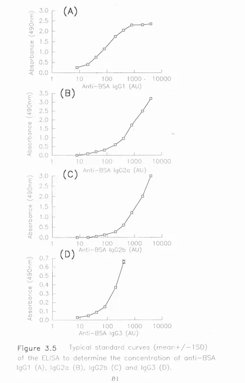
CHAPTER 3 DEVELOPMENT OF ENZYME LINKED 59-81
IMMUNOSORBENT ASSAYS
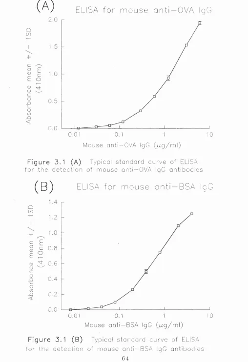
3.1 ELISA for Detection of Mouse Anti-OVA IgG Antibodies 60
3.1.1 Purification of mouse anti-OVA IgG antibodies 60-61
3.1.2 Development of an ELISA for mouse anti-OVA IgG
3.1.3 Comment 62
3.2 ELISA for Detection of Mouse Anti-BSA IgG Antibodies 65
3.2.1 Purification of mouse anti-BSA IgG antibodies 65
3.2.2 Development of an ELISA for mouse anti-BSA IgG
detection 65-66
3.2.3 Comment 66
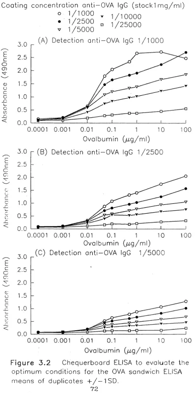
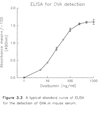
3.3 ELISA for Detection of OVA in Mouse Serum 68
3.3.1 Purification of rabbit anti-OVA IgG antibodies 68-69
3.3.2 Development of a sandwich ELISA for OVA detection 69-70
3.3.3 Comment 70
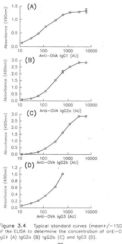
3.4 ELISA for Detection of Anti-OVA IgG Subclasses 74
3.4.1 Development of an ELISA for mouse anti-OVA
IgG subclass detection 74-74
3.4.2 Comment 75
3.5 ELISA for Detection of Anti-BSA IgG Subclasses 78
3.5.1 Development of an ELISA for mouse anti-BSA
IgG subclass detection 78-79
3.5.2 Comment 79
CHAPTER 4 INDUCTION OF ORAL TOLERANCE 82-94
TO OVA AND BSA
4.1 Induction of Oral Tolerance 83
4.1.1 Induction of oral tolerance to OVA in BALB/c mice 83-85
4.1.2 Induction of oral tolerance to BSA in BALB/c mice 85-87
4.1.3 Conclusions 87
4.2 Antigen Specificity of Oral Tolerance 87-88
4.2.1 Antigen specificity of oral tolerance to OVA 88-91
4.2.2 Antigen specificity of oral tolerance to BSA 91-93
CHAPTER 5 SERUM TRANSFER OF GUT PROCESSED 95-119 MATERIAL DERIVED FROM TWO
UNRELATED PROTEIN ANTIGENS
5.1 Ovalbumin 96
5.1.1 Serum transfer of gut processed OVA 97-100
5.1.2 Intestinal absorption of OVA 100-103
5.1.3 Serum transfer of gut processed OVA collected 5,
30 and 60 minutes after feeding. 103-105
5.1.4 Biological activity of OVA filtered through a membrane
(the peritoneum) other than the intestinal epithelium. 105-109
5.1.5 Conclusions 109-111
5.2 Bovine Serum Albumin 111
5.2.1 Serum transfer of gut processed BSA 111-115
5.2.2 Biological activity of BSA filtered through a biological
membrane other than the intestinal epithelium. 115-119
5.2.3 Conclusions 119
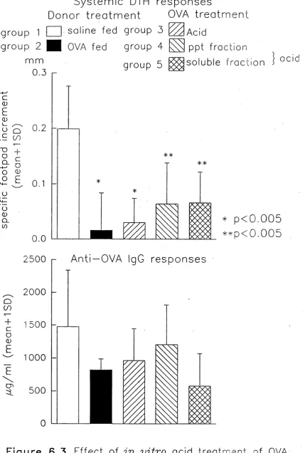
CHAPTER 6 EFFECTS OF IN VITRO DIGESTION 120-137
OF OVA AND BSA ON IMMUNOLOGICAL
ACTIVITY IN VIVO
6.1 Ovalbumin 121
6.1.1 Biological activity of pepsin digested OVA 121-126
6.1.2 Biological activity of acid treated OVA 126-129
6.1.3 Biological activity of heat aggregated OVA 129-130
6.1.4 Conclusions 131
6.2 Bovine Serum Albumin 131-132
6.2.1 Biological activity of pepsin digested BSA 132-134
6.2.2 Biological activity of heat aggregated BSA 135-136
CHAPTER 7 EFFECTS OF THE IMMUNOMODULATOR, 138-146 ISCOM, ON THE PRODUCTION OF A
TRANSFERABLE OVA TOLEROGEN
7.1 Comparison of the Kinetics of Intestinal Uptake of OVA
in Untreated and ISCOM Treated OVA Fed BALB/c Mice 140-142
7.2 Serum Transfer of Gut Processed OVA after ISCOM
Treatment of Donors 142-145
7.3 Conclusions 146
CHAPTER 8 BIOLOGICAL ACTIVITY OF GUT 147-172
PROCESSED OVA AND BSA BY Scid MICE AS
DETERMINED IN BALB/c RECIPIENTS
8.1 Serum Transfer of Gut Processed OVA from Scid Mice to
BALB/c Recipients 149-152
8.1.1 Conclusions 152-153
8.2 Kinetics of Intestinal Absorption of OVA in Scid Mice 153
8.2.1 Concentration of circulating immunoreactive OVA
after feeding OVA to Scid and BALB/c mice 153-155
8.2.2 Serum transfer of OVA dose adjusted Scid serum 155-156
8.2.3 Conclusions 156-158
8.3 Serum Transfer of Tolerance to OVA in Germ Free
BALB/c Mice 158-162
8.3.1 Conclusions 162
8.4 Serum Transfer of Gut Processed BSA from Scid Mice
to BALB/c Recipients 162-165
8.4.1 Conclusions 165
8.5 Histological and Morphological Analysis of the
Scid Mouse Gut 165-166
8.5.1 General morphology in Scid, BALB/c and
germ free BALB/c mice. 166-169
mouse types 169-172
8.5.3 Conclusions 172
CHAPTER 9 OVA TOLEROGEN DEPLETION STUDIES 173-191
9.1 Development of Methodology 175
9.1.1 Preparation of affinity columns 175
9.1.2 Verification of the specificity and capacity
of the affinity columns 175-177
9.1.3 Conclusions 177
9.2 Tolerogen Depletion of Serum from OVA Fed BALB/c Mice 178-183
9.3 Introduction of the Tolerogenic Fraction of Serum from
OVA Fed BALB/c Mice into Naive BALB/c Recipients 183-190
9.4 Summary and General Conclusions 190-191
CHAPTER 10 IDENTIFICATION OF A POTENTIAL 192-202
OVA TOLEROGEN
10.1 Purification of OVA from BALB/c Mouse Serum
Collected 5 and 60 Minutes After Feeding OVA 193-199
10.2 Purification of OVA from Scid Mouse Serum
Collected 60 Minutes After Feeding OVA 200-202
10.3 Conclusions 202
CHAPTER 11 CELLULAR TRANSFER OF ORAL 203-212
TOLERANCE
11.1 Cellular Transfer of Tolerance to OVA 205-207
11.1.1 Conclusions 207
11.2 Antigen Specificity of Tolerance Induced by
Transfer of Splenocytes from OVA Fed Donors 207-208
11.2.1 Conclusions 208-210
11.3 Cellular Transfer of Tolerance to BSA 210-212
CHAPTER 12 GENERAL DISCUSSION 213-240 12.1 Comparison of the Protein Structure of OVA and BSA 215-219
12.1.1 Is the lack of homology between OVA and BSA
responsible for the differing levels of tolerance seen
in mice to these two protein antigens? 220
12.1.2 Are the contrasting immunological outcomes achieved
on serum transfer following different manipulations of OVA and
BSA explained by the dissimilarities between the proteins? 221
12.2 Comparison of the Protein Structure of BSA and mouse serum
albumin (MSA) 222-223
12.2.1 Is BSA presented as a self antigen? 224
12.2.2 Does oral administration of BSA induce
active suppression or merely reactivate existing tolerance
networks specfic for MSA? 224-225
12.2.3 Why is parenteral administration of BSA tolerogenic? 226
12.3 IgG Subclass Regulation in Oral Tolerance to OVA and BSA 227-230
12.4 The Effect of Gastric Digestion of Soluble Protein
Antigens on Oral Tolerance Induction 231-232
12.5 Why Does Mitogenic Stimulation of the Mucosal
Immune System Abrogate the Production of an OVA Tolerogen? 232
12.6 Interpretation of the Function of Specific Immune Cells
(T and B Cells) in the Very Early Stages of Tolerance Induction 233-235
12.7 Tolerogen Depletion and Identification of a Possible
OVA Tolerogen 236-237
12.8 General Conclusions 237-239
12.9 Future Prospects 239-240
References 241-262
CHAPTER 1
GENERAL INTRODUCTION
1.1 Introduction 15
1.2 The Mucosal Immune System 16
1.2.1 Peyer's patches 16-17
1.2.2 Lamina propria 17-18
1.2.3 Intestinal epithelium 18-19
1.2.4 Intra-epithelial lymphocytes 19-22
1.3 Intestinal Permeability to Fed Antigens 22
1.3.1 Experimental evidence 22-24
1.4 O ral Tolerance 24-25
1.4.1 Immunological responses to fed protein antigens 25-27
1.4.2 Mechanisms of tolerance induction and
maintenance 27-30
1.4.3 The role of the gut in tolerance induction 30-34
1.4.4 Cellular control of oral tolerance maintenance 34-37
1.4.5 Genetic basis of oral tolerance 37-38
1.4.6 Consequences of breakdown of oral tolerance 38-39
1.1 Introduction
The gastrointestinal tract encounters large amounts of potentially harmful antigenic
material every day in the form of food proteins, normal gut flora (predominantly
gram negative bacilli) and invading pathogens comprising viruses, bacteria and
multicellular parasites. Despite this constant antigenic bombardment the local and
systemic immune systems maintain a state of immune unresponsiveness to all but
actively pathogenic stimulation. The ability of the mucosal immune system to distin
guish "friend from foe" is not due to proteolytic destruction of the immunogenic
properties of protein antigens in the gut by gastro-intestinal enzymes, since it has
been demonstrated repeatedly that after a feed of the protein antigen OVA to mice,
significant levels of the fed protein can be detected in the circulation (Halsey and
Benjamin 1976, Swarbrick et al 1979, Bruce and Ferguson 1986b, Peng et at 1990). The absorbed protein has been shown to be of similar molecular weight to the intact
protein and retains many of the native antibody epitopes (Bruce and Ferguson
1986b). This suggests that potentially antigenic material is present but that the
immune system is actively silenced (Hanson et at 1977, Swarbrick et al 1979, Bruce and Ferguson 1986a and b, Peng et al 1990).
These observations pose the tantalising question of why continuing systemic immune
u n re sp o n siv e n e ss is o b serv ed in the p re se n c e o f c irc u la tin g , a p p a re n tly
immunocompetent, antigen when the same dose delivered parenterally results in
significant priming of both the humoral and cellular limbs of the immune response
(Golub and Weigel 1969).
It has been hypothesised that this powerful tolerising response, elicited by orally
administered antigen, is present to prevent damaging local hypersensitivity reactions
in the gut against frequently encountered "passive" antigens. Breakdown of this
tolerance could result in a progressive destructive local immune response leading to
1.2 The Mucosal Immune System
The gastrointestinal tract in all mammals is composed of many different cell types
which all contribute to the efficient functioning of the mucosal immune system
(Brandtzaeg et al 1988). The alimentary tract acts as a physical barrier against invad ing pathogens as well as an excellent sampling organ for exogenous antigens
allowing subsequent presentation to the underlying immune organs of Peyer's patch
and mesenteric lymphnodes. The epithelium maintains constant cell to cell contact
with the intraepithélial lymphocytes and lymphoid cells of the lamina propria thereby
enabling constant immune surveillance of the antigens encountered in the gut lumen.
As a result of this close relationship between different cell types it is possible to
regard the mucosal immune system as comprising two parts:
(i) the professional lymphoid tissues of Peyer's patch, lamina propria and intra
epithelial lymphocytes (lEL) and (ii), the non professional gut epithelial cells or
enterocytes.
1.2.1 Peyer's patches
These lymphoid nodules, first described by Peyer in 1667, are the main organised
lymphoid tissue of the intestinal mucosa. They appear as lymphoid aggregates
arrayed along the small intestine like a "string of pearls". Peyer's patches are similar
to other peripheral lymphoid tissues such as the spleen and lymph nodes since they
are arranged into T and B cell dependent areas (Henry et al 1970). Each Peyer's patch in mice and rats is composed of 3 to 5 follicles separated from one another by
interfollicular areas (Owen and Jones 1974). The follicles are the B cell dependent
areas (50% of which have surface IgA) with a sparse scattering of T helper cells.
The interfollicular areas contain the majority of the T helper cells and a few
Cytotoxic T lymphocytes (CTL) (Ermak and Owen 1986).
Cells enter the Peyer's patches via the Peyer's patch high endothelial venules, this
be mediated via complementary receptors on recirculating lymphocytes and on the
organ's high endothelial venules. In the case of recirculation to the Peyer's patches in
the mouse, the adhesion molecule LPAM-1 is expressed on lymphocytes destined for
the Peyer's patches (in humans this may be regulated by VLA-4 on lymphocytes).
The LPAM-1 may interact with an organ specific molecule expressed on Peyer's
patch high endothelial venules thereby allowing selection of cells destined for the
mucosa (Holzman et al 1989). Antigen enters the patch via specialised membranous (M) cells which overlay the follicles and face into the lumen of the gut. These M
cells are scattered throughout a specialised epithelium, the dome epithelium, which
covers the lymphoid aggregate and is composed of these special M cells arrayed
between normal columnar absorptive epithelial cells, or enterocytes (Owen and Jones
1974). Unlike the columnar epithelial cells of the small intestine which have multiple
microvilli on their lumenal surface M cells are covered in microfolds which are
capable of specialised endocytosis of bacterial and macromolecules via clathrin
coated pits and vesicles (Owen 1977, Neutra et al 1987).
After antigen has entered the Peyer's patch through the M cells it first encounters
large mononuclear la positive veiled cells, with dendritic cell characteristics (Wilders
et al 1983, Mayrhofer et al 1983, Barr et al 1985) and macrophages (Owen et al
1980, Hume et al 1987). These cells are probably responsible for antigen processing and presentation to the T cells situated in the interfollicular areas of the patch
thereby initiating a specific immune response to intestinally derived antigen. It has
been postulated that the Peyer's patch is the site of initiation and regulation of specif
ic immune responses in the intestine (Kiyono et al 1982, Mayrhofer et al 1983).
1.2.2 Lamina propria
The intestinal lamina propria is situated between the epithelium and the muscularis
numbers of IgA producing plasma cells (85%) which may originate from the Peyer's
patches (Craig and Cebra 1971, Cebra et al 1977, Bienenstock et al 1974) or mesenteric lymph nodes (Guy-Grand et al 1974) or both (James et al 1990) the remainder of the B cells contained in the lamina propria are IgM (10%) and IgG
(5%) positive (Ernst et al 1987).
Specific and non specific immune cells of all types including T cells, macrophages,
mast cells, eosinophils and polymorphonuclear cells are found interspersed between
the plasma cells (Bartnik et al 1980, Davies and Parrott 1981). The T cells observed are mostly of the T helper phenotype (CD44-) which may be involved in activation
and control of the large numbers of B and plasma cells (Elson et al 1985). A much smaller proportion of cytotoxic lymphocytes (CD8+) are observed which may be
involved in pathogen control (particularly enteric viruses) at the mucosal surfaces
(Cerf-Bensussan et al 1983, James and Graeff 1987, Ruthlein and Heinze 1991). The macrophages observed in the lamina propria are MHC class II positive with no
dendritic or veiled cell characteristics. A large proportion are situated at the villus
tip (Hume 1985) where their prim ary function appears to be phagocytosis of
invading pathogens (Kujawa et al 1977, Muigai et al 1983).
1.2.3 Intestinal epithelium
The mucosal epithelium consists of large columnar epithelial cells (enterocytes), a
major function of these cells is to transport food molecules from the gut lumen to
underlying tissue for transport around the body. These cells have a specially adapted
brush border of microvilli facing into the lumen of the gut, thereby dramatically
increasing the absorptive capacity of each epithelial cell. These cells are derived
from rapidly dividing precursors situated in the crypts of Lieberkiihn at the foot of
the villus. The newly synthesised immature epithelial cells join the base of the villus
and mature as they ascend towards the villus tip. The length of the villus is con
trolled by the rate of division of the precursors in the crypt and by the mechanical
time for these cells is normally 2-3 days (Bland and Warren 1986a). Careful control
of epithelial cell turnover is necessary to maintain the integrity of the intestinal
epithelial barrier, the rate of turnover can increase during certain immunological
stimuli (Mason et al 1981, Barnard et al 1989). Interspersed between the epithelial cells are goblet cells which are also derived from the crypts; these cells provide the
mucus which coats the brush border of the epithelial cells and acts as an additional
barrier against pathogen penetration of underlying tissues.
It is obvious that the epithelial cells provide a physical barrier to pathogen invasion
of the mucosae. However, recent research has provided evidence for a more direct
role of these cells in the functioning and control of the mucosal immune system
(Geppert and Lipsky 1985, Bland and Warren 1986b). These cells have been shown
to express class II MHC in humans (Scott et al 1980, M ayer and Shlien 1987, Harvey et al 1990), rats (Bland and Warren 1986a), mice (Natalli et al 1981, Parr and MacKenzie 1979) and guinea pigs (Wiman et al 1979). In rats the top 2/3 of the intestinal villi are positive for class II MHC expression, this expression pattern may
reflect maturation of these cells as they move up the villi (Bland 1988). Expression
of class II MHC can be increased by various intestinal inflammatory responses and
is now thought to be mediated in rats and humans by tIFN (Barclay and Mason
1982, , Steiniger et al 1989, Heath et al 1991). The importance of this expression for antigen presentation to, and control of, the under
lying mucosal immune system is not fully understood and will be discussed later.
1.2.4 Intra-epithelial lymphocytes
95-98% of intraepithélial lymphocytes (lEL) are located on the basolateral enterocyte
membrane (Darlington and Rodgers 1966). They appear to be completely separated
from the epithelial cells with no desmosome or adhesion structures observed between
lEL and enterocyte (Ferguson 1977). The majority of lEL's are thymus dependent
and Scid mice (Ropke and Everett 1976, Fichtelius et al 1968, Croitoru et al 1990). The numbers of lEL in thymectomised and athymic mice can be increased, but not
to normal levels, on the introduction of gut flora (Fichtelius et al 1968, Ropke and Everett 1976). Specific homing of lEL to the epithelium appears to occur from the
mesenteric lymph nodes but not from other peripheral lymph nodes (Parrot and
Ferguson 1974, Guy-Grand et al 1974, Ferguson 1977) probably by a mechanism similar to that described for Peyer's patch lymphoid cells (see section 1.2.1).
Phenotypically lEL's are composed of T cells and up to 50% non specific granular
nul cells (Mowat 1983). The T cell compartment has a reversed ratio of C D 4+ to
CD8-I- cells (Klein 1986) compared with the lamina propria and, depending on the
species studied can have up to 70% of T cells with a granulated phenotype (Strobel
1990). Also, depending on the age and species studied the proportion of aBTcR to
tôTcR can vary dramatically (Bonneville et al 1988, Goodman and Lefrancois 1988, Lefrancois and Goodman 1989, Viney et al 1989, De Gens et al 1990, Viney et al
1990, Guy-Grande et al 1991a). Murine lE L 's are 80-90% CD84- (the classic cytotoxic lymphocyte phenotype) and can be either CD8 oilot^ i.e. homodimeric or they express the more usual heterodimeric CD8 a/6''' molecule (Maloy et al 1991) while the remainder are C D 4+ i.e. T helper cells or express natural killer cell
markers. Only 50% of lEL are Thyl 4- (Klein 1986, Ernst et al 1985, Petit and Ernst 1985) and these appear to be primarily the a6 T cell receptor expressors,
Thyl- lEL are predominately tÔ T cell receptor users (Viney and MacDonald 1992).
Recent evidence suggests that the lEL compartment may be a major extrathymic site
of T cell differentiation in mice (Mosley et al 1990, Viney and MacDonald 1990, Guy-Grand et al 1991a, Lefrancois 1991, Rocha et al 1991) and humans (Van Kerckhove et al 1992). The use of radiation chimeras in mice have demonstrated that T cells appear in the gut two weeks before they can be shown in the thymus and
spleen (Mosley and Klein 1992). The first cells to appear in the gut are tÔ T cells,
indicating oligoclonal T cell expansion in mice (Rocha et al 1991) and humans (Balk
et at 1991, Van Kerckhove et al 1992). The restricted gene usage is probably due to the limited number of antigens available in the foetal gut therefore limited antigen
dependent selection occurs. Furthermore these T cell appear not to have undergone
negative selection (normally occurs in the thymus) for self reactivity and can be
shown to express V13 genes which have been deleted in T cells of the periphery due
to their reactivity to self antigens (Murosaki et al 1991, Nagler-Anderson et al 1992, Poussier et al 1992, Rocha et al 1992b) i.e. T cells which have undergone conventional thymic education (Kappler et al 1987).
Functionally mammalian lEL have been shown to respond to T cell mitogens (Dillon
and MacDonald 1986, Dillon et al 1986, Wilson et al 1986, Mowat et al 1989, Viney and MacDonald 1992), anti CD2 antibodies (Ebert 1989), anti CD3 antibodies
(Mosley et al 1991, Viney and MacDonald 1992), allogeneic cells (Mowat 1986b), enteric viruses (Godson et al 1992) and m ulticellular parasites (Ebert 1989) displaying limited antigen specific (perhaps only to super antigens) (Dillon et al
1986, Godson et al 1992, Viney and MacDonald 1992) and antigen non specific cytotoxicity (Klein et al 1985, Klein and Kagnoff 1987, Lefrancois and Goodman 1989, Guy-Grand et al 1991b). They produce a variety of lymphokines including IL- 2, IL-3, IL-5, IL-6, interferon (IFN), mast cell growth factor (MCGF) and colony
stimulating factor (CSF) (Inle and Keller 1983, Cerf-Bennsussan 1984, Dillon et al
1986, Kiyono et al 1991, Taguchi et al 1991, B arrett et al 1992, Viney and MacDonald 1992). Their responses in vitro have shown dramatic differences de pending on the population of lEL studied. Thymodependent lEL (ail T cell receptor,
Thyl 4-) give very good responses in vitro whereas thymoindependent lEL (rô T cell receptor T hyl-) give very poor responses and appear to be inactive or anergic
Observations of the phenotype and functional properties of lELshave resulted in two
opposing hypotheses on the activation state of lEL's in vivo. It has been proposed that these cells are either fully activated in a situation o f constant antigenic
stimulation, explaining why in vitro they are poor responders to stimulation (Klein et al 1985, Bacca et al 1987, Mowat et al 1989), or that they are anergic and help to maintain tolerance to non pathogenic antigenic m aterial at mucosal surfaces
(Goodman and Lefrancois 1989, Mosley et al 1991). However it now appears that lELs are a heterogeneous population of lymphocytes which may have many different
roles in the mucosal immune system of both defence and immunoregulation.
1.3 Intestinal Permeability to Fed Antigens
Minute quantities of antigenic dietary proteins can be detected in the circulation of
both animals and humans after feeding (Gardner 1984). The uptake of these antigens
can be enhanced during intestinal immaturity and inflammation in mice (Hanson
1981), guinea-pig (Telemo et al 1987) and man (Jakobsson et al 1986, Jakobsson 1988). Transport of antigen from the lumen of the gut into the systemic circulation
can occur by three main routes, (i) transport through the specialised M cells of the
Peyer's patch (Owen 1977), (ii) active uptake and transport across columnar epithe
lial cells of the small intestine (Tagesson and Sjodahl 1984) and (iii) passive diffu
sion of molecules across the tight junctions (intracellular desmosomes) between the
epithelial cells of the intestine (Gardner 1984). Experimental evidence is available
for the utilisation of all these routes in animal models and some human studies.
1.3.1 Experimental evidence of gut permeability
Initial studies demonstrated that blood and urine collected from experimental animals
after feeding BSA or egg proteins was able to induce anaphylaxis in previously
sensitised animals (Schloss and Worthen 1916, Wilson and Walzer 1935), thereby
circulation after feeding.
The amount of each antigen which is detected in the circulation after feeding varies
widely for each antigen e.g. rats fed BSA have been reported to have between 2%
(Warshaw et al 1974) and 0.0001% (Peng 1989) of the total fed antigen in their circulation, similarly reported circulating OVA levels have ranged between 0.002%
(Swarbrick et al 1979) and 0.000125% (Peng et al 1990) of the total fed antigen. This apparent discrepancy in values can be explained by the timepoint of collection
after feeding since each antigen appears to have an optimum time for maximum
absorption (Peng 1989) and also the method for detection of antigen can dramatically
influence the concentration obtained.
The induction of intestinal anaphylaxis by administration of chemicals or parasitic
infections to mice or rats results in an increase in the absorption of intact dietary
protein (Reinhardt 1984). This increase in absorption of intact antigen is thought to
be due to an increase in intestinal permeability caused by the inflammatory response,
thereby allowing diffusion of high molecular weight molecules through the tight
junctions between the epithelial cells. Further evidence for this has been supplied by
the electron microscope studies of Rhodes and Kamovsky (1971) who reported that
after surgical trauma to the small intestine of guinea pigs HRP could be observed
diffusing intercellularly through the tight junctions but could not be demonstrated in
epithelial cells. Kleinman et al (1981) reported that intestinal anaphylaxis can lead to increased uptake of non-specific "bystander" intestinal antigens which can evoke an
IgG mediated reaction and therefore perpetuate intestinal inflammation leading to
chronic disease. Furthermore it was reported by Lens et al (1984) that arthritis mice fed collagen experienced a "flare up" of their arthritic condition suggesting
that immunoreactive collagen reached their arthritic joints after feeding. Recent
indirect evidence for this mechanism of transport of intact antigen in normal gut was
supplied by the observations of Peng (1989) who showed that intact OVA and BSA
could be demonstrated in the circulation of mice just 5 minutes after feeding, since 5
immunological effect of rapidly diffusing intact dietary antigen on the local and
systemic immune system is unclear.
Conventional antigen transport via absorptive epithelial cells and specialised M cells
has been reported for many different antigens (Owen 1977, Ducroc et al 1983, Neutra et al 1982). However the efficiency of these cells to transport specific anti gens appears to differ depending on the antigen measured. These differences may
reflect the immunological outcome of antigen entering the Peyer's patch via M cells
compared to antigen entering the lamina propria or intra-epithelial lymphocytes via
columnar epithelial cells or may simply reflect the ease of breakdown of each anti
gen by the gastrointestinal enzymes.
1.4 Oral Tolerance
Oral tolerance is defined as specific systemic immunologic hyporesponsiveness after
oral administration of that particular antigen and was first described by Dakin in
1829. He observed that South American indians ate poison ivy leaves to prevent
subsequent contact sensitivity reactions. This phenomenon appears to have been
ignored by the scientific community until the beginning of the 20^ century. In 1909
Bresredka described tolerance to milk protein on feeding to guinea pigs. Shortly
afterwards in 1911 Wells demonstrated that zein, the major protein component of
maize, when fed to guinea pigs prevented subsequent anaphylactic shock on parent
eral challenge. These observations were confirmed by Chase in 1946 using contact
sensitising agents to first establish the immunological basis of the response which
became known as the Sulzberger - Chase phenomenon.
In recent years the rise of interest in oral tolerance has been helped by the relative
ease of induction and measurement of oral tolerance in almost any laboratory animal
using a variety of antigens, particularly soluble protein antigens. This availability of
material encourages the use of this system for the study of fundamental mechanisms
in the immune response and immunoregulation and also for clinical application of
1.4.1 Immunological responses to fed protein antigens
All antigens capable of inducing tolerance on oral adm inistration are thymus
dependent antigens (Titus and Chiller 1981a), thymus independent antigens given
orally are likely to prime the immune system (Titus and Chiller 1981a). Tolerance
induction has been demonstrated using a wide variety of thymus-dependent antigens
with soluble proteins such as OVA and BSA being the antigens of preference since
they are readily available in the laboratory and experimental animals are likely to be
immunologically naive to them (Thomas and Parrott 1974 and reviewed in Mowat
1987). Other antigens used include heterologous RBC (André et al 1975, Kagnoff 1978 a and b, Kiyono et al 1982), contact sensitising agents (Asherson 1979, Newby
et al 1980, Gautam and Battisto 1985) and inactivated viruses and bacteria (Stokes et al 1979, Rubin et al 1981). This second group of antigens provides conflicting data which may help in elucidating the immunological basis of this phenomenon. When
live allogeneic cells or viruses are fed they prime both the local and systemic
immune systems mimicking the protective role of the local immune response on
pathogenic challenge (Rubin et al 1981, Klein and Kagnoff 1984).
The dose and frequency of administration of antigen also appears to be important to
the level of the tolerance induction. In mice and rats a single feed of a soluble
protein antigen in milligram quantities induces tolerance whereas a single microgram
dose of antigen is more likely to prime the animal (Jarrett and Haig 1976, Mowat
1987, Lamont et al 1989, Peng et al 1989a). However if the smaller priming dose is fed repeatedly over a short period of time priming does not occur and a strong
tolerogenic response is initiated (Peng et al 1989a). Furtherm ore it has been reported that with an increase in the frequency of oral exposure to antigen the
resulting tolerance becomes more marked and longer lasting (David 1979 and Peng
and Menamin 1989).
Oral tolerance to a soluble protein antigen affects all arms of the systemic immune
response in mice and rats. Suppression of antigen specific IgG and IgE antibodies
(Jarrett and Haig 1976, Vaz and Mara 1977, Ngan and Kind 1978, Rich man et al
1978, Swarbrick et al 1979, Challacombe and Tomasi 1980, Titus and Chiller 1981a, Mowat et al 1982) and marked tolerance of cell mediated immunity both in vitro, by cell proliferation assays, and measurement of in vivo delayed type hyper sensitivity reactions (DTK) (Miller et al 1979, Challacombe and Tomasi 1980, Titus and Chiller 1981a and b, Mowat et al 1982) have all been demonstrated. Conflicting reports exist on responses against the administered antigen by the local immune
system during tolerance induction. Early studies demonstrated induction of a specific
local immune response together with systemic hyporesponsiveness of both an
antibody and cellular nature (Challacombe and Tomasi 1980). However this system
used inactivated bacteria as antigen, which is probably not an ideal model for dietary
antigen and more closely resembles the presentation of a pathogenic antigen to the
local immune system. In more recent studies using soluble protein antigens both the
local and systemic immune responses were tolerised (Saklayen et al 1984).
The level and nature of the suppression achieved by oral administration of antigen is
heavily dependent on the param eters assessed. When antibody responses are
measured the IgE response appears to be the most sensitive to tolerance induction
and often no antigen specific IgE response can be detected locally or systemically
(Ngan and Kind 1978). Specific IgG responses can be detected but are significantly
lower than in parenterally primed animals; this tolerance lasts up to 3 months after
the initial feed (Strobel and Ferguson 1987). The cellular immune response assessed
by in vivo DTH responses to specific antigen challenge is profoundly suppressed for up to 17 months after the initial antigen exposure (Strobel and Ferguson 1987).
The observation that the IgE and DTH responses are profoundly tolerised after oral
administration of antigen when compared to IgG responses (Ngan and Kind 1978,
could suggests a possible function for oral tolerance. It would be advantageous for
the efficient functioning of the intestinal epithelium that the mucosal immune system
was not being continually activated by passive food antigens thereby causing local
hypersensitivity reactions, of which IgE and DTH responses are the principle media
tors of disruption of the mucosal architecture.
1.4.2 Mechanisms of tolerance induction and maintenance
The phenotype of any immunologically tolerant state results from an interaction of
many different immunoregulatory mechanisms and is therefore very difficult to
dissect. The situation is further complicated in this system because the nature of the
antigen used radically determines the immunological outcome on oral immunisation
(Titus and Chiller 1981a). As a result of this difficulty the research discussed will be
based on soluble protein antigens unless specifically stated.
To enable an antigen specific immune system to operate efficiently it is essential that
the immune response distinguishes self from nonself antigens and reacts accordingly.
The phenomenon of self tolerance is initiated when the immature T cell pool has
migrated to the thymus (Kisielow et al 1988). Two mechanisms have been proposed and experimentally demonstrated
1. Clonal deletion i.e. death of self reactive clones (Kappler et al 1987, Sha et al
1988)
2. Clonal anergy i.e. induced unresponsiveness of specific clones (De Franco 1989,
Goodnow et al 1989, Nossal 1989)
These processes and the experiments carried out to demonstrate self tolerance may
also be applicable to peripheral tolerance.
Primary self tolerance has been shown to occur in the thymus by a process of
negative selection. Immature T cells migrate from the bone marrow and /or foetal
liver to the thymus where they are first positively selected for recognition of self
The positively selected cells are then negatively selected on the basis of reactivity to
self antigens i.e. all T cell clones which react to self antigens presented by self MHC
are destroyed by apoptosis and therefore self tolerance is controlled initially by
clonal deletion of self reactive clones. However it would be realistically impossible
to imagine that tolerance to all self proteins, especially those with very restricted
tissue distribution, could occur in the local environment of the thymus. One might
envisage that incomplete tolerance to self proteins would lead to major autoimmune
disease in a large proportion of individuals.
Autoimmunity in children and adults is uncommon therefore T cell tolerance induc
tion must occur in the mature peripheral T cell pool after exiting from the thymus.
Naive T cells must be tolerised in the periphery to local self antigens with constant
reinforcement of that tolerance throughout the life of that T cell population therefore
memory T cells must continue to be actively suppressed or rendered anergic. Since
self reactive clones can be induced in vivo and in vitro by various mechanisms, it would appear that peripheral tolerance may be controlled through clonal anergy or
perhaps by active suppression. These observations reinforce the hypothesis that a
mechanism or mechanisms exist whereby external antigen presented to the peripheral
immune system, perhaps by "specialised" antigen presenting cells in a certain envi
ronment could allow tolerance rather than stimulation of the immune response
(Nossal 1989).
The question of B cell tolerance can be side stepped due to the requirement of T cell
help for a B cell response when most antigens are encountered (DeFranco 1987).
This appears to be true of oral tolerance since T dependent antigens are the only
effective tolerising antigens therefore T cell suppression would automatically prevent
a conventional B cell response by preventing T cell help (Titus and Chiller 1981a).
On oral administration of a hapten carrier conjugate the resulting tolerance is always
peripheral challenge with that antigen (Titus and Chiller 1981a). This is due to the
LPS, a mitogenic stim ulant for B cells, circum venting the T cell mediated
suppression. Although both the cellular and hum oral immune response are
su p p re sse d , o ral to le ra n c e ap p ea rs to be m ed iated by a sta te o f T cell
unresponsiveness with the B cell com partm ent rem aining potentially reactive
(discussed in section 1.4.4.). This observation is challenged by one report where
both active B and T cell tolerance has been demonstrated, using deaggregated human
gamma globulin (HGG) as the antigen (Vives et al 1980). This effect may be due to direct interaction of HGG with the Fc receptors on B cells. But recent observations
in another peripheral tolerance model have demonstrated that mature peripheral B
cells can become actively tolerised in the absence of specific T cells. This results in
a reduction of mIgM while maintaining levels of mIgD characteristic of a typical B
cell memory response (Goodman and Lefrancois 1988).
Mature naive helper T cells can become tolerised in the periphery as demonstrated
by oral tolerance where the T cell mediated phenomenon of antigen specific DTH,
IgG and IgE responses are suppressed. This tolerance decreases with time after
exposure to antigen (Strobel and Ferguson 1987) and if splenocytes are stimulated in vitro with a T cell mitogen antigen specific T cell activity can be detected (Richman
et al 1978, Miller and Hanson 1979). Therefore the mechanism of tolerance in this case is unlikely to be clonal deletion and specific clonal anergy is the more likely
mechanism. Induced T cell unresponsiveness in other peripheral tolerance systems as
well as oral tolerance seems to manifest itself in a T cell pool which is unable to
proliferate in vitro (Miller and Hanson 1979) or secrete IL-2 (Rammensee et al
1989) in response to antigen but on cell transfer to naive recipients can actively
recognise antigen and induce tolerance in the recipients (Miller and Hanson 1979).
The proposed mechanism for the maintenance of peripheral T cell anergy is a two hit
theory mediated by the antigen presenting cell (APC). T cells require two separate
signals from the APC for activation to occur with the second signal being the
State o f anergy is induced.
But what determines which signal the APC delivers? It has been suggested that this
is perhaps mediated by so called " non professional" APCs (Nossal 1989) which can
be induced to express class II Major Histocompatibility complex (MHC) and
therefore present antigen to T helper cells, but are subsequently unable to provide
the second signal for activation. The relevance of this observation to oral tolerance is
important and will be discussed later.
In oral tolerance the immunological status of the antigen presenting cell pool has
been shown to be influential in the induction of tolerance. Through modulation of
APC by oestradiol, which was thought to induce APC to present antigens enabling
activation of the specific T cell clones which were normally suppressed (Mowat and
Parrott 1983). More importantly there appears to be an active antigen specific T cell
compartment which can control tolerance induction and maintenance (Mowat 1986a,
Lamont et al 1988a).
1.4.3 The role of the gut in oral tolerance induction
The gut handles large amounts of potentially immunogenic material before it reaches
the peripheral immune system. The degradative and transport pathways of the
gastrointestinal tract have been implicated in the induction of oral tolerance through
utilisation of these processes by the local immune system.
Two mechanisms by which the gut could influence tolerance induction are:
1. Digestion of antigen in the lumen of the gut thereby permitting tolerogenic
epitopes to become exposed and subsequently presented to the local immune system
(Hanson and Morimoto 1980, Parks and Weigle 1980, Strobel et al 1983, Bruce and Ferguson 1986a and b).
2. Active antigen uptake, processing and presentation to the local immune system
1987).
Evidence fo r hypothesis L
It has been demonstrated that transfer of tolerance can occur if serum from OVA fed
mice is transferred into naive recipients one hour after a feed (Strobel et al 1983). This tolerance appears to correspond with the presence of OVA as demonstrated by
Bruce and Ferguson (1986b) who showed, that when OVA was removed from
"tolerogenic" serum by affinity chromatography the ability of the OVA depleted
serum to induce oral tolerance was abrogated. Further support for this hypothesis is
provided by the observation of Hanson et al (1979b) who injected mice with mouse anti-OVA antibodies during feeding with OVA. This resulted in an intermediate
tolerant state suggesting some loss of tolerance induction following removal of OVA
from the circulation of the immunised mice by immune complexing of antigen by
injected antibody.
The suppression described above is not observed if serum is transferred 5 minutes
after a feed even though measurable levels of OVA are present (Peng et al 1990). Furtherm ore if serum obtained 5 minutes after feeding is concentrated so that it
contains an equivalent dose of antigen as serum obtained 60 minutes after feeding
(i.e. tolerogenic serum) tolerance is still not transferred (Peng et al 1990). If the same dose of antigen is administeredparenterallyand allowed to circulate for one hour
in the donor before transfer of serum to the recipients, there is no subsequent
tolerance induction (Bruce and Ferguson 1986a). Therefore there appears to be a
qualitative difference in the nature of the antigen present which enables it to act as a
tolerogen rather than an immunogen, this qualitative difference in circulating antigen
appears to be unique to antigen which has entered the circulation via the gut. The
serum transfer model induces tolerance of DTH but not of antibody suggesting some
divergence in the mechanisms and thresholds of tolerance induction for different
treatment of mice with trasylol, a trypsin and chymotrypsin inhibitor, abolished the
induction of oral tolerance (Hanson and Morimoto 1980). It was suggested that this
may have been due to decreased production of an altered form of antigen or, that a
swamping effect occurred in the local immune system due to increased uptake of
intact protein from the gut (Hanson and Morimoto 1980). It has also been observed
that direct injection of antigen into the lumen of the lower ileum was two fold less
efficient at inducing tolerance than gastric or duodenal injection suggesting that
enzymes present in the small intestine were essential for the production of an OVA
tolerogen (Hanson and Morimoto 1980). However this finding is contested by the
reports of Strobel (1984) and Nagatomi et al (1980) who both reported that antigen administered rectally could also induce a tolerant state indistinguishable to oral
tolerance.
Another commonly used antigen in the induction of oral tolerance is BSA. It has
been shown that proteolytic digestion of BSA leads to production of tolerogenic
fragments (Muckerheide et al 1977, Dosa et al 1979, Ferguson et al 1983c) and that these fragments bind to specific T suppressor cells from orally tolerised mice (Zhang
et al 1987). These results appear to indicate that digestion of protein by the gut may play a significant contributory role in the suppression of immune responses following
oral administration of an antigen. However care should be taken not to extrapolate
from the results obtained with one particular protein antigen to provide a general
hypothesis since the effect of gastrointestinal modification of antigens may vary
dramatically depending on which antigen is used.
Evidence fo r hypothesis 2.
One function of intestinal epithelial cells is to transport food antigens, after digestion
in the lumen of the gut, from their luminal surfaces to the basal membrane and
consequently into the circulation for distribution around the body. They could,
therefore, be involved in selective antigen handling and presentation to the immune
system. The observation that intestinal epithelial cells constitutively express class II
Scott et al 1980, Mason et al 1981) suggested a role in antigen presentation (Geppert and Lipsky 1985) to the underlying lymphocytes of the lamina propria and lEL
perhaps to specifically induce oral tolerance. There is some evidence of intracellular
accumulation within the vesicular endosome compartment suggesting uptake of
antigen (Mayrhofer et al 1983). Recycling of the receptor has not been observed but the restricted surface distribution would only allow release of the ligand in an
endosome basolateral membrane direction (Bland 1988). All these observations
would suggest that intestinal epithelial cells could be efficient antigen presenters and
have been demonstrated to present soluble protein antigens in vitro to T cells and may selectively induce suppressor T cells (Bland and Warren 1986b, Bland and
Whiting 1989).
The level of expression is increased in association with inflammatory responses in
the mucosae (Mason et al 1981) probably as a result of rIFN production by lEL (Kiyono et al 1991, Taguchi et al 1991) or lamina propria lymphocytes (Barclay and Mason 1982) . Expression of Class II antigen is absent throughout fetal development
in the rodent and becomes detectable at various times after birth depending on the
species studied. The rat does not express class II on its enterocytes until weaning
(Bland and Warren 1986a) whereas mice express such antigens after the first week
of life (Natalli et al 1981).
There are many reasons why enterocytes could be involved in immune regulation in
the mucosal immune system, e.g.;
(1) Their position at the interface between the outside environment and cells of
mucosal immune system.
(2) Close spatial relationship with lEL and lamina propria lymphoid cells (Brandt-
zaeg et al 1988).
(3) They harbour terminal peptidases in the microvilli of their luminal surface.
It is unclear whether antigen undergoes processing while being transported across
the enterocyte (Bland 1988). However such cells are able in vitro to present antigen to specific T cells (Bland and Warren 1986a) and this presentation can be blocked by
anti I-A and I-E antibodies (Bland and Warren 1986a). Furthermore it has been
reported that enterocytes selectively stimulate functional T suppressor cells (Bland
and Warren 1986b). However for presentation to T cells most protein antigens
require processing to small oligopeptides so that they can be accommodated in the
peptide groove of the MHC molecule (Allen 1987). Therefore the antigens which
enterocytes are transporting and presenting via class II must be small fragments,
perhaps digested by the term inal peptidases present on their lum inal surface.
However, the serum transfer model of oral tolerance requires relatively large frag
ments of OVA, which still retain conformational epitopes recognisable by polyclonal
anti-OVA antibodies (Strobel et al 1983). This would not correspond with the conventional I oligopeptides: required for antigen presentation by Class II (Allen 1987).
1.4.4 Cellular control of tolerance maintenance
Active immunological suppression was first established in 1946 by Chase using
guinea pigs fed contact sensitising agents. Not until 1976 was it determined that T
cells were responsible for this suppression, using adoptive transfer studies of
splenocytes from bovine serum albumin (BSA) fed rats (Thomas and Ryan 1976). It
was also observed that this tolerance could not be abolished on administration of
naive spleen cells (Richman et al 1978). Subsequent research attempted to establish the phenotype of these cells. It was repeatedly shown that this adoptive transfer of
suppression could be abolished by prior treatment of tolerised splenocytes in vitro
with anti Thy-1.2 antibody and complement (Ngan and Kind 1978, Richman et al
1978, M iller and Hanson 1979, Mowat 1985 and Kay and Ferguson 1989).
Thymectomy was also shown to comprom ise the induction o f oral tolerance
from tolerant donors by prior passage over nylon wool had no effect on subsequent
transfer of tolerance (Richman et al 1978). This verified the hypothesis that T cells were important in oral tolerance induction and maintenance. However, adoptive
transfer of cells appeared only to transfer tolerance of cellular immunity when
soluble protein antigens were used as tolerogens (Mowat 1985), a finding which
corresponds with serum transfer of oral tolerance which also only transfers DTH
tolerance into naive recipients (Strobel et at 1983).
It has been established that this tolerant state is acutely radiosensitive and tolerance
is not maintained in irradiated animals when reconstituted with naive cells (Hanson
et al 1979a). Similarly treatment with cyclophosphamide (CY) (Mowat and Ferguson 1981, Mowat et al 1982, Mowat and Ferguson 1982, Strobel et al 1983) and 2'deoxyguanosine (Mowat 1986a), proposed inhibitors of T cells with suppressor
activity, have been shown to abolish oral tolerance of DTH but have very little effect
on antibody tolerance. However, these results require careful interpretation since the
gastrointestinal tract is very susceptible to damage by radiation and chemicals due to
its high cell turnover (Mowat 1986a).
In 1982 Green et al demonstrated that complement lysis of CD8 positive cells in vitro abrogated transfer of oral tolerance, an effect which was attributed to the presence of classical T suppressor cells. Subsequent research attempted to establish
an active suppressor cell network similar to that reported in other peripheral toler
ance models. Using sRBC as an antigen it was suggested that the presence of T
contrasuppressor cells in the mucosal microenvironment permitted induction of the
local immune response while Peyers patch T suppressor cells maintained systemic
unresponsiveness (Gershon et al 1981, Green et al 1982, Taylor 1986). The apparent absence of T suppressor cells in the Peyers patch of mutant LPS unresponsive mice
was proposed as an explanation of their failure to become orally tolerised using
sRBC as the antigen (Wannemeulher et al 1986). However oral tolerance induced using soluble protein antigens e.g. OVA fed to LPS unresponsive mice produced
1986c).
On the basis of this conflicting data it appears that particulate antigens such as sRBC
are perhaps not suitable antigens for demonstrating typical oral tolerance to common
food antigens. The mechanisms which maintain tolerance towards normal gut flora
and multi-antigenic particulate antigens (e.g. sRBC) may be very different to the
processes which maintain systemic tolerance to soluble protein antigens.
Hanson et al (1979b) demonstrated that splenectomy before the oral administration of antigen had no effect on the level of tolerance subsequently induced. However,
splenocytes transfer specific tolerance of cellular immunity very efficiently (Parks
and Weigle 1980, M iller and Hanson 1979, Richman et al 1978). These two observations can be explained by the migration of tolerising cells from the mucosal
immune tissues to the spleen after oral administration of antigen. If this is the case it
suggests that the induction of T suppressor cells occurs in the local environment of
the gut and then migrate to the periphery. This hypothesis is confirmed by the
observation that transfer of tolerance in mice by spleen cells is dependent on the time
of transfer after feeding the donor animal. Spleen cells taken earlier than day 7 after
feeding are unable to transfer tolerance although mesenteric lymph node cells can
transfer tolerance only 3 days after a feed (Miller and Hanson 1979). From this data
it would appear that these T suppressor cells are induced in the mucosal immune
system and subsequently migrate to the periphery via the mesenteric lymph nodes
into the spleen.
Although it has been repeatedly stated that T cells are a prerequisite for the induction
of oral tolerance, other immune cells and their status may also play a contributing
role in the level of tolerance induced and the length of time that tolerance is main
tained.
Treatment of mice with oestradiol abrogated oral tolerance induction with no
demonstatable effect on T cells (Mowat and Parrott 1983). This was thought to be
due to the activation of specialised antigen processing cells able to present an
immunogenic epitope of the antigen rather than a tolerogenic epitope to T cells. This