ABSTRACT
STERN, RACHEL ALLYSA. The Effect of Hyperammonemia on Myogenesis in Broiler Embryos. (Under the direction of Paul Mozdziak).
The embryonic environment and regulation of myogenesis is of particular interest in muscle biology because muscle fiber number is determined during embryonic growth. During embryonic myogenesis, growth is accomplished by the development of muscle fibers from progenitor cells of the somites. Myogenic regulatory factors are largely responsible for the specification and differentiation of somatic cells to become mature myocytes. Post-natal and post-hatch muscle growth is a result, exclusively, of increased muscle fiber size. Muscle fiber hypertrophy is dependent upon the addition of new myonuclei by satellite cell fusion, as the existing myonuclei are post-mitotic. Myogenesis and post-natal growth are intricately orchestrated by the myogenic regulatory factors, and satellite cell activity, all of which are inhibited by myostatin, a powerful negative regulator of muscle growth. Myostatin has been demonstrated to inhibit both myogenesis and post-natal growth, as myostatin-mutants reveal a severe hyperplasic and hypertrophic phenotype. Additionally, increased myostatin
was administered to the amniotic fluid of broiler eggs four times over 48 hours, beginning on embryonic day (ED) 15 or 17. Twelve hours after the last injection, serum and pectoralis muscle samples were obtained for analysis. The ammonium acetate administration protocol was successful in increasing serum ammonia concentration, more than four times higher than control samples, (P < 0.05) for both ED17 and ED19 collected samples. Pectoralis major samples were assessed for mRNA expression, determined by real-time PCR, of myostatin (MSTN), and myogenic regulatory factors, myogenic factor 5 (MyF5), myogenic
determination factor 1 (MyoD), myogenin (MYOG), and myogenic regulatory factor 4 (MRF4) was evaluated for experimental and sham-injected controls. A highly significant reduction (54% and 77%, respectively) (P < 0.01) in MSTN expression was observed in both ED17 and ED19 collected samples with increased serum ammonia concentrations. MyF5 expression was increased more than 100% (P < 0.05) in ED17 samples, which supports an increase in myoblast proliferation. Additionally, MRF4, which is expressed in mature myocytes, was decreased by more than 30% in both ED17 and ED19 samples (P ≤ 0.05), further suggesting that myoblast proliferation was prolonged. No significant difference was observed in the expression of MyoD or MYOG. MSTN downregulation was confirmed by Western blot analysis (P < 0.05). These data suggest that increasing serum ammonia
The Effect of Hyperammonemia on Myogenesis in Broiler Embryos
by
Rachel Allysa Stern
A thesis submitted to the Graduate Faculty of North Carolina State University
in partial fulfillment of the requirements for the degree of
Master of Science
Physiology
Raleigh, North Carolina 2014
APPROVED BY:
_______________________________ ______________________________
Jack Odle Christopher Ashwell
DEDICATION
This thesis is dedicated to my family, because without their support and
BIOGRAPHY
ACKNOWLEDGMENTS
There are many people I have to thank for helping me attain this milestone in my education, which I am very proud of:
First, and foremost, thanks to Dr. Mozdziak, my advisor, for this research opportunity and all the “teachable” moments along the way. You’ve helped me to discover and develop scientific skills I didn’t know I had, for which I am grateful.
To Dr. Petitte, for allowing me T.A., where I have learned a lot about cell culture and teaching. Both will serve me well as I move forward in my education and career.
To Dr. Ashwell, and Dr. Odle, for your involvement in my project and serving on my committee. I look forward to future opportunities to learn from you both.
To Dr. Shannon Pratt-Phillips, for continuing to serve as an adjunct mentor and offering guidance and advice that I greatly value.
To Elizabeth Harris, for your encouraging words, and friendship throughout this journey, it wouldn’t have been the same without you.
To Natasha Dillon, for always being there to solve the never ending issues. You have made such a difference in my experience in the Physiology program, and I know the same to be true for MANY other students. You are awesome.
TABLE OF CONTENTS
LIST OF TABLES ... vii
LIST OF FIGURES ... viii
CHAPTER I: LITERATURE REVIEW ...1
Introduction ...1
Skeletal Muscle Growth ...1
Myogenesis and Myogenic Regulatory Factors ...2
Myostatin and its Role in Myogenesis and Muscle Growth ...5
Myostatin Mutations in Livestock and Meat Production ...11
Myostatin in Muscle Wasting Diseases ...12
Hyperammonemia and Muscle Function ...13
Hyperammonemia and Embryogenesis ...15
Ammonia Detoxification and Excretion: Urea vs. Uric Acid ...16
Research Objectives ...18
REFERENCES ...19
CHAPTER II: THE EFFECT OF HYPERAMMONEMIA ON MYOSTATIN AND MYOGENIC REGULATORY FACTOR GENE EXPRESSION IN BROILER EMBRYOS ...28
ABSTRACT ...28
MATERIALS AND METHODS ...32
Injection of Eggs ...32
Collection of samples ...33
Serum ammonia analysis ...33
RNA extraction, reverse transcription, and quantitative real time-PCR ...34
Protein extraction, SDS-PAGE, and Western blot analysis ...36
Statistical Analysis...38
RESULTS ...39
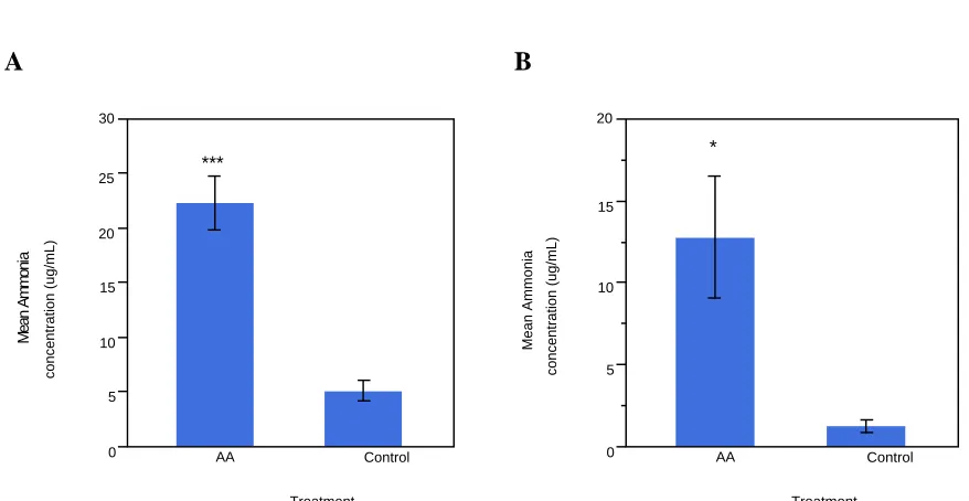
Ammonia assay ...39
qPCR...41
Western blot analysis ...43
DISCUSSION ...46
REFERENCES ...51
CHAPTER III: SUMMARY ...57
LIST OF TABLES
LIST OF FIGURES
Figure 1.1 Role of myogenic regulatory factors in embryonic myogenesis ...5
Figure 1.2 Myostatin mutations are highly conserved across species ...7
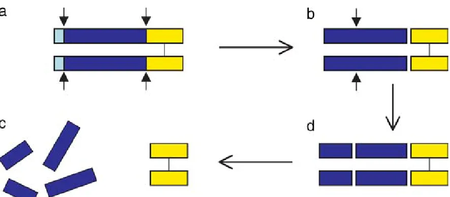
Figure 1.3 Processing of myostatin protein ...8
Figure 1.4 The urea cycle ...17
Figure 2.1 Serum ammonia concentrations ...40
Figure 2.2 mRNA expression of MSTN and MRFs ...42
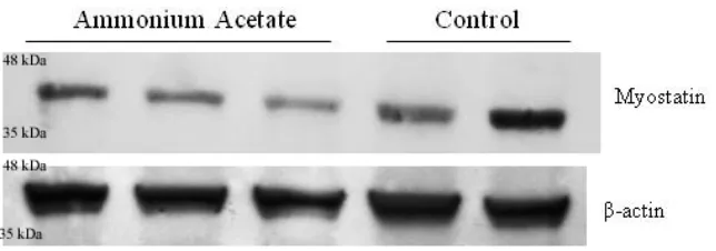
Figure 2.3 Western blot analysis of MSTN protein in ED17 collected samples ...44
CHAPTER I
LITERATURE REVIEW
Introduction
The chick embryo has been a model in developmental biology for many years, due to the solitary nature of their development. The avian model also provides a unique opportunity to observe effects of a manipulated embryonic environment on normal development that is difficult to achieve in mammalian species. While much of the focus in poultry production has focused on post-hatch strategies of optimizing meat production, there may be greater potential for improving muscle growth during embryonic myogenesis. Manipulation of growth and differentiation factors responsible for regulating muscle development is a logical strategy for improving embryonic myogenesis and may provide an opportunity to optimize meat production and quality that has yet to be considered.
Skeletal Muscle Growth
Skeletal muscle growth is characterized by two distinct phases: hyperplasia, an increase in cell number, and hypertrophy, an increase in cell size. Embryonic muscle
synthesis, and external factors, such as nutrition and mechanical load are responsible for post-natal muscle growth (Mozdziak et al., 1997; Schiaffino et al., 2013). The embryonic environment is crucial to muscle development and ultimate meat yield, as it establishes the framework for post-natal muscle growth.
Myogenesis and Myogenic Regulatory Factors
During embryogenesis, the mesoderm is the only germ layer that gives rise to skeletal muscle. More specifically, progenitor cells of the dermomyotome, a partition of epithelial somites from the paraxial mesoderm, are the precursor cells to myoblast cells (Pownall et al., 2002; Yusuf and Brand-Saberi, 2012). Somites can be identified by the expression of Pax genes, a family of developmentally regulated genes. Pax-3, a mammalian homolog of the Drosophila paired gene, is expressed in the unsegmented paraxial mesoderm and epithelial somite (Tajbakhsh et al., 1997). Pax-3 is expressed upstream of myogenic regulatory factors and is necessary for the migration of muscle precursor cells from the somites to developing limb buds, as murine Pax-3 mutants do not develop limb muscles (Tajbakhsh et al., 1997; Yusuf and Brand-Saberi, 2012). Pax-7 is expressed later than Pax-3 in the central
(Sabourin and Rudnicki, 2000; Pownall et al., 2002; Yusuf and Brand-Saberi, 2012). The MRFs are a subfamily of the basic helix-loop-helix superfamily of transcription factors.
The commitment of multipotent somite cells to myoblast cells is the first step in myogenesis and the major role of the primary MRFs. Myogenic factor 5 (MyF5) and myogenic determination factor1 (MyoD) are activated in all somite-derived muscle
progenitor lineages (Sabourin and Rudnicki, 2000; Pownall et al., 2002). In avian embryos, MyF5 and MyoD are activated in progenitor cells, while murine embryos express MyF5 in progenitor cells and MyoD in differentiated myotomes (Sassoon et al., 1989; Pownall et al., 2002). In the chick embryo, low levels of MyF5 and MyoD mRNA expression, without protein expression, have been detected prior to somatogenesis in the epiblast layer of the blastoderm, suggesting that initial regulation of myogenesis occurs many stages before terminal myoblast differentiation (Kiefer and Hauschka, 2001; Gerhart et al., 2007).
increase in MyF5 expression when compared to wild-type embryos (Rudnicki et al., 1992). While myoblast determination still occurs, further developmental investigations produced evidence that mice null for either MyF5 or MyoD exhibit delays in development of different muscle types, suggesting that they only play partially redundant roles and remain unique in their functions during normal development (Kablar et al., 1997). In addition to its role in myoblast specification, cell culture studies suggest that MyoD plays a role in preparing myoblasts for differentiation into myocytes. Ectopic MyoD causes inhibition of the cell cycle prior to the S-phase independently of induction of cellular differentiation (Crescenzi et al., 1990; Sorrentino et al., 1990).
Myoblast differentiation is marked by the fusion of multiple myoblasts into myocytes, which are the functional cellular units of mature muscle fibers. Myogenin, which is
expressed downstream of genes responsible for progenitor cell determination, is the major determinant of myoblast differentiation. In similar gene inactivation studies, mice lacking myogenin were observed to have a normal number of myoblasts, but completely lack myofibers, and therefore die at birth (Hasty et al., 1993; Nabeshima et al., 1993).
Additionally, myogenin was found to compensate for inactivation of myogenic regulatory factor 4 (MRF4), also known as herculin or MyF6, resulting in a normal muscle phenotype but abnormalities in rib formation (Zhang et al., 1995). Differently, in myogenin inactivated mice, MRF4 expression is low and normal myoblast differentiation does not occur,
suggesting that MRF4 plays a downstream role in the maturation of differentiated myocytes (Zhang et al., 1995; Olson et al., 1996). Though, MRF4 is normally the most highly
increased four-fold in adult muscles, further demonstrating its ability to compensate for MRF4 in muscular growth and maturation (Zhang et al., 1995). While there is some
evidence that myogenin can compensate for reduced or inactivated MRF4 expression, similar to the primary MRFs, the secondary MRFs maintain their individual importance in
embryonic development.
Figure 1.1 Role of myogenic regulatory factors in embryonic myogenesis. (edited from http://www.faculty.virginia.edu/mammgenetics/myogenesis.html)
Myostatin and its Role in Myogenesis and Muscle Growth
Myostatin, also called growth differentiation factor 8, is a member of the
Figure 1.2 Myostatin mutations are highly conserved across species. (a) the extreme
hyperplasic and hypertrophic phenotype in the forelimb of a MSTN knock-out mouse (right) compared to a wild-type mouse (left) (b) Belgian Blue bull demonstrating the double
muscling phenotype (Lee, 2004).
action or a decrease in pH releasing the active form of the myostatin protein (McPherron et al., 1997; Lee, 2008; Han and Mitch, 2012) (Figure 1.3). This suggests that the propeptide plays a regulatory role in the activity of myostatin.
Figure 1.3 Processing of myostatin protein. (a) Myostatin precursor (approx. 52 kDa) which must undergo two proteolytic events (b) Latent complex of myostatin (approx. 40 kDa) following the proteolytic removal of the N-terminal signal sequence, the propeptide (blue) and the disulfide linked C-terminal dimer (yellow) remain bound (d) the activation of the latent myostatin dimer occurs by proteolytic cleavage leaving (c) the myostatin dimer (approx. 26kDa) and dissociated propeptide fragments (Thomas et al., 2000; Lee, 2004).
As myostatin-null individuals illustrate both a hyperplasic and hypertrophic response, it can be assimilated that myostatin has inhibitory effects during embryonic myogenesis and post-natal muscle growth. During mouse embryogenesis, myostatin is expressed in
studies have shown that myostatin inhibits both myoblast proliferation and differentiation in C2C12 myoblasts (Thomas et al., 2000; Langley et al., 2002). Similar results have been noted in fetal bovine myoblasts and in chick limb bud development (Thomas et al., 2000; Amthor et al., 2002; Amthor et al., 2004). Specifically, myostatin has been found to downregulate MyF5, and Pax3, genes associated with proliferation, and increase the expression of p21, a cyclin-dependent kinase inhibitor, which has a negative effect on proliferation (Thomas et al., 2000; Amthor et al., 2002). Cyclin-dependent kinases (Cdks) catalyze transitions in the cell cycle. The p21 family inhibits all Cdks involved in G1/S phase transition, which has been observed in the growth of myostatin treated C2C12 myoblasts (Thomas et al., 2000). The negative effect of myostatin on myoblast
differentiation can be attributed to the downregulation of MyoD and myogenin (Langley et al., 2002; Rios et al., 2002). As previously mentioned, MyoD expression has been observed to cause growth arrest in cultured cells, and has been shown to inhibit the G0/S phase transition in quiescent NIH 3T3 cells stimulated to re-enter the cell cycle (Crescenzi et al., 1990; Sorrentino et al., 1990). By downregulating MyoD expression, myostatin has a negative effect on the differentiation of myoblasts by inhibiting cell cycle arrest (Langley et al., 2002).
1997). During normal muscle growth, satellite cells re-enter the cell cycle and proliferate to maintain a population of satellite cells available for myonuclear donation (Schultz, 1996).
Several studies, both in vivo and in vitro, suggest that increased myostatin inhibits satellite cell function by upregulating p21, which negatively regulates the cell cycle and prevents satellite cell mitosis (McCroskery et al., 2003; Dasarathy et al., 2004; Akita et al., 2013). Additionally, myostatin has been shown to downregulate MRFs, which are involved in muscle growth and serve as indicators of satellite cell proliferation and differentiation (Muroya et al., 2002; Dasarathy et al., 2004). Satellite cell activity is often assessed by administration of 5-bromo2-deoxyuridine (BrdU), a synthetic analog of thymidine that substitutes during DNA replication, which can then be detected by antibody staining to identify satellite cells that are actively undergoing mitosis. McCroskery et al. (2003) found that at multiple stages of growth (4 wk, 8 wk, and 6 mo) myostatin knockout mice had 33% more satellite cells in the S-phase of mitosis than wild-type counterparts after BrdU labeling, demonstrating that the presence of myostatin regulates satellite cell activation in normal growth. Further, a significantly higher amount of steady-state satellite cells per muscle fiber unit was observed in myostatin-null adult mice when compared to controls (McCroskery et al., 2003). Similarly to the regulatory role of myostatin in embryonic myogenesis, this evidence suggests that in post-natal muscle growth and maintenance of adult muscle,
Myostatin Mutations in Livestock and Meat Production
The hyperplasic and hypertrophic phenotype observed in myostatin mutated animals has been of particular interest in agricultural research, as efficiency of meat production and higher meat yield are constant goals of the industry. The higher percentage of muscle to carcass weight observed in myostatin mutants is desirable in meat-producing livestock. Additionally, myostatin mutant animals have been observed to have a lesser amount of connective tissue, which has been shown to positively impact meat tenderness (Arthur, 1995; Hope et al., 2013). However, there are many disadvantages that are associated with
myostatin mutations that have prevented commercial agricultural practices from employing this gene manipulation as a strategy for increased muscle yield. Firstly, there are
discrepancies on the impact of the myostatin mutation on meat quality in reference to flavor. McPherron and Lee (2002) found that the deletion of myostatin partially suppressed fat accumulation in mice. Similarly, reduced intramuscular fat content was observed in cattle and sheep with the myostatin mutation (Wiener et al., 2009; Masri et al., 2011; Hope et al., 2013). In these studies, there were varying reports of taste panel flavor scores. More
Myostatin in Muscle Wasting Diseases
In addition to muscular dystrophy, severe muscle wasting is a serious side effect that has been observed to accompany many diseases, including cancer, congestive heart failure, chronic kidney disease, diabetes, AIDS, chronic obstructive pulmonary disease (COPD), and liver diseases. In all of these disorders, regulation of myostatin expression is a targeted therapy for sarcopenia and cachexia (Dasarathy et al., 2004; Zhou et al., 2010; Han and Mitch, 2012). Clinical studies have shown that patients with COPD have increased serum myostatin concentrations compared to healthy age matched individuals (Ju and Chen et al., 2012). Using a murine model of cancer, Zhou et al. (2010) were able to prevent and reverse muscle wasting, even in animals whose tumor growth was not inhibited, by
pharmacologically blocking the myostatin signaling pathway. Similarly, a study of chronic kidney disease (CKD) revealed that infusion of a myostatin agonist into CKD mice
suppressed myostatin in the muscle, and both muscle weight and body weight were increased compared to CKD mice given sham infusions (Zhang et al., 2011). In cirrhosis, which occurs in advanced liver diseases, sarcopenia is common and causes significant
Hyperammonemia and Muscle Function
Hyperammonemia, or excess ammonia in the blood as a result of a metabolic disruption or insufficiency, has been observed in numerous diseases. Acute ammonia toxicity is marked by symptoms of neurophysiologic effects on the central nervous system. At high doses, hyperammonemia (1-5 mM) can produce variable shifts between excitatory and inhibitory neurotransmission causing seizures, coma, and death (Albrecht, 1998). Animal models of ammonia toxicity demonstrate similar responses to rapid increases of ammonia in the blood, though species differences have been reported. Wilson et al. (1968) examined the differences in toxicity of exogenous ammonia between uricotelic species and ureotelic species, using young chicks and mice. They found that through intravenous injection, chicks were more susceptible to ammonia toxicity when compared to mice (LD50 of 2.72 mmol/kg and 5.64 mmol/kg, for chicks and mice, respectively) (Wilson et al., 1968). Intraperitoneally, there was no significant difference in ammonia toxicities between chicks and mice (LD50 of 10.44 mmol/kg and 10.84 mmol/kg, for chicks and mice, respectively) (Wilson et al., 1968).
detoxification is inadequate (Holecek et al., 2000; Olde Damink et al., 2002 ). Specifically, significant ammonia uptake in the muscle is observed in patients with decompensated
cirrhosis and muscle wasting when compared to cirrhotic patients with normal compensation (Olde Damink et al., 2002). This further suggests the correlation between hyperammonemia and sarcopenia.
Other diseases that have been associated with hyperammonemia suggest that excess ammonia is released from the muscle during metabolic stress. For example, exercise-induced hyperammonemia has been observed in patients with COPD and chronic heart failure. In these studies, hyperammonemia was exacerbated by exercise and was used as indicator of metabolic stress and skeletal muscle ATP depletion (Andrews at al., 1997; Calvert et al., 2010). It is well understood that hyperammonemia during exercise, specifically, can be dominantly attributed to activation of the purine nucleotide cycle, in which deamination of adenosine monophosphate occurs, likely as a response to depleted ATP or lack of ATP resynthesis (Graham et al., 1992). In this case, ammonia is a byproduct of the purine
Hyperammonemia and Embryogenesis
Ammonia is generated in vivo during catabolism of amino acids and is generally considered a toxic waste product. This toxicity has been previously discussed in reference to adults. There is evidence that supports that ammonia also has negative effects on
embryogenesis in numerous in vitro studies. He et al. (2007) investigated effects on murine embryogenesis in glutamine synthetase (GS), the enzyme required for glutamine production from ammonia and glutamate, knock-out embryos in culture with and without
Ammonia Detoxification and Excretion: Urea vs. Uric Acid
Aside from its role in glucose synthesis and storage, the liver controls the availability of amino acids in the blood. Unlike glucose, amino acids cannot be stored and, therefore, must be used immediately for protein synthesis, or be broken down into α-keto acids and used for energy metabolism. The deamination of amino acids produces free ammonium ions, which as previously discussed are toxic at high concentrations in the blood. In bacteria and most mammalian species, ammonia is detoxified in hepatocytes, in a series of enzymatic processes termed the urea cycle, which produces non-toxic urea from ammonia and
Figure 1.4 The urea cycle. Through a series of enzymatic reactions (green boxes), excess ammonia is converted to urea in the hepatocytes. ARG, arginase 1; ASL, argininosuccinate lyase; ASS, argininosuccinate synthase; CPS1, carbamoyl phosphate synthase 1; NAG, N-acetylglutamate; NAGS, N-acetylglutamate synthase; ORNT1, ornithine transporter; OTC, ornithine transcarbamoylase (Nagamani et al., 2012).
Reptiles and avian species are uricotelic animals, meaning they excrete excess nitrogen in the form of uric acid. It is has been conceded that this mode of nitrogen
synthetase (ASS) were below the limit of detection. However, OTC and ASS activity have been reported in the kidney of chickens (Tamir and Ratner, 1960). Though their activity is not measurable in the liver, a genome mapping study identified all three of these enzymes, including CPS1, in the chicken genome which suggests that they may be physiologically relevant in some other metabolic process (Shimogiri et al., 2004).
Research Objectives
The overall objective of the current thesis research was to determine the effect of hyperammonemia on embryonic myogenesis. There doesn’t appear to be any data on ammonia toxicity in broiler embryogenesis. The primary objective was to induce
REFERENCES
Akita, Y., Y. Sumino, K. Mori, T. Nomura, F. Sato, and H. Mimata. 2013. Myostatin inhibits proliferation of human urethral rhabdosphincer satellite cells. Int. J. Urol. 20:522-529. Albrecht, J. 1998. Roles of neuroactive amino acids in ammonia neurotoxicity. J. Neurosci.
Res. 51:133-138.
Andrews, R., J.T. Walsh, A. Evans, S. Curtis, and A.J. Cowley. 1997. Abnormalities of skeletal muscle in patients with chronic heart failure: evidence that they are present at rest. Heart. 77:159-163.
Amthor, H., R. Huang, I. McKinnell, B. Christ, R. Kambadur, M. Sharma, and K. Patel. 2002. The regulation and action of myostatin as a negative regulator of muscle development during avian embryogenesis. Dev. Biol. 251:241-257.
Amthor, H., G. Nicholas, I. McKinnell, C.F. Kemp, M. Sharma, R. Kambadur, and K. Patel. 2004. Follistatin complexes myostatin and antagonises myostatin-mediated inhibition of myogenesis. Dev. Biol. 270:19-30.
Arthur, P.F. Double muscling in cattle: a review. 1995. Aust. J. Agric. Res. 46:1493-1515. Bishonga, C., J.J. Robinson, T.G. McEvoy, P. Findlay, R.P. Aitken, and I. Robertson. 1996.
Excess dietary urea intake in ewes and its effect on ovulation rate and embryo development. Jpn. J. Vet. Res. 44(3):139-151.
Calvert, L.D., M.C. Steiner, M.D. Morgan, and S.J. Singh. 2010. Plasma ammonia response to incremental cycling and walking tests in COPD. Respir. Med. 104:675-681. Crescenzi, M., T.P. Fleming, A.B. Lassar, H. Weintraub, and S.A. Aaronson. 1990. MyoD
Proc. Natl. Acad. Sci. 87:8442-8446.
Dasarathy, S., M. Dodig, S.M. Muc, S.C. Kalhan, and A.J. McCullough. 2004. Skeletal muscle atrophy is associated with and increased expression of myostatin and impaired satellite cell function in the portacaval anastamosis rat. Am. J. Physiol. Gastrointest. Physiol. 287:G1124-G1130.
Forbes, D., M. Jackman, A. Bishop, M. Thomas, R. Kambadur, and M. Sharma. 2006. Myostatin auto-regulates its expression by feedback loop through Smad7 dependent mechanism. J. Cell. Physiol. 206:264-272.
Gerhart, J., C. Neely, J. Elder, J. Pfautz, J. Perlman, L. Narciso, K.K. Linask, K. Knudsen, and M. George-Weinstein. 2007. Cells that express MyoD mRNA in the epiblast are stably committed to the skeletal muscle lineage. J. Cell Biol. 178(4):649-660.
Graham, T.E., and D.A. MacLean. 1992. Ammonia and amino acid metabolism in human skeletal muscle during exercise. Can. J. Physiol. Pharmacol. 70:132-141.
Hammon, D.S., S. Wang, and G.R. Holyoak. 2000. Ammonia concentration in bovine follicular fluid and its affect during in vitro maturation on subsequent embryo development. Anim. Reprod. Sci. 58:1-8.
Han, H.Q, and W.E. Mitch. 2011. Targeting the myostatin signaling pathway to treat muscle wasting diseases. Curr. Opin. Support Palliat. Care. 5(4):334-341.
Hasty, P., A. Bradley, J.H. Morris, D.G. Edmondson, J.M. Venuti, E.N. Olson, and W.H. Klein. 1993. Muscle deficiency and neonatal death in mice with a targeted mutation in the myogenin gene. Nature. 364:501-506.
Glutamine Synthetase is essential in early mouse embryogenesis. Dev. Dyn. 36:1865- 1875.
Holecek, M., L. Sprongl, and M. Tichy. 2000. Effect of hyperammonemia on leucine and protein metabolism in rats. Metab. 49(10):1330-1334.
Hope, M., F. Haynes, H. Oddy, M. Koohmaraie, A. Al-Owaimer, and G. Geesink. 2013. The effects of the myostatin g+6723G>A mutation on carcass and meat quality of lamb. Meat Sci. 95:118-122.
Ju, C.-R., and R.-C. Chen. 2012. Serum myostatin levels and skeletal muscle wasting in chronic obstructive pulmonary disease. Respir. Med. 106:102-108.
Kablar, B., K. Krastel, C. Ying, A. Asakura, S.J. Tapscott, and M.A. Rudnicki. 1997. MyoD and Myf-5 differentially regulate the development of limb versus trunk skeletal muscle. Dev. 124:4729-4738.
Kiefer, J.C., and S.D. Hauschka. 2001. Myf-5 is transiently expressed in nonmuscle and exhibits dynamic regional changes within the presegmented mesoderm and somites I-IV. Dev. Biol. 232:77-90.
Langley, B., M. Thomas, A. Bishop, M. Sharma, S. Gilmour, and R. Kambadur. 2002. Myostatin inhibits myoblast differentiation by down-regulating MyoD expression. J. Biol. Chem. 277:49831-49840.
Lee, S.-J. 2004. Regulation of muscle mass by myostatin. Annu. Rev. Cell Dev. Biol. 20:61- 86.
Lee, S.-J., and A.C. McPherron. 1999. Myostatin and the control of skeletal muscle mass. Curr. Opin. Genet. Dev. 9:604-607.
Mansouri, A., A. Stoykova, M. Torres, and P. Gruss. 1996. Dysgenesis of cephalic neural crest derivatives in Pax7-/- mutant mice. Dev. 122:831-838.
Masri, A.Y., N.R. Lambe, J.M. Macfarlane, S. Brotherstone, W. Haresign, and L. Bunger. 2011. Evaluating the effects of a single copy of a mutation in the myostatin gene (c.*1232 G>A) on carcass traits in crossbred lambs. Meat Sci. 87:412-418.
McCroskery, S., M. Thomas, L. Maxwell, M. Sharma, and R. Kambadur. 2003. Myostatin negatively regulates satellite cell activation and self-renewal. J. Cell Biol.
162(6):1135-1147.
McEvoy, T.G., J.J. Robinson, R.P. Aitken, P.A. Findlay, and I.S. Robertson. 1997. Dietary excesses of urea influence the viability and metabolism of preimplantation sheep embryos and may affect fetal growth among survivors. Anim. Reprod. Sci. 47:71-90. McPherron, A.C., A.M. Lawler, and S.-J. Lee. 1997. Regulation of skeletal muscle mass in
mice by a new TGF-β superfamily member. Nature. 387:83-90.
McPherron, A.C., and S.-J. Lee. 1997. Double muscling in cattle due to mutations in the myostatin gene. Proc. Natl. Acad. Sci. 94:12457-12461.
McPherron, A.C., and S.-J. Lee. 2002. Suppression of body fat accumulation in myostatin- deficient mice. J. Clin. Invest. 109:595-601.
Mora, J., J. Martuscelli, J. Ortiz-Pineda, and G. Soberon. 1965. The regulation of urea- biosynthesis enzymes in vertebrates. Biochem. J. 96:28-35.
determinant of avian skeletal muscle growth. Am. J. Physiol. Cell Physiol. 272: C565-C571.
Muroya, S., I. Nakajima, and K. Chikuni. 2002. Sequential expression of myogenic
regulatory factors in bovine skeletal muscle and the satellite cell culture. Anim. Sci. J. 73:375-381.
Nabeshima, Y., K. Hanaoka, M. Hayasaka, E. Esumi, S. Li, I. Nonaka, and Y. Nabeshima. 1993. Myogenin gene disruption results in perinatal lethality because of severe muscle defect. Nature. 364:532-535.
Nagamani, S.C.S., A. Erez, and B. Lee. 2012. Arginosuccinate lyase deficiency. Genet. Med. 14(5):501-507.
Olde Damink, S.W.M., N.E.P. Deutz, C.H.C. Dejong, P.B. Soeters, and R. Jalan. 2002. Interorgan ammonia metabolism in liver failure. Neurochem. Int. 41:177-188.
Olson, E.N., H.-H. Arnold, P.W.J. Rigby, and B.J. Wold. 1996. Know your neighbors: three phenotypes in null mutants of the myogenic bHLH gene MRF4. Cell. 85:1-4.
Pownall, M.E., M.K. Gustafsson, and C.P. Emerson Jr. 2002. Myogenic regulatory factors and the specification of muscle progenitors in vertebrate embryos. Annu. Rev. Cell Dev. Biol. 18:747-83.
Qiu, J., C. Tsien, S. Thapaliya, A. Narayanan, C.C. Weihl, J.K. Ching, B. Eghtesad, K. Singh, X. Fu, G. Dubyak, C. McDonald, A. Almasan, S.L. Hazen, S.V.N. Prasad, and S. Dasarathy. 2012. Hyperammonemia-mediated autophagy in skeletal muscle
Qiu, J., S. Thapaliya, A. Runkana, Y. Yang, C. Tsien, M.L. Mohan, A. Narayanan, B. Eghtesad, P.E. Mozdziak, C. McDonald, G.R. Stark, S. Welle, S.V.N. Prasad, and S. Dasarathy. 2013. Hyperammonemia in cirrhosis induces transcriptional regulation of myostatin by an NF-κB-mediated mechanism. Proc. Natl. Acad. Sci.
110(45):18162-18167.
Remignon, H., M.-F. Gardahaut, G. Marche, and F.-H. Ricard. 1995. Selection for rapid growth increases the number and the size of muscle fibers without changing their typing in chickens. J. Muscle Res. Cell. Motil. 16:95-102.
Rios, R., I. Carneiro, V. M. Arce, and J. Devesa. 2002. Myostatin is an inhibitor of myogenic differentiation. Am. J. Physiol. Cell Physiol. 282:C993-C999.
Rudnicki, M.A., T. Braun, S. Hinuma, and R. Jaenisch. 1992. Inactivation of MyoD in mice leads to up-regulation of the myogenic HLH gene Myf-5 and results in apparently normal development. Cell. 71:383-390.
Rudnicki, M.A., P.N.J. Schnegelsberg, R.H. Stead, T. Braun, H.-H. Arnold, and R. Jaenisch. 1993. MyoD of Myf-5 is required for the formation of skeletal muscle. Cell. 75:1351- 1359.
Sabourin, L.A., and M.A. Rudnicki. 2000. The molecular regulation of myogenesis. Clin. Genet. 57:16-25.
Sassoon, D., G. Lyons, W.E. Wright, V. Lin, A. Lassar, H. Weintraub, and M. Buckingham. 1989. Expression of two myogenic regulatory factors myogenin and MyoD1 during mouse embryogenesis. Nature. 341:303-307.
regulating skeletal muscle growth and atrophy. FEBS J. 280:4294-4314.
Schultz, E. 1996. Satellite cell proliferative compartments in growing skeletal muscles. Dev. Biol. 175:84-94.
Seale, P., L.A. Sabourin, A. Girgis-Gabardo, A. Mansouri, P. Gruss, and M.A. Rudnicki. 2000. Pax7 is required for the specification of myogenic satellite cells. Cell. 102:777- 786.
Shimogiri, T., N. Bosak, M. Morisson, S. Okamoto, K. Kawabe, Y. Maeda, A. Vignal, and H. Yasue. 2004. Assignment of CPS1, OTC, CRYD2, ARG2, and ASS genes to the chicken RH map. Genet. Sel. Evol. 36:593-599.
Shiokawa, K., Y. Kawazoe, H. Nomura, T. Miura, N. Nakakura, T. Horiuchi, and K. Yamana. 1986. Ammonium ion as a possible regulator of the commencement of rRNA synthesis in Xenopus laevis embryogenesis. Dev. Bio. 115:380-391. Shiokawa, K., M. Aso, T. Kondo, J.-I. Takai, J. Yoshida, T. Mishina, K. Fuchimukai,
T. Ogasawara, T. Kariya, K. Tashiro, and K. Igarashi. 2010. Effects of S- adenosylmethionine decarboxylase, polyamines, amino acids, and weak bases (amines and ammonia) on development and ribosomal RNA synthesis in Xenopus embryos. Amino Acids. 38:439-449.
Sorrentino, V., R. Pepperok, R.L. Davis, W. Ansorge, and L. Philipson. 1990. Cell
proliferation inhibited by MyoD1 independently of myogenic differentiation. Nature. 345:813-815.
Cell 89:127-138.
Tamir, H., and S. Ratner. 1963. Enzymes of arginine metabolism in chicks. Arch. Biochem. Biophys. 102:249-258.
Thomas, M., B. Langley, C. Berry, M. Sharma, S. Kirk, J. Bass, and R. Kambadur. 2000. Myostatin, a negative regulator of muscle growth, functions by inhibiting myoblast proliferation. J. Biol. Chem. 275:40235-40243.
Webb, D.J., and M. Charbonneau. 1987. Weak bases inhibit cleavage and embryogenesis in amphibians and echinoderms. Cell Differ. 20:33-44.
Wiener, P., J.A. Woolliams, A. Frank-Lawale, M. Ryan, R.I. Richardson, G.R. Nute, J.D. Wood, D. Homer, and J.L. Williams. 2009. The effects of a mutation in the myostatin gene on meat and carcass quality. Meat Sci. 83:127-134.
Wigmore, P.M.C., and N.C. Strickland. 1983. Muscle development in large and small pig fetuses. J. Anat. 137(2):235-245.
Wilson, R.P., M.E. Muhrer, and R.A. Bloomfield. 1968. Comparative ammonia toxicity. Comp. Biochem. Physiol. 25:295-301.
Yusuf, F., and B. Brand-Saberi. 2012. Myogenesis and muscle regeneration. Histochem. Cell Biol. 138:187-199.
Zhang, L., V. Rajan, E. Lin, Z. Hu, H.Q. Han, X. Zhou, Y. Song, H. Min, X. Wang, J. Du, and W.E. Mitch. 2011. Pharmacological inhibition of myostatin suppresses systemic inflammation and muscle atrophy in mice with chronic kidney disease. FASEB J. 25:1653-1663.
MRF4 results in up-regulation of myogenin and rib abnormalities. Genes Dev. 9:1388-1399.
CHAPTER II
THE EFFECT OF HYPERAMMONEMIA ON MYOSTATIN AND MYOGENIC REGULATORY FACTOR GENE EXPRESSION IN BROILER EMBRYOS
ABSTRACT
Myogenesis is facilitated by four myogenic regulatory factors (MRFs) and is significantly inhibited by myostatin (MSTN). The objective of the current study was to examine embryonic gene regulation of MSTN/MRFs, and subsequent manipulations of protein synthesis, in broiler embryos under induced hyperammonemia. Broiler eggs were injected with ammonium acetate solution (50 mmol/kg body weight) four times over 48 hours beginning on either embryonic day (ED) 15 or 17. Serum ammonia concentration was significantly increased, more than four-fold, (P < 0.05) in ammonium acetate injected
embryos for both ED17 and ED19 collected samples when compared to sham-injected controls. Expression of mRNA, extracted from pectoralis major of experimental and control embryos, was measured using real-time quantitative PCR for MSTN, and MRFs myogenic factor 5 (MyF5), myogenic determination factor 1(MyoD), myogenin (MYOG), and
No significant difference was seen in MyoD or MYOG expression for either age group. MSTN protein levels were evaluated by Western blot analysis, and also showed decreased myostatin expression (P < 0.05). Overall, it appears possible to inhibit myostatin expression through hyperammonemia, which is expected to increase embryonic myogenesis and
postnatal muscle growth.
INTRODUCTION
It is widely accepted in both agricultural and human research that embryonic and immediate post-natal environment plays an important role in muscle development. In poultry, it is well established that muscle fiber number is determined during embryonic development and that post hatch growth is dependent on hypertrophy of the existing fibers (Remignon et al., 1995; Mozdziak et al., 1997). Embryonic muscle hyperplasia, or increase in myoblast proliferation, is controlled by a group of four basic helix-loop-helix transcription factors called myogenic regulatory factors (MRFs).
al., 1993). Myogenin (MYOG), and myogenic regulatory factor 4 (MRF4), also known as MyF6 or herculin, are expressed later in embryonic development. MYOG is the major determinant of myoblast differentiation, while MRF4 is expressed in mature myocytes (Nabeshima et al., 1993).
Myostatin (MSTN), a transforming growth factor-β (TGF-β) family member, is the most powerful negative regulator of myogenesis, but is also expressed in adult muscles, suggesting it also inhibits postnatal muscle growth (McPherron et al., 1997; Lee and McPherron, 2001; Amthor et al., 2004). MSTN knockout mice display an extreme
hyperplasic and hypertrophic phenotype termed double muscling (McPherron and Lee 1997). Mutations in the MSTN gene were deemed responsible for the same phenotype observed, as a result of genetic selection for muscle growth, in Belgian Blue and Piedmontese cattle proving that the role of the MSTN gene is highly conserved across species (McPherron and Lee, 1997). MSTN has been shown to inhibit myogenesis by downregulating expression of the crucial growth factors MyoD, MyF5, and MYOG (Langley et al., 2002; Amthor et al., 2004; Dasarathy et al., 2004).
realistic opportunity to benefit from increased hyperplasia and hypertrophy observed with MSTN mutations (Wiener et al., 2009; Hope et al., 2013). It is therefore of particular interest for agricultural advancement to metabolically adjust the expression of MSTN, and
subsequently MRFs, without completely eliminating its role in normal regulation of muscle growth.
Increased MSTN expression has been noted in the investigation of diseases, such as cancer, heart and kidney failure, and cirrhosis, where muscle wasting is frequently a
secondary, but serious, side effect (Dasarathy et al., 2004; Han and Mitch, 2012; Qiu et al., 2013). Regulation of MSTN expression is targeted as a potential therapy for sarcopenia and cachexia (Dasarathy et al., 2004, Han and Mitch, 2012). In a recent study, Qiu et al. (2013), observed increased MSTN expression under induced hyperammonemia using rats as a model for cirrhosis. Additionally, hyperammonemia has been observed in other diseases including chronic obstructive lung disease and heart failure (Andrews et al., 1997; Calvert et al., 2010). These clinical and mechanistic observations suggest that hyperammonemia has an adverse effect on muscle structure and function in a broad range of disorders.
therefore inhibiting protein synthesis (Shiokawa et al., 1986; Shiokawa et al., 2010). Thus, the adverse effects of ammonia on the growing embryo are inconsistent in contrast to the consistent negative effects in adults. There is no data on the role of ammonia toxicity in the developing embryo of broilers. The objective of the present study was to investigate chick embryonic gene manipulation of MSTN and MRFs under induced hyperammonemia, and the effect of the gene manipulation on protein synthesis and growth.
MATERIALS AND METHODS
Injection of Eggs
Fertilized broiler eggs (Ross 708 x Ross 344) were incubated at 37oC to embryonic day (ED) 15 or 17. In late stage broiler embryos, secondary muscle fiber formation is occurring and a population of adult myoblasts, or satellite cells, is being established,
returned to the incubator. Blood and tissue samples were collected 12 hours after the last injection (i.e. for groups beginning injections in ED15, samples were harvested on ED17, and those beginning on ED17, harvesting was performed on ED19). There was no observed toxicity, or marked difference in health, in experimental embryos, as compared to controls, at the time of sampling. Additionally, upon sampling from the ED19 group, internal pipping was noted in both the experimental and control groups, indicating that chicks in each group were preparing for hatch.
Collection of samples
Blood samples were drawn directly from blood vessels using beveled glass capillary tubes and placed in 0.5 mM EDTA treated tubes. Samples were centrifuged at 12,000 rpm for 10 min, the serum was separated, and placed in fresh tubes for storage at -80oC. The embryos were removed from the shell, killed via decapitation and total body weight was recorded. Pectoralis major tissue was dissected and stored in RNAlater (Ambion Inc., Grand Island, NY) at -20oC for total RNA extraction, or snap frozen in liquid nitrogen and stored at -80oC for total protein extraction.
Serum ammonia analysis
working volume of a 96-well plate. Absorbance was measured at 340 nm 5 minutes after the reagent was mixed with standard, sample, or water. The diluted enzyme was mixed (10 µL) into each well and allowed to incubate at room temperature for 5 minutes. Absorbance readings (340 nm) were taken in 1 minute intervals for 6 minutes. Change in absorbance was calculated based on the manufacturers’ guidelines and ammonia concentration was calculated using the following equation provided for the ammonia assay kit (Sigma Aldrich, St. Louis, MO):
RNA extraction, reverse transcription, and quantitative real time-PCR
Total RNA was isolated using the RNeasy Mini Kit protocol (Qiagen, Venlo, Limburg). Approximately 30 g of RNAlater (Ambion Inc., Grand Island, NY) preserved pectoralis muscle was placed in 600 µL of the provided buffer for homogenization using a Mini-Beadbeater-1 (BioSpec Products, Bartlesville, OK). Total RNA concentration was determined by measuring absorbance at 260 nm. RNA quality was assessed using agarose gel electrophoresis. Reverse transcription was performed using the High Capacity cDNA Reverse Transcription Kit (Applied Biosystems Inc., Grand Island, NY) using the reverse transcriptase from the murine leukemia virus and random hexamers. After the reverse transcription reaction was complete, cDNA was diluted 1:20 before qPCR.
Five samples for each treatment (ED17 AA, ED17 C, ED19 AA, ED19 C) were prepared for qPCR analysis of MSTN, MyoD, MYOG, MyF5 and MRF4 genes. These genes
Δ(ΔA340)Test or Standard
= ΔA340(Test or Standard) – ΔA340(Blank)
mg of NH3/ml of original sample
were chosen because they are known to play a critical regulatory role in muscle development and differentiation. β-actin was used as an internal control for normalization of each sample
because our preliminary data showed no change in expression under our experimental conditions (data not shown). Primers were designed using Primer-BLAST (NCBI) which utilizes Primer3 software (Table 2.1). qPCR was performed at a volume of 20 µL, consisting of 1 µL diluted cDNA, 1 µL diluted primer (400 µM), 10 µL power SYBR Green PCR Master Mix (Applied Biosystems Inc., Grand Island, NY), and 8 µL ultra pure H2O. For each reaction, the thermocycler (Applied Biosystems Inc., Grand Island, NY) was set to the following cycle, based upon optimum temperatures identified by a gradient reaction: 95oC for 10 min, then 40 cycles of 95oC for 30 sec denaturing, 62oC for 30 sec annealing, 72oC for 5 min extension. Standard curves for amplification efficiency were produced for each set of primers by performing serial dilutions of pooled cDNA (1:5, 1:25, 1:125, 1:625). At each extension step, fluorescence was measured and the cycle threshold (Ct) was calculated by the StepOne software (version 2.1, Applied Biosystems Inc., Grand Island, NY). All
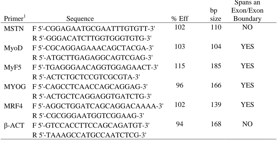
Table 2.1 Primer sequences for real-time qPCR
Primer1 Sequence % Eff bp size Spans an Exon/Exon Boundary MSTN F 5'-CGGAGAATGCGAATTTGTGTT-3' 102 110 NO
R 5'-GGGACATCTTGGTGGGTGTG-3'
MyoD F 5'-CGCAGGAGAAACAGCTACGA-3' 103 104 YES
R 5'-ATGCTTGAGAGGCAGTCGAG-3'
MyF5 F 5'-TGAGGGAACAGGTGGAGAACT-3' 115 185 YES
R 5'-ACTCTGCTCCGTCGCGTA-3'
MYOG F 5'-CAGCCTCAACCAGCAGGAG-3' 96 166 YES
R 5'-ACTGCTCAGGAGGTGATCTG-3'
MRF4 F 5'-AGGCTGGATCAGCAGGACAAAA-3' 102 139 YES
R 5'-CGCGGGAATGGTCGGAAG-3'
β-ACT F 5'-GTCCACCTTCCAGCAGATGT-3' 94 168 NO
R 5'-TAAAGCCATGCCAATCTCG-3'
1
Primers were designed using Primer-BLAST for myostatin (MSTN), myogenic
determination factor 1 (MyoD), myogenic factor 5 (MyF5), myogenin (MYOG), myogenic regulatory factor 4 (MRF4), and β-actin (β-ACT).
Protein extraction, SDS-PAGE, and Western blot analysis
measured using the Bio-Rad Protein Assay Dye Reagent Concentrate (Bio-Rad Laboratories Inc., Richmond, CA), which produces an absorbance shift relative to the amount of
solublized protein as Coomassie blue dye binds to protein, using Bovine Serum Albumin as a standard.
Loading volumes for each sample were calculated, considering a dilution factor of 5, to an amount of 20 µg protein per well. Samples were boiled 1:1 in Laemmli buffer (Bio-Rad Laboratories Inc., Richmond, CA) and separated by SDS-PAGE using a 10% Mini-PROTEAN TGX precast gel (Bio-Rad Laboratories Inc., Richmond, CA) with a 10-245 kDa Prism Ultra Protein Ladder (Abcam, Cambridge, MA). The protein was transferred to a polyvinylidene difluoride membrade (PVDF) via wet transfer, using a 0.02 M Tris/0.15 M glycine buffer with 18% methanol, at 400 mA for 1 hour. The membrane was Ponceau S stained to observe loading and transfer accuracy, then destained using ultra pure water.
activity was detected using chemiluminescent reagent (Bio-Rad Laboratories Inc., Richmond, CA). The membrane was stripped of the MSTN antibodies using an acidic glycine buffer, as outlined in the Abcam mild stripping protocol (Abcam, Cambridge, MA). Chemiluminescent reagent was used to confirm the antibodies had been removed from the membrane.
After the PVDF membrane was stripped of the antibodies detecting myostatin, the membrane was re-blocked with 5% goat serum, washed, and re-probed with β-actin for a loading control. The primary antibody for β-actin (Thermo Scientific, Rockford, IL) is a mouse monoclonal to β-actin N-terminal peptide. The β-actin antibody was diluted 1:1000 in
5% goat serum and incubated overnight at 4oC. Following primary antibody incubation, the membrane was washed three times with TBS-T, then probed with goat anti-mouse IgG secondary antibody conjugated with HRP (SouthernBiotech, Brimingham, AL), diluted 1:10,000 in 5% goat serum, for 1.5 hours on an orbital shaker at room temperature. Three washes with TBS-T were performed and HRP was detected using Chemiluminescent reagent. Semi-quantitative analysis of protein was determined by band intensity using Kodak 1D Scientific Imaging Systems (v.3.6.2, New Haven, CT). MSTN intensity of each band was normalized to β-actin prior to comparing experimental and control samples.
Statistical Analysis
RESULTS
Ammonia assay
A B
0 5 10 15 20 25 30
Mean Ammonia
concentration (ug/mL)
AA Control
Treatment ***
0 5 10 15 20
Mean Ammonia
concentration (ug/mL)
AA Control
Treatment *
qPCR
Mean Fold Change
Fold Change
0.0 0.5 1.5 2.0 2.5
normal expression 1.0
MSTN MyF5 MyF6/MRF4 MyoD MYOG
Gene
Embryonic Day 17 19
**
***
*
* *
Figure 2.2 mRNA expression of MSTN and MRFs. RNA was isolated from ED17 and ED19 collected pectoralis major samples after 48 hours of induced hyperammonemia (four
Western blot analysis
MSTN protein expression was detected by Western blot and normalized to β-actin for
relative quantification and statistical significance. The MSTN band assessed is
approximately 43 kDa. ED17 ammonium acetate injected samples had a lower amount of MSTN expression when compared to the control injected samples (Figure 2.3A). Relative intensity measurements confirm the visual observation of decreased MSTN protein (P < 0.05) in experimental samples (Figure 2.3B). Similarly, the ED19 experimental samples showed a lower amount of MSTN expression, when the β-actin loading control showed
A
B
0 0.25
0.5 0.75
1 1.25
1.5
Mean(MSTN/β-actin)
AA Control
Treatment
*
A
B
0 0.1 0.2 0.3 0.4 0.5 0.6 0.7 0.8 0.9
Mean(MSTN/β-actin)
AA Control
Treatment
*
DISCUSSION
The present study demonstrates that ammonia in broiler embryos may have an effect different from previous reports of murine models under hyperammonemic conditions because hyperammonemia in ovo resulted in a lower expression of MSTN (Dasarathy et al., 2004; Qiu et al., 2012; Qiu et al., 2013). Additionally, myogenic regulatory factor expression suggests an embryonic environment that supports myoblast proliferation and muscle growth. Agricultural and medical research greatly supports the importance of the embryonic
environment and MRF expression on myogenesis and ultimate muscle growth. During embryonic development, MyoD, MyF5, MYOG, and MRF4 are crucial for determination, proliferation, and differentiation of myoblasts (Rudnicki et al., 1992; Rudnicki et al., 1993; Olson et al., 1996). Though redundancy has been noted in their specific functions during myogenesis, each MRF plays an important role in muscle development and regulation of transcription factor expression (Rudnicki et al., 1993; Olson et al., 1996). In post-natal and adult muscles, MRFs are responsible for muscle growth and serve as indicators of satellite cell activity, specifically satellite cell proliferation and differentiation (Muroya et al., 2002; Dasarathy et al., 2004).
Conversely, previous research has suggested that MSTN is a powerful negative regulator of myogenesis and is highly expressed in muscle wasting conditions (McPherron and Lee, 1997; Langley et al., 2002; Dasarathy et al., 2004; Han and Mitch, 2012).
varying reports of impact on meat quality, selecting for MSTN-null animals for meat
production has not become a commercially relevant practice (Wiener et al., 2009; Hope et al., 2013). Highly increased expression of MSTN has been identified in adult muscles of
individuals exhibiting severe muscle atrophy (Dasarathy et al., 2004; Han and Mitch, 2012; Qiu et al., 2013). MSTN regulation, as it applies to medical research, could be a potential therapy for individuals suffering from sarcopenia and cachexia as a side effect of a number of diseases. Therefore, understanding the mechanisms that regulate MSTN expression is
exceedingly valuable for both agricultural and medical research.
The aim of this study was to investigate changes in chick embryonic myogenesis under conditions of increased serum ammonia concentration, as seen in cirrhotic patients. Based upon previous studies reporting increases in plasma ammonia concentration negatively effecting satellite cell proliferation, via increased MSTN expression and reduced expression of proliferation factors, the present study aimed to understand the effects of
4-week-old chicks as guidelines for an initial low (2 mmol/kg) and high (10 mmol/kg) amount of ammonium acetate to be administered (Wilson et al., 1968; Yonden et al., 2010). The average body weight (12g) of ten ED15 embryos was used to calculate dosages for all injections. Empirically, it was determined that a dose of 50 mmol/kg every twelve hours imparts a significant increase in serum ammonia concentration without any adverse effects on the embryo.
Previous studies, using portacaval anastamosis (PCA) rats as a model, investigating the mechanisms of muscle wasting secondary to cirrhosis have demonstrated a strong correlation between hyperammonemia and increased MSTN expression (Dasarathy et al., 2004; Qiu et. al., 2013). Therefore, it was expected that increasing serum ammonia in broiler embryos would upregulate MSTN expression and have a negative effect on muscle growth. The ammonium acetate administration protocol was successful in increasing serum ammonia concentration that is consistent with previous reports of inducing hyperammonemia in murine models (Kosenko et al., 2004; Qiu et al., 2013). These authors induced
hyperammonemia in adult animals and reported adverse effects. However, the current results show a significant downregulation of MSTN mRNA expression in both ED17 and ED19 collected samples under induced hyperammonemia. Western blot analysis on MSTN protein content confirmed the downregulation of mRNA observed by qPCR in pectoralis samples obtained from embryos with increased serum ammonia concentration.
Potential reasons for suppression of MSTN by hyperammonemia include the
hyperammonemia (Qiu et al. 2012). Also, the primary mechanism of excess ammonia excretion in mammals is producing urea to be eliminated as a urinary waste product. Avian species do not have a developed urea cycle and the principal mode of ammonia disposal is through uric acid generation (Wiggins et al., 1982). Administration of low levels of
ammonium acetate to the embryonic environment may have provided substrate, acetic acid, for acetyl coenzyme A synthesis, which is oxidized via the citric acid cycle for energy
production and increased nitrogen availability for protein synthesis. Additionally, expression of glutamine synthetase is high in avian tissue and the glutamine synthesized is used for purine and uric acid biosynthesis (Campbell and Vorhaben, 1976). Glutamine synthesis is driven by the reaction between ammonia and α-ketoglutarate from the tricarboxylic acid
cycle to generate glutamate that combines with another molecule of ammonia to generate glutamine (Hod et al., 1982). Glutamine synthesis is potentially a major mechanism of skeletal muscle ammonium detoxification (Thompson and Wu, 1991; He et al., 2010). Glutamine also plays an important role in the metabolic activity of muscle and has been reported to inhibit MSTN expression (Hickson et al., 1995; Salehian et al., 2006; Bonetto et al., 2011). Thus increased skeletal muscle glutamine synthesis in response to
hyperammonemia may be responsible for reduced MSTN expression in the broiler embryos. Furthermore, glutamine has been shown to upregulate gluconeogenesis via increased
In addition to downregulating MSTN expression, a significant increase in MyF5 was observed in ED17 collected samples. Increased expression of MyF5 during myogenesis supports myoblast proliferation, which is determined during embryonic development. The downregulation of MRF4, which is expressed in mature myocytes, further suggests that ammonium acetate administration increases the time that myoblasts and satellite cells are active. Increasing myoblast activity, and ultimately increasing myofiber number, during development not only provides a greater potential for postnatal muscle growth, but may be a more efficient manner to increase ultimate meat yield in poultry. Given these interesting results, it would be necessary to examine the functional impact of these transcriptional changes induced by hyperammonemia on skeletal muscle mass, protein content, translational efficiency, rates of protein synthesis and breakdown in the embryos.
The results of the current study suggest that increased serum ammonia concentration in developing broiler embryos leads to a reduction of MSTN. Muscle mass is positively affected by reductions in MSTN expression. Therefore, it is possible that a transient reduction in MSTN expression in the embryo can increase post hatch muscle mass. An investigation of the downstream effects on growth, and an understanding of the biochemical pathway that leads to a transient downregulation of MSTN expression are needed to
REFERENCES
Andrews, R., J.T. Walsh, A. Evans, S. Curtis, and A.J. Cowley. 1997. Abnormalities of skeletal muscle in patients with chronic heart failure: evidence that they are present at rest. Heart. 77:159-163.
Amthor, H., G. Nicholas, I. McKinnell, C.F. Kemp, M. Sharma, R. Kambadur, and K. Patel. 2004. Follistatin complexes myostatin and antagonises myostatin-mediated inhibition of myogenesis. Dev. Bio. 270:19-30.
Bishonga, C., J.J. Robinson, T.G. McEvoy, P. Findlay, R.P. Aitken, and I. Robertson. 1996. Excess dietary urea intake in ewes and its effect on ovulation rate and embryo development. Jpn. J. Vet. Res. 44(3):139-151.
Bonetto, A., F. Penna, V.G. Minero, P. Reffo, D. Costamagna, G. Bonelli, F.M. Baccino, and P. Costelli. 2011. Glutamine prevents myostatin hyperexpression and protein
hypercatabolism induced in C2C12 myotubes by tumor necrosis factor-α. Amino Acids. 40:585-594.
Calvert, L.D., M.C. Steiner, M.D. Morgan, and S.J. Singh. 2010. Plasma ammonia response to incremental cycling and walking tests in COPD. Respir. Med. 104:675-681. Campbell, J.W., and J.E. Vorhaben. 1976. Avian mitochondrial glutamine metabolism. J.
Biol. Chem. 251:781-786.
Hammon, D.S., S. Wang, and G.R. Holyoak. 2000. Ammonia concentration in bovine follicular fluid and its affect during in vitro maturation on subsequent embryo development. Anim. Reprod. Sci. 58:1-8.
Han, H.Q, and W.E. Mitch. 2011. Targeting the myostatin signaling pathway to treat muscle wasting diseases. Curr. Opin. Support Palliat. Care. 5(4):334-341.
Hartley, R.S., E. Bandman, and Z. Yablonka-Reuveni. 1992. Skeletal muscle satellite cells appear during late chicken embryogenesis. Dev. Biol. 153(2):206-216.
He, Y., T.B.M. Hakvoort, J.L.M. Vermeulen, W.H. Lamers, and M.A. Van Roon. 2007. Glutamine Synthetase is essential in early mouse embryogenesis. Dev. Dyn. 236:1865-1875.
He, Y., T.B.M. Hakvoort, S.E. Köhler, J.L.M. Vermeulen, D.R. de Waart, C. de Theije, G.A.M. ten Have, H.M.H. van Eijk, C. Kunne, W.T. Labruyere, S.M. Houten, M. Sokolovic, J.M. Ruijter, N.E.P. Deutz, and W.H. Lamers. 2010. Glutamine synthetase in muscle is required for glutamine production during fasting and extrahepatic
ammonia detoxification. J. Biol. Chem. 285(13):9516-9524.
Hickson, R.C., S.M. Czerwinski, and L.E. Wegrzyn. 1995. Glutamine prevents downregulation of myosin heavy chain synthesis and muscle atrophy from glucocorticoids. Am. J. Physiol. Endocrinal. Metab. 268: E730-E734.
Hod, G., M. Chaouat, Y. Haskel, O.Z. Lernau, S. Nissan, and M. Mayer. 1982. Ammonia uptake by skeletal muscle in the hyperammonaemic rat. Eur. J. Clin. Invest. 12(6):445-450.
The effects of the myostatin g+6723G>A mutation on carcass and meat quality of lamb. Meat Sci. 95:118-122.
Kosenko, E., C. Montoliu, G. Giordano, Y. Kaminsky, N. Venediktova, Y. Buryanov, and V. Felipo. 2004. Acute ammonia intoxication indices and NMDA receptor-mediated increase in poly(ADP-ribose) polymerase level and NAD+ metabolism in nuclei of rat brain cells. J. Neurochem. 89:1101-1110.
Langley, B., M. Thomas, A. Bishop, M. Sharma, S. Gilmour, and R. Kambadur. 2002. Myostatin inhibits myoblast differentiation by down-regulating MyoD expression. J. Biol. Chem. 277:49831-49840.
Lavoinne, A., A. Husson, M. Quillard, A. Chédeville, and A. Fairand. 1996. Glutamine inhibits the lowering effect of glucose on the level of phosphoenolpyruvate carboxykinase mRNA in isolated rat hepatocytes. Eur. J. Biochem. 242:537-543. Lee, S.-J., and A.C. McPherron. 2001. Regulation of myostatin activity and muscle growth.
Proc. Natl. Acad. Sci. 98(16):9306-9311.
Maier, A. 1993. Development of chicken intrafusal muscle fibers. Cell Tissue Res. 274:383- 391.
McEvoy, T.G., J.J. Robinson, R.P. Aitken, P.A. Findlay, and I.S. Robertson. 1997. Dietary excesses of urea influence the viability and metabolism of preimplantation sheep embryos and may affect fetal growth among survivors. Anim. Reprod. Sci. 47:71-90. McPherron, A.C., A.M. Lawler, and S.-J. Lee. 1997. Regulation of skeletal muscle mass
in mice by a new TGF-β superfamily member. Nature. 387:83-90.
myostatin gene. Proc. Natl. Acad. Sci. 94:12457-12461.
Mozdziak, P.E., E. Schultz, and R.G. Cassens. 1997. Myonuclear accretion is a major determinant of avian skeletal muscle growth. Am. J. Physiol. Cell Physiol. 272: C565-C571.
Muroya, S., I. Nakajima, and K. Chikuni. 2002. Sequential expression of myogenic
regulatory factors in bovine skeletal muscle and the satellite cell culture. Anim. Sci. J. 73:375-381.
Nabeshima, Y., K. Hanaoka, M. Hayasaka, E. Esumi, S. Li, I. Nonaka, and Y. Nabeshima. 1993. Myogenin gene disruption results in perinatal lethality because of severe muscle defect. Nature. 364:532-535.
Olson, E.N., H.-H. Arnold, P.W.J. Rigby, and B.J. Wold. 1996. Know your neighbors: three phenotypes in null mutants of the myogenic bHLH gene MRF4. Cell. 85:1-4.
Pfaffl, M.W. 2001. A new mathematical model for relative quantification in real-time RT- PCR. Nucleic Acids Res. 29(9):e45.
Qiu, J., C. Tsien, S. Thapaliya, A. Narayanan, C.C. Weihl, J.K. Ching, B. Eghtesad, K. Singh, X. Fu, G. Dubyak, C. McDonald, A. Almasan, S.L. Hazen, S.V.N. Prasad, and S. Dasarathy. 2012. Hyperammonemia-mediated autophagy in skeletal muscle
contributes to sarcopenia of cirrhosis. Am. J. Physiol. Endocrinol. Metab. 303:E983- E993.
regulation of myostatin by an NF-κB-mediated mechanism. Proc. Natl. Acad. Sci. 110(45):18162-18167.
Remignon, H., M.-F. Gardahaut, G. Marche, and F.-H. Ricard. 1995. Selection for rapid growth increases the number and the size of muscle fibers without changing their typing in chickens. J. Muscle Res. Cell. Motil. 16:95-102.
Rudnicki, M.A., T. Braun, S. Hinuma, and R. Jaenisch. 1992. Inactivation of MyoD in mice leads to up-regulation of the myogenic HLH gene Myf-5 and results in apparently normal development. Cell. 71:383-390.
Rudnicki, M.A., P.N.J. Schnegelsberg, R.H. Stead, T. Braun, H.-H. Arnold, and R. Jaenisch. 1993. MyoD or Myf-5 is required for the formation of skeletal muscle. Cell. 75:1351- 1359.
Salehian, B., V. Mahabadi, J. Bilas, W.E. Taylor, and K. Ma. 2006. The effect of glutamine on prevention of glucocorticoid-induced skeletal muscle atrophy is associated with myostatin suppression. Metab. Clin. Exp. 55:1239-1247. Shiokawa, K., Y. Kawazoe, H. Nomura, T. Miura, N. Nakakura, T. Horiuchi, and K.
Yamana. 1986. Ammonium ion as a possible regulator of the commencement of rRNA synthesis in Xenopus laevis embryogenesis. Dev. Bio. 115:380-391. Shiokawa, K., M. Aso, T. Kondo, J.-I. Takai, J. Yoshida, T. Mishina, K. Fuchimukai, T.
Ogasawara, T. Kariya, K. Tashiro, and K. Igarashi. 2010. Effects of
Stockdale, F.E., N. Raman, and H. Baden. 1961. Myosin light chains and the development origin of fast muscle. Proc. Natl. Acad. Sci. 78(2):931-935.
Terjesen, B.F., R.N. Finn, B. Norberg, and I. Ronnestad. 2002. Kinetics and fates of ammonia, urea, and uric acid during oocyte maturation and ontogeny of the atlantic halibut (Hippoglossus hippoglossus L.). Comp. Biochem. Physiol. 131:433-455. Thompson, J.R., and G. Wu. 1991. The effect of ketone bodies on nitrogen metabolism in
skeletal muscle. Comp. Biochem. Physiol. 100B(2):209-216.
Webb, D.J., and M. Charbonneau. 1987. Weak bases inhibit cleavage and embryogenesis in amphibians and echinoderms. Cell Differ. 20:33-44.
Wiener, P., J.A. Woolliams, A. Frank-Lawale, M. Ryan, R.I. Richardson, G.R. Nute, J.D. Wood, D. Homer, and J.L. Williams. 2009. The effects of a mutation in the myostatin gene on meat and carcass quality. Meat Sci. 83:127-134.
Wiggins, D., P. Lund, and H.A. Krebs. 1982. Adaptation of urate synthesis in chicken liver. Comp. Biochem. Physiol. 72B(4):565-568.
Wilson, R.P., M.E. Muhrer, and R.A. Bloomfield. 1968. Comparative ammonia toxicity. Comp. Biochem. Physiol. 25:295-301.
Yonden, Z., M. Aydin, A. Kilbas, H. Demrin, R. Sutcu, N. Delibas. 2010. Effects of
CHAPTER 3 SUMMARY
The embryonic environment is crucial to myogenesis and ultimately determines maximum potential skeletal muscle growth. The process of somatic progenitor cells becoming specified myoblasts, and subsequently differentiated myocytes is intricately controlled by Pax genes, myogenic regulatory factors, and inhibitors, such as myostatin. Initially expressed are the primary myogenic regulatory factors, myogenic factor 5 (MyF5) and myogenic determination factor 1 (MyoD), which are responsible for the commitment of somatic progenitor cells to become myoblasts (Rudnicki et al., 1993; Rudnicki et al., 1993). Without expression of Pax-3, the migration of somatic progenitor cells to the developing limb buds does not occur, and is therefore essential to skeletal muscle growth (Tajbakhsh et al., 1997). The secondary myogenic regulatory factors, myogenin and myogenic regulatory factor 4 (MRF4) are responsible for myoblast differentiation and maturation of myocytes, which are the functional units of mature muscles. This process, which only occurs in the developing embryo, determines the number of mature muscle fibers present in the adult animal. Post-natal muscle growth relies on an increase in myofiber size, or hypertrophy.
Post-natal hypertrophy occurs, largely, as a result of myonuclear accretion, or the increase in DNA units via satellite cell donation (Schultz, 1996; Mozdziak et al., 1997). Satellite cells, which become specified, in part, as a result of Pax-7 expression during
health and activity is as important to post-natal growth as myogenic regulatory factor and Pax expression is during myogenesis.
Myostatin, a gene predominantly expressed in skeletal muscle and a widely known inhibitor of muscle development and growth, has negative effects on myogenesis and post-natal growth via downregulation of myogenic regulatory factors and inhibition of satellite cell activity (McPherron et al., 1997; Thomas et al., 2000; McCroskery et al., 2003). Additionally, myostatin has been linked to muscle wasting conditions in a wide variety of diseases, including cancer, congestive heart failure, and cirrhosis (Dasarathy et al., 2004; Han and Mitch, 2012). In cirrhosis, specifically, a strong correlation has been drawn between hyperammonemia and sarcopenia, where increased myostatin expression has been observed, and is thought to be the cause (Dasarathy et al., 2004; Qui et al., 2012; Qiu et al., 2013). The effect of hyperammonemia on embryonic development has varying reports (Bishonga et al., 1996; McEvoy et al., 1997; Hammon et al., 2000; He et al., 2007).
mechanism that results in decreased myostatin expression, by manipulation of the embryonic environment, deserves further investigation.
The concept of developmental programming of the fetus and neonate has been a topic of general knowledge, and has been highly targeted in both human disease prevention and the maximization of various agricultural practices for many years. An inadequate fetal
environment, for even a short time during development, has the potential to permanently alter anatomical structure, physiology, and metabolism of the neonate through adulthood. Early ecological studies revealed that poor maternal nutrition is linked to high incidence of a variety of diseases in offspring, including coronary heart disease, diabetes, hypertension, and obesity (Langley-Evans, 2006). More recent studies have revealed that inadequate fetal environments can result in permanent changes in gene expression. For example, offspring of rats fed a low protein diets during gestation developed hypertension, which persisted through adulthood, due to reduced angiotensin II type 2 receptor expression (McMullen et al., 2004). Additionally, Markham et al. (2009) reports that increasing maternal feeding during
designated times of fetal primary and secondary muscle fiber formation can change the proportions of fiber-type, resulting in increased oxidative capacity. These studies are testament to the theory that the embryonic environment provides opportunity to have both deleterious and beneficial impacts on the fetus throughout life.


![Figure 2.2 mRNA expression of MSTN and MRFs. RNA was isolated from ED17 and ED19 collected pectoralis major samples after 48 hours of induced hyperammonemia (four injections of [50 mmol/kg] ammonium acetate solution)](https://thumb-us.123doks.com/thumbv2/123dok_us/1380291.1170711/53.612.90.505.146.493/expression-isolated-collected-pectoralis-hyperammonemia-injections-ammonium-solution.webp)