University of Windsor University of Windsor
Scholarship at UWindsor
Scholarship at UWindsor
Electronic Theses and Dissertations Theses, Dissertations, and Major Papers
2012
The function and evolution of egg colour in birds
The function and evolution of egg colour in birds
Daniel Hanley University of Windsor
Follow this and additional works at: https://scholar.uwindsor.ca/etd
Recommended Citation Recommended Citation
Hanley, Daniel, "The function and evolution of egg colour in birds" (2012). Electronic Theses and Dissertations. 382.
https://scholar.uwindsor.ca/etd/382
This online database contains the full-text of PhD dissertations and Masters’ theses of University of Windsor students from 1954 forward. These documents are made available for personal study and research purposes only, in accordance with the Canadian Copyright Act and the Creative Commons license—CC BY-NC-ND (Attribution, Non-Commercial, No Derivative Works). Under this license, works must always be attributed to the copyright holder (original author), cannot be used for any commercial purposes, and may not be altered. Any other use would require the permission of the copyright holder. Students may inquire about withdrawing their dissertation and/or thesis from this database. For additional inquiries, please contact the repository administrator via email
THE FUNCTION AND EVOLUTION OF EGG COLOURATION IN BIRDS
by
Daniel Hanley
A Dissertation
Submitted to the Faculty of Graduate Studies through Biological Sciences
in Partial Fulfillment of the Requirements for the Degree of Doctor of Philosophy at the
University of Windsor
Windsor, Ontario, Canada 2011
THE FUNCTION AND EVOLUTION OF EGG COLOURATION IN BIRDS
by
Daniel Hanley
APPROVED BY:
__________________________________________________ Dr. D. Lahti, External Examiner
Queens College
__________________________________________________ Dr. K. Drouillard
Department of Earth and Environmental Sciences/ Great Lakes Institute for Environmental Research
__________________________________________________ Dr. D. Mennill
Department of Biological Sciences
__________________________________________________ Dr. T. Pitcher
Department of Biological Sciences
__________________________________________________ Dr. S. Doucet, Advisor
Department of Biological Sciences
__________________________________________________ Dr. J. Frank, Chair of Defense
Faculty of Graduate Studies
iii DECLARATION OF CO-AUTHORSHIP
I hereby declare that this thesis incorporates material that is result of joint research. All data chapters were written with the guidance of my supervisor, Dr. Stéphanie Doucet
who provided valuable feedback and editorial input during the writing process. In
addition, Chapter 2 was financially supported by Dr. Stéphanie Doucet and was published in Behavioral Ecology and Sociobiology. Chapter 3 was prepared as a manuscript for
submission to the Journal of Applied Ecology and has been invited for resubmission. Chapters 4 and 5 are the joint effort of Dr. Stéphanie Doucet, Dr. Phillip Cassey and
myself. Dr. Phillip Cassey provided 31% of the data required for this project and intellectual guidance. The key ideas, primary contributions, experimental designs, data analysis were my own, while the interpretation was a product of a joint effort with Dr.
Stéphanie Doucet.
I am aware of the University of Windsor Senate Policy on Authorship and I certify that I have properly acknowledged the contribution of other researchers to my thesis, and have obtained written permission from each of the co-author(s) to include the above
material(s) in my thesis.
iv DECLARATION OF PREVIOUS PUBLICATION
This thesis includes two original papers that have been previously
published/submitted for publication in peer reviewed journals, as follows:
Dissertation Chapter
Publication title/ full citation Publication status Chapter 2 Hanley, D.& Doucet, S.M. 2009. Egg coloration in
ring-billed gulls (Larus delawarensis): a test of the sexual signaling hypothesis. Behavioral Ecology and Sociobiology 63, 719 - 729
published
Chapter 3 Hanley, D.& Doucet, S.M. submitted. Does environmental contamination influence egg colouration? A long-term study in herring gulls. Journal of Applied Ecology (JAPPL-2010-00575)
invited resubmission
I certify that I have obtained a written permission from the copyright owner(s) to include the above published material(s) in my thesis. I certify that the above material describes
work completed during my registration as graduate student at the University of Windsor. I declare that, to the best of my knowledge, my thesis does not infringe upon anyone‟s
copyright nor violate any proprietary rights and that any ideas, techniques, quotations, or
any other material from the work of other people included in my thesis, published or otherwise, are fully acknowledged in accordance with the standard referencing practices.
Furthermore, to the extent that I have included copyrighted material that surpasses the bounds of fair dealing within the meaning of the Canada Copyright Act, I certify that I have obtained a written permission from the copyright owner(s) to include such
material(s) in my thesis.
I declare that this is a true copy of my thesis, including any final revisions, as approved
v ABSTRACT
Animal colouration generally evolves via natural or sexual selection, or some
combination of the two. From a naturalist‟s perspective, the diversity of colour exhibited by avian eggs is particularly interesting, because much of this diversity has not been
thoroughly explained by either mode of selection. Until recently, a sexual selection mechanism for the evolution of egg colour was not known, and natural selection did not appear to be acting on some egg colours, most notably the unspotted white and
blue-green eggs laid in open nests. The goal of my dissertation is to investigate the functional significance and selective pressures facing the evolution of egg colour. In Chapter 2, I
investigate whether egg colour serves as signal of female quality. I find little support for this hypothesis and suggest that future research should examine other explanations for the evolution of egg colour. In Chapter 3, I find that environmental contaminants have a
significant influence on egg colour. This has important implications for employing eggshell pigmentation as a non-destructive bio-indicator. In Chapters 4 and 5, I conduct
large-scale comparative analyses that involve the reconstruction of a super-tree including representatives of all but one avian order. In Chapter 4, I find that predation is negatively related to ultraviolet chroma in open nests, and eggshell brightness is positively related to
predation pressure in species using open nests above the ground. In addition, the risk of brood parasitism is greatest in species with a high proportion of blue-green chroma, but
nest attendance is higher for these nests, suggesting that parents may behaviourally mitigate the risks of parasitism. I also find greater variation between clutches in species that experience high rates of parasitism; this presumably makes spotting a brood parasitic
vi eggshell brightness. I also find suggestive evidence that eggshell pigments could be adapted to protect the embryo from harmful solar radiation. In Chapter 6, I document and
vii DEDICATION
viii
ACKNOWLEDGEMENTS
Looking back over the last four years, it is clear that I have made considerable
progress in my scientific development. I would like to thank Stéphanie Doucet for her guidance and making this possible. In these formative years in my academic
development, she has helped me improve my presentation and writing skills, develop my teaching abilities, and become a well-rounded scientist. I‟m very proud of the work that we accomplished and of the many milestones I‟ve been able to achieve as her student.
I thank Dan Mennill for his unwavering support and enthusiasm. I‟m very
thankful for the support and valuable resources loaned to me by Ken Drouillard. I am
especially grateful for guidance, advice, and support from Trevor Pitcher. It‟s an honour to have David Lahti agree to serve as my external examiner. His work on egg colouration was valuable in my early interest in the topic.
I would never have been able to finish this dissertation had it not been for the support and help of Jessica Cuthbert. Jessica, you have always been my voice of reason
and the light at the end of the tunnel. I love you and I hope that you know what your support over the last few years has meant to me. I owe everything to my parents, Frank and Nadine Hanley. My parents taught me to think for myself and be a keen observer of
the natural world. In addition, they always encouraged me to ask questions which has and will continue to be a useful skill. This dissertation would literally not have been possible
ix I also wish to thank the undergraduate and graduate students at the University of Windsor for their help on my projects over the years: David Bradley, Karan Odom,
Roberto Sosa, Pierre-Paul Bitton, Melissa Abdellah, Celia Chui, Van La, Summer Ross, Michelle Bondy, and Allison Mistakidis. I‟m especially grateful for the hard work and
dedication of Fabio Castelli, who helped in the field and tirelessly searched through ornithological literature to help me, without once complaining. Dave Wilson, thank you for all your advice and help along the way.
I‟m very grateful to Janet Hinshaw, David Williard, John Bates, Joel Cracraft, Paul Sweet, and Peter Capainolo for help at the collections and for making those
collections available to me. I have enjoyed discussing egg colour function and evolution with Patricia Brennan and Phillip Cassey, and these conversations have improved this dissertation and also will be valuable in my continued development. I would also like to
acknowledge my funding sources, without which this research would not have been possible: American Museum of Natural History (Collection Study Grant and Frank M.
Chapman Memorial Fund), Field Museum (Visiting Scholarship), Explorers Club,
Animal Behavior Society, Sigma Xi, Natural Sciences and Engineering Research Council of Canada, Ontario Graduate Scholarship, American Ornithological Society, Chris
Wysiekierski Memorial Fund.
I also would like to thank H. Bruce Rinker, Irby J. Lovette, David W. Winkler,
x TABLE OF CONTENTS
DECLARATION OF CO-AUTHORSHIP ... III DECLARATION OF PREVIOUS PUBLICATION ... IV ABSTRACT ... V DEDICATION ... VII ACKNOWLEDGEMENTS ... VIII LIST OF TABLES ... XIII LIST OF FIGURES ... X
CHAPTER 1–GENERAL INTRODUCTION ...1
Sexual reproduction... 2
Formation of the avian egg ... 3
Pigment composition of avian eggs... 9
Genetic determination of eggshell pigments ... 11
Objective colour measurement ... 13
Colour ... 15
Visual systems of avian nest predators ... 17
Avian vision ... 19
Illustrating the diversity of avian egg colour ... 20
Concluding remarks ... 21
References ... 24
CHAPTER 2:EGG COLOURATION IN RING-BILLED GULLS (LARUS DELAWARENSIS): A TEST OF THE SEXUAL SIGNALING HYPOTHESIS ...37
Chapter summary ... 38
Introduction ... 39
Materials and Methods ... 42
Study species and study site ... 42
Egg colour quantification ... 43
Assessing laying order effects ... 46
Assessing female and offspring quality ... 46
Assessing male investment and experimental manipulation ... 47
Statistical Analyses ... 50
Results ... 51
Biliverdin as a limiting factor ... 51
Egg colouration as a signal of female quality ... 51
Egg colouration as a signal of offspring quality ... 52
Paternal investment ... 52
Discussion ... 53
Acknowledgements ... 59
xi
CHAPTER 3:DOES ENVIRONMENTAL CONTAMINATION INFLUENCE EGG COLOURATION?A
LONG-TERM STUDY IN HERRING GULLS ...70
Materials and methods ... 77
Long-term dataset ... 77
Egg colour assessment ... 78
Possible egg fading ... 79
Testing discriminability using visual modeling ... 80
Statistical analyses ... 82
Results ... 83
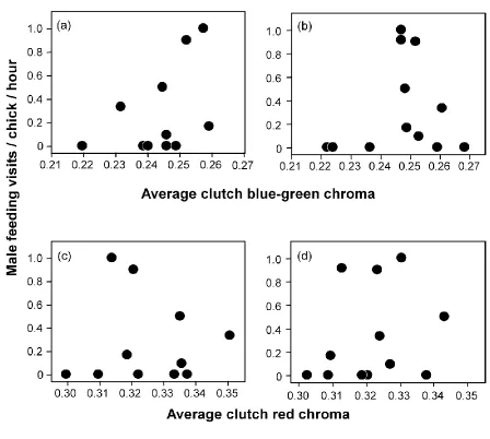
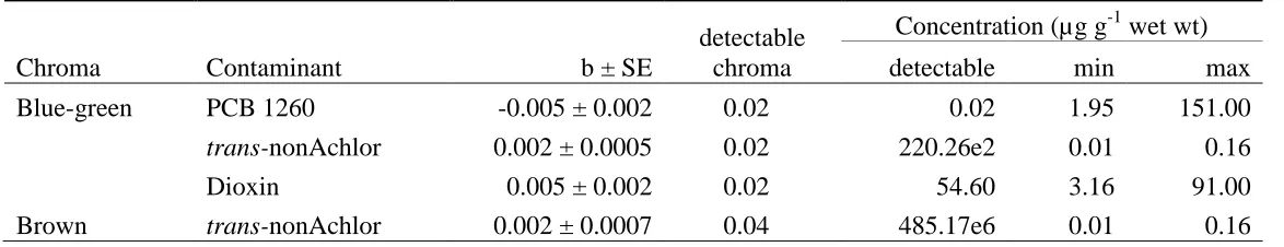
Do contaminant levels explain variation in egg colouration? ... 83
Are differences in chroma associated with contamination levels visually detectable in the field? ... 84
Discussion ... 85
Acknowledgements ... 90
References ... 91
CHAPTER 4-PARENTS, PREDATORS, PARASITES, AND THE EVOLUTION OF COLOUR IN EGGS .1 Summary ... 101
Methods ... 110
Egg reflectance ... 110
Influence of egg fading ... 113
Natural history data ... 114
Phylogenetic reconstruction & comparative analyses ... 115
Results ... 116
Crypsis and aposematism hypotheses ... 116
Blackmail hypothesis ... 116
Sensory bias hypothesis ... 117
Sexual signalling hypothesis ... 118
Parasitism recognition hypothesis ... 118
Coloniality recognition hypothesis ... 119
Discussion ... 119
Acknowledgments ... 128
References ... 128
CHAPTER 5-A COMPARATIVE TEST OF VISIBILITY, ANTI-MICROBIAL, AND SOLAR RADIATION HYPOTHESES FOR THE EVOLUTION OF EGG COLOUR IN BIRDS ...142
Summary ... 143
Introduction ... 144
Methods ... 149
Egg reflectance ... 149
Natural history data ... 151
Phylogenetic reconstruction and comparative analyses ... 152
Results ... 154
xii
Anti-microbial hypothesis ... 154
Solar radiation hypothesis ... 155
Discussion ... 155
Acknowledgments ... 161
References ... 161
CHAPTER 6-AVIAN EGGS PHOSPHORESCE ...175
Summary ... 176
Introduction ... 177
Materials and Methods ... 179
Results ... 180
Discussion ... 181
Acknowledgements ... 183
References ... 183
CHAPTER 7–GENERAL DISCUSSION ...189
Dissertation summary and implications ... 190
Areas of future research ... 195
References ... 197
APPENDIX 1–NATURAL HISTORY REFERENCES ...202
APPENDIX 2–PHYLOGENETIC RECONSTRUCTION ...265
APPENDIX 3-BOX-COX TRANSFORMATIONS ...275
APPENDIX 4–SUPPELEMENTARY VIDEO (SEE CD) ...277
APPENDIX 5- SUPPLEMETARY MATERIAL FOR CHAPTER 6 ...278
xiii LIST OF TABLES
Table 2.1 – Offspring quality ... 65
Table 2.2 – Parental investment ... 66
Table 3.1 – Model predicted chroma ... 98
Table 5.1 – Eggshell brightness and hatching success ... 169
Table 5.2 – Hatching success across microbial risk levels ... 170
x LIST OF FIGURES
Figure 1. 1 – Molecular structure of egg pigments ... 33
Figure 1. 2 – Pigment absorbance ... 34
Figure 1. 3 – Diversity of avian egg colour ... 36
Figure 2. 1 – Ring-billed gull egg reflectance ... 67
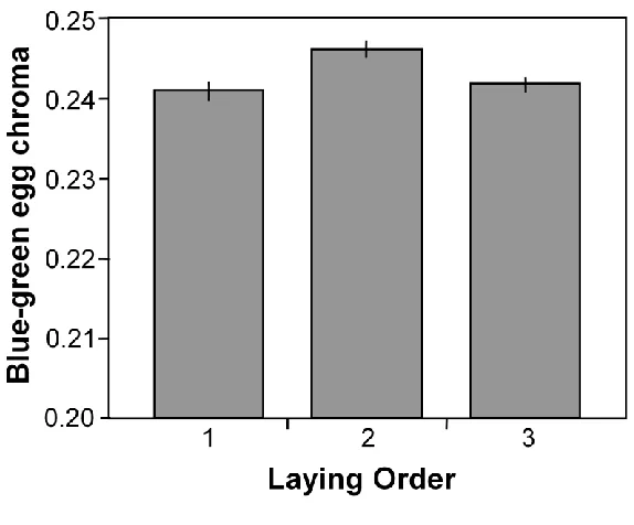
Figure 2. 2 – Blue-green egg chroma across the laying sequence ... 68
Figure 2. 3 – Paternal provisioning ... 69
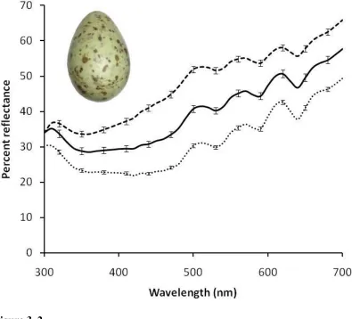
Figure 3. 1 – Herring gull egg reflectance ... 99
Figure 3. 2 – Great Lakes map ... 100
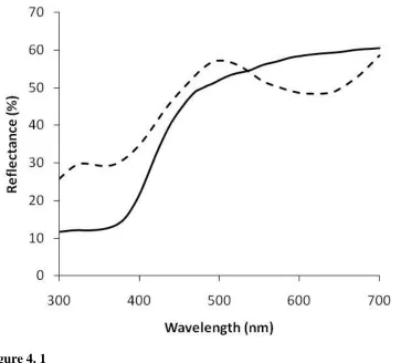
Figure 4. 1 – Proportional blue-green chroma ... 139
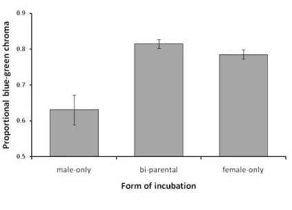
Figure 4. 2 – Proportional blue-green chroma by form of incubation ... 140
Figure 4. 3 – Blue-green egg chroma by form of parental care ... 141
Figure 5. 1 – Eggshell brightness across nest types ... 171
Figure 5. 2 – Relationship between hatching success and eggshell brightness ... 172
Figure 5. 3 – Eggshell ultraviolet chroma across nest types ... 173
Figure 5. 4 – Eggshell brightness and ultraviolet chroma across habitat types ... 174
Figure 6. 1 – Pied-billed grebe (Podilymbus podiceps) egg reflectance measuresd with multiple lamp types ... 187
Chapter 1 – General Introduction
2 Sexual reproduction
Sexual organisms are derived from the unification of parental gametes
(Gegenbaur 1859; Kökkiker 1899, as cited in Mayr 1982). One gamete, known as the ovum, is larger and generally less mobile than its smaller, highly motile counterpart,
known as sperm. This difference in gamete size, known as anisogamy, is maintained by the combined effect of competition of two or more sperm attempting to fertilize the ovum (sperm competition), and an increased likelihood of fertilization if one gamete is
numerous and small (Parker 1982). This distinction has important implications for parental investment. Specifically, males with motile gametes (sperm) invest in quantity,
while females with larger immobile gametes (eggs) invest more in the quality of the gamete (Trivers 1972). This difference between the sexes provides an opportunity for the female to provision the cell with more than just a haploid set of genes. Once fertilization
occurs, the developing zygote uses maternal resources allocated to the ovum. Since females have the opportunity to provision their offspring with resources, they have some
options available in terms of how they will allocate those resources across progeny. The differential allocation hypothesis suggests that a female mated to a high quality partner should increase her maternal investment (Burley 1986). Such maternal investment has
been found in the zebra finch (Taeniopygia guttata), where females add more testosterone to their eggs when mated to more attractive males (Gil et al. 1999).
However, these types of decisions about maternal allocation need not necessarily be in response to the perceived attractiveness of her mate. Females may also choose to invest more or less based on environmental conditions and to enhance the competitive ability of
Chapter 1 – General Introduction
3 Females incur a number of costs associated with egg production, which can limit when and how often a female will become fertile (Monaghan and Nager 1997; Monaghan
et al. 1998).Oviparity, or the production of eggs outside the body, restricts females to depositing eggs under only certain favourable conditions. For example, many conditions
are too harsh or unstable for the development of external eggs (Andrews and Mathies 2000). As females invest heavily into the production of the eggs themselves, they may face limitations on the number of eggs, quality of these eggs, or frequency with which
they lay (Monaghan and Nager 1997). Birds, in particular, display an interesting array of investment strategies, ranging from raising a single brood, raising multiple broods per
year, raising offspring in two separate nests, leaving eggs to hatch from the heating action of decomposing debris, and even laying their eggs within the nests of conspecifics (intra-specific brood parasitism) or hetero(intra-specifics (inter-(intra-specific brood parasitism), therby
evading their parental care responsibilities, with variable investment by the male within these strategies (Kendeigh 1952; Verner and Willson 1969).
Another important yet understudied female investment strategy lies in the
deposition of pigments into eggshells, which produces a vast array of colours and patterns across the class Aves (Kennedy and Vevers 1976; Kilner 2006; Walters 2006). My
dissertation research will investigate the functional significance and evolution of avian egg colouration.
Formation of the avian egg
As with most vertebrates, female birds are born with all of the gametes (oöcytes)
Chapter 1 – General Introduction
4 not occur until the proper hormonal triggers have begun the egg formation process. Although there are large interspecific differences in when females reach their age at first
reproduction (Møller 2006; Wasser and Sherman 2009), the process of egg formation is remarkably similar between species (Romanoff and Romanoff 1949). In birds for
example, environmental cues such as variation in day length are important hormonal triggers for egg formation (Bentley et al. 2000; Visser and Sanz 2009). One hormone integral to ovum development is the follicle stimulating hormone (Romanoff and
Romanoff 1949; Onagbesan et al. 1999). This hormone, in conjunction with an insulin-like growth factor, is responsible for the rapid growth of follicular ova, and the timing of
these processes are tied to a species-specific breeding cycle. Ova develop sequentially and the length of this process depends on the size of the bird and the size of the clutch it will lay (ranging from 4-5 days in Passeriformes to 16 days in Sphenisciformes).
Prolactin levels increase at the beginning of egg laying and inhibit further egg production, which presumably corresponds to a transition from laying to incubation behaviour (Burke
and Dennison 1980).
Prior to ovulation, while ova are still attached to the ovary, a vascularised follicle surrounds the primordial oöcytes and allows for the addition of yolk. Through this
process oöcytes develop into ova, which are attached to the ovary by a small structure known as the peduncle. The liver-produced proteins and lipids that form the yolk are then
transferred through the blood and accumulate in the yolk sac via receptor-mediated endocytosis (Romanoff and Romanoff 1949; Hirayama et al. 2003). In some species, this increase in ovum mass represents a greater than 1000% increase from its original size
Chapter 1 – General Introduction
5 ovulation, the peduncle is cleaved at its base, known as the stigma, and is released from the ovary into the oviduct. The region of the oviduct that receives the ovum is known as
the infundibulum, and it pulses back and forth towards each successive ovum. By the time the follicle breaks, the ovum is within the infundibulum where fertilization will
occur (for a more complete review, Romanoff and Romanoff 1949).
The structure of the avian oviduct allows a female to store sperm for long periods of time prior to fertilization (Birkhead and Møller 1992; Das et al. 2008). The sperm is
stored in sperm storage tubules that are located at the junction of the vagina and uterus, situated at the opposite end of the female‟s reproductive tract to the site of fertilization
(Bobr et al. 1964). During the laying period, sperm must be continuously secreted from the sperm storage tubules so that it can travel to the infundibulum where fertilization occurs (Baskt 1998). This mechanism facilitates insemination even if females have not
mated at the exact moment that would allow both the sperm and ovum to coincide within the infundibulum.
Once fertilization has occurred, the ovum moves further along the oviduct into the magnum, where the egg undergoes the process of albumen addition. There are actually four dehydrated layers of albumen, including the familiar layer of white twisted-looking
strands that is found on either end of the yolk. This layer comprises strands known as the chalazae, which take this form because the ovum is slowly rotated as this layer is secreted
around it. More specifically, the chalaza attached to the pointed end of the egg is longer, thicker, and more firmly attached to the albumen, and it is twisted in a counter clockwise direction. The chalaza at the blunt end of the egg is twisted in a clockwise direction as it
Chapter 1 – General Introduction
6 blastoderm oriented upwards and within the geometric center of the egg (Romanoff and Romanoff 1949; Rahman et al. 2007). After the chalazae are added, the remaining three
layers of albumen are added over top. The egg continues to move away from the
infundibulum into the isthmus where the porous inner- and outer- membranes are added.
The inner membrane is a fine mesh of keratin fibres, while the outer membrane is composed of a coarser mesh of keratin fibres. The inner keratinized membrane often appears pinkish, and is the reason why some white eggshells appear to have a pinkish
hue. These porous membranes allow for gas and liquid exchange after the egg is laid. It is the permeability of these membrane layers which allows the egg to take on its
characteristic shape. The albumen enclosed within these membranes becomes hydrated at this stage, through a process known as plumping. Now the egg has its ultimate shape and a firmer surface onto which the shell will adhere. In this form, the avian egg is
reminiscent of the eggs of some closely related taxa within Chelonia (turtles, tortoises, and terrapins) and Lepidosauria (scaled lizards) (Ewert 1979). The membrane-bound egg
then moves to the uterus where the process of shell formation begins.
The next step of complete calcification and pigmentation makes bird eggs unique. The evolution of shell calcification is believed to have been linked to selection pressures
caused by soil microbes because the common ancestor of birds and reptiles were likely at risk of microbial invasion (Packard and Packard 1980). This hypothesis proposes that the
calcified shell reduces permeability, and therefore provides greater protection for developing embryos. Nonetheless, there remains a great diversity in the degree of shell calcification found in reptiles (Ewert 1979; Packard and DeMarco 2004) and an
Chapter 1 – General Introduction
7 Within the uterus, eggshell pigments are added to the shell. This process results in the diversity of colours exhibited by avian eggs, which forms the basis of the chapters to
follow. Cone-shaped calcium carbonate structures are first laid over the outer membrane, and these ultimately form what is known as the mammillary layer of the egg. This layer
has the important function of providing calcium necessary for bone formation to the developing embryo (Dieckert et al. 1989). After this layer has been laid, a layer known as the palisade (or spongy) layer is placed over it. This layer is created by the interweaving
of collagen-like fibres and calcite, resulting in the hard dense layer which characterizes the outer surface of avian eggs (Romanoff and Romanoff 1949). It is within this palisade
layer that the eggshell ground colouration is added. Here, when I refer to eggshell ground colouration, I mean the colour that uniformly covers the shell‟s surface. Ground
colouration is created by two pigments that may be found independent of one another or
in combination: proto-porphyrin, which produces brown colours, and biliverdin, which produces blue-green colours (Romanoff and Romanoff 1949; Kennedy and Vevers 1976;
Miksik et al. 1994; Miksik et al. 1996; Gorchein et al. 2009). Although these two
pigments may also be circulating in the blood, those found within the shell originate from within the shell gland (Baird et al. 1975; Zhao et al. 2006). Recent research suggests that
the mechanism behind biliverdin deposition more specifically involves transportation of biliverdin from the shell gland into the uterus fluid; in blue-green eggs, biliverdin in the
shell gland was transferred to uterine fluid and then to the shell surface, while in white eggs, biliverdin was produced in the shell gland but was not present in the uterine fluid (Liu et al. 2010). This implies that once within the fluid, pigments may be easily
Chapter 1 – General Introduction
8 calcium matrix begins after the formation of the palisade layer, and therefore pigments are rarely found within the mammillary layer (Romanoff and Romanoff 1949). However,
there are exceptions to how far pigments penetrate into the shell, even within a single species (personal observation).
Many avian eggs also possess another layer known as the cuticle; however, this layer is not present in all species (e.g., gulls, Romanoff and Romanoff 1949). When present, this layer is comprised of two membranes and covers the entire shell surface,
including numerous pores in the shell. This outer layer is gas permeable, which allows gas exchange necessary to sustain the developing embryo, and is the last feature added to
the egg before laying. The properties of this layer determine the apparent texture of the eggshell (glossy, chalky, etc.).Within this layer, another form of porphyrin-based pigmentation is applied, which creates the familiar brown streaks, spots, and other
markings found atop the ground colouration in a variety of species. This layer is thickest where the pigments are deposited and is otherwise even across the unspotted areas
(Romanoff and Romanoff 1949). Some species, especially those with absent or thin cuticles, will create spots by intermixing pigments within the calcium matrix, known as shell pigments, while the spotting found within the cuticle is known as cuticular pigment
(Romanoff and Romanoff 1949).
Interestingly, spots are placed specifically where the shell is thinnest (Gosler et
al. 2005), which has been hypothesized to be due to a shared carrier protein between porphyrin and calcium (Solomon 1997). Such a mechanism would allow porphyrin to be carried to the shell whenever calcium is lacking.The deposition of pigment where the
Chapter 1 – General Introduction
9 2005). However, researchers have yet to determine the mechanism that allows pigments within this proteinaceous cuticle layer to bind to specific shell areas. For example, the
pigments forming dark eggshell spots could initially be evenly distributed throughout the cuticle layer and then become concentrated at thin parts of the shell. The thin parts of the
shell would then act as sinks for pigment concentration, leading to a patchy distribution of pigmentation in the cuticle layer. More research on dark eggshell spotting is also warranted because dark spots appear to have different photo-electric properties than
lighter speckling (Chapter 6), even though they should be produced by the same pigments (Kennedy and Vevers 1976). More precise analytical approaches will be necessary to
fully characterize the pigment composition of avian eggs. This point is timely because current extraction protocols do not necessarily isolate pigments found in specific areas of the egg; they usually homogenize pigments throughout the shell.
Pigment composition of avian eggs
Although researchers have been in almost unanimous agreement about the general composition and origin of eggshell pigments since the late 1800‟s (Sorby 1875), the
specific composition of pigments has long been debated (Liebermann 1878; Sorby 1878)
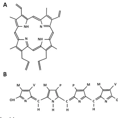
and remains contentious (Lang and Wells 1987; Gorchein et al. 2009). What is certain is that there are two main pigment classes involved in colouring birds‟ eggs: porphyrins and
verdins (Kennedy and Vevers 1976; Miksik et al. 1994; Miksik et al. 1996). These are biologically important pigments, and are intimately connected to the heme biosynthesis pathway, which is necessary for the formation of chlorophyll in plants and haemoglobin
Chapter 1 – General Introduction
10 comprised of four pyrrole subunits, arranged in a ring with substitutions around this ring perimeter (Figure 1A; McGraw 2006). This molecule is constructed by the binding of
identical colourless monopyrrole units. Chain-link polymerization of these pyrroles creates the highly planar, conjugated double bond system which produces the brilliantly
coloured and highly photo-sensitive porphyrin (Needham 1974). Porphyrin has multiple absorption peaks (Figure 2) and on the surface of avian eggs this pigment appears brown to reddish brown. In addition, porphyrin is the precursor to numerous important natural
colourants including chlorophyll and heme, a precursor to hemoglobin that is integral to the oxygenation of living tissues (Ponka 1999). The difference between heme and
chlorophyll begins with the addition of an iron ion (in the case of heme), and a
magnesium ion (in the case of cholorphyll). The porphyrin that precedes the addition of a metal ion is known as proto-porphyrin IX. The majority of investigations have only found
the iron-less proto-prophyrin in avian eggshells (Kennedy and Vevers 1976; Miksik et al. 1994; Miksik et al. 1996; Gorchein et al. 2009). However, some researchers have
detected other forms of natural porphyrins (Sorby 1875; With 1973; Baird et al. 1975), prompting questions about the possible presence of other forms of porphyrin in the eggshell (Lang and Wells 1987; Gorchein et al. 2009). In some cases, the detection of
other natural porphyrins may be the result of experimental contamination (Gorchein et al. 2009).
The second pigment found in avian eggs is biliverdin, which produces blue-green colouration. Researchers have been aware of this pigment‟s role for more than a hundred
years (Sorby 1875); however, biliverdin in avian eggs was known as oöcyan until 1945
Chapter 1 – General Introduction
11 pigment is formed through the oxidation of heme, a process which releases both an iron ion and a single molecule of carbon monoxide (Galbraith 1999). Biliverdin is an
open-chain tetrapyrrole molecule (Figure 1B), and along with its derivatives, is known to have powerful antioxidant capacities (Stocker et al. 1987; Kaur et al. 2003). Biliverdin is
characterized by two major absorption peaks in the 375-384 nm and 665-670 nm ranges (Figure 2; Ding and Xu 2002; Falchuk et al. 2002).
Genetic determination of eggshell pigments
For either natural or sexual selection to act on a trait, variation within the trait
needs to be heritable (Darwin 1871). Heritability, or the proportion of variation in a trait attributable to an organism‟s genes rather than environmental conditions, can be
calculated to determine if a trait meets this basic criterion for selection (Boag and Grant
1978). Considering the wealth of empirical and theoretical studies on egg colouration (Underwood and Sealy 2002; Kilner 2006; Cherry and Gosler 2010), there has been
surprisingly little research on the environmental and genetic control of egg colour. Nevertheless, our knowledge of the heritability of egg colour is expanding, and we are beginning to understand at least generally how several different forms of pigmentation
are inherited. The heritability of white and brown colours has been well studied in poultry (Wei et al. 1992; Francesch et al. 1997; Zhang et al. 2005); however, less effort has
focused on blue-green egg colour. It has been proposed that blue shell colouration is under simple autosomal dominance (Punnett 1933; Stevens 1991) that involves independent pairs of alleles at two loci (Collias 1993), although this may be an
Chapter 1 – General Introduction
12 as “white,” “emarld,” or “turquoise.” Although this work carefully describes what was
known about eggshell pigmentation at the time, these colour classifications do not
currently have an adequate pigment strategy to explain them, nor was there any attempt to use an analytical approach to quantify them. If future work should find other pigments
in avian eggs, this genetic control mechanism may provide an adequate explanation. More careful genetic studies outlined a similar system in the Japanese quail (Coturnix japonica) (Ito et al. 1993). An eggshell colour mutation, known as celadon, entered a
captive population and produced blue-green eggshells. This mutation was controlled by an autosomal recessive gene (ce) and is located on a different locus than the gene
controlling white eggshells in Japanese quail (we). These loci are not linked, but the phenotypic expression of ce is masked by the expression of we (Ito et al. 1993). In combination, these two studies provide evidence for a two-allele system for the genetic
control of egg colour.
A recent five-year study has established heritability measures for blue-green
eggshell colour in a population of pied flycatchers (Ficedula hypoleuca), and has shown that in this population, within-clutch standard deviation in blue-green chroma and egg brightness were the most heritable aspects of eggshell colouration (Morales et al. 2010).
In addition, investigations into the inheritance of eggshell spotting has shown that this trait is sometimes linked to the female W chromosome (Gosler et al. 2000), while in other
cases it is not (Mahler et al. 2008). These investigations establish that there is a genetic component to egg colouration on which selection may operate, despite there also being a significant environmental component (Avilés et al. 2007; Jagannath et al. 2008; Morales
Chapter 1 – General Introduction
13 Objective colour measurement
Although there are numerous methods for quantifying colour (Andersson and Prager 2006; Montgomerie 2006), and many different colour spaces in which colours
may be modeled (Wyszceki and Stiles 1982; Endler and Mielke 2005), I will restrict this discussion to the field of spectroscopy, which is the technique I used in the following chapters. Spectroscopy involves the quantification of light emitted from surfaces. The
reflectance of a surface is defined as the ratio of reflected light to incident light across a range of wavelengths (Wyszceki and Stiles 1982). In behavioural sciences, reflectance is
often expressed as a percentage relative to a white standard. The wavelengths of light are measured in nanometers (nm). A perfectly white object should reflect at 100% across all wavelengths, and the reflectance of other achromatic colours should be similarly even
across all wavelengths but at increasingly lower reflectance levels as you progress from white through grey to black. Throughout this dissertation I use a WS-1 Spectralon-based
white standard, which provides 96% reflectivity between 300 – 400nm, and 99% between 400 – 700nm (Ocean Optics, Dunedin, FL).
Reflectance is generally measured with a device known as a spectroradiometer.
This device measures radiometric quantities across a wavelength range (Wyszceki and Stiles 1982). A spectrophotometer measures both the reflectance and transmission of
light, while simultaneously examining the radiant power of an object at each wavelength relative to incident light. There has been confusion about the terminology regarding the equipment commonly employed by researchers measuring the reflectance of animal
Chapter 1 – General Introduction
14 source, and then percent reflectance across the wavelength range can be determined from these data. These conversions are conducted automatically with most end-user
applications (Ocean Optics, Dunedin, FL). Andersson and Prager (2006) provide a good general rule of thumb: if your instrument “measures the spectral composition of the
radiation as a function of wavelength, it is a spectroradiometer” (p. 50). However,
changes in how spectrometers operate, modern charge-couple device (CCD) spectrometer technology, and integration with computer software seems to be blurring the line between
spectroradiometer and spectrophotometer. This is most likely why companies such as Ocean Optics and many researchers opt for the more generic term spectrometer, which is
the term I use throughout this dissertation.
Throughout this dissertation I used an external light source which provides full spectrum light through a bifurcated fibre optic cable. This cable comprises six separate
fibre optic cables, with the light being delivered through the outer five cables of the bundle. The inner fibre optic cable carries the reflected light back to the spectrometer.
This returning light enters the unit and then is redirected to a diffraction grating. The grating of this component is specifically adjusted for each unit, and essentially separates the light much the way a prism would. This refracted light then is focused on a mirror
which shines the light on the CCD photo-diode array. These diodes are photosensitive and the light that falls on this array is registered as voltage differences across the
Chapter 1 – General Introduction
15 Colour
Our concept of colouration is necessarily anthropogenic. However, if we hope to
understand the function and evolution of colour signals across diverse taxa, it is necessary to have a broader and more generally applicable appreciation of colouration (Endler
1990; Bennett et al. 1994). In the past, perceptual biases dictated how researchers quantified variation in colour, and these biases influenced theories on animal colour perception (Bennett et al. 1994). This illustrates an important point, that colour is more
than just the spectral properties emitted by an object, it is actually a physiological experience for the receiver (Wyszceki and Stiles 1982). A good,
psychologically-grounded definition of colour should take this into account. One such definition is that colour is the perceptual ability of an observer to discriminate two equivalently
illuminated structures of equal size and shape by differences in the spectral composition
of reflected light alone (Wyszceki and Stiles 1982). This definition makes proper measurement difficult, and only recently have our technical abilities caught up with our
conceptual knowledge-base.
In terms of natural pigments, most colours are produced through the transfer of electrical charges from one ion to another. This operates under the general umbrella of
molecular orbital theory and applies to molecules with alternating single and double bonds (Needham 1974). Generally, larger molecules with multiple rings, or those
possessing side groups, have extended pi orbitals, which define the combined wave characteristics of the electrons comprising the molecule (Nassau 1997). These molecules exhibit absorption properties in the human-visible range. These properties are shared by
Chapter 1 – General Introduction
16 difference between the structures of porphyrin and biliverdin explain the variation in their absorption spectra. In addition, these differences in orbitals, conjugation, and resonance
explain differences in the luminescence properties of these two pigments. This point will be elaborated on more thoroughly in Chapter 6. Human perception has traditionally been
used to classify which molecules are considered pigments. For example, although simple benzene rings can be excited in the ultraviolet range (Nassau 1997), these are not
considered pigments because humans lack the ability to detect ultraviolet light.
Nevertheless, these molecules may be important for organisms with different perceptual abilities (see Avian Vision section, below).
Numerous terms are used to describe colour such as hue, saturation, chroma, and brightness. These are complicated by the colloquial usages of colour terms that are also used in a technical sense (MacAdam 1997). Hue represents the perception of
predominant wavelengths of colour (such as red, blue, yellow, etc.). Saturation and chroma can be thought of as the degree of purity of the colour, while brightness refers to
its value on a white to black scale (Kelber et al. 2003). In the human visual system, any colour can be explained by two chromatic (hue, saturation) and one achromatic
(brightness) aspect of colour. Variation in colours is detected by the combined output of
photoreceptors known as rods and cones. These receptors are activated at different thresholds of light. Rods are active in low light and are the predominant photoreceptors
used in scotopic conditions such as at night, whereas cones are activated at high light levels often experienced in full daylight (Jacobs 1981; Kelber et al. 2003). Furthermore, cones possess pigments, known as photopigments, which have specific absorptance
Chapter 1 – General Introduction
17 differentially absorb light across the spectrum based on the photopigment that they
possess, and these differences can be used to classify different cone types. To
discriminate between colours, a viewer must possess at least two distinct cone types (Jacobs 1981; Wyszceki and Stiles 1982; Kelber et al. 2003); however, possessing
multiple cone types does not necessarily equate to possessing colour vision (Chen et al. 1984; Chen and Goldsmith 1986). In addition to these reception prerequisites, the perception of colour is also dependent on subsequent neurological stages (Jacobs 1981).
Careful physiological, neurological, and behavioural experimentation are necessary to determine if an animal has colour vision (Jacobs 1981; Kelber et al. 2003). Such
experimentation has improved our understanding of both mammalian and avian colour vision and has contributed significantly to the study of animal behaviour (Vorobyev et al. 2001; Goldsmith and Butler 2003, 2005).
Visual systems of avian nest predators
An appreciation for the visual abilities of potential predators has important implications for avian egg colour (Ricklefs 1969; Bosque and Bosque 1995; Cain et al. 2006). Aside from birds, mammals and reptiles are important nest predators of birds
(Ricklefs 1969; Bosque and Bosque 1995; Weatherhead and Blouin-Demers 2004; Sinclair et al. 2005; Cain et al. 2006). Snakes may arguably be the most important avian
nest predators in some parts of the world (Weatherhead and Blouin-Demers 2004). Although the visual system of snakes remains poorly described, the photopigments of at least one species seem to be primarily adapted for low light vision and motion detection
Chapter 1 – General Introduction
18 important cue in prey detection, especially when used in combination with other signal reception modalities (de Cock Buning 1983). Mammals also rely heavily on non-visual
signaling modalities (Alberts 1992), although colour has been shown to act as an
important visual cue in this group (Wells and Lehner 1978; Jacobs 1993) and is therefore
worthy of being addressed. Variation in mammalian colour vision is quite high because mutations within the opsin gene that controls photopigment expression are common (Kelber et al. 2003). Unfortunately, little of this diversity has been subjected to rigorous
examination among mammals. Even when information on spectrally distinct cone types is available, mammalian visual abilities have not often been examined behaviourally. We
do have a general understanding of some commonalities in colour vision across this class. Generally, mammals are classified as dichromats, meaning that they have only two cone types, and this distinction results in marked differences from our own trichromatic vision.
When considering the six mammalian families representing the most important avian nests predators (Sinclair et al. 2005), there is variation in the sensitivity of both cone
types (Canidae: 429 and 555 nm, Felidae: 450 and 555 nm, : 444 and 543 nm in tree squirrels, 436 and 518 nm in ground squirrels, Muridae: 360 and 512 nm, Procyonidae: unknown and 560, Didelphidae: unknown and 560; reviewed in, Jacobs 1993). In
dichromats, the spectral sensitivities of both photopigments dictate which colours are differentiable. Primates are also common nest predators; however their visual systems
vary across the order, and even within a species between sexes. Colour vision is important for successful foraging in a number of primate species, and trichromacy is thought to be an adaptation for this lifestyle in some primates (Mollon 1989; Osorio and
Chapter 1 – General Introduction
19 tend to be dichromatic or trichromatic or a combination of both (Jacobs et al. 1996; Kelber et al. 2003). These colour vision abilities may explain the relatively high
occurrence of primate induced nest predation (Olmos 1990; Tarwater 1998; Robinson and Robinson 2001). Birds also possess excellent colour vision and are another important
source of avian nest predation, and the colour of nest contents appears to be an important factor regulating this pressure (Blanco and Bertellotti 2002; Castilla et al. 2007).
Avian vision
Birds possess four spectrally distinct photopigments and have tetrachromatic
vision (Bennett et al. 1994; Church et al. 2001; Hart 2001a; Maddocks et al. 2001; Bennett and Thery 2007). In birds, all four photopigments are involved in colour vision (Church et al. 2001). These photopigments are sensitive over a wide spectral range from
approximately 320 to 700 nm (Chen et al. 1984; Church et al. 1998; Withgott 2000; Hunt et al. 2001; Ödeen and Håstad 2003). The four classes of avian photopigments are
sensitive over different wavelength ranges, which include long-wave-sensitive (LWS; λmax 543 - 571 nm), medium-wave-sensitive (MWS; λmax 497 - 509 nm),
short-wave-sensitive (SWS; λmax 430 - 463 nm), and either violet-short-wave-sensitive (VS; λmax 402 - 426
nm) or ultraviolet-sensitive (UVS; λmax 355 - 376 nm). Although there are interspecific differences in the wavelength of maximum sensitivity for these visual pigments (Hart
2001b), the absorption characteristics of these photoreceptors are generally similar across all birds (Hart et al. 2000; Cuthill 2006). In addition to these photopigments, birds (as well as some fishes, amphibians, and reptiles) possess oil droplets that absorb lower
Chapter 1 – General Introduction
20 between cone type sensitivities, which ultimately improves discriminability between colours (Bowmaker et al. 1997; Hart 2001b).
Illustrating the diversity of avian egg colour
The colour of birds‟ eggs has captured the interest of artists, philosophers, and scientists
for millennia (Stagiritis 350 BC; Wallace 1889; Purcell et al. 2008). When examining the diversity of colours and forms of patterns found across species (Figure 3), it is no wonder
why people have been drawn to this trait. Although this diversity is believed to be
produced by only two pigment classes (Kennedy and Vevers 1976; Gorchein et al. 2009),
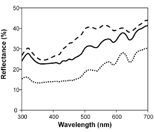
the dramatic variation in egg colour between species suggests that the mechanisms of colouration remain to be fully explained. Colours on the surface of avian eggs reflect many hues within the human visual range (400 – 700 nm). Reflectance spectra for species
that differ in visually perceived egg colour illustrate that the reflectance properties between these eggs are indeed quite different. The blue-green colouration commonly
found in avian eggs is generally similar across species; this colour varies most often in terms of chroma, with some species (Figure 3B) exhibiting higher and narrower reflectance peaks than other species (Figure 3A). As mentioned earlier, the ground
colouration can comprise a combination of biliverdin and porphyrin, which can result in olive, brown, or blue-green colours (Figure 3C). Although green eggs are rare, some
species such as the elegant crested tinamou (Eudromia elegans) exhibit remarkably green eggs (Figure 3D). These spectral curves have a fundamentally different spectral shape than those of blue-green eggs (Figures 3A -B), and of blue-green eggs created through a
Chapter 1 – General Introduction
21 produced by fine speckling creates the overall impression of a light brown colour (Figure 3E). Although the deep brown colour found in the eggs of Nothura boraquira are fairly
uncommon (Figure 3F), these colours do occur. Unlike many other egg colours, the deep chocolate brown colours found in this species are very dark and result in relatively low
reflectance across the spectrum.
Concluding remarks
Avian egg colouration is remarkably variable across species (Walters 2006). This variation is primarily produced by the differential contribution of two related pigment
classes that are integrated into the eggshell matrix while the eggs are in utero (Romanoff and Romanoff 1949). In this dissertation, I adopt a comprehensive approach to
understanding variation in egg colour, ranging from the properties of the pigments
themselves to the evolutionary factors influencing the evolution of egg colouration. Here, I provide a brief summary of the chapters that follow.
In Chapter 2, I test the hypothesis that blue-green egg colour may indicate female quality in the ring-billed gull (Larus delawarensis). To this end, I combine an
observational and experimental approach to evaluate multiple assumptions of the sexual
signalling hypothesis: 1) blue-green egg colour is limiting, 2) high quality females produce more chromatic eggs, 3) egg colour reflects offspring quality, 4) males exhibit
post-mating sexual selection based on this proposed egg colour signal.
In Chapter 3, I examine egg colouration in a non-signalling context in a related gull species, the herring gull (Larus argentatus). Specifically, I used the world‟s
Chapter 1 – General Introduction
22 contaminants, and their influence on avian populations of the Great Lakes. This
investigation is the first of its scale to examine the relationship between egg colour and
contaminants.
In Chapter 4, I investigate the role of egg colouration as a potential cue or signal
to conspecifics and heterospecifics. This large-scale comparative analysis involved the reconstruction of a super-tree that includes representatives of all avian orders (except sandgrouse, Pteroclidiformes). In this chapter I examine the hypothesis that egg colours
influence predation levels and found that in open-nesting species, predation pressure was positively related to eggshell brightness. I also examine the blackmail hypothesis we
recently proposed (Hanley et al. 2010), which suggests that females lay colourful eggs to coerce males into providing additional care. For this hypothesis to operate, risk needs to be associated with certain egg colours and parents need to compensate for this risk. In
addition I examine the sexual signalling hypothesis suggesting that egg colour indicates female quality and the sensory bias hypothesis suggesting that egg colour is selected
based on inherent colour preferences. Moreover, I examine the possibility that egg colour enhances egg recognition in the context of brood parasitism and dense coloniality.
In Chapter 5, I use the same comparative information to investigate whether broad
environmental and ecological factors, such has habitat type and the form of nest, are important selective agents for the evolution of egg colour. In particular, I test whether
eggs have been selected to be brighter in cavity nests, if egg pigments have evolved to protect eggs from microbial invasion, and whether egg pigments provide protection from direct solar radiation. Together with chapter 4, this research represents the largest
Chapter 1 – General Introduction
23 In Chapter 6, I provide the first documentation that avian eggshells phosphoresce, and provide evidence to suggest that porphyrin within the shell matrix is the source of
eggshell phosphorescence. This property does not appear to negatively influence eggshell reflectance; however, future egg colour research should utilize light sources that include
ultraviolet irradiance because this would best approximate natural lighting conditions. Phosphorescence has both applied and evolutionary implications. Specifically, this property may be diagnostic of the presence of proto-porphyrin within an egg, and is
Chapter 1 – General Introduction
24 References
Alberts, A. C. 1992. Constraints on the design of chemical communication systems in terrestrial vertebrates. The American Naturalist 139:S62-S89.
Andersson, S., and M. Prager. 2006. Quantifying colors. pp. 41 - 90 in G. E. Hill, and K. J. McGraw, eds. Bird Coloration. Harvard University Press, Cambridge,
Massachusetts.
Andrews, R. M., and T. Mathies. 2000. Natural history of reptilian development: constraints on the evolution of viviparity. Bioscience 50:227-238.
Avilés, J. M., B. G. Stokke, A. Moksnes, E. Røskaft, and A. P. Møller. 2007.
Environmental conditions influence egg color of reed warblers Acrocephalus scirpaceus and their parasite, the common cuckoo Cuculus canorus. Behavioral Ecology and Sociobiology 61:475-485.
Baird, T., S. E. Solomon, and D. R. Tedstone. 1975. Localisation and characterisation of egg shell porphyrins in several avian species. British Poultry Science 16:201-208. Baskt, M. R. 1998. Structure of the avian oviduct with emphasis on sperm storage in the
poultry. The Journal of Experimental Zoology 282:618 - 626.
Bennett, A. T. D., I. C. Cuthill, and K. J. Norris. 1994. Sexual selection and the mismeasure of color. American Naturalist 144:848-860.
Bennett, A. T. D., and M. Thery. 2007. Avian color vision and coloration: multidisciplinary evolutionary biology. American Naturalist 169:S1 - S6. Bentley, G. E., B. D. Spar, S. A. MacDougall-Shackleton, T. P. Hahn, and G. F. Ball.
2000. Photoperiodic regulation of the reproductive axis in male zebra finches, Taeniopygia guttata. General and Comparative Endocrinology 117:449-455. Birkhead, T. R., and A. P. Møller. 1992. Numbers and size of sperm storage tubules and
the duration of sperm storage in birds: a comparative study. Biological Journal of the Linnean Society 45:363-372.
Blanco, G., and M. Bertellotti. 2002. Differential predation by mammals and birds: implications for egg-colour polymorphism in a nomadic breeding seabird. Biological Journal of the Linnean Society 75:137-146.
Boag, P. T., and P. R. Grant. 1978. Heritability of external morphology in Darwin finches. Nature 274:793-794.
Chapter 1 – General Introduction
25 Bosque, C., and M. T. Bosque. 1995. Nest predation as a selective factor in the evolution
of developmental rates in altricial birds. American Naturalist 145:234-260. Bowmaker, J. K., L. A. Heath, S. E. Wilkie, and D. M. Hunt. 1997. Visual pigments and
oil droplets from six classes of photoreceptor in the retinas of birds. Vision Research 37:2183-2194.
Burke, W. H., and P. T. Dennison. 1980. Prolactin and luteinizing-hormone levels in female turkeys (Meleagris gallopavo) during a photoinduced reproductive cycle and broodiness. General and Comparative Endocrinology 41:92-100.
Burley, N. 1986. Sexual selection for aesthetic traits in species with biparental care. The American Naturalist 127:415-445.
Cain, J. W., K. S. Smallwood, M. L. Morrison, and H. L. Loffland. 2006. Influence of mammal activity on nesting success of passerines. The Journal of Wildlife Management 70:522-531.
Castilla, A. M., A. A. Dhondt, R. Díaz-Uriarte, and D. Westmoreland. 2007. Predation in ground-nesting birds: an experimental study using natural egg color variation. ACE-ÉCO 2:2.
Chen, D. M., J. S. Collins, and T. H. Goldsmith. 1984. The ultraviolet receptor of bird retinas. Science 225:337-340.
Chen, D. M., and T. H. Goldsmith. 1986. Four spectral classes of cone in the retinas of birds. Journal of Comparative Physiology A 159:473-479.
Cherry, M. I., and A. G. Gosler. 2010. Avian eggshell coloration: new perspectives on adaptive explanations. Biological Journal of the Linnean Society 100:753-762. Church, S. C., A. T. D. Bennett, I. C. Cuthill, and J. C. Partridge. 1998. Ultraviolet cues
affect the foraging behaviour of blue tits. Proceedings of the Royal Society of London Series B-Biological Sciences 265:1509-1514.
Church, S. C., A. S. L. Merrison, and T. M. M. Chamberlain. 2001. Avian ultraviolet vision and frequency-dependent seed preferences. The Journal of Experimental Biology 204:2491-2498.
de Cock Buning, T. 1983. Thermal sensitivity as a specialization for prey capture and feeding in snakes. American Zoologist 23:363-373.
Collias, E. C. 1993. Inheritance of egg-color polymorphism in the village weaver (Ploceus cucullatus). Auk 110:683-692.
Chapter 1 – General Introduction
26 Darwin, C. 1871. The descent of man, and selection in relation to sex. Murray, London. Das, S. C., N. Isobe, and Y. Yoshimura. 2008. Mechanism of prolonged sperm storage
and sperm survivability in hen oviduct: a review. American Journal of Reproductive Immunology 60:477-481.
Dieckert, J. W., M. C. Dieckert, and C. R. Creger. 1989. Calcium reserve assembly: a basic structural unit of the calcium reserve system of the hen egg shell. Poultry Science 68:1569-1584.
Ding, Z. K., and Y. Q. Xu. 2002. Purification and characterization of biliverdin IXα from
Atlantic salmon (Salmo salar) bile. Biochemistry (Moscow) 67:927-932.
Endler, J. A. 1990. On the measurement and classification of colour in studies of animal colour patterns. Biological Journal of the Linnean Society 41:315- 352.
Endler, J. A., and P. W. Mielke. 2005. Comparing entire colour patterns as birds see them. Biological Journal of the Linnean Society 86:405-431.
Ewert, M. A. 1979. The embryo and its egg: development and natural history. pp. 333-413 in M. Harless, and H. Morlock, eds. Turtles: perspectives and research. John Wiley & Sons, New York.
Falchuk, K. H., J. M. Contin, T. S. Dziedzic, Z. Feng, T. C. French, G. J. Heffron, and M. Montorzi. 2002. A role for biliverdin IXα in dorsal axis development of Xenopus laevis embryos. Proceedings of the National Academy of Sciences of the United States of America 99:251-256.
Francesch, A., J. Estany, L. Alfonso, and M. Iglesias. 1997. Genetic parameters for egg number, egg weight, and eggshell color in three catalan poultry breeds. Poultry Science 76:1627-1631.
Galbraith, R. 1999. Heme oxygenase: Who needs it? Proceedings of the Society for Experimental Biology and Medicine 222:299-305.
Gegenbaur, C. 1859. Grundzüge der Vergleichenden Anatomie Leipzig.
Gil, D., J. Graves, N. Hazon, and A. Wells. 1999. Male attractiveness and differential testosterone investment in zebra finch eggs. Science 286:126-128.
Goldsmith, T. H., and B. K. Butler. 2003. The roles of receptor noise and cone oil droplets in the photopic spectral sensitivity of the budgerigar, Melopsittacus undulatus. Journal of Comparative Physiology a-Neuroethology Sensory Neural and Behavioral Physiology 189:135-142.
Chapter 1 – General Introduction
27 Comparative Physiology a-Neuroethology Sensory Neural and Behavioral
Physiology 191:933-951.
Gorchein, A., C. K. Lim, and P. Cassey. 2009. Extraction and analysis of colourful eggshell pigments using HPLC and HPLC/electrospray ionization tandem mass spectrometry. Biomedical Chromatography 23:602-606.
Gosler, A. G., P. R. Barnett, and S. J. Reynolds. 2000. Inheritance and variation in eggshell patterning in the great tit Parus major. Proceedings of the Royal Society of London Series B-Biological Sciences 267:2469-2473.
Gosler, A. G., J. P. Higham, and S. J. Reynolds. 2005. Why are birds' eggs speckled? Ecology Letters 8:1105-1113.
Harris, M. P. 1964. Aspects of the breeding biology of the gulls Larus argentatus, L. fuscus, L. marinus. Ibis 106:432-456.
Hart, N. S. 2001a. Variations in cone photoreceptor abundance and the visual ecology of birds. Journal of Comparative Physiology a-Neuroethology Sensory Neural and Behavioral Physiology 187:685-697.
Hart, N. S. 2001b. The visual ecology of avian photoreceptors. Progress in Retinal and Eye Research 20:675-703.
Hart, N. S., J. C. Partridge, I. C. Cuthill, and A. T. D. Bennett. 2000. Visual pigments, oil droplets, ocular media and cone photoreceptor distribution in two species of passerine bird: the blue tit (Parus caeruleus L.) and the blackbird (Turdus merula L.). Journal of Comparative Physiology a-Sensory Neural and Behavioral
Physiology 186:375-387.
Herger, B. 2010. XDrawChem v 1.9.9 http://xdrawchem.sourceforge.net/
Hirayama, S., T. A. Bajari, J. Nimpf, and W. J. Schneider. 2003. Receptor-mediated chicken oocyte growth: Differential expression of endophilin isoforms in developing follicles. Biology of Reproduction 68:1850-1860.
Hunt, S., I. C. Cuthill, A. T. D. Bennett, S. C. Church, and J. C. Partridge. 2001. Is unltraviolet waveband a special communicaiton channel in avian mate choice? The Journal of Experimental Biology 204:2499-2507.
Ishikawa, S., K. Suzuki, E. Fukuda, K. Arihara, Y. Yamamoto, T. Mukai, and M. Itoh. 2010. Photodynamic antimicrobial activity of avian eggshell pigments. FEBS Letters 584:770 -774.
Chapter 1 – General Introduction
28 Jacobs, G. H. 1993. The distribution and nature of color vision among the mammals.
Biological Reviews of the Cambridge Philosophical Society 68:413-471.
Jacobs, G. H., M. Neitz, J. F. Deegan, J. Neitz. 1996. Trichromatic colour vision in New World monkeys. Nature 382:156-158
Jagannath, A., R. F. Shore, L. A. Walker, P. N. Ferns, and A. G. Gosler. 2008. Eggshell pigmentation indicates pesticide contamination. Journal of Applied Ecology 45:133-140.
Kaur, H., M. N. Hughes, C. J. Green, P. Naughton, R. Foresti, and R. Motterlini. 2003. Interaction of bilirubin and biliverdin with reactive nitrogen species. FEBS Letters 543:113-119.
Kelber, A., M. Vorobyev, and D. Osorio. 2003. Animal colour vision - behavioural tests and physiological concepts. Biological Reviews 78:81-118.
Kendeigh, S. C. 1952. Parental care and its evolution in birds. Illinois Biological Monographs 22:1 - 356.
Kennedy, G. Y., and H. G. Vevers. 1976. A survey of avian eggshell pigments. Comparative Biochemistry and Physiology 55:117-123.
Kilner, R. M. 2006. The evolution of egg colour and patterning in birds. Biological Reviews 81:383-406.
Kökkiker, A. 1899. Erinnerungen aus meinem Leben. Engelmann, Leipzig.
Lang, M. R., and J. W. Wells. 1987. A review of eggshell pigmentation. Worlds Poultry Science Journal 43:238-246.
Liebermann, C. 1878. Ueber die färbungen der vogeleierschalen. Berichte der deutschen chemischen Gesellschaft 11:606 - 610.
Liu, H. C., M. C. Hsiao, Y. H. Hu, S. R. Lee, and W. T. K. Cheng. 2010. Eggshell pigmentation study in blue-shelled and white-shelled ducks. Asian-Australasian Journal of Animal Sciences 23:162 - 168.
MacAdam, D. L. 1997. The physical basis of color specification. pp. 33 - 63 in A. Bryne, and D. R. Hilbert, eds. Readings on color, the science of color. the MIT Press, Cambridge, Massachusetts.
Chapter 1 – General Introduction
29 Mahler, B., V. A. Confalonieri, I. J. Lovette, and J. C. Reboreda. 2008. Eggshell spotting
in brood parasitic shiny cowbirds (Molothrus bonariensis) is not linked to the female sex chromosome. Behavioral Ecology and Sociobiology 62:1193-1199. Mayr, E. 1982 The growth of biological thought: diversity, evolution, and inheritance.
Belknap Press, Cambridge, Massachusetts.
McDonagh, A. F. 2001. Turning green to gold. Nature Structural Biology 8:198-200. McGraw, K. J. 2006. Mechanics of uncommon colors: pterins, porphyrins, and
psittacofulvins. pp. 354- 398 in G. E. Hill, and K. J. McGraw, eds. Bird Coloration. Harvard University Press, Cambridge, Massachusetts.
Miksik, I., V. Holan, and Z. Deyl. 1994. Quantification and variability of eggshell pigment content. Comparative Biochemistry and Physiology, Series A 109:769-772.
Miksik, I., V. Holant, and Z. Deyl. 1996. Avian eggshell pigments and their variability. Comparative Biochemistry and Physiology 113B:607-612.
Møller, A. P. 2006. Sociality, age at first reproduction and senescence: comparative analyses of birds. Journal of Evolutionary Biology 19:682-689.
Mollon, J. D. 1989. „Tho‟ she kneel‟d in that place where they grew…‟ The uses and origins of primate colour vision. Journal of Experimental Biology 146:21-38 Monaghan, P., and R. G. Nager. 1997. Why don't birds lay more eggs? Trends in Ecology
& Evolution 12:270-274.
Monaghan, P., R. G. Nager, and D. C. Houston. 1998. The price of eggs: increased investment in egg production reduces the offspring rearing capacity of parents. Proceedings of the Royal Society of London Series B-Biological Sciences 265:1731-1735.
Montgomerie, R. 2006. Analyzing colors. pp. 90 - 148 in G. E. Hill, and K. J. McGraw, eds. Bird Coloration. Harvard University Press, Cambridge, Massachusetts. Moore, M. R. 1998. The biochemistry of heme synthesis in porphyria and in the
porphyrinurias. Clinics in Dermatology 16:203-223.
Morales, J., S. Y. Kim, E. Lobato, S. Merino, G. Tomas, J. Martinez-de La Puente, and J. Moreno. 2010. On the heritability of blue-green eggshell coloration. Journal of Evolutionary Biology 23:1783-1791.
Morales, J., A. Velando, and R. Torres. 2011. Biliverdin-based egg coloration is
Chapter 1 – General Introduction
30 Nassau, K. 1997. The causes of color in A. Byrne, and D. R. Hilbert, eds. Readings on
color, the science of color. The MIT Press, Cambridge, Massachusetts.
Needham, A. R., ed. 1974. The significance of zoochromes. Springer-Verlag, New York. Ödeen, A., and O. Håstad. 2003. Complex distribution of avian color vision systems
revealed by sequencing the SWS1 opsin from total DNA. Molecular Biology and Evolution 20:855-861.
Olmo, F. 1990. Nest predation of plumbeous ibis by capuchin monkeys and greater black hawk. Wilson Bulletin 102: 169-170.
Onagbesan, O. M., B. Vleugels, N. Buys, V. Bruggeman, M. Safi, and E. Decuypere. 1999. Insulin-like growth factors in the regulation of avian ovarian functions. Domestic Animal Endocrinology 17:299-313.
Osorio, D., M. Vorobyev. 1996. Colour vision as an adaptation to frugivory in primates. Proceedings of the Royal Society 263B:593-599.
Packard, G. C., and M. J. Packard. 1980. Evolution of the cleidoic egg among reptilian antecedents of birds. American Zoologist 20:351-362.
Packard, M. J., and V. G. DeMarco. 2004. Eggshell structure and formation in eggs of oviparous reptiles. pp. 53-70 in D. C. Deeming, and M. W. J. Ferguson, eds. Egg incubation: its effects on embryonic development in birds and reptiles. Cambridge University Press, Cambridge.
Parker, G. A. 1982. Why are there so many tiny sperm - sperm competition and the maintenance of two sexes. Journal of Theoretical Biology 96:281-294. Ponka, P. 1999. Cell biology of heme. American Journal of the Medical Sciences
318:241-256.
Punnett, R. C. 1933. Genetic studies in poultry. IX. The blue egg. Journal of Genetics 27:465-470.
Purcell, R., L. S. Hall, and R. Corado. 2008. Egg and nest. The Belknap Press of Harvard University Press, Cambridge.
Rahman, M. A., Baoyindeligeer, A. Iwasawa, and N. Yoshizaki. 2007. Mechanism of chalaza formation in quail eggs. Cell and Tissue Research 330:535-543.
Ricklefs, R. E. 1969. An analysis of nesting mortality in birds. Smithsonian Contribution to Zoology 9:1 - 48.