Page 1 of 9 Review Article
Dendritic cell therapy in melanoma
Carmen Alvarez-Dominguez1, Ricardo Calderón-Gonzalez1, Hector Terán-Navarro1, David Salcines-Cuevas1, Almudena Garcia-Castaño2, Javier Freire3, Javier Gomez-Roman3, Fernando Rivera2
1Instituto de Investigación Marqués de Valdecilla (IDIVAL), Santander 39011, Cantabria, Spain; 2Servicio de Oncología Médica, 3Servicio de Anatomía Patológica, Hospital Universitario Marqués de Valdecilla, Santander 39008, Cantabria, Spain
Contributions: (I) Concepcion and design: All authors; (II) Administrative support: None; (III) Provision of study materials or patients: None; (IV) Collection and assembly of data: All authors; (V) Data analysis and interpretation: All authors; (VI) Manuscript writing: All authors; (VII) Final approval of manuscript: All authors.
Correspondence to: Carmen Alvarez-Dominguez. Instituto de Investigación Marqués de Valdecilla (IDIVAL) and Hospital Universitario Marqiués de Valdecilla, Santander 39011, Cantabria, Spain. Email: [email protected].
Abstract: Dendritic cell (DC) vaccines are cancer vaccines used currently as melanoma therapies. They act as adjuvants initiating the immune responses, but not only as they can also have effector activities redirecting cytotoxic CD8+ T cells against melanoma. Ex vivo preparation of monocyte derived DCs has been implemented to produce large numbers of DCs for clinical therapy, highlighting the necessity of activate DC s to produce Th1 cytokines, especially TNF-a and IL-12 with potent anti-tumour actions. Several clinical trials both in the European Union and USA are open currently using DC vaccines, alone or in combination with other immunotherapies. The type of antigen is also an active area of investigation involving tumour antigens and bacterial epitopes, both providing good responses. Bacterial epitopes presented the advantage versus tumour antigens that they can prepare the vaccination site as they induce innate and specific immune responses as they are potent recall antigens that expand cytotoxic responses.
Keywords: Dendritic cells (DCs); melanoma; Listeria; vaccines
Submitted Apr 14, 2017. Accepted for publication May 10, 2017. doi: 10.21037/atm.2017.06.13
View this article at: http://dx.doi.org/10.21037/atm.2017.06.13
Introduction
Dendritic cells (DCs) are central cells of the immune system at the interphase of innate and specific immunity, the latter also known as adaptive immunity. Therefore, they stimulate naive T cells to initiate the immune responses,
but also, they interact with and influence the responses of
other innate immune cells, such as macrophages or natural killer cells (NK). To initiate the immune responses, DC cells encounter and degrade antigens to small molecules called epitopes. Next, these epitopes bind to either class I major histocompatibility molecules (MHC-I) or class II MHC (MHC-II) to present them to CD8+ T cells (MHC-I)
or to CD4+ T cells (MHC-II) by interaction with the T
cell receptor. This whole process of antigen processing and presentation gives a first signal to T cells (signal 1). This
antigen specific signal 1 in T cells is not sufficient for T cell
activation. T cell activation requires that CD28 molecules
Alvarez-Dominguez et al. Dendritic vaccines for melanoma Page 2 of 9
The function of DC in vivo is the surveillance of the organism to phagocyte and destroy intruders such as pathogens or tumour cells. They can act in two processes of the immune responses, in the priming phase as well as in the effector phase. In the priming phase, DC recognize molecular patterns on pathogens or in tumour cells using different Toll-like receptors (TLR) that activates signalling pathways that remodel cell surface molecules and produce different types of cytokines. Cell surface molecules suffering deep remodelling are those involved in antigen presentation, MHC-I, MHC-II molecules and co-stimulatory molecules, CD40, CD80 and CD86. In this regard, mature DC of any source, either plasmacytoid DC or monocyte derived DC
(MoDC), show significant expression of I or
MHC-II molecules but low or absent expression of co-stimulatory molecules. While activated MoDC present high expression of molecules involved in antigen presentation, MHC-I, MHC-II and high expression of all or some co-stimulatory molecules, CD40, CD80 or CD86. Also mature or activated MoDC produce different types of cytokines. The cytokines produced by DC depend on the signals they receive from the environment, being these cytokines classified as Th1 cytokines/chemokines with a pro-inflammatory pattern, TNF-α, MCP-1, IL-1β, IFN-α/β and IL-12p40/IL-12p70
or Th2 cytokines with predominant anti-inflammatory profiles as IL-6, IL-10 or TGF-β (Figure 2). However, the MoDC cytokine profile in each situation varies and the levels of each one of these Th1 or Th2 patterns determine immune-stimulation or immune-suppression
profile, being in general Th1 profiles of MoDC considered immune stimulatory and Th2 profiles, immune suppressor.
Mature MoDC in general, produce a mixture of Th1 and Th2 cytokines, while activated MoDC produced almost exclusively Th1 cytokines.
Next, MoDC can also act in the effector phase of the immune response, either CD4+ or CD8+ T cells, that are
usually IFN-γ producers. The type of predominant effector cell would depend on the pathogen or tumour cell. In general, pathogens activate both CD4+ and CD8+ effector
T cells that destroy the pathogens either by induction of neutralizing antibodies or activating cytotoxic T cells that lysed those cells infected with the pathogens. In the case of tumour cells, CD8+ T cells are mainly stimulated by DC as
well as NK cells in order they kill directly the tumour cells (2).
Vaccines
Cancer vaccines are expected to activate the immune system
as adjuvants and deliver antigens to T cells. Cancer vaccines function different than prophylactic vaccines as they are expected to expand only cytotoxic CD8+ T cells to cause
tumour regression. It is suitable that cancer vaccines also decrease the number or activating responses of tumour immunosuppressor signals of T regulatory (Treg) cells or
myeloid derived suppressor cells (MDSC). The different types of cancer vaccines approved by FDA are autologous, peptide, dendritic or vector-based vaccines.
Autologous vaccines are composed of tumour cells extracted from each patient. They may contain tumour-associated antigens (TAA) of each tumour but whole cells are not different than normal cells and therefore, the response they generate might not be specific enough to cause impact in the tumour size. For this reason, autologous
vaccines are modified to improve their immunogenicity.
A variation of autologous vaccines is allogenic whole tumour cell vaccines expressing GM-CSF and irradiated (GVAX) (1). This approach uses a tumour cell line that expresses a plurality of shared tumour antigens such as tyrosinase, gp100, MART-1/melan-A, and MAGE-A3 and
genetically modified to secrete GM-CSF, a potent adjuvant
that induces DC maturation. Peptide vaccines target the
above-mentioned tumour antigens but identifying those peptides unique to cancer cells. While generating specific immune responses, they do not implement survival.
Vector-based vaccines, either bacterial or viruses induce specific as well as substantial immune responses that affect clinical results. They are based in the assumption that the primary function of the immune system is to protect against foreign pathogens. However, they present several disadvantages, as the immune responses against the vector are restricted and they cause pathogenesis or be mutagenized. In this regard, attenuated mutants of Listeria monocytogenes either deficient in two virulence factors ActA
and Internalin B, known as LADD mutants (Aduro Biotech)
(3,4), or deficient in a partial sequence of the main virulence
factor listeriolysin O (LLO) (Advaxis) (5), are constructed to express different tumour antigens, such as prostate PSV or mesothelin (CRS-207) alone or in combination with GVAX (6), supposed a great expectation as these bacteria induced not only strong innate immune responses but also cytotoxic immune responses. However, recently CRS-207 in combination with GVAX failed in trials against pancreatic cancer, getting patients worse and develop symptoms associated with the bacterial pathogen.
Figure 2 Preparation of monocyte derived dendritic cells (MoDC) for therapies and differentiation between mature and activated dendritic
cells: markers and cytokines. The figure describes briefly the procedure to produce MoDC and the markers and cytokines that differentiate
Alvarez-Dominguez et al. Dendritic vaccines for melanoma Page 4 of 9
Dendritic vaccines are potent antigen-presenting cells with high capacity to induce T cell immune responses through eliciting pro-inflammatory cytokine responses and stimulating cytotoxic T cells. However, tumours are immunosuppressive environments and might damp the effectiveness of DC. DC are in very low frequencies in the peripheral blood and tissues and therefore, they should be expanded either in vivo or ex vivo in order to use them as
therapies. The first developing approach using DC vaccines
consisted on infusing immature DC that collected antigens in vivo. Next, MoDC were harvested from patients and
generated at large scale to yield sufficient numbers of cells
for clinical application. Next, MoDC were pulsed ex vivo with TAA, tumour lysates, whole tumour cells, tumour RNA or apoptotic tumour cells or bacteria derived reagents to induce MoDC maturation. Mature MoDC are re-infused
back in patients with the purpose to induce tumour-specific
immune responses.
Current ex vivo expansion of MoDC is performed after incubation with GM-CSF and IL-4 to induce full maturation of DC. The procedure implies first Ficoll isolation of leukocytes, CD45+ cells. Second, monocytes
are selected as CD14+ positive cells in MACS columns
(MACSTM, Myltenyi) to provide large numbers of mature
MoDC. Mature MoDC are next activated with different agents such as bacteria as Mycobacterium BCG strain (BCG) or Listeria monocytogenes (Listeria) that signal through TLR-2 or TLR-4 or bacterial oligonucleotides (ODN) with CpG motifs (Figure 2). Trials with MoDC have used indistinctively either mature or activated DC, but they produce different cytokines that can generate different immune responses. Therefore, it is important DC vaccines produce Th1 cytokines with tumorigenic abilities, such as TNF-α or IL-12p40/IL-12p70, otherwise they would not induce cytotoxic immune responses and be ineffective as anti-tumour therapies. In this regard, ex vivo mature MoDC obtained from patients with malignancies produced predominantly Th2 phenotypes (7). However, if they are activated with adjuvants such as Listeria peptides in nanoparticle platforms, MoDC of these patients shifted
their Th2 immune responses towards Th1 profiles (8).
DC can also be activated in vivo requiring the use of non-specific adjuvants such as GM-CSF, CpG, stimulating cytokines as IFN-α/β, STING-activating cyclic dinucleotides (9,10), bacteria as BCG or Listeria or bacterial products. In this regard, IFN-α is a common adjuvant therapy applied in melanoma patients after surgery to prevent or delay tumour relapses (11-13).
Bacterial toxins can also present anti-tumour activity such as LLO, a pore-forming cytotoxin of Listeria and high
capacity to expand non-specific immune responses, binds
to TLR-2 costimulatory molecules as CD14 and act as potent adjuvant (14).
DC vaccines in melanoma therapy
DC vaccines have been used to activate the immune system in autologous and allogenic vaccination strategies using peptides or tumour lysates (15-18). However, the response rates using DC vaccines have been usually low ranging from 10% to 20%, although they are the only immunotherapy so far that increases patient survival rates. DC vaccines can stimulate the immune system to respond to one specific antigen or carbohydrate moiety or to elicit an immune response against multiple tumour antigens, incorporating either allogenic whole cells, autologous tumour cells, recombinant proteins or tumour lysates. Moreover, DC vaccines are safe immunotherapies compared with other adjuvants as IFN-α, or immune check point inhibitors that cause immunological adverse events and have low number of responders.
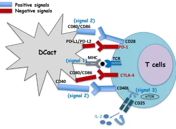
In general, the success of DC vaccines against melanoma is explained by their participation in two stages of the induction of immune responses (Figure 3). First, as adjuvants they act in the priming stage of the immune responses, decreasing the activation of Treg cells localized in
the tumour environment. Moreover, as mentioned before, activated DC vaccines produce large amounts of TNF-α, IFN-α/β and of IL-12p40/IL-12p70 cytokines with anti-neoplastic abilities. These pro-inflammatory cytokines act as feedback mechanisms, activating cytotoxic CD8+ T cells.
Second, as immune stimulators in the effector phase of the
immune responses, activated DC interacted efficiently with
cytotoxic CD8+ T cells and expanded melanoma specific
CD8+ T cells. Finally, melanoma specific CD8+ T cells can
attack melanoma and benefit tumour regression.
Since the approval in 2010 of the first DC vaccine for prostate cancer, sipuleucel-T (Provenge), this early design has improved currently (19) and DC vaccines have raised a huge focus in melanoma treatment. In this regard, trials for human leukocyte antigen (HLA)-A2+ advanced melanomas
have been conducted (20,21), presenting significant clinical responses, albeit the number of patients benefiting of these
Figure 3 Sites of action of dendritic cell vaccines against melanoma. The figure shows real melanoma cells and the two sites of action of
dendritic cell vaccines in the activation of the immune response, at the priming stage also known as adjuvant effect and at the effector stage
also called immunostimulation. The figure also shows the negative signals involved in both stages.
DC and induced high production of IFN-γ. This approach was also used in experimental models of melanoma and observed great reduction of tumour responses. In fact, mice presented 65% of tumour remission and developed effective immune responses. These DC matrices were valid approaches that interacted with immune cells, including lymphocytes (22). One of the first trials using MoDC pulsed with GM-CSF and IL-4, evaluated sixteen patients of melanoma treated with autologous MoDC pulsed with a
cocktail of specific MART-1, MAGE-1, MAGE-3, gp100 or tyrosinase peptides to fit into especial class I HLA molecules
of the patients. This trial also included KLH as an antigen that induced non-specific CD4+ T cells to interact with
CD8+ T cells and DC vaccination was in lymph nodes
non-involved in the tumour. Five out of sixteen patients presented tumour regression and two completed responses
that lasted over fifteen months (15).
Later on, similar approaches were used with a similar peptide cocktail of melanoma antigens, MART-1, Melan-A,
gp100 or tyrosinase to load MoDC in cancer vaccines in phase I and phase II trials (20,21). In fact, peptides, tyrosinase386-406, MART-1/Melan-A51-73 or gp10044-59 showed
high induction of immune responses. Twenty-three patients out of thirty-seven showed antibody and T cell responses,
two patients antibody response only and five showed T cell
response only.
Electroporation of DC with mRNA encoding different activating markers such as CD40L, CD70 and a constitutive active form of TLR-4, referred as TriMix (23) constituted a potent DC-based cellular therapy.
Alvarez-Dominguez et al. Dendritic vaccines for melanoma Page 6 of 9
the use of attenuated Mycobacterium strains as BCG is a frontline treatment in several melanoma trials (24,25) as well as other tumours as bladder cancer. However, the action of BCG implies non-specific immunostimulation of the immune responses and not direct killing of cancer cells. A different action supposes the use of Listeria against melanoma. This rare human bacterial pathogen induced apoptosis of melanoma cells, even at very low doses and expands CD8+ T cell cytotoxic responses, benefiting
also the expansion of melanoma specific cytotoxic responses (26). Moreover, DC loaded with LLO91–99
peptides can act as recall antigens that pre-condition the vaccination sites and induced anti-tumour immune responses (27). This effect appeared similar to the action reported by the potent recall antigen and bacterial toxin, tetanus toxoid loaded into DC vaccines, that induced effect responses against glioblastoma (28).
Another highly active area is the use of biomaterials or nanoparticles to improve DC and T cell function at various tissues for the adjuvant treatment of melanoma (29-32). The use of biomaterials and nanomaterials implies solving several issues with adjuvants as the short half-lives of antigens plus adjuvants or the loss of viability upon in vivo transplantation of activated cell products. Therefore, biomaterials are used to extend the duration of the immune responses as well as controlling antigen localization in tissues or targeting to DC cells. The use of scaffolds of macroporous poly(lactide-co-glycolide)
(PLG) matrices can regulate the trafficking and activation
of DC in the tissues (33). These matrices can be implanted in vivo and potentiate the immune responses. They are fabricated with GM-CSF, a CpG ODN and tumour lysate. When they were implanted in experimental mice, they recruited DC in a melanoma model and increase the cytotoxic responses, causing melanoma regression (34). Other types of materials are gold glyconanoparticles that bear carbohydrates in their structures and therefore are targeted to DC and other antigen presenting cells as macrophages (29,35).
DC loaded with peptides of bacterial toxins able to elicit anti-tumour immune responses as 91–99 LLO peptide, are able to act as efficient adjuvants and induce strong innate and cytotoxic responses, expanding melanoma specific cytotoxic responses and preventing melanoma growth, dissemination and formation of metastasis (27). LLO91–99
peptide loaded into DC is able to induce effector CD8+
T cells that localize in the tumour, inducing apoptosis of melanoma. These DC vaccines also decrease Treg in the
tumours and induce NK cells, therefore, promoting anti-melanoma immune responses, but also increase the positive signals between DC and T cells and control tumour growth and dissemination.
Trials with DC vaccines for melanoma
One of the great advantages of using DC vaccines for melanoma therapy is their safety and potent ability to combine with other immunotherapies as immunological check point inhibitors, either in patients or in experimental models (36-40).
The current trials with DC vaccines for melanoma have been approved by FDA in USA as well as by the European agency of the medicaments and they are compiled in Table 1. Some of these trials involved DC vaccines and other strategies as tumour infiltrating lymphocytes (trial 2006-005238-19 in Spain) or uses electroporated DC with mRNA (trial 2011-001410-33 in Belgium) or combination with other adjuvants as pegylated IFN-α (trial 2005-002636-97 in Great Britain) or the use of the TriMix DC formula combined with ipilimumab (trial 2010-023058-35 in Belgium). Some the trials are for advanced melanoma, others for early stages melanoma IIA, IIB, IIIB, IIIC and others for uveal melanoma. In all cases, DC vaccines are prepared ex vivo using mature MoDC. A large amount of trials haw requirements of HLA-A2 expression as well as other melanoma specific antigens such as gp100 or tyrosinase (Table 1).
Conclusions
DC cell vaccines because of their safety and dual action into the two arms of the immune responses, the priming and effector stages are versatile immunotherapies that can be used to stimulate the immune system to fight against melanoma, both early stages and metastatic melanoma and even uveal melanoma. They also accept combination with all types of immunotherapies currently been used for melanoma, immunological check point inhibitors as ipilimumab or adjuvant therapies as pegylated IFN-a. Their high induction of TH1 cytokines as TNF-a and IL-12p40/IL-12p70 and activation of anti-melanoma cytotoxic CD8+ T cells are their best features to be used
as adjuvants, with specific antigens or with polyvalent vaccines. In this regard, bacterial epitopes loaded into DC as LLO91–99 peptides are also emerging as potent
Table 1 Trials in European Union and United States of America involving dendritic cells
Trials with dendritic cells (number,
sponsor, country code and ID) Title Starting date Medical condition
2007-007847-28
(Universitätsklinikum Erlangen-DE) (DERMA-ER-DC08)
A multicenter, randomized, two-armed, open-label phase III study to evaluate the adjuvant vaccination with tumor RNA-loaded autologous dendritic cells versus observation of patients with resected mel…..
2011-09-16 Uveal melanoma
2015-005322-19 (Radboud University Nijmegen Medical Centre-NL) (MIND-DC)
A randomized, double-blind, placebo-controlled phase III study to evaluate active immunization in adjuvant therapy of patients with stage IIIB and IIIC melanoma with natural dendritic cells pulsed...
2016-01-21 Melanoma with regional lymph nodes metastases (stage III)
2012-001410-41 (Istituto Scientifico Romagnolo per lo studio e la cura dei tumori-IT) (IRST172.02)
Vaccination with autologous dendritic cells loaded with autologous tumor lysate or homogenate combined with immunomodulating radiotherapy and/or preleukapheresis IFN-alfa in patients with metastati...
2013-05-30 Metastatic melanoma
2009-015737-73 (Radboud University Nijmegen Medical Centre-NL) (2009-015737-73)
Single-step antigen loading and TLR activation of dendritic cells by mRNA electroporation for vaccination in stage III and IV melanoma patients
2010-02-04 Melanoma patients
TLR ligand matured dendritic cell vaccination in melanoma patients: the key towards a more potent immune induction?
2009-04-27 HLA-A2.1 positive
mRNA-transfected dendritic cell vaccination in high risk uveal melanoma patients
2009-04-14 HLA-A2.1 positive patients at high risk of uveal
Immunochemotherapy: do platin-based chemotherapeutics enhance dendritic cell vaccine efficacy in melanoma patients?
A two-stage phase II study of autologous TriMix-DC
therapeutic vaccine in combination with ipilimumab in patients with previously treated unresectable stage III or IV melanoma
2011-01-04 Previously treated unresectable stage III or IV melanoma
2005-002636-97 (Immuno-Designed Molecules-GB) (DC-MEL-203)
An open-label, phase II study of matured dendritic cells pulsed ex vivo with 3 melanoma cell line lysates (IDD-3) with or without peginterferon alpha-2b (PegIFN-a 2b) in patients with resected sta...
2005-09-19 Patients with cutaneous melanoma (stage IIc, IIIb, IIIc)
2011-001410-33 (UZ Brussel-BE) (UZB-VUB-11-01)
Randomized phase II clinical trial on mRNA electroporated autologous dendritic cells for stage III/IV melanoma in patients who are free from measurable tumor lesions following the local treatment o...
2016-03-10 Stage III/IV melanoma free of measurable tumour
A single-centre, single arm, open-label, exploratory trial of interleukin-2 administered subcutaneously as neo-adjuvant treatment prior to sentinel lymph node biopsy(SLNB)/ complete lymph node disse...
2012-01-02 Melanoma patients stage III
2006-005238-19 (Instituto Cientifico y Tecnologico de Navarra-ES)
Phase II study with immunotherapy with dendritic cells and tumour infiltrating lymphocytes in solid tumours
2007-07-26 Melanoma,
Dendritic cell activating scaffold in melanoma, immunotherapy (vaccine), dendritic cell (phase I)
2012-12-17 Melanoma with recurrent stage IIIC or with stage IV
NCT02334735 (Icahn School of Medicine at Mount Sinai-USA)
A comparison of matured dendritic cells and montanide® in
study subjects with high risk of melanoma recurrence (phase II)
2015-01-06 (recruiting)
Alvarez-Dominguez et al. Dendritic vaccines for melanoma Page 8 of 9
Acknowledgements
Authors acknowledge the technical assistance of lab technician M. Sierra (IDIVAL) and Innovation coordinator P. Zorilla (IDIVAL).
Funding: This study is funded by MINECO (grant: SAF2012-34203) and IDIVAL to C Alvarez-Dominguez (grant: INNVAL15/01).
Footnote
Conflicts of Interest: The authors have no conflicts of interest
to declare.
References
1. Lipson EJ, Sharfmann WH, Chen S, et al. Safety and immunologic correlates of melanoma GVAX, a GM-CSF allogenic melanoma secreting vaccine administered in the adjuvant setting. J Transl Med 2015;13:214.
2. Berraondo P, Minute L, Ajona D, et al. Innate immune mediators in cancer: between defense and resistance. Immunol Rev 2016;274:290-306.
3. Shahabi V, Maciag PC, Rivera S, et al. Live, attenuated strains of Listeria and Salmonella as vaccine vectors in cancer treatment. Bioengineered Bugs 2010;1:235-43. 4. Wood LM, Pan ZK, Shahabi V, et al. Listeria derived ActA is an effective adjuvant for primary and metastatic tumour immunotherapy. Cancer Immunol Immunother 2010;59:1049-58.
5. Pan ZK, Weiskirch LM, Paterson Y. Regression of established B16F10 melanoma with a recombinant Listeria monocytogenes vaccine. Cancer Res 1999;59:5264-9. 6. Le DT, Wang-Guillan A, Picozzi V, et al. Safety and
survival with GVAX pancreas prime and Listeria monocytogenes–expressing mesothelin (CRS-207) boost vaccines for metastatic pancreatic cancer. J Clin Oncol 2015;33:1325-33.
7. Calderon-Gonzalez R, Teran-Navarro H, Marimon JM, et al. Biomarker tools to design clinical vaccines determined from a study of annual listeriosis incidence in northern Spain. Front Immunol 2016;7:541.
8. Calderon-Gonzalez R, Teran-Navarro H, Salcines-Cuevas D, et al. Gold glyconanoparticles coupled to lysteriolysin O 91-99 peptide serve as adjuvant therapy against melanoma. Nanoscale 2017. In Press. doi: 10.1039/ C7NR02494K.
9. Dubensky TW, Kanne DB, Leong ML. Rationale,
progress and development of vaccines utilizing STING-activating cyclic dinucleotide adjuvants. Ther Adv Vaccines 2013;1:131-43.
10. Fu J, Kanne DB, Leong M, et al. STING agonist formulated cancer vaccines can cure established tumors resistant to PD-1 blockage. Sci Transl Med 2015;7:283ra52.
11. Moreno Nogueira JA, Valero-Arbizu M, Perez-Temprano R. Adjuvant treatment of melanoma. ISRN Dermatology 2013;2013:545631.
12. Parker BS, Rautela J, Hertzog PJ. Antitumor actions of interferons: implications for cancer therapy. Nat Rev Cancer 2016;16:131-44.
13. Espinosa E, Soriano V, Malvehy J, et al. Treatment patterns of adjuvant interferon-α2b for high-risk melanoma: a retrospective study of the Grupo Español Multidisciplinar de Melanoma - Prima study. Melanoma Res 2016;26:278-83.
14. Sun R, Liu Y. Listeriolysin O as a strong immunogenic molecule for the development of new anti-tumor vaccines. Hum Vaccin Immunother 2013;9:1058-68.
15. Nestle FO, Alijagic S, Gilliet M, et al. Vaccination of melanoma patients with peptide-or tumor lysate-pulsed dendritic cells. Nat Med 1998;4:328-32.
16. Butterfield LH. Dendritic cells in cancer immunotherapy
clinical trials: are we making progress? Front Immunol 2013;4:454.
17. Berrocal A, Cabañas L, Espinosa E, et al. Melanoma: diagnosis, staging, and treatment. Consensus group recommendations. Adv Ther 2014;31:945-60.
18. de Rosa F, Ridolfi L, Ridolfi R, et al. Vaccination with
autologous dendritic cells loaded with autologous tumor lysate or homogenate combined with immunomodulating radiotherapy and/or preleukapheresis IFN-α in patients with metastatic melanoma: a randomised "proof-of-principle" phase II study. J Transl Med 2014;12:209. 19. Wei XX, Fong L, Small EJ. Prostate cancer
immunotherapy with Sipuleucel-T: surrent standards and future directions. Expert Rev Vaccines 2015;14:1529-41. 20. Akiyama Y, Tanosaki R, Inoue I, et al. Clinical response in
japanese metastatic melanoma patients treated with peptide cocktail-pulse dendritic cells. J Transl Med 2005;3:4. 21. Oshita C, Takikawa M, Kume A, et al. Dendritic
cell-based vaccination in metastatic melanoma patients: Phase II clinical trial. Oncol Rep 2012;28:1131-8.
Oncotarget 2016;7:39894-906.
23. Van Lint S, Wilgenhof S, Heirman C, et al. Optimized dendritic cell-based immunotherapy for melanoma: the TriMix formula. Cancer Immunol Immunother 2014;63:959-67.
24. Aris M, Barrio MM. Combining immunotherapy with oncogene-targeted therapy: a new road for melanoma treatment. Front Immunol 2015;6:46.
25. Aris M, Bravo AA, Barrio MM, et al. Inoculation site from a cutaneous melanoma patient treated with an allogenic therapeutic vaccine: a case report. Front Immunol 2015;6:144.
26. Bronchalo-Vicente L, Calderon-Gonzalez R, Freire J, et al. A novel therapy for melanoma developed in mice: transformation of melanoma into dendritic cells with Listeria monocytogenes. PLoS One 2015;10:e0117923. 27. Calderon-Gonzalez R, Bronchalo-Vicente L, Freire J, et al. Exceptional anti-neoplastic activity of a dendritic-cell-targetted vaccine loaded with a Listeria peptide proposed against metastatic melanoma. Oncotarget 2016;7:16855-65. 28. Mitchell DA, Batich KA, Gunn MD, et al. Tetanous toxoid
and CCL3 improve dendritic cell vaccines in mice and gliobastoma patients. Nature 2015;519:366-9.
29. Ojeda R, de Paz JL, Barrientos AG, et al. Preparation of multifunctional glyconanoparticles as a platform for potential carbohydrate-based anticancer vaccines. Carbohydr Res 2007;342:448-59.
30. Brinãs RP, Sundgren A, Sahoo P, et al. Design and synthesis of multifunctional gold nanoparticles bearing tumor-associated glycopeptide antigens as potential cancer vaccines. Bioconjug Chem 2012;23:1513-23.
31. Silva JM, Videira M, Gaspar R, et al. Immune system targeting by biodegradable nanoparticles for cancer vaccines. J Control Release 2013;168:179-99.
32. Irvine DJ, Hanson MC, Rakhra K, et al. Synthetic Nanoparticles for Vaccines and Immunotherapy. Chem Rev 2015;115:11109-46.
33. Bencherif SA, Sands RA, Ali OA, et al. Injectable cryogel-based whole cell cancer vaccines. Nat Commun 2015;6:7556.
34. Ali OA, Doherty E, Mooney D, et al. Relationship
of vaccine efficiency to the kinetics of DC and T cell
responses induced by PLG-based cancer vaccines. Biomatter 2011;1:66-75.
35. Rojo J, Diaz V, De la Fuente JM, et al. Gold
glyconanoparticles as new tolos as anti-adhesive therapy. Chembiochem 2004;5:291-7.
36. Grosso JF, Jure-Kunkel MN. CTLA-4 blockade in tumor models: an overview of preclinical and translational research. Cancer Immun 2013;13:5.
37. Selby MJ, Engelhardt JJ, Johnston RJ, et al. Preclinical Development of Ipilimumab and Nivolumab Combination Immunotherapy: Mouse Tumor Models, In Vitro
Functional Studies, and Cynomolgus Macaque Toxicology. PLoS One 2016;11:e0161779.
38. Ali OA, Verbeke C, Johnson C, et al. Identification of
immune factors regulating anti-tumour immunity using polymeric vaccines with multiple adjuvants. Cancer Res 2014;74:1670-81.
39. Sanmamed MF, Carranza-Rua O, Alfaro C, et al. Serum
interleukin-8 reflects tumor burden and treatment
response across malignancies of multiple tissue origins. Clin Cancer Res 2014;20:5697-707.
40. Márquez-Rodas I, Marin Gonzalez M, Nagore E, et al., Frequency and characteristics of familial melanoma in Spain: the FAM-GEM-1 Study. PLoS One
2015,10:e0124239.