Drug Design, Development and Therapy
Dove
press
O r i g i n a l r e s e a r c h
open access to scientific and medical research
Open access Full Text article
controlled release of optimized electroporation
enhances the transdermal efficiency of sinomenine
hydrochloride for treating arthritis in vitro and
in clinic
shun Feng,1,* lijun Zhu,1,*
Zhisheng huang,2 haojia
Wang,1 hong li,1 hua
Zhou,3 linlin lu,1 Ying
Wang,1 Zhongqiu liu,1,3
liang liu1,3
1international institute for Translational chinese Medicine, guangzhou University of chinese Medicine, guangzhou, guangdong, 2Department of acupuncture and rehabilitation, guangzhou hospital of integrated Traditional chinese and Western Medicine, guangzhou, guangdong, 3state Key laboratory of Quality research in chinese Medicine, Macau University of science and Technology, Macau, People’s republic of china
*These authors contributed equally to this work
Abstract: Sinomenine hydrochloride (SH) is an ideal drug for the treatment of rheumatoid arthritis and osteoarthritis. However, high plasma concentration of systemically administered SH can release histamine, which can cause rash and gastrointestinal side effects. Topical delivery can increase SH concentration in the synovial fluid without high plasma level, thus minimizing systemic side effects. However, passive diffusion of SH was found to be inefficient because of the presence of the stratum corneum layer. Therefore, an effective method is required to com-pensate for the low efficiency of SH passive diffusion. In this study, transdermal experiments in vitro and clinical tests were utilized to explore the optimized parameters for electropora-tion of topical delivery for SH. Fluorescence experiment and hematoxylin and eosin staining analysis were performed to reveal the mechanism by which electroporation promoted perme-ation. In vitro, optimized electroporation parameters were 3 KHz, exponential waveform, and intensity 10. Using these parameters, transdermal permeation of SH was increased by 1.9–10.1 fold in mice skin and by 1.6–47.1 fold in miniature pig skin compared with passive diffusion. After the electroporation stimulation, the intercellular intervals and epidermal cracks in the skin increased. In clinical tests, SH concentration in synovial fluid was 20.84 ng/mL after treatment with electroporation. Therefore, electroporation with optimized parameters could significantly enhance transdermal permeation of SH. The mechanism by which electroporation promoted permeation was that the electronic pulses made the skin structure looser. To summarize, elec-troporation may be an effective complementary method for transdermal permeation of SH. The controlled release of electroporation may be a promising clinical method for transdermal drug administration.
Keywords: electroporation, sinomenine hydrochloride, transdermal drug delivery, arthritis
Introduction
Rheumatoid arthritis (RA) is a refractory and chronic inflammatory disease, while osteoarthritis (OA) is the most common disorder of the locomotor system. RA and OA can damage joint function, induce joint injury and arthromeningitis, and lead to
premature death.1 Reports have shown that approximately 1% of the world
popula-tion is suffering from RA and that OA affects mainly the elderly.2,3 Nonsteroidal
anti-inflammatory drugs (NSAIDs) have been reported to be widely used in RA and
OA therapy.4,5 However, NSAIDs can induce renal and gastrointestinal toxicities
because they inhibit prostaglandin synthesis, thus limiting the clinical application of
NSAIDs.6 Among the side effects, NSAID-induced enteropathy could increase small
correspondence: Zhongqiu liu; liang liu no 232, Waihuandong road, guangzhou University of chinese Medicine, guangzhou higher education Mega center, Panyu District, guangzhou, guangdong, 510006, People’s republic of china
Tel +86 20 3935 8061 Fax +86 20 3935 8071 email [email protected]; [email protected]
Journal name: Drug Design, Development and Therapy Article Designation: Original Research
Year: 2017 Volume: 11
Running head verso: Feng et al
Running head recto: Electroporation enhances sinomenine hydrochloride transdermal efficiency DOI: http://dx.doi.org/10.2147/DDDT.S136313
Drug Design, Development and Therapy downloaded from https://www.dovepress.com/ by 118.70.13.36 on 21-Aug-2020
For personal use only.
Number of times this article has been viewed
This article was published in the following Dove Press journal: Drug Design, Development and Therapy
Dovepress Feng et al
intestine bleeding and ulcers.7 Thereby, choosing a drug to
reduce RA and OA symptoms and prevent future adverse effects is necessary.
Sinomenine (C19H23NO4), extracted from the stems and
roots of Sinomenium acutum, is a kind of pharmaceutically
active alkaloid with few side effects.8 Previous
pharma-cological studies have demonstrated that sinomenine has
analgesic,9 anti-inflammatory,10 cartilage protection,11 and
immune suppression effects.12 For decades, sinomenine has
been widely used to treat RA and OA in China.13 Compared
with NSAIDs, sinomenine is more effective in amelioration of morning stiffness, painful joints, and erythrocyte
sedimen-tation rate, with less adverse effects on the digestive system.14
Therefore, sinomenine is a valuable remedy to treat RA and OA in clinical practice.
Sinomenine hydrochloride (SH, Figure 1) is designed to
provide better efficacy with daily application.15 SH tablets
and injections have been used effectively for RA and OA therapy. The anti-RA and anti-OA effect of SH might be closely connected with its antiproliferative effect on inflam-matory indexes on articular tissue and intervention of gristle
degeneration and cartilage cell apoptosis.16,17 Hence, the
articular tissue is the primary therapeutic target of SH. SH concentration within the articular tissues, such as synovial fluid (SF) or synovial membrane, is important. Nevertheless, SH tablets and injections are systemically administered. Moreover, the concentration of systemically administered
SH in plasma was much higher compared with that in SF.18
Therefore, comparatively higher plasma concentration is necessary for achieving adequate amounts of SH in SF via systemic administration. Previous studies have demonstrated that high plasma concentration of SH will lead to potent release of histamine in association with the degranulation of
mast cells in mammalian tissues,19 and this could cause rash
and gastrointestinal side effects (eg, nausea, diarrhea, gas-tralgia, and occasionally vomiting). Research has also found that the adverse effects of SH appearing in some animals after dosing might be related to high plasma concentration of
rapid SH distribution to the internal organs.20 Undoubtedly,
topical transdermal drug delivery targeting the joint tissue is a promising therapeutic method for RA and OA patients. On one hand, it decreases drug plasma concentration and SH dis-tribution into the internal organs, which could alleviate side effects. On the other hand, it increases drug concentration in SF, which could enhance drug efficiency. Concurrently, transdermal drug delivery might enable the drug to bypass the first pass effect (oral administration) and might provide
relatively steady drug concentration in SF for long periods
of time.21 Although transdermal delivery of SH has been
reported, the application of the transdermal delivery system is often limited due to the stratum corneum (SC) layer, which
is the primary barrier of the outermost skin layer.22 The
transdermal delivery did not enable the passive diffusion of SH into the skin, thereby reducing SH efficacy.
At present, some physical means, such as microneedles,23
ionophoresis,24 sonophoresis,25 and electroporation,26
have been utilized to enhance transdermal drug delivery. Electroporation has been considered as an efficient physical technique of promoting skin permeability of small molecules
and biomacromolecules.18,27 Electroporation can
compen-sate for the defects of passive diffusion and significantly increase transdermal efficiency by using a series of short,
high-voltage electric pulses.28,29 Furthermore, electroporation
uses only a small instrument, is safe, and is convenient for self-administration by patients.
However, the mechanism by which electroporation improves transdermal efficiency is not clearly understood. For example, why and how could electroporation enhance permeation is yet to be determined. In this study, fluores-cence method and HE staining were chosen to explore the transdermal enhancement mechanism of electroporation. The selection of appropriate parameters of electroporation is one of the keys to high transdermal enhancement effect of electroporation. Three parameters, namely, pulse frequency, pulse waveform, and pulse intensity, of electroporation were selected as the typical parameters to explore the above rela-tionship. The transdermal enhancement effect of electropo-ration was confirmed by the using optimized parameters for electroporation, facilitating clinically transdermal permeation tests. Highly efficient transdermal delivery by using the optimized parameters of electroporation is recommended to enhance SH transdermal administration method in future clinical application.
Materials and methods
Materials
SH solution (25 mg/mL, injection) was provided by Zhengqing Pharmaceutical Group Co., Ltd. (Hunan, People’s Republic of China). SH (Standard substance) was purchased from Dalian Meilun Biotech Co., Ltd. (Dalian, People’s
Republic of China). CaCl2, KCl, NaCl, and NaHCO3 were
purchased from Sigma-Aldrich (St Louis, MO, USA). Tes-tosterone, used as internal standard (IS), was purchased from Nacalai Tesque Company (Kyoto, Japan). Sodium fluorescein
Drug Design, Development and Therapy downloaded from https://www.dovepress.com/ by 118.70.13.36 on 21-Aug-2020
Dovepress Electroporation enhances sinomenine hydrochloride transdermal efficiency
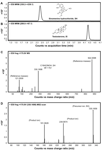
Figure 1 UhPlc–Ms/Ms method for analysis of sh in ringer’s solution and sF.
Notes: The compound-dependent parameters were in MrM method. (A) The transition of sh was 330.2/239.1. (B) The transition of testosterone (is) was 289.0/97.1. (C) and (D) were the Ms spectra and the Ms/Ms spectra of sh, respectively.
Abbreviations: SH, sinomenine hydrochloride; IS, internal standard; MRM, multiple reaction monitoring; MS, mass; ESI, electron spray ionization; SF, synovial fluid;
UhPlc, ultra high performance liquid chromatography.
&
(6,IUDJ 906
5HIHUHQFHPDVVHV
5HIHUHQFHPDVVHV
&+126+ 0+
&RXQWVYVPDVVFKDUJHUDWLRP]
î
'
(6,IUDJ 906VFDQ
3URGXFWLRQ 3URGXFWLRQ
3UHFXUVRULRQ6+
&RXQWVYVPDVVFKDUJHUDWLRP]
î
î
&RXQWVYVDFTXLVLWLRQWLPHPLQ
(6,050±!
$
%
(6,050±!
î
2
+2
2 2
1 + Â+&,
6LQRPHQLQHK\GURFKORULGH6+
7HVWRVWHURQH,6
2
2+ +
+ +
Drug Design, Development and Therapy downloaded from https://www.dovepress.com/ by 118.70.13.36 on 21-Aug-2020
Dovepress Feng et al
(NaFluo) was from GBCBIO Biological Technology Co., Ltd. (Guangzhou, People’s Republic of China). Urethane was purchased from Sinopharm Chemical Reagent Co., Ltd. (Beijing, People’s Republic of China). The water used for the ultra-high performance liquid chromatography–mass spectroscopy/mass spectroscopy (UHPLC–MS/MS) analysis was obtained from a Milli-Q water purification system produced by Millipore (Billerica, MA, USA). HPLC-grade acetonitrile, methanol, and formic acid were acquired from Merck (Darmstadt, Germany). Other reagents were of ana-lytical grade.
animals
All experiments were performed in accordance with the National Institutes of Health Guide for the Care and Use of Laboratory Animals (1996). The animal protocol used in the study was approved by the Institutional Animal Care and Use Committee of the Guangzhou University of Chinese Medicine. Male Kunming mice (weighing 18–22 g) were used in the current experiments. All mice were purchased from the Laboratory Animal Center of Guangzhou University of Chinese Medicine (Guangzhou, People’s Republic of China) and were raised in the animal houses of the Labora-tory Animal Center of Guangzhou University of Chinese Medicine. Four mice were housed per cage in a clean animal cabinet with ad libitum access to nutritionally adequate food and uncontaminated drinking water throughout the experimental periods. The mice were housed in an animal cabinet with unidirectional air flow under 40%–70%
rela-tive humidity, 20°C–24°C, and 12 h light/dark cycle. The
abdominal skins of Bama miniature pigs (weighing 13 kg) were purchased from and prepared at Taizhou Taihe Biotech Co., Ltd. (Taizhou, People’s Republic of China). All animals were killed by cervical luxation after anesthesia with an intraperitoneal injection of urethane (50%, w/v).
skin preparation
The back skins of mice and the abdominal skins of Bama miniature pig were cut using surgical instruments and washed with Ringer’s solution or normal saline. The skins were
divided into 3 cm2 size and stored at −80°C. The pieces of
skins were used within 3 months. The skins were thawed at room temperature and then soaked in Ringer’s solution (pH 7.4) for 1 h before transdermal experiments.
Passive diffusion study
Percutaneous experiments in vitro were done on YB-P6 intel-ligent transdermal diffusion instrument, which was purchased
from Tianjin Pharmacopoeia Standard Instrument Factory (Tianjin, People’s Republic of China), with the perpendicular
Franz-type diffusion cells. The diffusion area was 1.96 cm2.
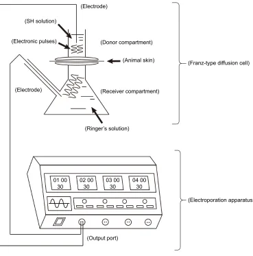
The pieces of skin tissue were sandwiched between the receiver and donor compartments, and the epidermal side was facing up (Figure 2). Diffusion was continued for 8 h
at 37°C±0.5°C.30 A total of 2 mL SH solution was added to
the donor compartment. The mouths of donor compartments were covered with parafilm (Bemis Company, Oshkosh, USA) to avoid liquid evaporation. The receiver compart-ments were filled with 17 mL Ringer’s solution (pH 7.4) and stirred at 400 rpm. The samples (3 mL) were extracted from the receiver compartments for UHPLC–MS/MS analysis and then rapidly replaced with the same quantity (3 mL) of
fresh Ringer’s solution at 37°C±0.5°C at 1, 2, 3, 4, 6, and
8 h. Each experiment was repeated six times.
electroporation study
An electroporation system (Yuandao Digital Electronics Co., Ltd., Shenzhen, People’s Republic of China) was used to provide a series of electronic pulses. The electrode com-ponents, which were made of metal and shaped like a small stick, were placed in the donor and the receptor compartments (Figure 2). Other procedures were the same as described in “Passive diffusion study” section.
Transdermal enhancement regulation
with different electroporation parameters
and different drug concentrations
The relationship between the different frequency, waveform, and intensity of electroporation and the amount of SH that permeated in the in vitro transdermal experiments (mice and miniature pig skins) were studied under the fixed drug concentration.
To study the amount of SH that permeated at differ-ent drug concdiffer-entrations, in vitro transdermal experimdiffer-ents (mice and miniature pig skins) were performed using fixed parameters of electroporation.
Transdermal enhancement mechanism
of electroporation
hematoxylin and eosin staining
Transdermal permeation experiments were done in vitro. Then, the mice and miniature pig skins were immediately col-lected and soaked in 4% paraformaldehyde solution for 24 h
at 4°C. The skin was then dehydrated using gradient alcohol
and xylene, and then embedded in paraffin according to
standard processes. Serial 5 μm-thick sections of skins were
Drug Design, Development and Therapy downloaded from https://www.dovepress.com/ by 118.70.13.36 on 21-Aug-2020
Dovepress Electroporation enhances sinomenine hydrochloride transdermal efficiency
obtained using a Leica RM2235 microtome (Oberkochen, Germany). The histological properties of the skin were ana-lyzed using hematoxylin and eosin (HE) staining and were observed with a Leica DM750 Microscopy.
Transdermal permeation experiments of sodium fluorescein
NaFluo (2 mg) was added to Ringer’s solution (2 mL) and continually vortexed until it dissolved. Transdermal perme-ation experiments (on mice and miniature pig skins) were done using the NaFluo aqueous solution with or without electroporation following the method described in “Passive
diffusion study” Section.31 Then, the skins were immediately
collected and frozen in liquid nitrogen. The skins were then cut using a freezing microtome (Leica CM1860 UV, Leica
Camera AG, Solms, Germany) into 5 μm-thick sections. The
sections were washed thrice with Ringer’s solution (pH 7.4) to wash off free NaFluo and observed with a Leica TSC
SP8 Laser Confocal Microscopy (Oberkochen, Germany). The conditions were as follows: 488 nm excitation wave-length, 515 nm emission wavewave-length, 0 to 255 fluorescence
intensity.32
Transdermal treatment of sh with
optimized electroporation in clinical tests
Patients and study designThe clinical tests were done at Guangzhou Hospital of Inte-grated Traditional Chinese and Western Medicine. These tests did not change the original treatment plan of patients. All patients in the tests were diagnosed with arthritic dis-eases (RA or OA) according to a detailed medical history for knee pain, a comprehensive physical exam, and imag-ing studies. The inclusion criteria were as follows: male or
female 40 years old; presence of knee inflammation, knee
pain, or any other symptom connected with knee arthritis
diseases; and Kellgren–Lawrence radiological grade I–III.33
Figure 2 Flow diagram of transdermal experiment in vitro.
Notes: The skin tissues of mice and miniature pig were fixed between the donor and receiver compartments. Diffusion was continued for 8 h at 37°c±0.5°c.
Abbreviation: sh, sinomenine hydrochloride.
2XWSXWSRUW
(OHFWURSRUDWLRQDSSDUDWXV )UDQ]W\SHGLIIXVLRQFHOO (OHFWURGH
(OHFWURGH (OHFWURQLFSXOVHV
6+VROXWLRQ
$QLPDOVNLQ 'RQRUFRPSDUWPHQW
5HFHLYHUFRPSDUWPHQW
5LQJHU¶VVROXWLRQ
Drug Design, Development and Therapy downloaded from https://www.dovepress.com/ by 118.70.13.36 on 21-Aug-2020
Dovepress Feng et al
Meanwhile, the exclusion criteria were as follows: presence of severe cardiovascular disease, immunodepression, coagu-lopathy, liver disease, infection, and hematological disease; and pregnancy. In patients with bilateral arthritis, only the knee that showed more advanced disease was considered. The protocol was approved by Guangzhou Hospital of Inte-grated Traditional Chinese and Western Medicine Ethical Committee, and informed consent was obtained from all patients. All programs were performed in conformity to the ethical standards of the institutional research committee and in accordance with the 1964 Helsinki Declaration and its latter amendments.
Evaluating the therapeutic efficacy and detecting the concentration of sh in the patient sF in clinical tests
For evaluating clinical efficacy, the test group had nine patients, and the positive control group had six patients. The therapeutic program in the test group was transdermal treatment of SH solution with optimized electroporation combined with standard therapy (eg, celecoxib). Meanwhile, the therapeutic program in the positive control group was only the standard therapy (eg, celecoxib). Clinical efficacy parameters were assessed after 8 days. The parameters for evaluating clinical efficacy were joint pain degree (Visual Analog Scale/Score [VAS]) and joint movement degree. The clinical improvement rate was calculated according to Eq (3). The transdermal treatment method using SH solution with electroporation was as follows: 4 mL SH solution (25 mg/mL, injection) was mixed with 5 mL normal saline and subse-quently vortexed. After vortexing to allow homogenization, the mixture was absorbed by two pieces of cotton. The two pieces of cotton containing the liquid mixture were placed in two electrodes, and the two electrodes were subsequently placed on one knee of the patients. Clinical transdermal tests were done using the liquid mixture with electroporation for 30 minutes. The electroporation conditions were as follows: 3 KHz, exponential waveform, and intensity 10.
For detecting the concentration of SH in the patient SF, the test group had nine patients and the control group had six patients. The clinical transdermal drug delivery in the test group was done using the liquid mixture with electropora-tion for 30 minutes. The clinical transdermal drug delivery in the control group was done using the liquid mixture without electroporation for 30 minutes. After transdermal administration, the patient SF samples were collected by the hospital staff using the SF collection equipment. The con-centration of SH in the patient SF samples were determined by an UHPLC–MS/MS system. The transdermal treatment
method using SH solution with electroporation was done as described earlier.
sample preparation for in vitro
transdermal experiments
Samples for in vitro transdermal experiments (200 μL)
were mixed with 10 μL IS solution (0.5 μmol/L
testos-terone in methanol) and subsequently vortexed for 5 min. After vortexing to allow homogenization, the mixture was
centrifuged at 19,357 g for 30 min. A total of 5 μL of the
supernatant layer was injected into the UHPLC–MS/MS column for analysis.
Patient sF sample preparation
Patient SF samples (200 μL) were mixed with 5 μL IS solution
(25 nmol/L testosterone in methanol) and 1,000 μL methanol
solution (5:1), which was added to extract the analytes and remove proteins, and subsequently vortexed for 5 min. After vortexing to homogenize, the mixture was centrifuged at 19,357 g for 30 min. The supernatant layer was transferred into a new tube and evaporated to dryness in a thermostatic vacuum drier (EYELA Vacuum oven VOS-301SD, Tokyo,
Japan). The residues were redissolved using 100 μL of 50%
methanol aqueous solution and centrifuged at 19,357 g for
30 min. A total of 5 μL of the supernatant layer was injected
into the UHPLC–MS/MS column for analysis.
Analysis and identification of drugs
in the receptors
UHPLC–MS/MS was performed on an Agilent 6540 accurate-mass Q-TOF MS system and 6460 Triple Quadrupole accurate-mass system with an electrospray ionization source (Agilent Tech-nologies, Santa Clara, CA, USA). The data were recorded, and the system was controlled using the MassHunter software (version B.06.00, Agilent Technologies). The conditions
were as follows: column, Zorbax SB-C18, 3.0×100 mm,
1.8 μm (Agilent Technologies), with the column temperature
maintained at 25°C; mobile phase A, 0.1% formic acid water;
mobile phase B, 100% acetonitrile; flow rate, 0.35 mL/min; gradient, 0–1.5 min, 15%–15% B, 1.5–2 min, 15%–90% B, 2–4.2 min, 90%–68% B, 4.2–4.7 min, 68%–15% B; and
injection volume, 5 μL.
The main MS/MS working parameters in the positive mode (1–4.7 min) were set as follows: 3.5 kV capillary
voltage, 500 V nozzle voltage, 300°C gas temperature, 300°C
sheath temperature, 12 L/min sheath gas flow, and 10 L/min gas flow. Quantification was performed via the multiple
reaction monitoring of the transitions of m/z 330.2→239.1
Drug Design, Development and Therapy downloaded from https://www.dovepress.com/ by 118.70.13.36 on 21-Aug-2020
Dovepress Electroporation enhances sinomenine hydrochloride transdermal efficiency
and m/z 330.2→181.0 for SH34 and m/z 289.0→97.1 for IS.
Collision energy was set at 26 for SH and 18 for IS. The low limit of quantitative analysis was 0.023 ng/mL for SH.
Data analysis
Cumulative drug permeation amount (Qn, μg/cm2) was the
cumulative amount of drug transdermally delivered in the receiver compartments, and it was computed according to Eq (1).
Q C V V C
A
n
n i i
n
= × + =
−
∑
0 1
1
(1)
In Eq (1), A (cm2) is the transdermal area, V
0 (mL) is the
bulk of samples extracted from the receiver compartments, V (mL) is the bulk of Ringer’s solution in the receiver
com-partment, and Ci (μg/mL) and Cn (μg/mL) are the
concentra-tion of sample i and n.
Steady-state transdermal flux (Jss, μg⋅cm−2⋅s−1) at time t
represented the slope of the line graphs of cumulative drug
permeation amount (Qn) versus time (t, s). The flux
enhance-ment ratio (ER) induced by electroporation was computed according to Eq (2).
ER Flux with electroporation Flux without electroporation
=
= +
− J J
ss
ss
(2)
Clinical improvement rate was calculated according to Eq (3).
Clinical improvement rate
(Pretreatment value Posttreatmen =
− tt value)
Pretreatment value ×100% (3)
Data were represented as mean ± standard error of mean.
One-way analysis of variance was used to evaluate statisti-cal differences. A difference was considered statististatisti-cally
significant at P0.05.
Results and discussion
Analysis and identification of SH
in samples
UHPLC–MS/MS was performed to identify the structure and analyze SH levels in samples. The results of UHPLC–MS/ MS were as follows: in the representative MRM chromato-gram, retention time of SH was 2.4 min (Figure 1A) and that of IS was 4.2 min (Figure 1B); the high-resolution mass
spectrometry of SH indicated that the [M + H]+=330.1698
ion (Figure 1C), and the MS/MS analyses of SH product the m/z 181.0628 and 239.0674 (Figure 1D). Taken together, the data obtained for the SH in samples was in accordance with the standard substance.
effect of electroporation parameters and
drug concentration on sh penetration in
mice and miniature pig skins
Transdermal enhancement by varying electroporation parameters in mice skin
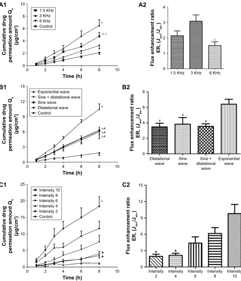
1.5, 3, and 6 KHz were chosen to study the effect of pulse frequency on SH penetration in mice skins. The results
dem-onstrated that the total Qn of 3 KHz was 6.50±0.94 μg/cm2
(Figure 3A1). The ER of 3 KHz was 3.09±0.41, which was
more than the ER seen at other pulse frequencies (Figure 3A2). Taken together, 3 KHz was found to be the optimal pulse fre-quency that had the most significant enhancement-promoting
effect on SH penetration. Interestingly, we found total Qn
differed in the following manner: Qn induced by 3 KHz
Qn induced by 1.5 KHz Qn induced by 6 KHz. The
situa-tion above was exactly opposite to our primary hypothesis that 6 KHz might induce the strongest transdermal enhance-ment. The reason for this phenomenon might be that 6 KHz produced too many pulses, and 1.5 KHz produced too little pulses, which were not in the optimal range inducing change
for skin structure. Thereby, Qn induced by 6 KHz and 1.5 KHz
were less than that induced by 3 KHz, which could produce the exact pulses required for changing the skin structure. To summarize, electroporation did enhance the SH penetration in mice skins, and 3 KHz was the optimal pulse frequency.
Different pulse waveforms produced different Qn in
mice skins. The total Qn of exponential waveform was
10.48±1.08 μg/cm2 (Figure 3B1). The ER of the exponential
waveform was 6.40±0.64 (Figure 3B2). That is, exponential
pulse produced more intense SH permeation. A previous study showed that exponential waveform was one of the
most commonly used pulse waveforms in electroporation.35
Generally, increasing pulse amplitude, duration, and number resulted in increased skin permeability, allowing for better
drug transport.36 The threshold-induced change in the skin
structure depends on the pulse duration. An increase in the electronic pulse duration leads to a decrease in the critical threshold, and so pulse duration of exponential pulse was
long.37 Therefore, we concluded that the exponential
wave-form was the optimal pulse wavewave-form.
The total Qn and ER at intensity 10 were 18.32±2.98 μg/cm2
(Figure 3C1) and 9.79±1.69, respectively, (Figure 3C2), and
Drug Design, Development and Therapy downloaded from https://www.dovepress.com/ by 118.70.13.36 on 21-Aug-2020
Dovepress Feng et al
Figure 3 Transdermal enhancement of pulse frequencies, waveforms, and intensity on sh in in vitro mice skins.
Notes: The effect of pulse frequencies on Qn–time curves (A1) and er (A2). The other fixed parameters of electroporation: sine wave and intensity 4. The effect of pulse
waveforms on Qn–time curves (B1) and er (B2). The other fixed parameters of electroporation: 1.5 KHz and intensity 4. The effect of pulse intensities on Qn–time curves
(C1) and er (C2). The other fixed parameters of electroporation: sine wave and 1.5 KHz. *P0.05 versus the control groups. P0.05 versus the 3 Khz groups. #P0.05
versus the exponential waveform groups. P0.05 versus the intensity 10 groups. Data are represented as mean ± seM. n=6.
Abbreviations: sh, sinomenine hydrochloride; Qn, cumulative drug permeation amount; er, enhancement ratio; seM, standard error of mean.
7LPHK &XPXODWLYHGUXJ SHUPHDWLRQDPRXQW4 Q JFP .+] .+] .+] &RQWURO U
$
)OX[HQKDQFHPHQWUDWLR (5-VV -VV± .+] .+] .+] U$
7LPHK &XPXODWLYHGUXJ SHUPHDWLRQDPRXQW4 Q JFP ([SRQHQWLDOZDYH 6LQHGLODWDWLRQDOZDYH 6LQHZDYH 'LODWDWLRQDOZDYH &RQWURO%
)OX[HQKDQFHPHQWUDWLR (5-VV -VV± 'LODWDWLRQDOZDYH ZDYH6LQH GLODWDWLRQDO6LQH ZDYH ([SRQHQWLDO ZDYH
%
7LPHK &XPXODWLYHGUXJ SHUPHDWLRQDPRXQW4 Q JFP Ÿ Ÿ &RQWURO ,QWHQVLW\ ,QWHQVLW\ ,QWHQVLW\ ,QWHQVLW\ ,QWHQVLW\&
)OX[HQKDQFHPHQWUDWLR (5-VV -VV ± ,QWHQVLW\ ,QWHQVLW\ ,QWHQVLW\ ,QWHQVLW\ ,QWHQVLW\ Ÿ Ÿ&
Qn increased in an intensity-dependent manner in the intensity
range 2–10 (Figure 3C1 and C2). Therefore, enhanced pulse intensity could enhance skin permeability. For our electropo-ration equipment, intensity 10 was the maximum intensity that
could be given to humans without harm, and for animals it was 4. Using higher intensity might result in excessive stimula-tion of the animal and make them feel uncomfortable. The
total Qn and ER of intensity 10 was more than those seen at
Drug Design, Development and Therapy downloaded from https://www.dovepress.com/ by 118.70.13.36 on 21-Aug-2020
Dovepress Electroporation enhances sinomenine hydrochloride transdermal efficiency
other pulse intensities. As expected, intensity 10 was found to be the optimal pulse intensity for humans. Above all, pulse frequency, waveforms, and intensity could affect the SH pen-etration in mice skins, and the optimal parameters were 3 KHz, exponential waveform, and intensity 10 for humans.
Transdermal enhancement of electroporation in miniature pig skins by varying the parameters
The total Qn of 3 KHz, exponential waveform, and intensity
10 were 8.67±0.87, 8.67±0.87, and 53.92±5.07 μg/cm2,
respectively (Figure 4A1, B1, and C1). Meanwhile, ER for 3 KHz, exponential waveform, and intensity 10 were
8.12±0.92, 8.12±0.92, and 48.17±3.93, respectively
(Figure 4A2, B2, and C2). These results showed that Qn and
ER induced by electroporation parameters 3 KHz, expo-nential waveform, and intensity 10 were higher compared with that induced by other electroporation parameters. On analysing the overall situation, the results in miniature pig skins were in accordance with that in mice skins, and the optimal parameters were also same in both mice and min-iature pig skins, and the optimal electroporation parameters were 3 KHz, exponential waveform, and intensity 10 for humans. In practical clinical application, we could choose different parameters of electroporation to achieve different kinds of enhancement in transdermal efficiency so as to achieve the purpose of controlled release drugs.
effect of different drug concentration of sh on cumulative drug permeation amount in mice and miniature pig skins
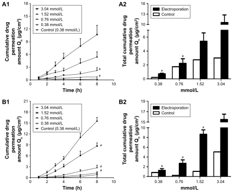
The total Qn of different groups were 0.73±0.08, 2.22±0.67,
5.41±1.21, and 10.57±2.80 μg/cm2 in mice skins (Figure 5A2),
and 1.28±0.35, 2.71±0.71, 8.67±0.87, and 15.50±1.01 μg/cm2
in miniature pig skins (Figure 5B2). We also found that Qn
increased gradually with the increase of the drug concentration in vitro in mice and miniature pig skin permeation experiments with electroporation (Figure 5A1 and 5B1). According to these
results, the total Qn of 3.04 mmol/L was more apparent than
that of other drug concentrations in in vitro mice and miniature pig skins. In brief, drug concentration was one of the key fac-tors for electroporation to promote transdermal penetration of
SH, and Qn values increased in a drug-dependent manner.
Drug penetration routes include intercellular route,
intracellular route, and eccrine route (eg, sweat glands).38 Cell
gap fillers and skin are usually hydrophobic;39 therefore,
pen-etrating through the skin into the blood circulation is difficult
for the hydrophilic SH. As shown above, the Qn of the passive
diffusion transdermal experiments without electroporation were all so low that concentration of SH might not reach the
targeted therapeutic concentration. Through inducing the change of skin structure, electroporation could enable SH to overcome transdermal defects caused by its hydrophilic
property. All Qn values in the four drug concentrations with
electroporation were more than that in control without elec-troporation, and this was a strong proof that electroporation significantly enhanced the transdermal permeation of SH.
Transdermal enhancement mechanism
of electroporation in mice and miniature
pig skins
The epidermis is the outermost skin layer that consists of that
hypodermis, hydrophilic dermis, and epidermis.40 The layers
of the epidermis are stratum spinosum, stratum granulosum, and SC, which protects the skin from damage and is the main
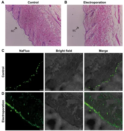
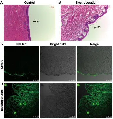
transdermal barrier of the skin.41 On HE staining analysis, the
nucleus was blue and the cytoplasm was red (Figures 6A and B, 7A and B). In normal skin tissues, the skin structures were tight and integrated, and the flattened cells of the epidermis were tightly arranged, and the edge of cells was difficult to distinguish (Figures 6A and 7A). After electroporation stimulation, the skin structures were loose, the intercellular intervals and epidermal cracks of skin increased, and the distribution of stratified cells in SC was chaotic (Figures 6B and 7B). Taken together, electroporation did change the skin structure, increased the skin permeability, and enhanced transdermal drug delivery.
NaFluo is a fluorescent agent with a similar structure to SH. The molecular weight of NaFluo is 376.27. SH and NaFluo are both hydrophilic. NaFluo is only distributed in the outmost SC after passive diffusion without electroporation (Figures 6C and 7C). However, when electroporation was applied to the skins, the stratum spinosum, stratum granulo-sum, and SC were observed to have very strong fluorescence of NaFluo (Figures 6D and 7D), and this meant that NaFluo
deeply permeated into epidermis.42 Therefore, electroporation
could significantly promote NaFluo transdermal permeation. In summary, the mechanism by which electroporation pro-moted permeation was that electronic pulses made the skin looser and increased the intercellular spaces in the skin. Also, a schematic mechanism of electroporation enhancing SH transdermal delivery is shown in Figure 8. In addition, we found that the skin permeability enhanced by electroporation in miniature pig skin was greater than that in the mice skin in this study. This might be because the electroporation thresh-olds and electrical resistance of miniature pig abdomen skin was smaller than that of skin taken from the backs of mice; thus, the electronic pulse could better penetrate the miniature pig skin and cause the skin structure to change.
Drug Design, Development and Therapy downloaded from https://www.dovepress.com/ by 118.70.13.36 on 21-Aug-2020
Dovepress Feng et al
Figure 4 Transdermal enhancement of pulse frequencies, waveforms, and intensity on sh in in vitro miniature pig skins.
Notes: The effect of pulse frequencies on Qn–time curves (A1) and er (A2). The other fixed parameters of electroporation are as follows: exponential wave and intensity 4.
The effect of pulse waveforms on Qn–time curves (B1) and er (B2). The other fixed parameters of electroporation: 3 KHz and intensity 4. The effect of pulse intensities on
Qn–time curves (C1) and er (C2). The other fixed parameters of electroporation: exponential wave and 3 KHz. *P0.05 versus the control groups. P0.05 versus the
3 Khz groups. #P0.05 versus the exponential waveform groups. P0.05 versus the intensity 10 groups. Data are represented as mean ± seM. n=6.
Abbreviations: sh, sinomenine hydrochloride; Qn, cumulative drug permeation amount; er, enhancement ratio; seM, standard error of mean.
7LPHK &XPXODWLYHGUXJ SHUPHDWLRQDPRXQW4 Q JFP &RQWURO 'LODWDWLRQDOZDYH 6LQHZDYH 6LQHGLODWDWLRQDOZDYH ([SRQHQWLDOZDYH 7LPHK &XPXODWLYHGUXJ SHUPHDWLRQDPRXQW4 Q JFP U U .+] .+] .+] &RQWURO 7LPHK &XPXODWLYHGUXJ SHUPHDWLRQDPRXQW4 Q JFP Ÿ Ÿ Ÿ Ÿ &RQWURO ,QWHQVLW\ ,QWHQVLW\ ,QWHQVLW\ ,QWHQVLW\ ,QWHQVLW\
$
%
&
)OX[HQKDQFHPHQWUDWLR (5-VV -VV ± .+] .+] .+] U U$
)OX[HQKDQFHPHQWUDWLR (5-VV -VV± 'LODWDWLRQDOZDYH ZDYH6LQH GLODWDWLRQDO6LQH ZDYH ([SRQHQWLDO ZDYH
%
)OX[HQKDQFHPHQWUDWLR (5-VV -VV± ,QWHQVLW\ ,QWHQVLW\ ,QWHQVLW\ ,QWHQVLW\ ,QWHQVLW\ Ÿ Ÿ Ÿ Ÿ&
Drug Design, Development and Therapy downloaded from https://www.dovepress.com/ by 118.70.13.36 on 21-Aug-2020
Dovepress Electroporation enhances sinomenine hydrochloride transdermal efficiency
Figure 5 relationship between drug concentration and transdermal amount of sh.
Notes: The Qn increased as drug concentration rose in the skin taken from the back of mice (A1) and in miniature pig abdominal skins (B1). Total Qn for different drug
concentrations in mice skins (A2) and in miniature pig abdominal skins (B2). Electroporation condition: exponential wave, 3 KHz, and intensity 4. #P0.05 versus the 3.04 mmol/L groups. Data are represented as mean ± seM. electroporation groups, n=6. control groups, n=1.
Abbreviations: sh, sinomenine hydrochloride; Qn, cumulative drug permeation amount; seM, standard error of mean.
$
$
7LPHK
&XPXODWLYHGUXJ SHUPHDWLRQ DPRXQW4
Q
JFP
PPRO/ PPRO/ PPRO/ PPRO/ &RQWUROPPRO/
PPRO/
7RWDOFXPXODWLYHGUXJ
SHUPHDWLRQ
DPRXQW4
Q
JFP
(OHFWURSRUDWLRQ &RQWURO
%
%
&XPXODWLYHGUXJ SHUPHDWLRQ DPRXQW4
Q
JFP
7LPHK
PPRO/
&RQWUROPPRO/ PPRO/ PPRO/ PPRO/
7RWDOFXPXODWLYHGUXJ
SHUPHDWLRQ
DPRXQW4
Q
JFP
PPRO/
(OHFWURSRUDWLRQ &RQWURO
effect of optimized parameter
electroporation in clinical tests
In the test group, the improvement percentage in joint
move-ment degree was 94.75%±6.84% (Figure 9B), and the
effec-tive percentage of joint pain relief (VAS) was 79.39%±4.63%
(Figure 9C). Compared with the test group, the above two parameters were lower in positive control group, with the improvement percentage of joint movement degree being
52.41%±12.51% (Figure 9D) and the effective percentage
of joint pain relief (VAS) being 62.40%±7.54% (Figure 9E).
Moreover, the improvement percentage of joint movement
degree was statistically significant (P0.05).
The concentration of SH in the patient SF of the test group and the control group was examined. In the test group, the maximum and minimum concentrations of SH were approxi-mately 20.84 and 0.39 ng/mL, respectively (Figure 9A). In
the test group, we found that the concentration fluctuation of SH between different individuals was a little high due to the existence of high personal differences. The concentration of SH in patient SF samples of the control group without elec-troporation was below limit of quantification, so the results of the control group are not shown in this paper.
Based on the above results, high SH concentration in SF might provide better joint pain relief and joint mobility improvement. Therefore, to ensure therapeutic efficacy, we predicted that the minimum therapeutic concentration of SH in SF should be 20.84 ng/mL. As previously reported,
the maximum concentration (Cmax) of transdermally
deliv-ered sinomenine (50 mg/kg) with electroporation was
29.33±4.87 ng/mL in rabbit plasma and 58.87±7.50 ng/mL
in rabbit SF, and these were lower compared with oral
and injection administration.18 However, the C /C
Drug Design, Development and Therapy downloaded from https://www.dovepress.com/ by 118.70.13.36 on 21-Aug-2020
Dovepress Feng et al
$
1D)OXR
P P
P
P P P
%ULJKWILHOG 0HUJH
&RQWURO
(OHFWURSRUDWLRQ
%
&
'
&RQWURO (OHFWURSRUDWLRQ
6&
P
6&
P
Figure 6 effect of electroporation on mice skin structure.
Notes: The analysis of he staining in control groups (A) and in electroporation groups (B). Distribution of naFluo was in mice skins, and the control and electroporation groups are displayed in (C) and (D), respectively. All experiments were performed in triplicate. Magnification ×100.
Abbreviations: NaFluo, sodium fluorescein; HE, hematoxylin and eosin; SC, stratum corneum.
(concentration of sinomenine in SF versus that in plasma)
by electroporation was 3:1, and the CSF/CPlasma of systemic
administration routes was approximately 1:3. In this study,
the Cmax of transdermally delivered SH (11.11 mg/mL,
9 mL, approximately 1.67 mg/kg) with electroporation was
20.84 ng/mL in patient SF. The result revealed that the Cmax
of SH in patient plasma that produced systemic side effects might be low. Hence, the side effects might easily be relieved. Concurrently, high SH concentration in SF could enhance the therapeutic effect of the target joint. In short, transdermal delivery of SH with electroporation could be selected as a promising treatment regimen of RA and OA to improve efficacy and reduce side effects.
Electroporation has some advantages compared with other transdermal enhancing methods. The chemical trans-dermal enhancers have some side effects, such as irritation and irreversible skin damage. However, no skin damage is caused in transdermal treatment with electroporation. Another widely used physical means, ie, sonophoresis, is not convenient due to large machines needed and the necessity for skilled nurses. Electroporation is excellent because it uses small machines and is convenient. In gen-eral, the major advantages of electroporation on improv-ing transdermal drug delivery include high transdermal efficiency, simple performance, safety, and the small instrument needed. Therefore, electroporation could be
Drug Design, Development and Therapy downloaded from https://www.dovepress.com/ by 118.70.13.36 on 21-Aug-2020
Dovepress Electroporation enhances sinomenine hydrochloride transdermal efficiency
1D)OXR
P P P
P P P
%ULJKWILHOG 0HUJH
&RQWURO
(OHFWURSRUDWLRQ
&
'
$
&RQWURO%
(OHFWURSRUDWLRQ6&
P P
6&
Figure 7 effect of electroporation on miniature pig skin structure.
Notes: The analysis of he staining in control groups (A) and in electroporation groups (B). Distribution of naFluo was in miniature pig skins, and the control and electroporation groups are displayed in (C) and (D), respectively. All experiments were performed in triplicate. Magnification ×100.
Abbreviations: NaFluo, sodium fluorescein; HE, hematoxylin and eosin; SC, stratum corneum.
$
&RQWURO
6LQRPHQLQHK\GURFKORULGH
6NLQV
(OHFWURQLFSXOVHV
(OHFWURGH
%
(OHFWURSRUDWLRQ
3XOVHIUHTXHQF\ 3XOVHZDYHIRUP
3XOVHLQWHQVLW\
Figure 8 The mechanism of electroporation enhancing sh transdermal delivery.
Notes: (A) in control, the overall skin was tight and integrated. (B) The intercellular intervals and epidermal cracks of skin were increased after the electroporation stimulation, which would result in that sh had an enhanced permeation in the skin.
Drug Design, Development and Therapy downloaded from https://www.dovepress.com/ by 118.70.13.36 on 21-Aug-2020
Dovepress Feng et al
$
7KHFRQFHQWUDWLRQ
RI6+LQSDWLHQW6)QJP/
3DWLHQWV
3DWLHQWV 3DWLHQWV
3DWLHQWV 3DWLHQWV
%
'
&
(
-RLQWSDLQ
RISUHWUHDWPHQW
-RLQWSDLQ
RISUHWUHDWPHQW
-RLQWPRYHPHQWGHJUHH RISUHWUHDWPHQW
-RLQWPRYHPHQWGHJUHH RISUHWUHDWPHQW
Figure 9 Optimized electroporation increased sh concentration in patient sF and improved the therapeutic effect of sh against arthritis.
Notes: (A) The sh concentration in patient sF of the test group that tested the combination of celecoxib with electroporation. (B) The improvement percentage of joint movement degree in test group. (C) The effective percentage of joint pain relief in test group. (D) The improvement percentage of joint movement degree in the positive control group that was treated with celecoxib without electroporation. (E) The effective percentage of joint pain relief in the positive control group. (B–E) the percentage of clinical symptom improvement is shown as patient self-control. electroporation condition: 3 Khz, exponential waveform, and intensity 10.
Abbreviations: SH, sinomenine hydrochloride; SF, synovial fluid.
selected for promoting transdermal drug delivery of SH in clinical applications.
Conclusion
One of the most important factors determining the trans-dermal enhancement effect of electroporation is the
parameters of electroporation. The relationship between electroporation parameters (pulse frequency, waveform, and intensity) and the transdermal enhancement effect of electroporation was deeply investigated in this study. We found that exponential waveform, 3 KHz, and intensity 10 were the optimum parameters for electroporation in
Drug Design, Development and Therapy downloaded from https://www.dovepress.com/ by 118.70.13.36 on 21-Aug-2020
Dovepress Electroporation enhances sinomenine hydrochloride transdermal efficiency
humans. The results of our study demonstrated that with optimized parameters, electroporation exhibited significant enhancing effects on percutaneous permeation of SH. In vitro electroporation-promoting permeation experiments showed
that the Qn values increased gradually as the drug
concen-tration (0.38–3.04 mmol/L) increased. Therefore, the drug concentration was also one of the key factors that promoted transdermal penetration of SH by electroporation. The mech-anism by which electroporation promoted permeation was that electronic pulses made the SC looser and increased the intercellular spaces in the skins. The electroporation-induced change of skin structure reduced skin barrier properties and promoted SH permeation. In clinical transdermal test, SH combined with the optimized parameter electropora-tion produced high SH concentraelectropora-tion in SF. The optimized parameters of electroporation improved the efficacy of SH through enhancing efficiency of SH transdermal delivery. At the same time, electroporation is safe, uses only small instruments, and is convenient for self-administration by patients. In general, electroporation is an ideal technology for enhancing the transdermal permeation of SH. Electropora-tion using optimized parameters can be a basis for further improvement of the clinical transdermal application of SH.
Acknowledgments
This work was supported by the grants from the National Natural Science Foundation of China (81473410 and 81503466), Science and Technology Project of Guangzhou City (201509010004), and Guangdong Natural Science Founda-tion Province (2015AD030312012 and 2016A050502052).
Disclosure
The authors report no conflicts of interest in this work.
References
1. Auger I, Charpin C, Balandraud N, Martin M, Roudier J. Autoantibodies to PAD4 and BRAF in rheumatoid arthritis. Autoimmun Rev.2012; 11(11):801–803.
2. Pugner KM, Scott DI, Holmes JW, Hieke K. The costs of rheumatoid arthritis: an international long-term view. Semin Arthritis Rheum.2000; 29(5):305–320.
3. Roczniak W, Brodziak-Dopierala B, Cipora E, Jakobik-Kolon A, Kluczka J, Babuska-Roczniak M. Factors that affect the content of cadmium, nickel, copper and zinc in tissues of the knee joint. Biol Trace Elem Res.Epub 2017 Jan 9.
4. Tacheci I, Bradna P, Douda T, et al. Small intestinal injury in NSAID users suffering from rheumatoid arthritis or osteoarthritis. Rheumatol Int.2016;36(11):1557–1561.
5. Majima T, Inoue M, Kasahara Y, Onodera T, Takahashi D, Minami A. Effect of the Japanese herbal medicine, Boiogito, on the osteoarthritis of the knee with joint effusion. Sports Med Arthrosc Rehabil Ther Technol. 2012;4:3.
6. Bournia VK, Kitas G, Protogerou AD, Sfikakis PP. Impact of non-steroidal anti-inflammatory drugs on cardiovascular risk: is it the same in osteoarthritis and rheumatoid arthritis? Modern Rheumatol.2016:1–11. 7. Tacheci I, Bradna P, Douda T, et al. NSAID-Induced enteropathy
in rheumatoid arthritis patients with chronic occult gastrointestinal bleeding: a prospective capsule endoscopy study. Gastroenterol Res Pract.2013;2013:268382.
8. Zhang HC, Liu MX, Wang EP, Lin Z, Lv GF, Chen X. Effect of sinome-nine on the expression of rheumatoid arthritis fibroblast-like synovio-cytes MyD88 and TRAF6. Genet Mol Res.2015;14(4):18928–18935. 9. Gao T, Hao J, Wiesenfeld-Hallin Z, Wang DQ, Xu XJ. Analgesic effect of sinomenine in rodents after inflammation and nerve injury. European J Pharmacol.2013;721(1–3):5–11.
10. Qiu J, Wang M, Zhang J, et al. The neuroprotection of Sinomenine against ischemic stroke in mice by suppressing NLRP3 inflammasome via AMPK signaling. Int Immunopharmacol.2016;40:492–500. 11. Ju XD, Deng M, Ao YF, et al. Protective effect of sinomenine on
carti-lage degradation and chondrocytes apoptosis. Yakugaku Zasshi.2010; 130(8):1053–1060.
12. Zhang MF, Zhao Y, Jiang KY, et al. Comparative pharmacokinetics study of sinomenine in rats after oral administration of sinomenine monomer and Sinomenium acutum extract. Molecules.2014;19(8): 12065–12077.
13. Zhao XX, Peng C, Zhang H, Qin LP. Sinomenium acutum: a review of chemistry, pharmacology, pharmacokinetics, and clinical use. Pharm Biol.2012;50(8):1053–1061.
14. Xu M, Liu L, Qi C, Deng B, Cai X. Sinomenine versus NSAIDs for the treatment of rheumatoid arthritis: a systematic review and meta-analysis.
Planta Med.2008;74(12):1423–1429.
15. Jiang Y, Gao M, Wang W, et al. Sinomenine hydrochloride protects against polymicrobial sepsis via autophagy. Int J Mol Sci.2015;16(2): 2559–2573.
16. Zhou H, Wong YF, Wang J, Cai X, Liu L. Sinomenine ameliorates arthritis via MMPs, TIMPs, and cytokines in rats. Biochem Biophys Res Commun.2008;376(2):352–357.
17. Nishida S, Satoh H. In vitro pharmacological actions of sinomenine on the smooth muscle and the endothelial cell activity in rat aorta. Life Sci.2006;79(12):1203–1206.
18. Yan H, Yan M, Li HD, Jiang P, Deng Y, Cai HL. Pharmacokinetics and penetration into synovial fluid of systemical and electropora-tion administered sinomenine to rabbits. Biomed Chromatogr.2015; 29(6):883–889.
19. Mo ZX, An SL, Zhou JY. [Effects of Caulis Sinomenii and sinomenine on morphine-induced place preference and brain histamine level in mice]. Nan fang yi ke da xue xue Bao.2006;26(12):1709–1713. 20. Liu ZQ, Chan K, Zhou H, et al. The pharmacokinetics and tissue
dis-tribution of sinomenine in rats and its protein binding ability in vitro.
Life Sci.2005;77(25):3197–3209.
21. Wang J, Ruan J, Zhang C, Ye Y, Cai Y, Wu Y. Development and evalu-ation of the Sinomenine transdermal patch. Pak J Pharm Sci.2008; 21(4):407–410.
22. Liu X, Chen T, Liu X, Chen Y, Wang L. Penetration effect of ostrich oil as a promising vehicle on transdermal delivery of sinomenine.
J Oleo Sci.2013;62(9):657–664.
23. Nguyen J, Ita KB, Morra MJ, Popova IE. The influence of solid microneedles on the transdermal delivery of selected antiepileptic drugs.
Pharmaceutics.2016;8(4).
24. Kolli CS, Chadha G, Xiao J, Parsons DL, Babu RJ. Transdermal ionto-phoretic delivery of selegiline hydrochloride, in vitro. J Drug Target. 2010;18(9):657–664.
25. Rangsimawong W, Opanasopit P, Rojanarata T, Panomsuk S, Ngawhirunpat T. Influence of sonophoresis on transdermal drug deliv-ery of hydrophilic compound-loaded lipid nanocarriers. Pharm Dev Technol.2017;22(4):597–605.
26. Pavselj N, Zorec B, Miklavcic D, Becker S. Experimental factors to be considered in electroporation-mediated transdermal diffusion experi-ments. J Biomech Eng.2015;137(12):124501.
Drug Design, Development and Therapy downloaded from https://www.dovepress.com/ by 118.70.13.36 on 21-Aug-2020
Drug Design, Development and Therapy
Publish your work in this journal
Submit your manuscript here: http://www.dovepress.com/drug-design-development-and-therapy-journal Drug Design, Development and Therapy is an international,
peer-reviewed open-access journal that spans the spectrum of drug design and development through to clinical applications. Clinical outcomes, patient safety, and programs for the development and effective, safe, and sustained use of medicines are the features of the journal, which
has also been accepted for indexing on PubMed Central. The manu-script management system is completely online and includes a very quick and fair peer-review system, which is all easy to use. Visit http://www.dovepress.com/testimonials.php to read real quotes from published authors.
Dovepress
Dove
press
Feng et al
27. Blagus T, Markelc B, Cemazar M, et al. In vivo real-time monitoring system of electroporation mediated control of transdermal and topical drug delivery. J Control Release.2013;172(3):862–871.
28. Ita K. Perspectives on transdermal electroporation. Pharmaceutics. 2016;8(1).
29. Mohammad EA, Elshemey WM, Elsayed AA, Abd-Elghany AA. Elec-troporation parameters for successful transdermal delivery of insulin.
Am J Ther.2016;23(6):e1560–e1567.
30. Zhao YZ, Lu CT, Zhang Y, et al. Selection of high efficient transdermal lipid vesicle for curcumin skin delivery. Int J Pharm.2013;454(1): 302–309.
31. Garland MJ, Caffarel-Salvador E, Migalska K, Woolfson AD, Donnelly RF. Dissolving polymeric microneedle arrays for electrically assisted trans-dermal drug delivery. J Control Release.2012;159(1):52–59. 32. Subongkot T, Pamornpathomkul B, Rojanarata T, Opanasopit P,
Ngawhirunpat T. Investigation of the mechanism of enhanced skin penetration by ultradeformable liposomes. Int J Nanomed.2014;9: 3539–3550.
33. Kijowski R, Blankenbaker D, Stanton P, Fine J, De Smet A. Arthroscopic validation of radiographic grading scales of osteoarthritis of the tibiofemoral joint. AJR Am J Roentgenol.2006;187(3):794–799. 34. Chen T, Xiang Z, Dong G, et al. An ultra-high performance liquid
chromatographic-tandem mass spectrometric method for the determina-tion of sinomenine in human plasma after transdermal delivery of the zhengqing fengtongning injection. Molecules.2015;20(4):6454–6465. 35. Yarmush ML, Golberg A, Sersa G, Kotnik T, Miklavcic D.
Electroporation-based technologies for medicine: principles, applica-tions, and challenges. Ann Rev Biomed Eng.2014;16:295–320.
36. Pavselj N, Miklavcic D. A numerical model of permeabilized skin with local transport regions. IEEE Trans Biomed Eng.2008;55(7): 1927–1930.
37. Prausnitz MR, Bose VG, Langer R, Weaver JC. Electroporation of mammalian skin: a mechanism to enhance transdermal drug delivery.
Proc Natl Acad Sci U S A.1993;90(22):10504–10508.
38. Alkilani AZ, McCrudden MT, Donnelly RF. Transdermal drug delivery: innovative pharmaceutical developments based on disruption of the barrier properties of the stratum corneum. Pharmaceutics.2015;7(4): 438–470.
39. Berko S, Szucs KF, Balazs B, et al. Electroporation-delivered transder-mal neostigmine in rats: equivalent action to intravenous administration.
Drug Des Devel Ther.2016;10:1695–1701.
40. Petchsangsai M, Rojanarata T, Opanasopit P, Ngawhirunpat T. The combination of microneedles with electroporation and sonophoresis to enhance hydrophilic macromolecule skin penetration. Biol Pharm Bull.
2014;37(8):1373–1382.
41. Balazs B, Sipos P, Danciu C, et al. ATR-FTIR and Raman spectroscopic investigation of the electroporation-mediated transdermal delivery of a nanocarrier system containing an antitumour drug. Biomed Opt Express. 2016;7(1):67–78.
42. Subongkot T, Ngawhirunpat T. Effect of liposomal fluidity on skin per-meation of sodium fluorescein entrapped in liposomes. Int J Nanomed. 2015;10:4581–4592.
43. Manda P, Angamuthu M, Hiremath SR, Raman V, Murthy SN. Iontopho-retic drug delivery for the treatment of scars. J Pharm Sci.2014;103(6): 1638–1642.
Drug Design, Development and Therapy downloaded from https://www.dovepress.com/ by 118.70.13.36 on 21-Aug-2020