Dissertation zur Erlangung des Doktorgrades
der Fakultät für Biologie
der Ludwig-Maximilians-Universität München
Establishment of Primary Culture Models of
Multiple System Atrophy Based
on Expression of
-Synuclein in Oligodendrocytes:
Analysis of
-Synuclein Aggregation
and Associated Pathologies
vorgelegt von
Gwenaëlle Fillon
aus Dreux, Frankreich
Ehrenwörtliche Versicherung.
Ich versichere hiermit ehrenwörtlich, daß die Dissertation von mir selbständig, ohne unerlaubte Beihilfe angefertigt ist,
München, den _____________
___________________________ (Gwenaëlle Fillon)
Erklärung
Hiermit erkläre ich, daß ich mich anderweitig einer Doktorprüfung ohne Erfolg nicht unterzogen habe.
München, den _____________
___________________________ (Gwenaëlle Fillon)
Dissertation eingerichtet am
1. Gutachter : Prof. Dr. Thomas Cremer 2. Gutachter : Prof. Dr. Stefan Jentsch
A mon grand-père, Amand,
dont l’amour et la maladie m’ont amenée très jeune à vouloir
consacrer ma carrière aux neurosciences
Experience is not what happens to you;
it is what you do with what happens to you.
SUMMARY
Multiple system atrophy (MSA) is a neurodegenerative syndrome characterized by (oligodendro)glial cytoplasmic inclusions (GCIs) composed of -synuclein. I have developed cell culture models of MSA based on overexpression of human -synuclein in
TABLE OF CONTENTS
SUMMARY ... I
TABLE OF ABBREVIATIONS ... V
CHAPTER 1: GENERAL INTRODUCTION ... 1
1 STRUCTUREANDPHYSIOLOGICALFUNCTIONSOF -SYNUCLEIN ... 1
1.1 The synuclein protein family ... 1
a α-synuclein ... 1
b -synuclein ... 2
c -synuclein... 2
1.2 Structural properties of α-synuclein ... 3
1.3 Functions of α-synuclein ... 5
a α-Synuclein effects on synapse organization and on synaptic vesicle maintenance 5 b α-Synuclein effects on synaptic vesicle recruitment ... 6
c α-Synuclein effects on neurotransmission ... 8
d α-Synuclein and neuroprotection ... 10
e α-Synuclein: a molecular chaperone protein ... 11
2 ROLEOF -SYNUCLEININNEURODEGENERATIVEDISEASES ... 12
2.1 α-Synuclein aggregation and toxicity ... 12
2.2 Possible role of proteasome inhibition on α-synuclein fibril formation ... 16
2.3 α-Synuclein aggregation in neuronal and glial diseases: ... 19
α-synucleinopathies ... 19
a Parkinson‟s disease (PD) ... 19
b Dementia with Lewy Bodies (DLB) ... 21
c Neurodegeneration with Brain Iron Accumulation Type 1 (NBIA-1) ... 22
3 MULTIPLESYSTEMATROPHY(MSA) ... 23
3.1 Clinical characteristics and etiology of MSA ... 23
3.2 Neuropathological features of MSA ... 25
3.3 Animal models of MSA ... 28
4 OLIGODENDROCYTES:POTENTIALPRIMARYTARGETSINMSA ... 33
4.1 Oligodendrocyte functions in the CNS ... 34
4.2 Origin of oligodendrocytes ... 35
a Neuron-glia decision ... 35
b Oligodendrocyte specification... 36
c Spatial origin of oligodendrocytes ... 37
4.3 Oligodendrocyte precursor cell (OPC) proliferation and differentiation ... 38
a Oligodendrocyte precursor proliferation ... 38
b Oligodendrocyte precursor differentiation and maturation ... 39
CHAPTER 2: SPECIFIC AIMS ... 43
CHAPTER 3: EXPERIMENTAL PROCEDURES... 45
Animals... 45
Antibodies... 45
Purification and Culturing of Oligodendrocyte Precursor Cells ... 46
Lentiviral vector and primary oligodendrocyte transduction ... 47
Immunostaining ... 48
Proteasome Inhibition ...52
Quantification of -synuclein Inclusions ...52
Quantification of Ubiquitinated Cytoplasmic Inclusions ...52
Treatment with Caspase Inhibitors ...53
Treatment with Death Ligands...53
Caspase-3 Activation ...54
Visualization of Apoptotic Nuclei ...54
Fractionation of -synuclein Aggregates ...55
Biochemistry for Fas expression ...56
Proteasome activity ...57
Statistical Analysis ...57
CHAPTER 4: RESULTS ...58
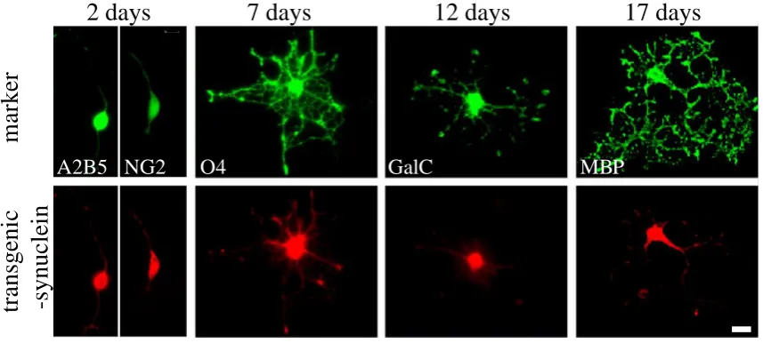
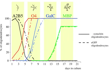
1 TRANSGENIC -SYNUCLEIN EXPRESSION IN PRIMARY OLIGODENDROCYTES DERIVED FROM (PLP)--SYNUCLEIN MICE ...58
2 CELLULAR CONSEQUENCES OF -SYNUCLEIN EXPRESSION ON TRANSGENIC OLIGODENDROCYTES ...59
3 INFLUENCE OF UPSIMPAIRMENT ON OLIGODENDROGLIAL -SYNUCLEINOPATHY ...62
3.1 Proteasome inhibition causes time- and dose-dependent formation of α-synuclein inclusions in transgenic oligodendrocytes ...62
3.2 Proteasome inhibition elevates α-synuclein levels and induces the formation of detergent insoluble α-synuclein inclusions ...64
4 HIGH LEVELS OF -SYNUCLEIN ARE SUFFICIENT TO INDUCE THE FORMATION OF INCLUSIONS IN PRIMARY OLIGODENDROCYTES ...66
5 INCLUSIONS FORMED IN CULTURE RESEMBLE HUMAN PATHOLOGICAL GCIS ...71
5.1 Characterization of the inclusions formed in vitro ...71
5.2 Pathological modifications of α-synuclein in proteasome-inhibited transgenic oligodendrocytes ...76
6 CELLULAR CONSEQUENCES OF -SYNUCLEIN AGGREGATION FOR AFFECTED OLIGODENDROCYTES ...80
6.1 Sensitization of α-synuclein transgenic oligodendrocytes to proteasome inhibitor mediated apoptosis...80
7 LENTIVIRAL DELIVERY OF -SYNUCLEIN SUPPRESSES THE FORMATION OF -SYNUCLEIN INCLUSIONS AND APOPTOSIS IN PROTEASOME-INHIBITED TRANSGENIC OLIGODENDROCYTES ...84
8 MOLECULAR MECHANISMS INVOLVED IN OLIGODENDROGLIAL CELL DEATH...86
8.1 Both the intrinsic (mitochondrial) and the extrinsic (death receptor) pathways are involved in α-synuclein-sensitized oligodendrocyte apoptosis ...86
8.2 Expression of α-synuclein in oligodendrocytes specifically sensitizes to Fas-mediated apoptosis via Fas upregulation ...89
8.3 Fas upregulation is observed in MSA ...91
CHAPTER 5: DISCUSSION ...95
CHAPTER 6: CONCLUSION AND SIGNIFICANCE ...113
REFERENCES ...115
Table of Abbreviations
AR = Adrenegic Receptor
bFGF = basic Fibroblast Growth Factor bHLH = basic Helix-Loop-Helix Protein BS = Bottenstein-Sato medium
CNP = 2',3'-Cyclic Nucleotide 3'-Phosphodiesterase CNS = Central Nervous System
DA=Dopamine
DAT=DopamineTransporter
DLB = Dementia with Lewy Bodies
DMEM = Dulbecco's Modified Eagle Medium DMSO = DiMethyl SulfOxide
eGFP = enhanced Green Fluorescent Protein FCS = Fetal Calf Serum
GalC = Galactocerebroside
GCI = Glial Cytoplasmic Inclusion GFP: Green Fluorescent Protein HNE = 4-Hydroxy-2-NonEnal hsp = heat shock protein IgG = Immunoglobulin G kDa = kilo Dalton
LB = Lewy Body LN = Lewy Neurites
LTR = Long Terminal Repeat
MAG = Myelin-Associated Glycoprotein MBP = Myelin Basic Protein
MOG = Myelin Oligodendrocyte oligoprotein MPP+ = 1-Methyl-4-PhenylPyridinium ion
MPTP = 1-Methyl 4-Phenyl 1,2,3,6-TetrahydroPyridine mRNA = messenger Ribonucleic Acid
MSA = Multiple System Atrophy
MSA-C = Multiple System Atrophy with predominant cerebellar ataxia‟s symptoms MSA-P = Multiple System Atrophy with predominant Parkinsonism symptoms NAC = non amyloid component
NBIA-1 = Neurodegeneration with Brain Iron Accumulation type 1 NCI = Neuronal Cytoplasmic Inclusion
NGF = Nerve Growth Factor NNI = Neuronal Nuclear Inclusion OPC = Oligodendrocyte Precursor Cells OPCA = OlivoPontoCerebellar Atrophy PBS = Phosphate Buffered Saline PD = Parkinson‟s Disease
PDGF = Platelet Derived Growth Factor
PLP = ProteoLipid Protein
PVDF = polyvinylidene difluoride RT = room temperature
SDS = Sodium Dodecyl Sulfate
SDS-PAGE = Sodium DodecylSulfate Polyacrylamide Gel Shh = Sonic hedgehog
TBS = Tris Buffered Saline TNF = Tumor Necrosis Factor-
TRAIL = TNF-Related Aptosis-Inducing Ligand UPS = Ubiquitin Proteasome System
Chapter 1:
GENERAL INTRODUCTION
1
STRUCTURE AND PHYSIOLOGICAL FUNCTIONS OF
-SYNUCLEIN
1.1 The synuclein protein family
a α-synuclein
In 1988, a novel protein was isolated from the electric lobe of the Pacific ray, Torpedo californica, and from rat brain (Maroteaux et al., 1988). Due to its distribution on portions of the nuclear membrane and its presence in high concentrations in presynaptic nerve terminals, this protein was named α-syn (synapse) -nuclein (nucleus); however, localization of mammalian synucleins to the nucleus was not confirmed by subsequent studies.
associated with vesicular membranes (George et al., 1995; Irizarry et al., 1996; Jensen et al., 1998). α-synuclein is enriched in synaptosomal preparations (but is not found in highly purified synaptic vesicle fractions) (George et al., 1995; Irizarry et al., 1996; Kahle et al., 2002b). Immunogold electron microscopy showed that α-synuclein is localized to the inner face of plasma membranes in close proximity, but loosely associated with, synaptic vesicles at axonal termini (Clayton and George, 1998; Iwai et al., 1995; Jenco et al., 1998a; Maroteaux et al., 1988). Thus α-synuclein exists in both cytoplasmic and membrane bound forms, most likely in a dynamic equilibrium (Kahle et al., 2000; Nuscher et al., 2004).
b -synuclein
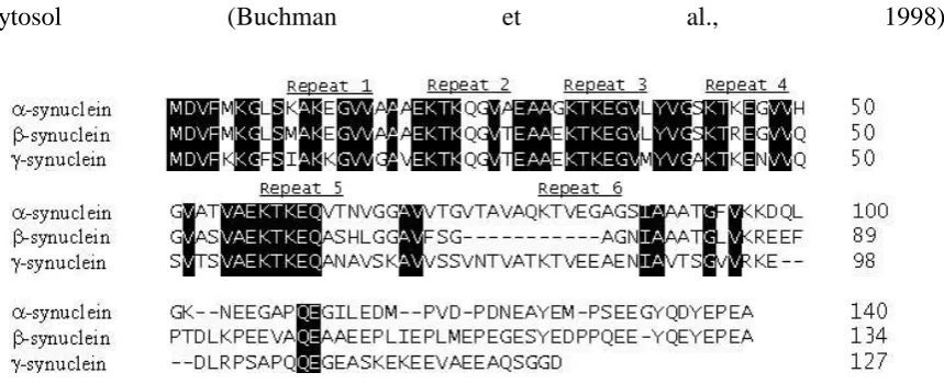
The second member of the synuclein protein family, initially named phosphoneuroprotein-14 (Nakajo et al., 1993), was identified in bovine brain. Because of its high homology with α-synuclein at the amino acid sequence level (62%), this protein was named -synuclein (Jakes et al., 1994). -synuclein, the most conservedof the synuclein proteins, is 134 amino acids long and contains five repeats of the EKTKEGV consensus sequence (Figure 1).
Like α-synuclein, -synuclein is predominantly expressed in neuronal axon termini of CNS neurons (Jakes et al., 1994; Nakajo et al., 1990; Nakajo et al., 1994) but its distribution throughout the brain is more even than that of α-synuclein (Abeliovich et al., 2000; Nakajo et al., 1994). Similarities in sequence and predominant localization in presynaptic terminals may suggest that α- and -synuclein share similar functions.
Another synuclein homologue, originally called breast cancer-specific gene-1 protein, was identified in 1997 in metastatic brain cancer tissue (Ji et al., 1997). Due to its significant sequence homology with α-synuclein (55%), this protein was named -synuclein (Lavedan et al., 1998b) and is also refered to as persyn (Ninkina et al., 1998) or synoretin (Surguchov et al., 1999).
-synuclein is 127 amino acids long and like α-synuclein, it contains six repeats of the EKTKEGV consensus sequence. -synuclein is expressed in CNS as well as in the spinal cord, but it is most abundant in the peripheral nervous system including neurons of the dorsal root ganglia and trigeminal ganglia (Buchman et al., 1998; Lavedan et al., 1998a). Whereas α-synuclein and -synuclein are concentrated in synaptic vesicles, with little staining in cell bodies and dendrites, -synuclein is distributed throughout the neuronal
cytosol (Buchman et al., 1998).
Figure 1: Amino acid sequence alignment of the human synuclein proteins. The imperfect EKTKEGV repeats are identified. The black background highlights amino acid residues conserved
between α- and -synucleins (Jakes et al., 1994), and -synuclein (Ji et al., 1997). The very high
conservation between species for EKTKEGV consensus sequence suggests that the repeats have arisen
from the duplication of a single domain within an ancestral synuclein gene.
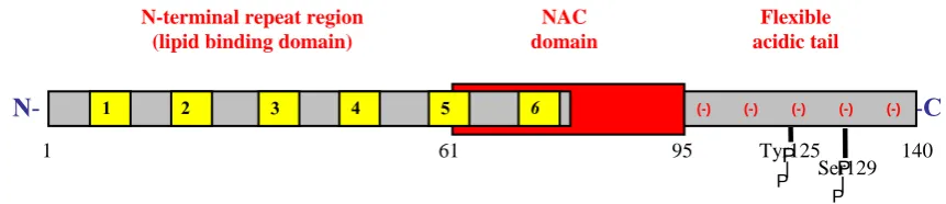
α-Synuclein consists of three major regions (figure 2):
(1) The highly conserved N-terminal region contains 60 amino acid residues and four repeats, with each repeat containing an imperfectly conserved KTKEGV motif (Clayton and George, 1998; George, 2002). These repeats form amphipathic α-helices similar to apolipoprotein class A2 molecules through which membrane binding becomes stabilized (Davidson et al., 1998; Eliezer et al., 2001; Perrin et al., 2000).
(2) Residues 61–95 form the hydrophobic and amyloidogenic central region, referred to as the non-amyloid component (NAC) of the amyloid precursor protein (see part 2.3), which includes two additional EKTKEGV repeats. This domain is the building block of α-synuclein aggregation (Giasson et al., 2001; Kahle et al., 2002a), with critical residues for the fibrillization of the protein being residues 66 to 74 (Du et al., 2003). -synuclein lacks 11 amino acids in the region corresponding to NAC (Figure 1). β-synuclein is a non-amyloidogenic homolog of synuclein and may act as a physiological inhibitor of α-synuclein aggregation (see part 2.1; (Hashimoto et al., 2001).
Figure 2: Schematic diagram of α-synuclein structure
The structure of α-synuclein allows the molecule to exhibit different conformations depending on its interacting environment (Uversky, 2003). This protein is natively unfolded in solution (Weinreb et al., 1996), adopts an α-helical conformation upon binding to lipid vesicles (Davidson et al., 1998), or forms β-pleated sheets in its aggregated form, suggesting highly dynamic structural changes depending upon the local cellular milieu. Nuclear magnetic resonance and electron paramagnetic resonance spectroscopy analysis indicates that binding of α-synuclein to lipids induces a reorganization of the repeats into 3 complete helical turns (Bussell and Eliezer, 2003; Jao et al., 2004).
1.3 Functions of α-synuclein
As mentioned above, α-synuclein exists physiologically in both soluble and membrane-bound states, in natively unfolded and α-helical conformation, respectively. The physiological function of α-synuclein appears to require its translocation between these subcellular compartments and interconversion between the 2 conformations. However, the general functions of α-synuclein under physiological conditions remain unclear and many roles for this protein have been proposed.
a α-Synuclein effects on synapse organization and on synaptic vesicle maintenance
P P
-C
N-terminal repeat region (lipid binding domain)
N
N
-Flexible
acidictail
1 2 3 4 5 6 (-)
NAC domain
1 61 95 140
Ser129 Tyr125
P P
Much research has been carried out in humans, mice, and chickens to study the role of α-synuclein in neuronal development/synaptic plasticity (Eells, 2003; Sidhu et al., 2004b) and synaptic vesicle formation (Lotharius et al., 2002; Lotharius and Brundin, 2002). α-synuclein is initially present in the perikarya of mature neurons and is translocated to axon terminals between 15 and 18 weeks of gestation (Galvin et al., 2001; Murphy et al., 2000). Therefore, α-synuclein does not appear to play a role in initial synapse formation since it is expressed later in development and localizes to synapses after they are formed (Murphy et al., 2000; Withers et al., 1997). Abeliovich et al. developed mice homozygously deleted for α-synuclein by targeted gene disruption. These mice were viable and fertile; they exhibited no morphological deficits and possessed a normal complement of neuronal cell bodies, fibers, and synapses (Abeliovich et al., 2000; Cabin et al., 2002), providing further evidence that α-synuclein is not necessary for synaptic development. However, these mice exhibited significant impairments in synaptic response to tetanic stimulation, suggesting that α-synuclein may regulate synaptic vesicle mobilization at nerve terminals (Abeliovich et al., 2000).
b α-Synuclein effects on synaptic vesicle recruitment
activity of phospholipase D2 (PLD2) (Ahn et al., 2002; Jenco et al., 1998a), an enzyme localized primarily along the plasma membrane (Colley et al., 1997). Activation of PLD2 in the plasma and endosomal membranes, regulates the recycling of synaptic vesicles at or near the plasma membrane or endosomal compartments in response to external stimuli, and is instrumental in vesicle formation, through production of phosphatidic acid which recruits adaptor molecules, which, in turn, trigger the building of vesicles from donor membranes (Lotharius et al., 2002; Lotharius and Brundin, 2002). The regulatory effect of α-synuclein on synaptic vesicles recycling may be tightly regulated by various serine/threonine or tyrosine protein kinases (Lotharius and Brundin, 2002; Sidhu et al., 2004b). Hence, by inhibiting PLD2, α-synuclein may play a role in the control of synaptic vesicle cycling (Jenco et al., 1998b). Phosphorylation of membrane-bound α-synuclein by G-protein coupled receptor kinases, lowers its ability to inhibit PLD2 activity (Lotharius and Brundin, 2002). Binding of phosphorylated -synuclein to phospholipids is reduced (Sidhu et al., 2004b) and as a consequence monomeric α-synuclein is released into the cytoplasm (Leng et al., 2001; Pronin et al., 2000). Thus, through reduction of its tonic inhibition of PLD2, phosphorylated α-synuclein might promote vesicle recycling during periods of high neuronal activity and favor synaptic plasticity, whereas non-phosphorylated α-synuclein may suppress synaptic vesicle formation during periods of low neuronal activity (Sidhu et al., 2004b).
recycling (Lotharius et al., 2002; Lotharius and Brundin, 2002). These fatty acid-binding protein properties of α-synuclein rely on two observations:
its N-terminal lipid (and vesicles) binding domain (residues 7-87; figure 2) bears significant homology with the lipid-binding class A apolipoproteins A2 and C (George, 2002; Goedert, 2001; Maries et al., 2003), proteins implicated in lipid transport (Sharon et al., 2001).
short amino acyl stretches in α-synuclein N- and C-termini share more than 55 to 67% identity with a cytosolic fatty acid-binding motif of fatty acid-binding proteins (Sharon et al., 2001).
c α-Synuclein effects on neurotransmission
Many studies suggest the important role of α-synuclein in the regulation of neurotransmission and in the physiological maintenance of dopamine (DA) homeostasis in dopaminergic neurons of the substantia nigra pars compacta (Abeliovich et al., 2000; Lotharius et al., 2002; Lotharius and Brundin, 2002; Perez et al., 2002; Sidhu et al., 2004b).
(Abeliovich et al., 2000). In a second study, ultrastructural examination of synapses of α-synuclein knock-out mice also showed a reduction in the reserve-resting pool of synaptic vesicles in the hippocampus (Cabin et al., 2002). These data were consistent with previous
in vitro data (Murphy et al., 2000). After lowering the amount of α-synuclein in cultured rat hippocampal neurons using antisense oligonucleotides, Murphy et al. (2000) detected a decrease in the number of resting-reserve synaptic vesicles, suggesting that α-synuclein may be required for the genesis and/or maintenance of a subset of presynaptic vesicles, those in the „reserve‟ or „resting‟ pools (Cabin et al., 2002).
A more specific link to DA neurotransmission was established by the findings that -synuclein binds to tyrosine hydroxylase, the rate-limiting enzyme for DA synthesis (Perez et al., 2002) and to dopamine transporter (DAT) (Lee et al., 2002; Wersinger and Sidhu, 2005), thereby controlling the extravesicular cytoplasmic levels of DA (Sidhu et al., 2004b). Disruption of this function of α-synuclein can result in abnormal intracellular and extracellular DA content, which upon autoxidation and enzymatic metabolization can generate reactive oxygen species, ultimately leading to cell death. α-Synuclein interacts directly with the DAT (Lee et al., 2001a) through its NAC domain (Wersinger and Sidhu, 2003). It was recently demonstrated that in the presence of α-synuclein, DAT is dynamically trafficked away from the plasma membrane into the cytoplasm (Wersinger et al., 2003; Wersinger and Sidhu, 2003). These findings suggest that α-synuclein may act to tether the DAT to a cytoplasmic compartment, thereby keeping it away from the cell surface.
specifically only through the DAT in an energy-dependent manner, reversed the inhibitory effects of α-synuclein on DAT (Wersinger et al., 2003). The presence of α-synuclein enhances the vulnerability of cells to MPP+ exposure (Kanda et al., 2000), whereas α-synuclein null-mice are essentially resistant to 1-methyl-4-phenyl-1,2,3,6-tetrahydropyridine (MPTP)-induced degeneration of dopaminergic neurons (Dauer et al., 2002). Schluter et al. generated α-synuclein-deficient mice by homologous recombination. Upon acute MPTP challenge, α-synuclein knockout mice were partly protected from chronic depletion of nigrostriatal DA when compared with littermates of the same genetic background (Schluter et al., 2003).
d α-Synuclein and neuroprotection
resistance was not due to abnormalities of the DAT, which appeared to function normally in the null mice.
e α-Synuclein: a molecular chaperone protein
2
ROLE
OF
-SYNUCLEIN
IN
NEURODEGENERATIVE
DISEASES
α-synuclein is the major building block of pathological inclusions that characterize many neurodegenerative disorders, including Parkinson‟s disease (PD), dementia with Lewy bodies (DLB), neurodegeneration with brain iron accumulation type 1 (NBIA-1) and multiple system atrophy (MSA), collectively termed α-synucleinopathies (Trojanowski and Lee, 2002). Pathological aggregates specifically contain α-synuclein but not -and -synucleins. The factors leading to aggregation of α-synuclein are of critical importance as potential mechanisms of pathogenesis.
2.1 α-Synuclein aggregation and toxicity
detected (Rockenstein et al., 2001). Thus the balance between both synucleins in vivo
might be an important factor regulating α-synuclein aggregation.
The formation of α-synuclein fibrils is greatly accelerated in vitro above a critical concentration of purified recombinant α-synuclein (Conway et al., 2000a; Giasson et al., 1999; Hashimoto et al., 1998; Serpell et al., 2000) and by increased expression levels of α-synuclein in transgenic animals (Giasson et al., 2002; Lee and Lee, 2002; Neumann et al., 2002). However, since α-synuclein is ubiquitiously expressed in the brain it is unlikely that high expression levels of synuclein are the sole criteria to cause protofibril and/or α-synuclein aggregation in α-synucleinopathies (see part 2.3 for definition). In fact, many other factors can also influence the fibrillization of α-synuclein. Catecholamines, including DA, can form covalent adducts with α-synuclein and thereby increase the number of α-synuclein protofibrils (Conway et al., 2001). This might explain the vulnerability of dopaminergic neurons of the substantia nigra pars compacta in PD.
α-synuclein aggregates and signs of degeneration in PD and MSA respectively (see part 2.3.a and 3.1).
Post-translational modifications such as phosphorylation at Ser129 has been also shown to induce or accelerate α-synuclein aggregation, both in vivo and in vitro (Chen et al., 2005; Fujiwara et al., 2002; Smith et al., 2005). Only 4% of α-synuclein is Ser129 phosphorylated under steady state physiological conditions, contrasting with the massively disproportionate concentration of phosphorylated α-synuclein (90%) in proteinaceous inclusions characteristic of specific neurodegenerative diseases (respectively part 2.3 and 3.1). This findings suggest that extensive phosphorylation at Ser129 of α-synuclein in the brain is a highly pathological event. However, these correlative studies did not answer the question whether Ser129-phosphorylation is a cause or rather a secondary effect of -synuclein fibrillization in vivo.
Furthermore, α-synuclein monomer containing a tissue transglutaminase crosslinked intramolecular bond was extracted from PD substantia nigra pars compacta (Andringa et al., 2004). The presence of this abnormal internal bond may impair the ability of α-synuclein to interconvert between its α-helical configuration when bound to membrane and its unstructured cytoplasmic form, a process that appears to be necessary for its normal function. Concentration of this isoform increases with disease progression and might serve as a nucleation site that could initiate α-synuclein filament assembly.
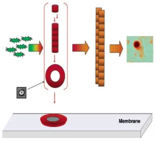
leak (Figure 3), elevating intracellular levels of potential cytotoxins such as calcium and DA (Conway et al., 1998; Conway et al., 2000b; Conway et al., 2001; Lashuel et al., 2002a; Rochet et al., 2000; Volles et al., 2001).
Figure 3: Putative model for pathological α-synuclein inclusion formation. Disordered monomeric (green starburst) α-synuclein oligomerizes to form a heterogeneous population of -sheet rich protofibrils (red), which includes pore-like structures (electron micrograph, image-averaged, 10-12nm
outer diameter, from (Lashuel et al., 2002b). Insertion of the protofibril pore into a membrane, which is
thought to account for protofibril permeabilizing activity, is shown schematically. The protofibril
population dissipates as amyloid fibrils (orange rods) are formed. Eventually, the fibrils coalesce to
form a unique cytosolic proteinaceous inclusion. A Lewy body (see part 2.3) is shown in the tissue
section at the far right. [Modified from (Volles and Lansbury, 2003)]
of non degradable protein aggregates would be predicted to crowd cellular machinery and impair cellular transport, thereby inducing cell death.
In addition it is conceivable that formation of protofibrils and aggregation of α-synuclein during the pathological process of α-synucleinopathies reduces the bioavailability of the physiological form of α-synuclein and may dampen the “anti-apoptotic” and neuroprotective effects of physiological concentrations of α-synuclein.
2.2 Possible role of proteasome inhibition on α-synuclein fibril formation
including ubiquitin carboxyl-terminal hydrolase, a deubiquitinating enzyme, chymotrypsin A and proteasome subunits (Lowe et al., 1990). The common appearance of components of the UPS machinery in α-synuclein aggregates suggests an unsuccessful attempt of degradation by the proteasome (Gai et al., 2000; Sharma et al., 2001). Finally, studies have shown that PD patients have significantly reduced levels of proteasomal subunits in the substantia nigra pars compacta compared to age-matched healthy controls, and proteasomal function is impaired in idiopathic PD, further strengthening the links between UPS dysfunction and α-synuclein mismetabolism (McNaught et al., 2003; McNaught and Jenner, 2001; McNaught and Olanow, 2003).
The level of protofibrils and aggregated forms of α-synuclein is likely to be modulated through the ubiquitin-proteasome pathway (Giasson and Lee, 2003), although a causal link between a decrease in proteasomal degradation and high amounts of α-synuclein aggregates has not yet been demonstrated in vivo. The UPS is clearly saturable and excess aggregated protein leads to saturation of proteolysis systems, inducing feed-forward toxicity (Bence et al., 2001). In fact, α-synuclein overexpression potentiates proteasome-inhibitor mediated apoptosis (Tanaka et al., 2001) and its aggregation is enhanced by proteasome inhibition (Dawson and Dawson, 2003; Maries et al., 2003; Rideout et al., 2001).
proteasome pathway may contribute to the pathophysiology of the neurodegenerative diseases defined by the mismetabolism of α-synuclein.
Figure 4: Postulated mechanisms of α-synuclein mediated toxicity. The presence of cytosolic aggregates in -synucleinopathies is caused by a failure to properly dispose of α-synuclein. α-synuclein
protofibrils and intracellular protein aggregates have been shown to impair the proteasome potentially
leading to enhanced accumulation. Solid arrows indicate more established pathways, whereas dotted
arrows indicate largely hypothetical mechanisms. Mutations (see part 2.3)
Genomic multiplications (see part 2.3)
Post-translational modification Aberant -synuclein expression
Aberrant
-synuclein
Soluble oligomers Insoluble oligomers
(protofibrils)
Fibrillar aggregates
Fibrillar inclusions
Proteasomal dysfunction
D
2.3 α-Synuclein aggregation in neuronal and glial diseases:
α-synucleinopathies
α-synuclein was first associated with a neurodegenerative disease when a fragment of -synuclein corresponding to the NAC domain was detected in amyloid plaques in Alzheimer‟s disease patient brains (Iwai et al., 1996; Masliah et al., 1996; Takeda et al., 1998a; Takeda et al., 1998b; Uéda et al., 1993). This finding stimulated great interest among researchers in a potential role for this protein in Alzheimer‟s disease pathogenesis. However, further research failed to confirm that the NAC peptide is an integral component of senile plaques (Bayer et al., 1999; Culvenor et al., 1999).
The discovery of α-synuclein in Lewy bodies in PD was followed quickly by its detection in cellular inclusions in several other neurodegenerative diseases including DLB (Baba et al., 1998; Spillantini et al., 1998a; Spillantini et al., 1998b), Hallervorden-Spatz syndrome, now known NBIA-1 (Arawaka et al., 1998; Saito et al., 2000), and MSA (Arawaka et al., 1998; Arima et al., 1998b; Fujiwara et al., 2002; Gai et al., 1999; Saito et al., 2000; Tu et al., 1998; Wakabayashi et al., 1998). Collectively, these neurodegenerative diseases that share synuclein pathology as a primary feature have come to be known as α-synucleinopathies (Arawaka et al., 1998; Arima et al., 1998b; Fujiwara et al., 2002; Saito et al., 2000; Spillantini et al., 1998b; Tu et al., 1998; Wakabayashi et al., 1998)
a Parkinson’s disease (PD)
disturbances. As the disease progresses, many patients develop cognitive dysfunction, which includes dementia (Lim et al., 2002). In contrast to other neurodegenerative disorders, there is relatively efficient symptomatic therapy for PD. This mainly consists of DA replacement and surgical therapy (deep brain stimulation) that relieves most motor symptoms. However, there is no proven therapy to prevent cell death or restore sick neurons to a normal state. Dopaminergic neurons contain intracytoplasmic inclusions called Lewy bodies (LBs), which contain mainly -synuclein fibrils (10nm) (Spillantini et al., 1998b). It is believed that α-synuclein, in patients with PD, undergoes a conformational change acquiring a predominantly -pleated sheet structure that facilitates polymerization of α-synuclein into amyloid fibrils (McNaught and Jenner, 2001). Dystrophic ubiquitin-positive neurites associated with PD pathology, known as Lewy neurites (LNs), are also α-synuclein positive (Braak et al., 1999; Spillantini et al., 1998b; Spillantini et al., 1997). Immunoelectron microscopy studies of the ultrastructure of synuclein in LBs, have demonstrated that LBs consisted of α-synuclein forming radially filaments surrounding an unstructured α-synuclein core.
There is some evidence that a dinucleotide repeat polymorphism located at a significant distance upstream in the promoter region of the α-synuclein gene may be associated with increased risk of PD in Caucasians (Farrer et al., 2001; Kruger et al., 1999). Similar observations concerning the variability of these repeats and allelic length as a risk for PD have been obtained by independent studies in an Asian population (Tan et al., 2000).
Another study showed that this region may play a role in transcriptional regulation of α-synuclein (Chiba-Falek and Nussbaum, 2001); thus α-α-synuclein may also play a role in PD pathogenesis through alterations in its transcriptional control.
The recent identification of genomic triplication in α-synuclein gene suggests that overexpression of α-synuclein causes pathology in neurons. Singleton and colleagues reported a triplication in a region of chromosome 4 that includes the α-synuclein locus and an estimated 17 other genes (Singleton et al., 2003). Carriers of this triplication are predicted to have four functional copies of α-synuclein, arguing for an increased gene dosage as a cause for PD. -synuclein locus duplication has also been reported to cause PD phenotype which, by contrast with -synuclein triplication families, closely resembles idiopathic PD with late age-of-onset, slow progression, and no prominent cognitive decline nor dementia (Ibanez et al., 2004).
b Dementia with Lewy Bodies (DLB)
parkinsonian and psychiatric symptoms. However, antiparkinsonian drugs that may help to reduce tremor and loss of muscle movement may actually worsen such symptoms as hallucinations and delusions. Recently, several clinical trials using cholinesterase inhibitors have shown its efficacy in the symptomatic treatment of cognitive impairment in DLB patients (Aarsland et al., 2004; Werber and Rabey, 2001).
Pathologically, DLB patient brains contain a large number of α-synuclein-positive LBs in neurons from the substantia nigra pars compacta like in PD and in the cerebral cortex, which is less common in PD (McKeith et al., 1996; Spillantini et al., 1997). The number of LBs correlates with the severity of the dementia (Hurtig et al., 2000). In addition to LB pathology, LNs are fairly abundant in the striatum of DLB brains, and these inclusions may contribute to the parkinsonian symptoms in DLB (Duda et al., 2002). Because LBs and LNs in DLB are so similar to those found in PD and are also mostly composed of filamentous α-synuclein protein (Baba et al., 1998; Crowther et al., 2000; Spillantini et al., 1998b; Spillantini et al., 1997), PD and DLB may share a common disease mechanism.
c Neurodegeneration with Brain Iron Accumulation Type 1 (NBIA-1)
found mostly throughout the cortex, subcortical regions, and brain stem (Arawaka et al., 1998; Saito et al., 2000; Tu et al., 1998).
3
MULTIPLE SYSTEM ATROPHY (MSA)
3.1 Clinical characteristics and etiology of MSA
The median age of onset is 55 years, but ranges from 33 to 76 years of age and affects more men than women (Wenning et al., 1995). The symptoms of MSA progress faster than those of PD with an average patient survival of 6 to 7 years (Wenning et al., 2004). Approximately 0.31 % of people over 65 years old suffer of MSA (Trenkwalder et al., 1995). Assuming that there exist worldwide 600 million people older than 65, we extrapolate that 1.7 million may suffer from MSA. However, the true prevalence of MSA has been underestimated, since many patients remain undiagnosed or misdiagnosed. In fact, MSA is a difficult diagnosis (especially early in the clinical course) and is clinically commonly confounded with PD at the initial stage of illness. Approximately 10% of patients diagnosed in life as having PD are found at autopsy to have MSA (Hughes et al., 1992). MSA is suggested when (1) disability progresses rapidly, (2) patients are poorly responsive to levodopa, (3) autonomic symptoms, ranging from impotence to urinary incontinence to orthostatic hypotension (drop in blood pressure when patient stands upright), are pronounced, (4) rigidity and bradykinesia are out of proportion to tremor and (5) severe dementia is absent. Mild dementia is not uncommon, but severe dementia is atypical in MSA (Wenning et al., 1997b).
The cause of MSA remains unknown and no current therapy can reverse or halt progression of the disease. A genetic component seems unlikely and screening studies for candidate genes revealed no risk factors (Bandmann et al., 1997; Nicholl et al., 1999). Autoimmune mechanisms and environmental toxins have been suggested to be involved in MSA pathogenesis (Hanna et al., 1999), but evidence for these etiologies is weak
3.2 Neuropathological features of MSA
Histopathological findings include moderate depigmentation of the substantia nigra pars compacta and locus ceruleus, and demonstrate neuronal and oligodendroglial cell loss as well as gliosis in striatum (mainly putamen), substantia nigra pars compacta, inferior olives, pons, cerebellum in addition to the intermediolateral cell columns and Onuf‟s nucleus in the spinal cord (Wenning et al., 1997a). Microvacuolation within the involved neuronal systems has also been reported. These findings are present to a lesser extent even in regions where atrophy is not noted.
Increasing evidence indicates that the formation of GCIs leads to cellular dysfunction. It has been proposed that GCIs often entrap cytoplasmic organelles (eg, mitochondria, secretory vesicles) and disrupt normal protein and organelle trafficking within affected cells (Arima et al., 1998a; Tu et al., 1998). These chronic alterations may lead to glial cell dysfunction and death and may impair trophic function between oligodendrocytes and axons, thereby causing neuronal damage.
It was shown recently that extensive myelin abnormalities occur in the white matter of the involved regions in MSA. Matsuo and colleagues reported extensive myelin degeneration in MSA brains (Matsuo et al., 1998) using antibodiesrecognizing degenerating myelin but not normal appearing myelin (Matsuo et al., 1997). Thepresence of unusual myelin basic protein epitopes in MSA, in both affected and unaffected brain regions, substantiates the notion of widespread oligodendroglial dysfunction in MSA and highlights white matter disease as an integral component. The dysmyelination observed in MSA results from dysfunction of the oligodendrocytes, the cells which produce and maintain myelin in the CNS (see part 4.1).
Figure 5: -synuclein pathology in multiple system atrophy. Immunostaining for -synuclein in paraffin sections (A, B, and C) in MSA. Note many GCI in white matter in basal ganglia (A and inset).
In the pons (B and C) was a mixture of glial and neuronal inclusions. The neuronal inclusions were
(rhodamine filter, red) and -synuclein (fluorescein filter, green). Note the absence of colocalization of
the two signals. Inset shows a GCI that is double labeled with -synuclein (fluorescein filter, green) and
C4d (rhodamine filter, red) [from Dickson, 1999]
3.3 Animal models of MSA
The adverse consequences of α-synucleinopathy in MSA are not understood to date. In order to shed light on the molecular pathology of α-synuclein in these diseases, several animal models have been generated (Fillon and Kahle, 2005; Stefanova et al., 2005c). Animal models of MSA are urgently required as test-bed for the evaluation of novel therapeutic interventions in the disorder. Initial attemps to mimic MSA pathology in rodents relied on the destruction of brain regions that degenerate in human MSA-P (Wenning et al., 1999). The nigral and striatal “double-lesion” rat model is based on sequential injection of 6-hydroxydopamine and quinolinic acid into the medial forebrain bundle and ipsilateral striatum, respectively (“double toxin-double lesion” approach). Intrastriatal injections of 3-nitropropionic acid and MPP+ in rodents result in secondary excitotoxic striatal lesions and subtotal neuronal degeneration of substantia nigra pars compacta, thus producing MSA-P like pathology by a simplified “single toxin-double lesion” approach. However, such models do not show the neuropathological hallmarks of MSA. Thus, chemical lesion models are of limited use for the investigation of MSA etiopathogenesis. To gain further insight into the molecular mechanisms of MSA, transgenic mouse models are currently being developed.
In situ hybridization with transgene-specific probes directly demonstrated that the glial synuclein protein was expressed from glial mRNA (Rockenstein et al., 2002). Glial α-synuclein was not observed in mice that expressed transgenic α-α-synuclein under the control of the highly neuron-specific Thy1 promoter (Kahle et al., 2000; Rockenstein et al., 2002; van der Putten et al., 2000). These mouse model data do not support the hypothesis that the neuronal protein α-synuclein transmigrates from axons into oligodendrocytes in MSA patients.
factors than aged neurons. One such risk factor may be exposure to environmental toxins. Transgenic (PLP)-SYN mice exposed to the neurotoxin 3-nitropropionic acid displayed exacerbated neuronal loss, astrogliosis and microglial activation (Stefanova et al., 2005b). The MSA-like pathology in this combined -synuclein / 3-nitropropionic acid mouse model caused severe motor impairment.
Another risk factor might be concomitant tauopathy. Tau protein, the microtubule-associated protein that is the major structural component of neurofibrillary tangles in Alzheimer‟s disease and progressive supranuclear palsy, is like -synuclein a natively unfolded protein with little secondary structure that forms pathologic filaments with unusual solubilityproperties and protease resistance (Dickson et al., 1999). When a 2‟,3‟-cyclic nucleotide 3‟-phosphodiesterase (CNP) promoter was employed to express human α-synuclein in transgenic mouse oligodendrocytes, no particular pathology and phenotype were initially noted unless these animals were cross-bred with mice that express [P301L]tau in oligodendrocytes (Giasson et al., 2003a). A subset of bigenic oligodendrocytes developed α-synuclein and tau fibrillar inclusions, as demonstrated by thioflavin S staining. Such amyloid formation coincided with motor impairments. It is tempting to speculate that in oligodendrocyte cytosol, α-synuclein and tau synergistically form individual fibrils, as was shown in vitro (Giasson et al., 2003a). Tau pathology has been noted in some MSA individuals (Giasson et al., 2003b; Jaros and Burn, 2000; Piao et al., 2001a) but more systematic studies are warranted to distinguish the potentially synergistic co-fibrillization of tau and α-synuclein from, e.g., co-morbid MSA and progressive supranuclear palsy.
α-synuclein accumulation in oligodendrocytes in an age-dependent manner, leading to a primary loss of the glial cells and a secondary neuronal degeneration (Yazawa et al., 2005). The insoluble fibrillar α-synuclein inclusions show striking resemblance to GCIs. The (CNP)-α-synuclein mouse has a normal life span; however, starting at three months of age, it begins to lose motor skills and paw strength. The decline is associated with brain atrophy especially severe at 2 years compared to littermates. In addition, neuronal and oligodendroglial cell loss is observed in the spinal cord. Electon microscopy analysis reveals degenerating glial cells and autophagocytosis of myelin. Neurons likewise show markers of injury and structural evidence of degeneration. Interestingly, neurons in older (CNP)-α-synuclein mouse express higher levels of endogenous mouse α-synuclein in their axons, presumably in response to oligodendrocyte degeneration.
(Shults et al., 2005). This model further suggests that accumulation of insoluble filamentous α-synuclein in oligodendrocytes, leads to a primary loss of the glial cells and a secondary neuronal
degeneration.
The major breakthrough demonstrated with the (CNP)-α-synuclein and (MBP)-α-synuclein mice is the identification of a secondary neurodegeneration following oligodendrocyte α-synucleinopathy. This provides evidence that neuronal degeneration can occur as a direct consequence of oligodendrocytic GCI-like pathologies and suggests that aberrant expression of α-synuclein in oligodendrocytes could be sufficient to cause MSA. However, the mechanisms leading to oligodendrocyte loss and subsequent neurodegeneration in this model remain unknown. Although it remains to be shown that oligodendrocytes induce the α-synuclein gene in human MSA patients, the (CNP)-synuclein and (MBP)-(CNP)-synuclein mice may allow the study how oligodendroglial α-synucleinopathy impairs myelination and axonal integrity. Moreover, as models of glial-driven neurodegeneration, they may be useful to test novel therapeutic approaches to treat MSA.
4
OLIGODENDROCYTES: POTENTIAL PRIMARY TARGETS IN
MSA
1996; Trapp et al., 1998). Furthermore, the critical trophic influences that are described between oligodendrocytes and axons (Kaplan et al., 1997) indicate that oligodendroglial pathology would likely affect neuronal function.
4.1 Oligodendrocyte functions in the CNS
Glial cells represent the vast majority of cells in the CNS and outnumber neurons by 10 to 1 and occupy half of the CNS space (Kettenmann 1995; Zhang 2001). These cells help to construct the nervous system during embryonic development and maintain its functions. Rio Hortega introduced the term oligodendroglia to describe neuroglial cells with few processes in material stained by metallic impregnation techniques (Baumann and Pham-Dinh, 2001). An oligodendrocyte extends many processes, each of which contacts and repeatedly envelopes a stretch of axon with subsequent condensation of this multispiral membrane-forming myelin (Bunge et al., 1962; Bunge, 1968) (Figure 6). The main and evident function of oligodendrocytes is the formation of a myelin sheath around most axons in the CNS (Figure 6). Myelin functions as an insulator of the axons, and its structure facilitates rapid transmission of impulses. Oligodendrocytes are able to myelinate up to 50 axonal segments, depending on the region of the CNS. On the same axon, adjacent myelin segments belong to different oligodendrocytes.
Figure 6: The oligodendrocyte and the mature myelin sheath. An oligodendrocyte sends a glial process forming compact myelin spiraling around an axon. Cytoplasm is trapped occasionally in the
compact myelin. In transverse sections, this cytoplasm is confined to a loop of plasma membrane, but
along the internode length, it forms a ridge that is continuous with the glia cell body. In the longitudinal
plan, every myelin unit terminates in a separate loop near a node. Within these loops, glial cytoplasm is
also retained. The tight wrapping of myelin prevents any ionic exchange or spread of electric current,
therefore the action potential can only occur at the nodes of Ranvier which are devoid of myelin. As
shown above, when the action potential is present at one node, the influx of Na+ ions causes the
displacement of K+ ions down the axon. Thus the action potential jumps from node to node. Saltatory
conduction increases conduction velocity and is very energy efficient, as only a small part of the axon is
involved in the exchange of ions, much fewer ions need to be pumped back after the action potential has
passed. [Adapted from Bunge et al., 1968]
Although the oligodendrocyte is mainly a myelin-forming cell, there are also satellite oligodendrocytes that may not be directly connected to the myelin sheath. Satellite oligodendrocytes are located in the grey matter, perineuronal and regulate the microenvironment around neurons(Ludwin, 1997).
4.2 Origin of oligodendrocytes
a Neuron-glia decision
Both intracellular factors as well as extracellular factors may exert influence on whether a neural cell becomes neuron or glia. Accumulating evidence suggests that the Notch receptor promotes glial development at least partially via repressing the neurogenic basic helix-loop-helix protein (bHLH) transcription factors, promoters of neuronal fate (reviewed in (Kageyama and Nakanishi, 1997). Notch signaling instructively commit CNS stem cells into the astroglial lineage, thus fulfilling the criterion for a molecular switch between neuronal and glial fate (Morrison et al., 2000; Tanigaki et al., 2001). Neurons are generated before glial cells (Lu et al., 2000; Morrison, 2000; Yu et al., 1994; Zhou et al., 2000). Environmental factors present during early embryonic development favour neuronal development whereas those present at later stages favour glial development. Once a cell fate is chosen, other mechanisms presumably will step in to permanently lock the cell in this fate. In addition, neural stem cells may contain an internal timer that counts time or cell division and that determines the sequential production of neuron and glia (Morrison et al., 2000).
b Oligodendrocyte specification
Oligodendrocyte precursors originate from neuroepithelial cells of the ventricular zones, at very early stages during embryoniclife. As mentioned before, neurogenic bHLH factors control the neuron-glia decision. The bHLH transcription factors Olig2 and Olig1 are absolutely required for oligodendrocyte fate determination (Balasubramaniyan et al., 2004; Lu and Sloan, 2002; Lu et al., 2000; Zhou and Anderson, 2002). Olig1,2 double null mutants mice or neurosphere cultures fail to give rise to any oligodendrocytes (Lu et al., 2002; Zhou and Anderson, 2002).
generation in both rostral and caudal neural tube (Alberta et al., 2001; Nery et al., 2001; Orentas et al., 1999; Park et al., 2002; Tekki-Kessaris et al., 2001b). Shh exerts its pro-oligodendrocyte functions by inducing Olig genes (Lu et al., 2000; Nery et al., 2001). However Olig genes cannot substitute for Shh, as shown in zebrafish where forced Olig expression largely failed to rescue the oligodendrocyte failure in the absence of Shh signalling (Park et al., 2002). Shh therefore must induce additional genes that cooperate with Olig to promote oligodendroglia fate.
Members of the bone morphogenetic proteins, on the contrary, repress Olig gene transcription even in the presence of Shh and thereby act as potent inhibitors of oligodendrocyte fate (Gross et al., 1996; Mabie et al., 1999; Mehler et al., 2000; Mekki-Dauriac et al., 2002).
In summary, oligodendroglia fate specification requires the absence of general glial repressors such as neurogenic bHLH factors and the induction of Olig genes and their partners.
c Spatial origin of oligodendrocytes
(Olivier et al., 2001b; Spassky et al., 1998). The initial restricted localization of oligodendrocyte precursors in the ventral plate of the neural tube appears not to belimited to the mid-and forebrain (Spassky et al., 1998). Earlier this year Cai et al., and Vallstedt et al., provided compelling evidence for a second dorsal origin of oligodendrocyte precursors in the hindbrain and spinal cord while oligodendrocyte precursors have previously been localized to the ventral midline (Cai et al., 2005; Vallstedt et al., 2005).
The subventricular zone is a germinal matrix of the forebrain that first appears during the later third of murine embryonic development (Doetsch et al., 1997). It enlarges during the peak of gliogenesis, between 5 and 20 days in postnatal life,and then shrinks but persists into adulthood. The majority of progenitors withinthis germinal matrix are glial precursors that generate astrocytesor oligodendrocytes and a rare cell will develop into both neurons andglia (Levison et al., 1993; Levison and Goldman, 1993; Luskin et al., 1988; Price et al., 1988). In cerebrum and cerebellum, oligodendrocytesarise postnatally from the SVZ of the lateral ventricles (Reynolds and Wilkin, 1988) (Levison et al., 1993; Zerlin et al., 1995). Oligodendrocyte progenitors then migrate long distances away from these zones and populatethe developing brain to form white matter throughout the brain.
4.3 Oligodendrocyte precursor cell (OPC) proliferation and differentiation
a Oligodendrocyte precursor proliferation
et al., 1995; Timsit et al., 1992; Yu et al., 1994; Zhou et al., 2000). OPCs are actively proliferating andpossess migratory properties.
A number of extracellular factors have been found to influence the proliferation of OPC. While OPC proliferates extensively in culture in the presence of PDGF, the absence or withdrawal of PDGF induces immediate terminal differentiation (Noble et al., 1988; Raff et al., 1988). Overexpression of PDGF-A in transgenic mice resulted in increased proliferation of OPC (Calver et al., 1998) whereas both the ligand (PDGF-A) and receptor (PDGFRα) knockout showed a severe loss of oligodendrocytes (Fruttiger et al., 1999; Klinghoffer et al., 2002). These experiments firmly established PDGF-PDGFRα ligand-receptor pair as one major regulator of OPC proliferation. The basic fibroblast growth factor (bFGF) is also a major inducer of OPCs proliferation (Villa et al., 2000) and the combined action of PDGF and bFGF can keep OPC in a proliferative state almost indefinitely in vitro (Bogler et al., 1990).
b Oligodendrocyte precursor differentiation and maturation
Because mature oligodendrocytes cannot migrate,preventing premature differentiation of progenitors is crucialfor ensuring that they successfully make it to their final destination. Premature oligodendrocyte differentiation is effectively prevented by an inhibition mechanism recently shown to occur in gliogenesis,the Notch pathway (Wang et al., 1998). Notch activation generally leads to the expression of Hes family inhibitory basic bHLH transcription factors which inhibit terminal differentiation of optic nerve OPC (reviewed by Kageyama and Nakanishi 1997). Thus, Notch acts as a negative signal for OPC differentiation in vivo. The expression of Jagged, a neuronal Notch ligand, is downregulated while embryonic development progresses, allowing myelination to occur in a controlled manner (Wang et al., 1998).
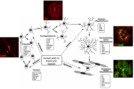
Two transcription factors necessary for oligodendrocyte terminal differentiation, Sox10 and Nkx2.2, have recently been discovered. (Stolt et al., 2002; Xu et al., 2000; Zhou et al., 2000). Studies in knockout mice revealed that loss of either Sox10 or Nkx2.2 results in hypomyelination and loss of mature oligodendrocyte markers such as PLP and MBP without affecting the development of oligodendrocyte precursors (Qi et al., 2001; Stolt et al., 2002).
Figure 7: Developmental stages of cells of the oligodendrocyte lineage. Schematic drawing of the morphological and antigenic progression from precursor cells to myelinating mature oligodendrocytes,
through progenitors, preoligodendrocytes, and immature non myelinating oligodendrocytes. Timing of
neuronal and astrocytic signaling is indicated. Stage-specific markers are boxed. RNAs are in italics.
Chapter 2:
SPECIFIC AIMS
In contrast to the well-studied neuronal α-synuclein pathology, both causes and effects of oligodendroglial -synucleinopathy in MSA are more enigmatic. While the presence of -synuclein in GCIs was identified almost one decade ago, we still do not know with certainty if and how -synuclein provokes the disease. In mature brain, oligodendrocytes express undetectable levels of α-synuclein (Miller et al., 2005; Solano et al., 2000a). Thus, ectopic expression of α-synuclein could be causally linked to MSA pathology. To test the hypothesis that α-synuclein expression in oligodendrocytes leads to the formation of GCIs, transgenic mouse models were developed. Although key features of MSA pathology were recapitulated in transgenic animal models, neither model as yet yields any specifics on how α-synuclein aggregates in MSA and selectively injures or kills oligodendrocytes. In particular, questions remain regarding the role of α-synuclein fibrils, protofibrils, oligomers, GCIs, ubiquitination, and α-synuclein-interacting proteins in oligodendrocyte dysfunction and cell death.
The aim of my PhD thesis was to further clarify these complex issues and to investigate the molecular mechanisms leading to oligodendroglial α-synucleinopathy and its cellular consequences. Therefore, I developed an experimental system based on overexpression of
1. When during oligodendrocyte differentiation is transgenic α-synuclein expressed in
primary mouse oligodendrocytes derived from (PLP)-α-synuclein transgenic mice?
2. What are the cellular consequences of α-synuclein expression on transgenic oligodendrocytes?
3. What is the contribution of an impairment of the oligodendroglial ubiquitin-proteasome system to the pathogenesis of MSA?
4. Do high α-synuclein levels reached by lentiviral delivery lead to the aggregation of ectopic α-synuclein?
5. To what extent do α-synuclein aggregates in culture resemble GCIs found in MSA patients?
6. What are the cellular consequences of α-synuclein aggregation for affected oligodendrocytes?
7. Can anti-aggregative gene therapy reduce α-synuclein mediated cytotoxicity?
8. Which molecular mechanisms induce death of affected oligodendrocytes in culture? Could these mechanisms also be responsible for glial cell loss in MSA?
Chapter 3:
EXPERIMENTAL PROCEDURES
Animals
Homozygous transgenic (PLP)--synuclein mice were generated in which human wild-type -synuclein was driven by a PLP promoter (Kahle et al., 2002b). Homozygous transgenic mice overexpressing the enhanced green fluorescent protein under the PLP promotor (PLP-eGFP) mice and wild-type mice with the same genetic background were used as controls.
Antibodies
2000), 1:200 for cytochemistry and 1:1000 for western blot), rabbit polyclonal anti-4-Hydroxy-2-Nonenal (HNE) Michael adducts (Calbiochem, 1:200), polyclonal anti-hsp 70 (Stressgen, 1:200), rabbit polyclonal anti-hsp 40 (Stressgen, 1:200), mouse monoclonal anti B-crystallin (MBL, 1:100), rabbit polyclonal anti 14-3-3 antibody (Cell signaling, 1:100), rabbit polyclonal p25 antibody (gift from PH. Jensen, 1:100), rabbit polyclonal activated caspase 3 (PharMingen, 1:300), monoclonal anti-GFP antibody (Clontech, 1:1000), rabbit polyclonal anti-Fas antibody (C20, Santa Cruz, 1:50 for
immunohistochemistry), mouse monoclonal anti-Fas antibody (1:1000 for western blot, BD Transduction Laboratories).
Secondary fluorescent antibodies were Alexa Fluor 488- and 594-conjugated goat anti-mouse, rabbit or rat antibodies (Molecular Probes, 1:1000). Peroxidase-conjugated anti-mouse, -rat and -rabbit IgGs were purchased from Sigma (1:5000).
Purification and Culturing of Oligodendrocyte Precursor Cells
resuspended in Dulbecco's modified Eagle medium (DMEM) (Invitrogen) containing 10% fetal calf serum (FCS) (Eurobio). The cells were plated out onto 14-mm-poly-L-lysine (Sigma) coated glass coverslips (OSI) or plastic 24-well plates (Costar) at a density of 50x103 cells/well and cultured in a 37°C 5% CO2 incubator. For biochemical analysis of
α-synuclein aggregates, oligodendrocytes were plated in 35mm diameter wells, at a density of 800x103cells/well.
The purified oligodendrocyte progenitors were cultured in Bottenstein and Sato medium (BS; (Bottenstein and Sato, 1979), supplemented with 1% FCS, 1% penicillin-streptomycin (ATGC Biotechnologie), 10ng/ml recombinant PDGF-AA (Peprotech). After 7 days in culture, the medium was switched to a differentiating medium (proliferating medium without PDGF).
The purity of oligodendroglial cultures assessed by immunostaining with O4 and GalC antibodies ascertained that 2 days after plating, 85% of the cells were O4 positive immature oligodendrocytes.
Lentiviral vector and primary oligodendrocyte transduction
Lentiviral vectors were produced using a triple transfection system in 293T cells with the following plasmids:
1. A packaging plasmid encoding structural viral proteins and enzymes. The packaging signal has been deleted.
3. A transfer plasmid encoding the transgene under control of a promoter. It contains cis-acting signals for encapsidation (), reverse transcription and integration flanked by two long terminal repeats (LTR)
The vector particles are released in the supernatants and were isolated by ultracentrifugation. The vectors are pseudotyped with the envelope of VSV to increase tropism and stability of the vectors. Lentiviral vectors produced by this method are able to transduce non-dividing cells such as neurons and oligodendrocytes. The backbone of recombinant lentiviral vectors expressing eGFP (LV-eGFP), -synuclein (LV-SYN) and
-synuclein (LV-SYN) under the mouse phosphoglycerate kinase promoter (PGK) was as follow:
Viral stocks were concentrated 10-fold and the biologic titers of concentrated vector preparations ranged between 1-3x10⁸ Infecting Unit/mL. After 7 days in culture, when about 80% of the cells were O4-positive, oligodendrocyte progenitors were exposed to lentiviral vectors at the doses of 50ng/L (p24 protein concentration). The transduction efficiency of oligodendrocytes was about 70%. 2 days after transduction, 10 of proteasome inhibitor was added to the oligodendrocyte cultures for 24h.
Immunostaining
eGFP/ / -synuclein
Mouse PGK1 SI
N
For double immunofluorescence staining of immature oligodendrocytes cultures, involving surface and cytoplasmic antigens, oligodendrocytes grown on glass coverslips were washed once with phosphate-buffered saline (PBS), and fixed with 4% paraformaldehyde (Sigma) in PBS for 5min at RT. After washing with PBS, fixed cells were blocked with 10% FCS in PBS for 30min and incubated overnight at 4°C with the primary antibody (A2B5, O4 or GalC) diluted in the blocking solution. The following day, the cells were washed with PBS and then incubated 1h with the secondary antibody at RT. After washing, cultures were fixed 10min with 4% paraformaldehyde, washed in PBS, and incubated with primary antibody 1h at RT with the primary antibody diluted in blocking solution containing 0.02% Triton-X-100 (Sigma) for cytosolic staining. Cells were washed, incubated for 1h at RT with the secondary antibody and counterstained with the nuclear dye Hoechst 33342 (Sigma, 1g/ml). After washing in PBS, coverslips were mounted in Fluoromount (Southern Biotechnology Associates).
For double immunostaining of mature oligodendrocytes involving only cytoplasmic antigens, cells were fixed with 4% paraformaldehyde in PBS for 15min at RT. After washing with PBS, fixed cells were blocked with 10% FCS in PBS for 30min and incubated 1h at RT with the primary antibody diluted in blocking solution containing 0.02% Triton-X-100. Cells were washed with PBS, incubated with the secondary antibody 1h at RT and counterstained with Hoechst dye before mounting.
To demonstrate simultaneous immunocytochemical localization, cells were visualized using 63X or 100X oil-immersion objectives with axioplan 2 imaging Zeiss Microscope or with LSM510 Zeiss confocal microscope. Images were acquired with FluoUp Mercator software (Explora Nova) and LSM 10 Meta (Zeiss), respectively.
Thioflavin S Staining
Cells were washed once with PBS, fixed 30min with 4% paraformaldehyde and subjected to immunofluorescence staining as described above. After three PBS washes, cells were incubated with 0.005% thioflavin S (diluted in 70% ethanol, Sigma) for 8min, washed three times in ethanol 70%, once in H2O, and then mounted.
Histochemistry on human brains
anti-mouse (Molecular Probes, 1:1000) as secondary antibodies. Autofluorescence was blocked by incubating the sections with 0.3 % Sudan Black in 70% ethanol (Romijn et al., 1999). Sections were mounted in Vectorshield with DAPI (Vector Laboratories).
Immunoelectron Microscopy
cell layer was reached, ultra-thin sections (50-60nm) were laid on a copper grid and counterstained with conventional techniques (uranyl acetate and lead citrate).
Sections were examined with a JEOL 1200EX II transmission electron microscope. Images were analyzed with Analysis Docu Soft Imaging System.
Proteasome Inhibition
Differentiated oligodendrocytes were exposed to 0.01-10 epoxomicin or MG-132 (Calbiochem) for 2-24h. After the indicated time, cells were fixed and stained with 15G7 for visualization and quantification of -synuclein aggregates. The composition of -synuclein aggregates was characterized by double staining for -synuclein (normal, nitrated or phosphorylated -synuclein), HNE, ubiquitin, 20S proteasome subunits, hsp70, hsp40, B crystallin, 14-3-3 chaperone protein, tubulin polymerization promoting protein TPPP/p25 protein. Cells were counterstained with Hoechst nuclear dye as described above.
Quantification of -synuclein Inclusions
Punctate aggregates were detected as many small sized--synuclein positive inclusions throughout the cytosol, whereas the GCI-like inclusions were single, compact and perinuclear. For each condition, 500 oligodendrocytes from randomly selected fields were analyzed in triplicates and each experiment was repeated at least three times.