Review
1
The functional implication for endothelial gap
2
junction and cellular mechanics in vascular
3
angiogenesis
4
Takayuki Okamoto 1*, Haruki Usuda 1, Tetsuya Tanaka 1, Koichiro Wada 1,
5
and Motomu Shimaoka 2,
6
1 Department of Pharmacology, Faculty of Medicine, Shimane University, 89-1 Enya-cho, Izumo-city,
7
Shimane 693-8501, Japan; [email protected] (T.O.), [email protected] (H.U.),
8
[email protected] (T.T.), [email protected] (K.W.)
9
2 Department of Molecular Pathobiology and Cell Adhesion Biology, Mie University Graduate School of
10
Medicine, 2-174 Edobashi, Tsu-city, Mie 514-8507, Japan; [email protected] (M.S.)
11
* Correspondence: [email protected]; Tel.: +81-853-20-2132
12
13
Abstract: Angiogenesis, the sprout and growth of new blood vessels from existing vasculature, is
14
an important process of tumor development for the supply of oxygen and nutrition to cancer cells.
15
Endothelial cell is a critical player in angiogenic process by modulating cell proliferation, cell
16
motility, and cell morphology in the response to pro-angiogenic factors and environments provided
17
by tumor and cancer cells. Recent in vivo and in vitro studies have revealed that gap junction of
18
endothelial cells also participates in the promotion of angiogenesis. Pro-angiogenic factors
19
modulate gap junction function and connexins expression in endothelial cells, whereas endothelial
20
connexins involve in angiogenic tube formation and cell migration of endothelial cells via both gap
21
junction channel function dependent or independent mechanisms. In particular, connexin might
22
have the potential to regulate cell mechanics such as cell morphology, cell migration, and cellular
23
stiffness that are dynamically changed during angiogenic processes. Here, we review the
24
implication for endothelial gap junction and cellular mechanics in vascular angiogenesis.
25
Keywords: gap junction; connexin; angiogenesis; cell mechanics; cell migration; cellular stiffness
26
27
1. Introduction
28
The vascular network which supplies oxygen and nutrition is necessary for the tumor growth
29
and cancer cell proliferation. In order to promote angiogenesis from existing blood vessels, tumor
30
and cancer cells secret high levels of pro-angiogenic factors and provide pro-angiogenic hypoxic
31
environments [1, 2]. In the response to these pro-angiogenic factors and environments, vascular
32
endothelial cells (ECs) initiate angiogenic process including vascular sprouting, cell proliferation, cell
33
migration, tube formation, and vascular stabilization [3, 4]. Notably, during these angiogenic
34
process, ECs dynamically changes of cell mechanics that are mechanical and physiological characters
35
determined by cytoskeletal rearrangement [5], focal adhesion formation [6], and contractile force [7],
36
have been also observed.
37
Gap junctions (GJs) are consisted of connexin (Cx) family protein which has four transmembrane
38
domains and two extracellular loop domains [8, 9]. The hexametric Cx forms a hemichannel
39
(connexon) that docks to another connexon on the adjacent cell via extracellular domains resulting in
40
the formation of GJ channel [8, 9]. GJ channel directly connects each cytoplasm of adjacent cells and
41
allow the intercellular movement of small molecules and electron coupling [10]. Thus, GJ
42
intercellular communication (GJIC) is essential for the transfer and synchronization of the
43
intracellular environment between adjacent cells. It has been considered that GJ-mediated transfer
44
and synchronization of intracellular mediators such as ions, amino acids, small metabolites, and
45
secondary messengers are essential for orchestration of multicellular responses [10]. In addition,
46
the C-terminal domain of Cx protein interacts with several intracellular protein such as signaling
47
molecules [11], cytoskeletal proteins [12], and cell junctional proteins [13], indicating the possibility
48
of GJ- and Cx-mediated regulation of cell mechanics and mechanotransduction.
49
EC plays a critical role in the regulation of vascular inflammation [14], blood coagulation [14]
50
[16, 17], leukocyte adhesion and extravasation [15] [18], and angiogenesis [16] [19], thereby, the EC
51
dysfunction is a conceivable cause of the development of cardiovascular diseases [17]. ECs
52
predominantly express three Cxs: Cx37, Cx40, and Cx43 [18, 19] and essentially regulate GJ function
53
and Cx expression in the response to pro-inflammatory stimuli [20, 21]. Conversely, alteration of
54
GJ function and Cx expression in ECs is able to influence on a multiple EC functions under
55
physiological and pathological condition [20, 22, 23]. Recent studies have indicated that
56
abnormality of GJ and Cx expression in vascular component cells including ECs, smooth muscle cells
57
and monocytes/macrophages contributes to atherosclerosis associated with excessive inflammation
58
and vascular remodeling [22, 23]. In addition, more than a decade of research on GJ in ECs and
59
angiogenesis has provided evidences of the interplay between endothelial Cxs and angiogenesis.
60
Here, we mainly focus on GJs and Cxs in ECs and will discuss the implications of cellular mechanics
61
for vascular angiogenesis.
62
2. Endothelial Cx expression and its role in vascular diseases
63
Cx expression pattern in ECs is dependent upon vessel type, be it arteries, veins, or lymphatic
64
vessels. Cx37 and Cx40 are co-expressed in arterial ECs of the healthy vessels [24], whereas Cx43
65
has been observed characteristically in ECs of the microvasculature and at branch points of arteries
66
that experience turbulent blood flow [24]. Cx32, Cx37 and Cx40 are present in venous ECs [25, 26].
67
In vitro studies have demonstrated Cx32, Cx37, Cx40, and Cx43 expression in both cultured human
68
vein and artery ECs [27-29]. It has been known that alteration of each Cx expression and GJ function
69
in ECs upon inflammatory stimuli is closely correlated with EC activation. Indeed,
pro-70
inflammatory tumor necrosis factor-α (TNF-α) reduces GJ function in EC at early phase (4hours) and
71
then decreases the expression of Cx32, Cx37 and Cx40, but not Cx43 at late phase (24 hours) [21, 30].
72
LPS, is an important activator of inflammation in ECs via toll-like receptor 4, also induces
serine-73
dephosphorylated Cx40 [31] and reduced GJ function between microvascular ECs [31, 32].
Pro-74
coagulant factor thrombin, which is a major trigger of thrombus formation and increased vascular
75
endothelial permeabilization, induces rapid and acute internalization of Cx43-mediated GJ in
76
primary pulmonary artery ECs [33]. On the other, opposite effect by which thrombin induces Cx43
77
expression and GJ function associating with the disruption of the endothelial barrier has been
78
reported [34]. In this way, although some different phenotypes have been observed, these results
79
have indicated the dynamic regulation of GJ function and Cxs expression in ECs upon
pro-80
inflammatory stimuli at both post-translational modifications and transcriptional level.
81
Several studies have revealed the contribution of aberrant GJs function and Cxs expression in
82
ECs to the promotion of endothelial dysfunction and vascular inflammatory diseases such as
83
atherosclerosis. For example, Cx37 and Cx40 are decreased in early stage of atherosclerosis [20],
84
while deletion of Cx40 from ECs in mice, as well as the dysfunction of Cx37, can promote the
85
development of atherosclerosis by enhancing both monocyte adhesion and transmigration [22, 35].
86
Moreover, Cx37-deficient mice enhance the expression of a number of pro-inflammatory genes
87
involved in advanced atherosclerosis [36]. Cx43 is increased in early stage of atherosclerosis [20],
88
whereas reduced expression of Cx43 by smooth muscle cells inhibits the formation of atherosclerotic
89
lesions [37]. Furthermore, endothelium specific deletion of Cx43 modulates renin secretion, thereby
90
inducing hypertension [38]. A Cx43 mutation in patients with cardiac infarction has been identified
91
as a risk factor [39]. We have previously shown not only that reduced Cx32 expression in HUVECs
92
facilitates pro-inflammatory cytokines expression upon inflammation [30], but also that
Cx32-93
deficient mice enhances activation of vascular inflammation and blood coagulation in septic model
94
expression may be a trigger of various endothelial dysfunction leading to the development of
96
atherosclerosis and vascular inflammatory diseases.
97
3. Alteration of GJ function and Cxs expression in ECs under pro-angiogenic stimuli
98
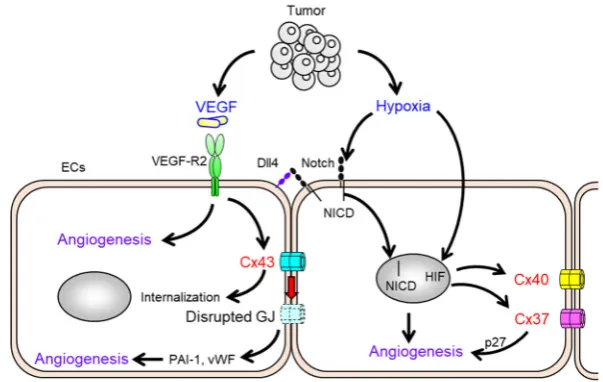
Pro-angiogenic factors have also been likely to modulate GJ function and Cx expression of ECs
99
[41] (Fig.1). Vascular endothelial growth factor (VEGF), which plays a central role in vasculogenesis
100
and angiogenesis [42], implicates in diverse physiologic processes including tumor angiogenesis [43,
101
44], diabetic retinopathy [45], wound healing [46], and tissue repair following ischemic injury [47].
102
VEGF-induced VEGF-receptor 2 (VEGF-R2) activation of ECs in existing vasculature is primarily an
103
initiation step of angiogenesis and then induces sprouting, cell proliferation, and cell migration of EC
104
[48]. In vitro model experiments, VEGF-induced c-Src tyrosine kinase and MAP kinases activation
105
results in the rapid disruption of GJ function of ECs [41], and increases paracellular endothelial
106
permeability associating with reduction of cell-cell junction [49]. Furthermore, it has been reported
107
that the VEGF-induced disruption of GJ function correlates with the rapid internalization of Cx43
108
and Cx43 tyrosine phosphorylation in rat coronary capillary endothelium [50, 51]. Therefore,
pro-109
angiogenic VEGF stimulation negatively modulate GJ function and Cxs expression in ECs in a
110
consequence of angiogenesis-related signaling.
111
In addition to VEGF, basic fibroblast growth factor (bFGF) and hypoxia are well-known as the
112
pro-angiogenic factor and environment. It has been reported that microvascular ECs facilitate GJ
113
function and Cx43 expression in the response to bFGF stimulation [52]. The stimulation with bFGF
114
not only increases Cx43 mRNA expression but also facilitates Cx43 localization at cell-cell interface
115
[52]. Hypoxia condition observed in tumor tissue activates HIF pathways and induces the
116
expression of a number of pro-angiogenic genes in cancer cells [1]. In the case of ECs, hypoxia
117
upregulates the Notch ligand Dll4 expression and promotes activation of Notch signaling which is
118
an essential pathway for vascular development and stabilization [53, 54]. The upregulation of Cx40
119
expression has been reported under hypoxia-mediated Notch signaling in ECs [54]. Recent study
120
has shown that a Notch-Cx37-p27 axis promotes EC cycle arrest leading to vascular regeneration
121
under shear stress [13]. These suggest that endothelial Cx and Notch might coordinate the
122
appropriate EC proliferation and angiogenesis.
123
124
Figure 1. Alteration of GJ function and Cxs expression in ECs under pro-angiogenic stimuli. VEGF
125
is an essential initiator of angiogenesis. ECs induce internalization and disruption of GJ formed by
126
Cx43 under VEGF-VEGF-R2 signaling. Hypoxic condition in tumor tissue activates Notch and HIFs
127
in EC. Notch signal including the nuclear translocation of the intracellular domain of the notch
128
protein (NICD) induces EC function and cell mechanics that involved in angiogenesis. HIF
129
pathways are angiogenic-related genes expression in ECs. Both signaling pathways results in
130
Although endothelial GJ function and Cx expression are assuredly regulated by pro-angiogenic
132
factors and environments, and further, it has been reported that Cxs expression and GJ function in
133
tumor cell [55], myocadiac cell [56], and mesenchymal stem cell [57], tightly link to VEGF expression
134
from these cells. For example, Cx43 knock-down in tumor cell lines increases VEGF expression and
135
enhanced the proliferation of ECs [55]. Thus, in order to understanding the role of GJ and Cx in
136
angiogenesis, it is necessary to elucidate the basic biology of GJ and Cx in these type cells at the
137
interplay of angiogenesis and tumor development.
138
4. The impact of endothelial Cxs in vascular endothelial angiogenesis
139
Several groups have investigated the impact of Cxs for development of cardiovascular system
140
which is closely related to angiogenesis. Mutations in the gene for Cx43 (GJA1) were found to cause
141
a hypoplastic left heart syndrome [58]. Cx43-deficient mice, which die at birth from connatural
142
heart malformations, have shown a reduction in the distal branching complexity and length of
143
coronary arteries [59]. In Cx40-deficient mice, cardiac malformations have been observed [60].
144
Additionally, both endothelial Cx40- and Cx37-knockout mice develop severe abnormalities of the
145
vascular function and structure [61]. Recently, loss of endothelial Cx40 leads to a reduction in
146
vascular growth and capillary density in the neovascularization of the mouse neonatal retina [62].
147
We have also demonstrated that aortic vascular tissue from Cx32-deficient mice exhibit suppressed
148
vascular sprouting of ECs [28]. Cx37 knock-out mice enhance vasculogenesis and remodeling
149
resulting in improvement from an ischemic hindlimb injury [63]. These studies have indicated the
150
contribution of endothelial Cxs to angiogenesis in the physiological or pathological condition.
151
Some reports have shown the relevance of endothelial Cxs expression and vascular angiogenic
152
potential in ECs in vitro angiogenesis assay. Knockdown of Cx43 using specific siRNAs reduces
153
tube formation and cell proliferation of human aortic ECs [64]. The downregulation of Cx43
154
increases angiogenesis-related factors [64], such as plasminogen activator inhibitor-1 [65] and von
155
Willebrand factor [66], suggesting that Cx43 might directly and/or indirectly contributes to
156
angiogenesis. Knockdown of Cx37, Cx40, or Cx43 using siRNAs has shown suppressed endothelial
157
angiogenesis including the branching of HUVECs, elongation of cell length, and tube formation by
158
an in vitro matrigel assay [67]. In gain-of-function experiments employed stable Cx-transfectants,
159
we have demonstrated that increased expression of Cx32 markedly enhances tube length and the
160
number of branching of EA. hy926 cells which is ECs line derived from HUVECs in matrigel tube
161
formation [28]. On the other hand, Cx37- or Cx43-transfected EA. hy926 cells impairs tube length
162
and the number of branching [28].
163
These studies have provided many evidences that endothelial Cxs expression modulate
164
angiogenesis, however the specific role of each Cxs on angiogenesis remains unclear. Notably, it
165
has been considered that any endothelial Cx expression may modifies other Cxs expression [28, 67,
166
68]. Indeed, Cx43 siRNA induces increased both Cx37 and Cx40 expression in aortic ECs. In
167
HUVECs, Cx43 siRNA does not alter the expression of other Cxs, whereas Cx40 siRNA and Cx37
168
siRNA reduce Cx43 and Cx40 expression, respectively [67]. In addition, Cx32-transfected EA. hy926
169
cells reduce Cx43 expression and has exhibited highly angiogenic potential such as tube formation
170
and branching [28]. Gain-of function and loss-of function assay remain to be experimentally tested,
171
however these have indicated that alteration Cx expression patterns and their relative network of Cx
172
expression may elicit different ECs phenotypes during angiogenic processes. This interrelated Cx
173
regulatory network have make difficult to understand specific role of each endothelial Cxs in
174
angiogenesis.
175
5. Endothelial Cxs-mediated regulation of cell migration in angiogenesis
176
ECs dynamically change cell mechanics such as cell morphology, cell proliferation, and cell
177
migration during angiogenesis process [69, 70]. EC activation by pro-angiogenic factors allows tip
178
cells to extend filamentous actin (F-actin)-rich filopodial protrusions migrating toward the required
179
site [3, 71]. Tip cells are the leading cells of the sprouts and guide following stalk cell which
180
proliferation of stalk cells is crucial for angiogenesis [4]. Notably, the implication of endothelial Cxs
182
in the control of EC migration has been progressively known. We have shown impaired cell
183
migration of ECs both in vitro wound healing assay by using Cx32 blocking ECs and in vivo matrigel
184
plaque implant assay in Cx32-deficient mice [28]. Other groups have reported that GJIC and Cx43
185
expression are increased in the region of cell migration and at localized to cells at the wound edge by
186
using wounded monolayer repair assay [72]. Cx43 specific siRNA markedly suppresses cell
187
migration of endothelial progenitor cells by transwell chamber migration assay that allowed cells to
188
migrate through the filter membrane upon pro-angiogenic factors [73]. In addition to ECs, several
189
type of cell such as leukocyte, epithelial cell, and tumor cell also regulate their migration via GJ
190
channel dependent and independent function (reviewed by Matsuuchi [74] and Kameritsch [75]).
191
192
Figure 2. Endothelial Cxs-mediated regulation of cell migration. Extracellular ATP released by
Cx-193
hemicannels activates P2Y receptors which trigger cell migration. GJ-mediated propagation of
194
calcium waves has been required for collective cell migration. The interaction of Cx and GJ with
195
cytoskeletal proteins or intracellular proteins orchestrate cytoskeletal rearrangement and cell
196
migration.
197
Both GJ mediated cell-cell interaction and hemichannel function have involved in the regulation
198
of cell migration in a number of cell type (Fig.2). Cultured adrenocortical cells have shown to exert
199
intact GJIC between cells during collective cell migration [76]. In a wound assay, Cx43 expression
200
in immortalized ECs is positively associated with cell migration and wound closure [77]. Moreover,
201
GJ-mediated propagation of calcium waves has been required for smooth muscle cell polarization
202
and migration [78], therefore, it is conceivable that GJIC in a cell cluster could play an important role
203
in coordination of the migration [79]. Extracellular ATP-induced calcium signaling has been shown
204
to modulate neuronal proliferation and migration of neuronal cells [80]. Cx hemichannels have
205
been known as a pathway of ATP release from intracellular space to extracellular space. ATP release
206
has been observed in glioma, glioblastoma and HeLa cells being transfected with Cx26, Cx32, or Cx43
207
[81]. Macrophage also releases ATP via Cx37 resulting in cell adhesion to endothelium [22]. It has
208
been considered that ATP release via Cx hemichannels from cells may induce cell migration through
209
calcium signaling following P2Y receptors activation in neighboring cells [81].
210
Intracellular domain of Cxs protein interacts with other proteins that involve in being structural
211
stability of cell-cell junction sustained by cytoskeletal scaffolds [10]. Due to the ubiquitous distribution
212
of Cx43, many studies have been performed focusing on Cx43 and their interacting proteins. The
213
carboxyl tail of Cx43 is indeed interacting with several cytoskeletal proteins such as F-actin,
α-/β-214
tubulins, cadherins, and cortactin) [82-85]. For example, the membrane expression of N-cadherin or
215
of ZO-1 is dominantly localized in the existing site of Cx43 protein [84]. The interaction of Cx43 with
216
intracellular signaling. Interaction of Cx43 and cadherins coordinates activation of Rho GTPases
218
which are promoting cell motility and invasion [86, 87]. Moreover, Rac1 in migrating cell is
219
dominantly found in forming actin-rich structures which in conjunction with E-cadherin are considered
220
responsible for the generation of traction forces of germ cells in vivo [88]. Intracellular carboxyl tail of
221
Cx43 has a number of interaction partners, thereby, the cell expressed a Cx43 lacking carboxyl tail
222
impairs cell migration [89]. Cx43 deficiency causes an impaired polarization caused by a
non-223
directional alignment of the microtubule organizing center. As a consequence, a loss of directionality
224
of cell migration and then an impaired development of coronary arteries can be observed in Cx43
225
deficient mice. A Cx43 mutant with lack of the tubulin binding site in the carboxyl tail has shown the
226
similar phenotypic pattern with Cx43 deficient [89], suggesting that interaction between Cx43 and
227
cytoskeletal protein may coordinate cell mechanics and behavior.
228
Interestingly, Cx43 seems indeed to be important for the stability of leading processes of the
229
neuronal cells determining the migratory pathway along the glial fibers [90]. Interesting mechanism
230
has been elicited that control the localization of Cx43 in the cellular extensions of migrating neurons in
231
a way that radial migration along the glia becomes possible [90]. Additionally, Watanabe and
232
colleagues have shown that fish GJ and Cx involves in fish morphological diversity, including skin
233
pattern formation and body shape determination [91]. Their studies have indicated that Cxs in
234
pigment cells, xanthophore and melanophore, dictate aggregation and separation of cells resulting in
235
pattern formation [92]. These suggest the possibility of Cxs dependent regulation of directional cell
236
migration.
237
6. Cxs-mediated regulation of cellular stiffness and cell migration
238
The interaction between Cx and cytoskeletal proteins correlatively contributes to the regulation
239
of cellular stiffness which is defined as the physical property of a cell to resist deformation in the
240
response to any applied force. A contraction force which generated by the actomyosin cytoskeleton
241
and F-actin has been inseparably connected with the regulation of cellular stiffness [93, 94].
242
Activation of the Rho-actomyosin signaling pathway enhances the formation of actin bundles, stress
243
fibers, and tensile actomyosin structures [95], all of which correlate with cellular stiffness [96, 97].
244
Thus, interplay between endothelial Cxs and Rho family has implicated in the regulation of cellular
245
stiffness. We have found that proinflammatory stimulation increased endothelial cellular stiffness
246
associating with impaired GJ function, cytoskeletal remodeling, and focal adhesion formation [98].
247
Moreover, blockade of GJs induces the cellular stiffening associated with focal adhesion formation
248
and cytoskeletal rearrangement, and prolonges TNF-α-induced endothelial cellular stiffening [98].
249
This study has provided first evidence that endothelial GJ contributes to the regulation of endothelial
250
cellular stiffness via interaction with cytoskeletal rearrangements.
251
It has been considered that endothelial cellular stiffness may be a determinant factor of leukocyte
252
adhesion to endothelium. In general, leukocyte senses the stiffness of extracellular substrate by
253
integrin-ligand interaction and adheres more strongly to stiff substrate [99]. ECs materially work
254
as a substrate during leukocyte adhesion and migration process. Leukocyte integrin assumes both
255
selective and cohesive adhesion via the binding to distinct endothelial adhesion receptors such as the
256
intercellular adhesion molecule 1 (ICAM1) [93]. Integrin increases the binding avidity to ligands
257
correlated with the endothelial cellular stiffness, while integrin-focal adhesion complex generates the
258
contractile force in cell and transduces the force into a mechanosignaling [100, 101]. These
259
suggested the possible mechanism which regulates leukocyte adhesion and activation via physical
260
endothelial cellular stiffness [102].
261
In addition to leukocyte, it has been reported that ECs themselves also modulate their migration,
262
proliferation, and morphological changes in the response to extracellular substrate stiffness [103, 104].
263
Thus, it has been shown a possibility that stiffening ECs in existing vasculature is favor to recruit
pro-264
angiogenic tip cells and stalk cells at the sprouting spots (Fig.3). Of note, VEGF-induced cytoskeletal
265
rearrangement and impaired GJ function might be supposed to increases EC stiffness. Stiff ECs may
266
recruit endothelial progenitor cells and support the cell proliferation and elongation of stalk cells.
267
angiogenic process of recruited ECs by being the activator of mechanosensing and transduction
269
pathway.
270
271
Figure 3. Potential role of endothelial cellular stiffening in angiogenesis. VEGF-induced GJ reduction
272
increases the stiffness of ECs in sprout initiation phase. Stiff ECs provide the favorable environment
273
for recruitment of endothelial progenitor cells, while stiff ECs support adjacent stalk cell proliferation
274
and elongation.
275
7. Conclusions
276
We are beginning to understand that GJ and Cx in ECs might be a center for connection between
277
biological function and cell mechanics in the context of angiogenesis. In this review, we provide an
278
overview of the endothelial GJ function and Cxs expression found in pro-angiogenic condition and
279
the functional role of endothelial GJ and Cxs in cell mechanics during the angiogenic process. Cell
280
mechanics-based mechanisms hold promise the better understanding for physiological and
281
pathological angiogenesis. Although several studies have demonstrated GJ-/Cx-dependent
282
regulation of angiogenesis, the mechanisms are still speculative and controversial. Additionally,
283
GJ- and Cx-mediated interactions in a number of other type cells such as vascular smooth muscle cell,
284
pericyte, fibroblast, macrophage, and tumor cell also contribute to tumor angiogenesis through the
285
expression of pro-angiogenic factors. Thus, further studies in the basic biology of GJ and Cx in these
286
type cells would be required for elucidation with a particular emphasis on the interplay of
287
angiogenesis and tumor development. We have speculated that GJ and Cx targeting approaches
288
may be relevant to the development of the treatment of cancer patients.
289
Author Contributions: Conceptualization, T.O.; writing—original draft preparation, T.O.; writing—review and
290
editing, T.O., H.U., T.T., K.W., M.S.;
291
Funding: Please add: This work was supported by the Japan Society for the Promotion of Science (JSPS)
292
KAKENHI Grant Number JP16K09513, JP16K15759, and JP25461125.
293
Conflicts of Interest: The authors declare no conflict of interest.
294
References
295
1. Shweiki, D.; Itin, A.; Soffer, D.; Keshet, E., Vascular endothelial growth factor induced by
296
hypoxia may mediate hypoxia-initiated angiogenesis. Nature 1992, 359, (6398), 843-5.
297
2. Poon, R. T.; Fan, S. T.; Wong, J., Clinical implications of circulating angiogenic factors in
298
cancer patients. J Clin Oncol 2001, 19, (4), 1207-25.
299
3. Gerhardt, H.; Golding, M.; Fruttiger, M.; Ruhrberg, C.; Lundkvist, A.; Abramsson, A.;
300
Jeltsch, M.; Mitchell, C.; Alitalo, K.; Shima, D.; Betsholtz, C., VEGF guides angiogenic
301
sprouting utilizing endothelial tip cell filopodia. J Cell Biol 2003, 161, (6), 1163-77.
302
4. Jakobsson, L.; Franco, C. A.; Bentley, K.; Collins, R. T.; Ponsioen, B.; Aspalter, I. M.;
303
Endothelial cells dynamically compete for the tip cell position during angiogenic sprouting.
305
Nat Cell Biol 2010, 12, (10), 943-53.
306
5. Cao, J.; Ehling, M.; Marz, S.; Seebach, J.; Tarbashevich, K.; Sixta, T.; Pitulescu, M. E.;
307
Werner, A. C.; Flach, B.; Montanez, E.; Raz, E.; Adams, R. H.; Schnittler, H., Polarized actin
308
and VE-cadherin dynamics regulate junctional remodelling and cell migration during
309
sprouting angiogenesis. Nat Commun 2017, 8, (1), 2210.
310
6. Abedi, H.; Zachary, I., Vascular endothelial growth factor stimulates tyrosine
311
phosphorylation and recruitment to new focal adhesions of focal adhesion kinase and
312
paxillin in endothelial cells. J Biol Chem 1997, 272, (24), 15442-51.
313
7. Hu, J.; Qiu, J.; Zheng, Y.; Zhang, T.; Yin, T.; Xie, X.; Wang, G., AAMP Regulates Endothelial
314
Cell Migration and Angiogenesis Through RhoA/Rho Kinase Signaling. Ann Biomed Eng
315
2016, 44, (5), 1462-74.
316
8. Kumar, N. M.; Gilula, N. B., The gap junction communication channel. Cell 1996, 84, (3),
317
381-8.
318
9. Saez, J. C.; Berthoud, V. M.; Branes, M. C.; Martinez, A. D.; Beyer, E. C., Plasma membrane
319
channels formed by connexins: their regulation and functions. Physiol Rev 2003, 83, (4),
320
1359-400.
321
10. Harris, A. L., Connexin channel permeability to cytoplasmic molecules. Prog Biophys Mol
322
Biol 2007, 94, (1-2), 120-43.
323
11. Chang, S. F.; Chen, L. J.; Lee, P. L.; Lee, D. Y.; Chien, S.; Chiu, J. J., Different modes of
324
endothelial-smooth muscle cell interaction elicit differential beta-catenin phosphorylations
325
and endothelial functions. Proc Natl Acad Sci U S A 2014, 111, (5), 1855-60.
326
12. Chen, C. H.; Mayo, J. N.; Gourdie, R. G.; Johnstone, S. R.; Isakson, B. E.; Bearden, S. E.,
327
The connexin 43/ZO-1 complex regulates cerebral endothelial F-actin architecture and
328
migration. Am J Physiol Cell Physiol 2015, 309, (9), C600-7.
329
13. Fang, J. S.; Coon, B. G.; Gillis, N.; Chen, Z.; Qiu, J.; Chittenden, T. W.; Burt, J. M.; Schwartz,
330
M. A.; Hirschi, K. K., Shear-induced Notch-Cx37-p27 axis arrests endothelial cell cycle to
331
enable arterial specification. Nat Commun 2017, 8, (1), 2149.
332
14. Esmon, C. T., The interactions between inflammation and coagulation. Br J Haematol 2005,
333
131, (4), 417-30.
334
15. Reglero-Real, N.; Marcos-Ramiro, B.; Millan, J., Endothelial membrane reorganization
335
during leukocyte extravasation. Cell Mol Life Sci 2012, 69, (18), 3079-99.
336
16. Folkman, J.; Merler, E.; Abernathy, C.; Williams, G., Isolation of a tumor factor responsible
337
for angiogenesis. J Exp Med 1971, 133, (2), 275-88.
338
17. Godo, S.; Shimokawa, H., Endothelial Functions. Arterioscler Thromb Vasc Biol 2017, 37,
339
(9), e108-e114.
340
18. Larson, D. M.; Haudenschild, C. C.; Beyer, E. C., Gap junction messenger RNA expression
341
by vascular wall cells. Circ Res 1990, 66, (4), 1074-80.
342
19. Yeh, H. I.; Rothery, S.; Dupont, E.; Coppen, S. R.; Severs, N. J., Individual gap junction
343
plaques contain multiple connexins in arterial endothelium. Circ Res 1998, 83, (12),
1248-344
63.
345
20. Kwak, B. R.; Mulhaupt, F.; Veillard, N.; Gros, D. B.; Mach, F., Altered pattern of vascular
346
225-30.
348
21. van Rijen, H. V.; van Kempen, M. J.; Postma, S.; Jongsma, H. J., Tumour necrosis factor
349
alpha alters the expression of connexin43, connexin40, and connexin37 in human umbilical
350
vein endothelial cells. Cytokine 1998, 10, (4), 258-64.
351
22. Wong, C. W.; Christen, T.; Roth, I.; Chadjichristos, C. E.; Derouette, J. P.; Foglia, B. F.;
352
Chanson, M.; Goodenough, D. A.; Kwak, B. R., Connexin37 protects against atherosclerosis
353
by regulating monocyte adhesion. Nat Med 2006, 12, (8), 950-4.
354
23. Wagner, C.; de Wit, C.; Kurtz, L.; Grunberger, C.; Kurtz, A.; Schweda, F., Connexin40 is
355
essential for the pressure control of renin synthesis and secretion. Circ Res 2007, 100, (4),
356
556-63.
357
24. Gabriels, J. E.; Paul, D. L., Connexin43 is highly localized to sites of disturbed flow in rat
358
aortic endothelium but connexin37 and connexin40 are more uniformly distributed. Circ Res
359
1998, 83, (6), 636-43.
360
25. Okamoto, T.; Akiyama, M.; Takeda, M.; Gabazza, E. C.; Hayashi, T.; Suzuki, K., Connexin32
361
is expressed in vascular endothelial cells and participates in gap-junction intercellular
362
communication. Biochem Biophys Res Commun 2009, 382, (2), 264-8.
363
26. Inai, T.; Shibata, Y., Heterogeneous expression of endothelial connexin (Cx) 37, Cx40, and
364
Cx43 in rat large veins. Anat Sci Int 2009, 84, (3), 237-45.
365
27. Van Rijen, H.; van Kempen, M. J.; Analbers, L. J.; Rook, M. B.; van Ginneken, A. C.; Gros,
366
D.; Jongsma, H. J., Gap junctions in human umbilical cord endothelial cells contain multiple
367
connexins. Am J Physiol 1997, 272, (1 Pt 1), C117-30.
368
28. Okamoto, T.; Akita, N.; Kawamoto, E.; Hayashi, T.; Suzuki, K.; Shimaoka, M., Endothelial
369
connexin32 enhances angiogenesis by positively regulating tube formation and cell
370
migration. Exp Cell Res 2014, 321, (2), 133-41.
371
29. Ebong, E. E.; Kim, S.; DePaola, N., Flow regulates intercellular communication in HAEC by
372
assembling functional Cx40 and Cx37 gap junctional channels. Am J Physiol Heart Circ
373
Physiol 2006, 290, (5), H2015-23.
374
30. Okamoto, T.; Akiyama, M.; Takeda, M.; Akita, N.; Yoshida, K.; Hayashi, T.; Suzuki, K.,
375
Connexin32 protects against vascular inflammation by modulating inflammatory cytokine
376
expression by endothelial cells. Exp Cell Res 2011, 317, (3), 348-55.
377
31. Bolon, M. L.; Kidder, G. M.; Simon, A. M.; Tyml, K., Lipopolysaccharide reduces electrical
378
coupling in microvascular endothelial cells by targeting connexin40 in a tyrosine-, ERK1/2-,
379
PKA-, and PKC-dependent manner. J Cell Physiol 2007, 211, (1), 159-66.
380
32. Lidington, D.; Tyml, K.; Ouellette, Y., Lipopolysaccharide-induced reductions in cellular
381
coupling correlate with tyrosine phosphorylation of connexin 43. J Cell Physiol 2002, 193,
382
(3), 373-9.
383
33. Baker, S. M.; Kim, N.; Gumpert, A. M.; Segretain, D.; Falk, M. M., Acute internalization of
384
gap junctions in vascular endothelial cells in response to inflammatory mediator-induced
G-385
protein coupled receptor activation. FEBS Lett 2008, 582, (29), 4039-46.
386
34. O'Donnell, J. J., 3rd; Birukova, A. A.; Beyer, E. C.; Birukov, K. G., Gap junction protein
387
connexin43 exacerbates lung vascular permeability. PLoS One 2014, 9, (6), e100931.
388
35. Chadjichristos, C. E.; Scheckenbach, K. E.; van Veen, T. A.; Richani Sarieddine, M. Z.; de
389
Kempen, M. J.; Coenjaerts, F. E.; Miquerol, L.; Deutsch, U.; Jongsma, H. J.; Chanson, M.;
391
Kwak, B. R., Endothelial-specific deletion of connexin40 promotes atherosclerosis by
392
increasing CD73-dependent leukocyte adhesion. Circulation 2010, 121, (1), 123-31.
393
36. Derouette, J. P.; Wong, C.; Burnier, L.; Morel, S.; Sutter, E.; Galan, K.; Brisset, A. C.; Roth,
394
I.; Chadjichristos, C. E.; Kwak, B. R., Molecular role of Cx37 in advanced atherosclerosis: a
395
micro-array study. Atherosclerosis 2009, 206, (1), 69-76.
396
37. Kwak, B. R.; Veillard, N.; Pelli, G.; Mulhaupt, F.; James, R. W.; Chanson, M.; Mach, F.,
397
Reduced connexin43 expression inhibits atherosclerotic lesion formation in low-density
398
lipoprotein receptor-deficient mice. Circulation 2003, 107, (7), 1033-9.
399
38. Haefliger, J. A.; Krattinger, N.; Martin, D.; Pedrazzini, T.; Capponi, A.; Doring, B.; Plum, A.;
400
Charollais, A.; Willecke, K.; Meda, P., Connexin43-dependent mechanism modulates renin
401
secretion and hypertension. J Clin Invest 2006, 116, (2), 405-13.
402
39. Yamada, Y.; Izawa, H.; Ichihara, S.; Takatsu, F.; Ishihara, H.; Hirayama, H.; Sone, T.;
403
Tanaka, M.; Yokota, M., Prediction of the risk of myocardial infarction from polymorphisms
404
in candidate genes. N Engl J Med 2002, 347, (24), 1916-23.
405
40. Okamoto, T.; Akita, N.; Hayashi, T.; Shimaoka, M.; Suzuki, K., Endothelial connexin 32
406
regulates tissue factor expression induced by inflammatory stimulation and direct cell-cell
407
interaction with activated cells. Atherosclerosis 2014, 236, (2), 430-7.
408
41. Suarez, S.; Ballmer-Hofer, K., VEGF transiently disrupts gap junctional communication in
409
endothelial cells. J Cell Sci 2001, 114, (Pt 6), 1229-35.
410
42. Ferrara, N.; Gerber, H. P.; LeCouter, J., The biology of VEGF and its receptors. Nat Med
411
2003, 9, (6), 669-76.
412
43. Kim, K. J.; Li, B.; Winer, J.; Armanini, M.; Gillett, N.; Phillips, H. S.; Ferrara, N., Inhibition
413
of vascular endothelial growth factor-induced angiogenesis suppresses tumour growth in
414
vivo. Nature 1993, 362, (6423), 841-4.
415
44. Ferrara, N.; Davis-Smyth, T., The biology of vascular endothelial growth factor. Endocr Rev
416
1997, 18, (1), 4-25.
417
45. Aiello, L. P.; Avery, R. L.; Arrigg, P. G.; Keyt, B. A.; Jampel, H. D.; Shah, S. T.; Pasquale, L.
418
R.; Thieme, H.; Iwamoto, M. A.; Park, J. E.; et al., Vascular endothelial growth factor in
419
ocular fluid of patients with diabetic retinopathy and other retinal disorders. N Engl J Med
420
1994, 331, (22), 1480-7.
421
46. Brown, L. F.; Yeo, K. T.; Berse, B.; Yeo, T. K.; Senger, D. R.; Dvorak, H. F.; van de Water, L.,
422
Expression of vascular permeability factor (vascular endothelial growth factor) by epidermal
423
keratinocytes during wound healing. J Exp Med 1992, 176, (5), 1375-9.
424
47. Maniscalco, W. M.; Watkins, R. H.; Finkelstein, J. N.; Campbell, M. H., Vascular endothelial
425
growth factor mRNA increases in alveolar epithelial cells during recovery from oxygen injury.
426
Am J Respir Cell Mol Biol 1995, 13, (4), 377-86.
427
48. Waltenberger, J.; Claesson-Welsh, L.; Siegbahn, A.; Shibuya, M.; Heldin, C. H., Different
428
signal transduction properties of KDR and Flt1, two receptors for vascular endothelial
429
growth factor. J Biol Chem 1994, 269, (43), 26988-95.
430
49. Antonetti, D. A.; Barber, A. J.; Hollinger, L. A.; Wolpert, E. B.; Gardner, T. W., Vascular
431
endothelial growth factor induces rapid phosphorylation of tight junction proteins occludin
432
retinopathy and tumors. J Biol Chem 1999, 274, (33), 23463-7.
434
50. Nimlamool, W.; Andrews, R. M.; Falk, M. M., Connexin43 phosphorylation by PKC and
435
MAPK signals VEGF-mediated gap junction internalization. Mol Biol Cell 2015, 26, (15),
436
2755-68.
437
51. Thuringer, D., The vascular endothelial growth factor-induced disruption of gap junctions is
438
relayed by an autocrine communication via ATP release in coronary capillary endothelium.
439
Ann N Y Acad Sci 2004, 1030, 14-27.
440
52. Pepper, M. S.; Meda, P., Basic fibroblast growth factor increases junctional communication
441
and connexin 43 expression in microvascular endothelial cells. J Cell Physiol 1992, 153, (1),
442
196-205.
443
53. Favre, C. J.; Mancuso, M.; Maas, K.; McLean, J. W.; Baluk, P.; McDonald, D. M., Expression
444
of genes involved in vascular development and angiogenesis in endothelial cells of adult lung.
445
Am J Physiol Heart Circ Physiol 2003, 285, (5), H1917-38.
446
54. Lanner, F.; Lee, K. L.; Ortega, G. C.; Sohl, M.; Li, X.; Jin, S.; Hansson, E. M.; Claesson-Welsh,
447
L.; Poellinger, L.; Lendahl, U.; Farnebo, F., Hypoxia-induced arterial differentiation requires
448
adrenomedullin and notch signaling. Stem Cells Dev 2013, 22, (9), 1360-9.
449
55. Wang, W. K.; Chen, M. C.; Leong, H. F.; Kuo, Y. L.; Kuo, C. Y.; Lee, C. H., Connexin 43
450
suppresses tumor angiogenesis by down-regulation of vascular endothelial growth factor via
451
hypoxic-induced factor-1alpha. Int J Mol Sci 2014, 16, (1), 439-51.
452
56. Pimentel, R. C.; Yamada, K. A.; Kleber, A. G.; Saffitz, J. E., Autocrine regulation of myocyte
453
Cx43 expression by VEGF. Circ Res 2002, 90, (6), 671-7.
454
57. Fan, X.; Teng, Y.; Ye, Z.; Zhou, Y.; Tan, W. S., The effect of gap junction-mediated transfer of
455
miR-200b on osteogenesis and angiogenesis in a co-culture of MSCs and HUVECs. J Cell Sci
456
2018, 131, (13).
457
58. Huang, G. Y.; Xie, L. J.; Linask, K. L.; Zhang, C.; Zhao, X. Q.; Yang, Y.; Zhou, G. M.; Wu, Y.
458
J.; Marquez-Rosado, L.; McElhinney, D. B.; Goldmuntz, E.; Liu, C.; Lampe, P. D.; Chatterjee,
459
B.; Lo, C. W., Evaluating the role of connexin43 in congenital heart disease: Screening for
460
mutations in patients with outflow tract anomalies and the analysis of knock-in mouse
461
models. J Cardiovasc Dis Res 2011, 2, (4), 206-12.
462
59. Ya, J.; Erdtsieck-Ernste, E. B.; de Boer, P. A.; van Kempen, M. J.; Jongsma, H.; Gros, D.;
463
Moorman, A. F.; Lamers, W. H., Heart defects in connexin43-deficient mice. Circ Res 1998,
464
82, (3), 360-6.
465
60. Gu, H.; Smith, F. C.; Taffet, S. M.; Delmar, M., High incidence of cardiac malformations in
466
connexin40-deficient mice. Circ Res 2003, 93, (3), 201-6.
467
61. Simon, A. M.; McWhorter, A. R., Vascular abnormalities in mice lacking the endothelial gap
468
junction proteins connexin37 and connexin40. Dev Biol 2002, 251, (2), 206-20.
469
62. Haefliger, J. A.; Allagnat, F.; Hamard, L.; Le Gal, L.; Meda, P.; Nardelli-Haefliger, D.; Genot,
470
E.; Alonso, F., Targeting Cx40 (Connexin40) Expression or Function Reduces Angiogenesis
471
in the Developing Mouse Retina. Arterioscler Thromb Vasc Biol 2017, 37, (11), 2136-2146.
472
63. Fang, J. S.; Angelov, S. N.; Simon, A. M.; Burt, J. M., Cx37 deletion enhances vascular
473
growth and facilitates ischemic limb recovery. Am J Physiol Heart Circ Physiol 2011, 301,
474
(5), H1872-81.
475
Activation of endothelial cells to pathological status by down-regulation of connexin43.
477
Cardiovasc Res 2008, 79, (3), 509-18.
478
65. Bacharach, E.; Itin, A.; Keshet, E., In vivo patterns of expression of urokinase and its
479
inhibitor PAI-1 suggest a concerted role in regulating physiological angiogenesis. Proc Natl
480
Acad Sci U S A 1992, 89, (22), 10686-90.
481
66. Xu, H.; Cao, Y.; Yang, X.; Cai, P.; Kang, L.; Zhu, X.; Luo, H.; Lu, L.; Wei, L.; Bai, X.; Zhu, Y.;
482
Zhao, B. Q.; Fan, W., ADAMTS13 controls vascular remodeling by modifying VWF reactivity
483
during stroke recovery. Blood 2017, 130, (1), 11-22.
484
67. Gartner, C.; Ziegelhoffer, B.; Kostelka, M.; Stepan, H.; Mohr, F. W.; Dhein, S., Knock-down
485
of endothelial connexins impairs angiogenesis. Pharmacol Res 2012, 65, (3), 347-57.
486
68. Johnson, T. L.; Nerem, R. M., Endothelial connexin 37, connexin 40, and connexin 43
487
respond uniquely to substrate and shear stress. Endothelium 2007, 14, (4-5), 215-26.
488
69. Kliche, K.; Jeggle, P.; Pavenstadt, H.; Oberleithner, H., Role of cellular mechanics in the
489
function and life span of vascular endothelium. Pflugers Arch 2011, 462, (2), 209-17.
490
70. Eilken, H. M.; Adams, R. H., Dynamics of endothelial cell behavior in sprouting angiogenesis.
491
Curr Opin Cell Biol 2010, 22, (5), 617-25.
492
71. Stapor, P.; Wang, X.; Goveia, J.; Moens, S.; Carmeliet, P., Angiogenesis revisited - role and
493
therapeutic potential of targeting endothelial metabolism. J Cell Sci 2014, 127, (Pt 20),
4331-494
41.
495
72. Simpson, K. J.; Selfors, L. M.; Bui, J.; Reynolds, A.; Leake, D.; Khvorova, A.; Brugge, J. S.,
496
Identification of genes that regulate epithelial cell migration using an siRNA screening
497
approach. Nat Cell Biol 2008, 10, (9), 1027-38.
498
73. Wang, H. H.; Su, C. H.; Wu, Y. J.; Li, J. Y.; Tseng, Y. M.; Lin, Y. C.; Hsieh, C. L.; Tsai, C. H.;
499
Yeh, H. I., Reduction of connexin43 in human endothelial progenitor cells impairs the
500
angiogenic potential. Angiogenesis 2013, 16, (3), 553-60.
501
74. Matsuuchi, L.; Naus, C. C., Gap junction proteins on the move: connexins, the cytoskeleton
502
and migration. Biochim Biophys Acta 2013, 1828, (1), 94-108.
503
75. Kameritsch, P.; Pogoda, K.; Pohl, U., Channel-independent influence of connexin 43 on cell
504
migration. Biochim Biophys Acta 2012, 1818, (8), 1993-2001.
505
76. Friedl, P.; Gilmour, D., Collective cell migration in morphogenesis, regeneration and cancer.
506
Nat Rev Mol Cell Biol 2009, 10, (7), 445-57.
507
77. Kwak, B. R.; Pepper, M. S.; Gros, D. B.; Meda, P., Inhibition of endothelial wound repair by
508
dominant negative connexin inhibitors. Mol Biol Cell 2001, 12, (4), 831-45.
509
78. Espinosa-Tanguma, R.; O'Neil, C.; Chrones, T.; Pickering, J. G.; Sims, S. M., Essential role
510
for calcium waves in migration of human vascular smooth muscle cells. Am J Physiol Heart
511
Circ Physiol 2011, 301, (2), H315-23.
512
79. Defranco, B. H.; Nickel, B. M.; Baty, C. J.; Martinez, J. S.; Gay, V. L.; Sandulache, V. C.;
513
Hackam, D. J.; Murray, S. A., Migrating cells retain gap junction plaque structure and
514
function. Cell Commun Adhes 2008, 15, (3), 273-88.
515
80. Weissman, T. A.; Riquelme, P. A.; Ivic, L.; Flint, A. C.; Kriegstein, A. R., Calcium waves
516
propagate through radial glial cells and modulate proliferation in the developing neocortex.
517
Neuron 2004, 43, (5), 647-61.
518
Naus, C. C.; Nedergaard, M., Connexins regulate calcium signaling by controlling ATP
520
release. Proc Natl Acad Sci U S A 1998, 95, (26), 15735-40.
521
82. Giepmans, B. N., Role of connexin43-interacting proteins at gap junctions. Adv Cardiol 2006,
522
42, 41-56.
523
83. Theiss, C.; Meller, K., Microinjected anti-actin antibodies decrease gap junctional
524
intercellular commmunication in cultured astrocytes. Exp Cell Res 2002, 281, (2), 197-204.
525
84. Wei, C. J.; Francis, R.; Xu, X.; Lo, C. W., Connexin43 associated with an
N-cadherin-526
containing multiprotein complex is required for gap junction formation in NIH3T3 cells. J
527
Biol Chem 2005, 280, (20), 19925-36.
528
85. Vitale, M. L.; Akpovi, C. D.; Pelletier, R. M., Cortactin/tyrosine-phosphorylated cortactin
529
interaction with connexin 43 in mouse seminiferous tubules. Microsc Res Tech 2009, 72, (11),
530
856-67.
531
86. Xu, X.; Li, W. E.; Huang, G. Y.; Meyer, R.; Chen, T.; Luo, Y.; Thomas, M. P.; Radice, G. L.; Lo,
532
C. W., Modulation of mouse neural crest cell motility by N-cadherin and connexin 43 gap
533
junctions. J Cell Biol 2001, 154, (1), 217-30.
534
87. Xu, X.; Francis, R.; Wei, C. J.; Linask, K. L.; Lo, C. W., Connexin 43-mediated modulation of
535
polarized cell movement and the directional migration of cardiac neural crest cells.
536
Development 2006, 133, (18), 3629-39.
537
88. Kardash, E.; Reichman-Fried, M.; Maitre, J. L.; Boldajipour, B.; Papusheva, E.;
538
Messerschmidt, E. M.; Heisenberg, C. P.; Raz, E., A role for Rho GTPases and cell-cell
539
adhesion in single-cell motility in vivo. Nat Cell Biol 2010, 12, (1), 47-53; sup pp 1-11.
540
89. Rhee, D. Y.; Zhao, X. Q.; Francis, R. J.; Huang, G. Y.; Mably, J. D.; Lo, C. W., Connexin 43
541
regulates epicardial cell polarity and migration in coronary vascular development.
542
Development 2009, 136, (18), 3185-93.
543
90. Elias, L. A.; Wang, D. D.; Kriegstein, A. R., Gap junction adhesion is necessary for radial
544
migration in the neocortex. Nature 2007, 448, (7156), 901-7.
545
91. Watanabe, M., Gap Junction in the Teleost Fish Lineage: Duplicated Connexins May
546
Contribute to Skin Pattern Formation and Body Shape Determination. Front Cell Dev Biol
547
2017, 5, 13.
548
92. Watanabe, M.; Sawada, R.; Aramaki, T.; Skerrett, I. M.; Kondo, S., The Physiological
549
Characterization of Connexin41.8 and Connexin39.4, Which Are Involved in the Striped
550
Pattern Formation of Zebrafish. J Biol Chem 2016, 291, (3), 1053-63.
551
93. Schaefer, A.; Te Riet, J.; Ritz, K.; Hoogenboezem, M.; Anthony, E. C.; Mul, F. P.; de Vries, C.
552
J.; Daemen, M. J.; Figdor, C. G.; van Buul, J. D.; Hordijk, P. L., Actin-binding proteins
553
differentially regulate endothelial cell stiffness, ICAM-1 function and neutrophil
554
transmigration. J Cell Sci 2014, 127, (Pt 20), 4470-82.
555
94. Stroka, K. M.; Aranda-Espinoza, H., Effects of Morphology vs. Cell-Cell Interactions on
556
Endothelial Cell Stiffness. Cell Mol Bioeng 2011, 4, (1), 9-27.
557
95. Chrzanowska-Wodnicka, M.; Burridge, K., Rho-stimulated contractility drives the formation
558
of stress fibers and focal adhesions. J Cell Biol 1996, 133, (6), 1403-15.
559
96. Wojciak-Stothard, B.; Potempa, S.; Eichholtz, T.; Ridley, A. J., Rho and Rac but not Cdc42
560
regulate endothelial cell permeability. J Cell Sci 2001, 114, (Pt 7), 1343-55.
561
Stamenovic, D., Cell prestress. I. Stiffness and prestress are closely associated in adherent
563
contractile cells. Am J Physiol Cell Physiol 2002, 282, (3), C606-16.
564
98. Okamoto, T.; Kawamoto, E.; Takagi, Y.; Akita, N.; Hayashi, T.; Park, E. J.; Suzuki, K.;
565
Shimaoka, M., Gap junction-mediated regulation of endothelial cellular stiffness. Sci Rep
566
2017, 7, (1), 6134.
567
99. Oakes, P. W.; Patel, D. C.; Morin, N. A.; Zitterbart, D. P.; Fabry, B.; Reichner, J. S.; Tang, J.
568
X., Neutrophil morphology and migration are affected by substrate elasticity. Blood 2009,
569
114, (7), 1387-95.
570
100. Huveneers, S.; Daemen, M. J.; Hordijk, P. L., Between Rho(k) and a hard place: the relation
571
between vessel wall stiffness, endothelial contractility, and cardiovascular disease. Circ Res
572
2015, 116, (5), 895-908.
573
101. Stroka, K. M.; Aranda-Espinoza, H., Endothelial cell substrate stiffness influences
574
neutrophil transmigration via myosin light chain kinase-dependent cell contraction. Blood
575
2011, 118, (6), 1632-40.
576
102. Schaefer, A.; Hordijk, P. L., Cell-stiffness-induced mechanosignaling - a key driver of
577
leukocyte transendothelial migration. J Cell Sci 2015, 128, (13), 2221-30.
578
103. Sack, K. D.; Teran, M.; Nugent, M. A., Extracellular Matrix Stiffness Controls VEGF
579
Signaling and Processing in Endothelial Cells. J Cell Physiol 2016, 231, (9), 2026-39.
580
104. Browning, M. B.; Guiza, V.; Russell, B.; Rivera, J.; Cereceres, S.; Hook, M.; Hahn, M. S.;
581
Cosgriff-Hernandez, E. M., Endothelial cell response to chemical, biological, and physical