Scholarship@Western
Scholarship@Western
Electronic Thesis and Dissertation Repository
12-11-2013 12:00 AM
Investigating the Effects of Custom Made Orthotics on Brain
Investigating the Effects of Custom Made Orthotics on Brain
Forms: A Pilot Study
Forms: A Pilot Study
Lindsay CareyThe University of Western Ontario Supervisor
Dr. Colin Dombroski
The University of Western Ontario Joint Supervisor Dr. Jeff Holmes
The University of Western Ontario
Graduate Program in Health and Rehabilitation Sciences
A thesis submitted in partial fulfillment of the requirements for the degree in Master of Science © Lindsay Carey 2013
Follow this and additional works at: https://ir.lib.uwo.ca/etd
Part of the Neurosciences Commons, and the Orthotics and Prosthetics Commons
Recommended Citation Recommended Citation
Carey, Lindsay, "Investigating the Effects of Custom Made Orthotics on Brain Forms: A Pilot Study" (2013). Electronic Thesis and Dissertation Repository. 1792.
https://ir.lib.uwo.ca/etd/1792
This Dissertation/Thesis is brought to you for free and open access by Scholarship@Western. It has been accepted for inclusion in Electronic Thesis and Dissertation Repository by an authorized administrator of
(Thesis format: Monograph)
by
Lindsay Carey
Graduate Program in Health and Rehabilitation Sciences
A thesis submitted in partial fulfillment of the requirements for the degree of
Master of Science
The School of Graduate and Postdoctoral Studies The University of Western Ontario
London, Ontario, Canada
ii
Abstract
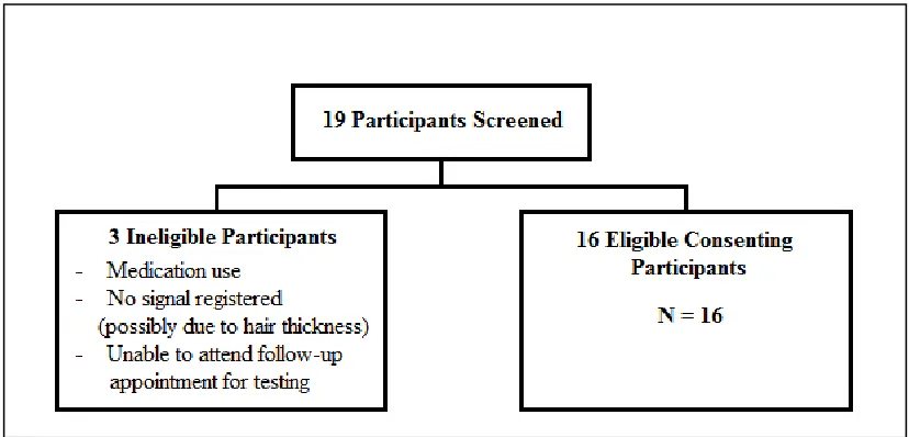
OBJECTIVES: To determine (1) the feasibility of this novel approach and technique of
recording brain activity, wirelessly and continuously, during human gait, and (2) if custom
made orthotics will alter the brain activity patterns recorded. METHODS: Gait trials were
performed on 16 participants walking with and without orthotic devices in their shoes while
simultaneously collecting EEG data through the Emotiv wireless neuroheadset. RESULTS:
The Emotiv neuroheadset was capable of detecting changes in brain activity between the two
gait trials. The differences in brain activity identified between conditions were not
statistically significant. CONCLUSION: The findings suggest the Emotiv EEG device is
sensitive enough to detect changes in brain activation patterns during human gait. Further
research is required before definite conclusions can be made about this novel device, or about
what effects, if any, orthotics have on brain activation patterns during gait.
iii
Co-Authorship Statement
This thesis is the primary work of Lindsay Carey. The creation of the research topic and
design of the intervention was created by Dr. Colin Dombroski through collaboration with
Dr. Jeff Holmes and Dr. Andrew Johnson. The moulding and fabrication of the orthotic
devices were completed by Dr. Dombroski and SoleScience Laboratories. The process of
data collection was facilitated by both Dr. Dombroski and Lindsay Carey. The raw EEG data
produced from the Emotiv neuroheadset was processed and analyzed through the use of EEG
lab within MAT lab by James Desjardins from Brock University. I wrote the original draft of
this thesis in its entirety, including the interpretation of the statistical results (with the
assistance of James Desjardins). The drafts were then sent to Dr. Dombroski and Dr. Holmes
iv
Acknowledgments
The completion of this thesis would not have been possible without the many people who
provided tremendous support and assistance along the way.
First and foremost, thank you to Dr. Colin Dombroski for his knowledge, guidance and
support throughout this very condensed research project.
Thank you to Dr. Jeff Holmes and Dr. Andrew Johnson for their valuable input and
assistance throughout this process.
To all of the SoleScience staff, thank you for arranging appointment times (Betty Ann) and
fabricating the orthotic devices used for testing.
I would like to acknowledge James Desjardins for his expertise and contributions involving
the processing and analysis of the EEG data.
Thank you to all other faculty and students in the Interdisciplinary Movement Disorders Lab
for their feedback and participation in this project.
I would like to express my gratitude towards Dr. Lisa Fischer, for her incredible support and
advice during the last two years.
My sincere thanks and appreciation goes out to my parents, sister, family and friends.
Without your never-ending support, encouragement and motivation, this journey would never
have been possible.
Finally, I would like to thank Mike and Kelly Ravenek. Your continued persistence and
guidance allowed me to discover the field of Health and Rehabilitation Sciences and become
v
Table of Contents
Abstract ... ii
Co-Authorship Statement... iii
Acknowledgments ... iv
Table of Contents ...v
List of Tables ... viii
List of Figures ... ix
List of Appendices ...x
List of Abbreviations...xi
Chapter 1 ...1
1 Introduction ...1
Chapter 2 ...5
2 Literature Review ...5
2.1 Introduction ...5
2.2 Anatomy of the Brain and Central Nervous System ...6
2.2.1 Cerebrum ...6
2.2.2 Cerebellum ... 11
2.2.3 Brain Stem and Spinal Cord ... 12
2.3 Central Pattern Generators ... 13
2.4 Gait ... 15
2.5 Sensory, Motor and Proprioceptive Feedback ... 16
2.5.1 Receptors ... 17
2.5.2 Signal Pathways ... 18
2.5.3 Plantar Surface of the Foot... 19
vi
2.6.1 Electroencephalography (EEG) ... 22
2.6.2 Magnetic Resonance Imaging (MRI) / Functional Magnetic Resonance Imaging (fMRI) ... 24
2.6.3 Computed Tomography Scan (CT) & Positron Emission Tomography Scan (PET) ... 26
2.6.4 Single-Photon Emission Computed Tomography (SPECT)... 28
2.6.5 Near-Infrared Spectroscopy (NIRS) ... 30
2.6.6 Transcranial Magnetic Stimulation (TMS) ... 32
2.6.7 Real Gait vs. Gait Imagery ... 34
2.7 New Neuroheadset Technology ... 35
2.8 Use of Orthotics ... 37
2.9 Summary ... 40
Chapter 3 ... 43
3 Methodology ... 43
3.1 Study Design... 43
3.2 Subject Recruitment and Eligibility Criteria ... 43
3.3 Testing Procedures ... 45
3.3.1 Testing Session 1 – Informed Consent & Orthoses Moulding ... 45
3.3.2 Testing Session 2 – Headset Calibration and Gait Trials ... 46
3.4 Sample Size ... 49
3.5 Outcome Measures ... 49
3.6 Statistical Analysis ... 52
Chapter 4 ... 54
4 Results ... 54
4.1 Demographic Information ... 54
4.2 Primary Outcome Measure ... 54
vii
4.4 Feasibility Results ... 63
4.4.1 Recruitment ... 63
4.4.2 Methods ... 64
4.4.3 Novel Technique of Data Capture and Intervention ... 64
4.4.4 Outcome Measures ... 65
Chapter 5 ... 66
5 Discussion ... 66
5.1 Feasibility Study Strengths ... 69
5.1.1 Recruitment ... 69
5.1.2 Methods ... 69
5.1.3 Novel Technique of Data Capture and Intervention ... 70
5.2 Feasibility Study Limitations... 70
5.2.1 Recruitment ... 70
5.2.2 Methods ... 71
5.2.3 Outcome Measures ... 71
5.3 Directions for Future Research ... 72
5.4 Conclusion ... 74
References ... 75
Appendices ... 88
viii
List of Tables
Table 1: Participant Demographics... 54
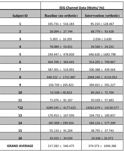
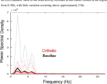
Table 2: Power Spectral Density Data for "high" Cluster ... 56
ix
List of Figures
Figure 1: Flow Diagram of Participants Screened ... 44
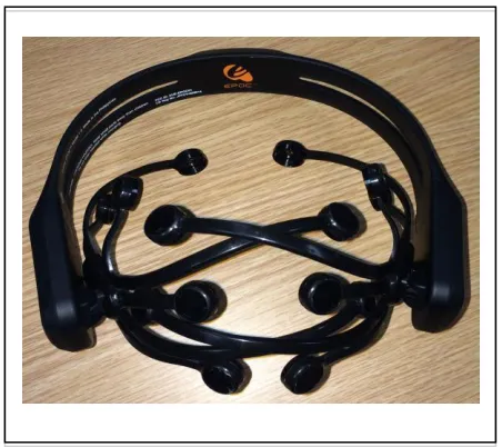
Figure 2: Emotiv Wireless Neuroheadset ... 47
Figure 3: Sensor Location Schematic ... 47
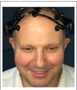
Figure 4: Headset Placement ... 48
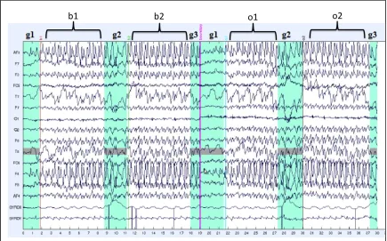
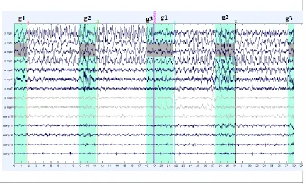
Figure 5: Raw EEG Data from a Single Subject During both Gait Conditions ... 51
Figure 6: EEG Output from Single EEG Channel after Filtering and ICA ... 52
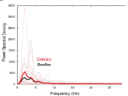
Figure 7: Power Spectral Density Graph for ‘high’ Cluster ... 57
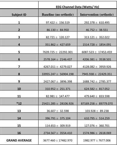
Figure 8: Power Spectral Density Graph for ‘low’ Cluster...59
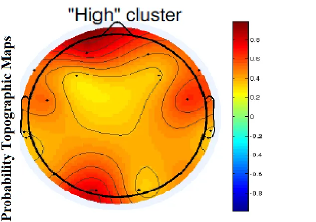
Figure 9: Probability Topographic Map for ‘high’ Cluster... 60
Figure 10: Probability Topographic Map for ‘low’ Cluster ... 61
Figure 11: Topographic Map for ‘high’ Cluster Depicting Baseline and Orthotic Signal Activity ... 62
x
List of Appendices
Appendix A: Ethics Approval Form ... 88
Appendix B: Letter of Information and Consent Form ... 89
Appendix C: Demographic and Contact Information ... 93
xi
List of Abbreviations
99m
Tc- HM-PAO - Technetium-99m-hexamethyl-propyleneamine oxime
AL - anterolateral system
b1 – first section of baseline continuous gait
b2 – second section of baseline continuous gait
BCI – brain computer interface
BOLD - blood oxygen level-dependent signal
C.D. – primary investigator Colin Dombroski
CNS – central nervous system
CPG - central pattern generator
CT – computed tomography scan
DCML - dorsal-column medial-lemniscus system
EEG – electroencephalography
EMG – electromyogram
ERP – event related potentials
fMRI – functional magnetic resonance imaging
fNIRS – functional near infrared spectroscopy
g1 – gait initiation
g2 – turning events during gait
xii H2O-15 – oxygen-15 labelled water
‘high’ – smaller amplitude rhythmic oscillations (resembling stationary EEG)
Hz – hertz
ICA - independent components analysis
‘low’ – larger amplitude rhythmic oscillations (resembling movement artifacts)
MRI – magnetic resonance imaging
NIRS – near infrared spectroscopy
o1 – first section of orthotic continuous gait
o2 – second section of orthotic continuous gait
PET – positron emission tomography scan
PMC – primary motor cortex
PSC – primary somatosensory cortex
PSD – power spectral density
rCBF – regional cerebral blood flow
SMA – supplementary motor area
SPECT – single photon emission computed tomography
Chapter 1
1
Introduction
Human gait is a complex task that requires the coordination of multiple systems in
the body, most importantly, the musculoskeletal and nervous systems. The bulk of the
current knowledge pertaining to the neural mechanism of walking has been derived from
studies on the nervous system of animals (Whittle, 2007). It is, however, unclear to what
extent these findings may be applied to the control of human gait. Historically, human
gait has been considered an automatic process involving little or no higher cortical input
(Wang et al., 2009). It has been suggested that human gait is controlled by central pattern
generators (CPG’s) in the spinal cord that produce the repetitive oscillation movements of
the lower limbs. Armstrong (1988), however, suggested that supraspinal inputs play a
major role not only in initiating locomotion, but also in adapting the locomotor pattern to
environmental and motivational conditions. Through advancements in technology, recent
studies have demonstrated that human gait involves much more than just CPG’s and the
spinal cord. For example, it is speculated that many areas are involved in this complex
process, including the cerebral cortex, basal ganglia, cerebellum (Fukuyama et al., 1997),
and supraspinal centers in the brainstem (Jahn et al., 2004). Futhermore, there is
important information relayed from peripheral sensory receptors, as well as integrated
information from the visual, vestibular and auditory senses (Rossignol et al., 2006).
Although this research has contributed to a greater comprehension of the complexities
involved with gait, an understanding of the neuronal circuits involved in the process of
human gait is still largely unknown and requires further investigation.
The brain-scanning techniques that allow researchers to observe the levels of
activity in an individual’s brain are known as neuroimaging. Neuroimaging techniques
have facilitated a greater understanding of the human brain. Prior to neuroimaging, the
only way to learn about the human brain was by cataloguing a set of impairments that a
group of patients would exhibit when they suffered damage to a specific brain region
(Sadava, Heller, Orians, Purves & Hillis, 2008). Over the years there have been
developments in non-invasive brain imaging techniques that have allowed researchers to
have used either functional magnetic resonance imaging (fMRI) or positron emission
tomography (PET) scans, both of which have associated limitations. The most important
limitation being that a person is incapable of performing walking activities due to the
physical restrictions with the technology (ie. individuals must lie down inside a confined
space) (Wang et al., 2009).
To overcome this shortcoming, some research has used repetitive foot movements
as a surrogate to gait (Miyai et al. 2001).For example, while inside an fMRI machine, a
participant would perform alternating foot flexion-extension movements as recording of
the brain activity occurs. Similarly, other research has addressed this issue by adopting
procedures that allow for the recording of cerebral activity while performing imaginary
gait tasks using motor imagery (mental simulation of gait without actual execution, or
watching a video of another person walking) and planning (recording just prior to gait
initiation, after receiving an external cue to start walking) (Jahn et al., 2004; Wang et al.,
2008; Hanakawa, 2006; Iseki et al., 2008). Additionally, these techniques do not take
into account the simple task of un-supported upright stance, the effects of gravity on the
body, balance control, or the coordination of multiple limbs and joints. The complex task
of integrating vestibular, visual and somatosensory signals while moving in the
environment is also not considered.
To address these limitations, the usage of near-infrared spectroscopic (NIRS)
imaging and single photon emission computed tomography (SPECT) have been
investigated as they allow for the acquisition of data during dynamic activities such as
gait. NIRS allows for visualization of cortical activities during dynamic movements, by
detecting changes in oxygenated hemoglobin, deoxygenated hemoglobin and total
hemoglobin within a few centimeters’ depth of the skull surface (Mayai et al., 2011).
Mayai et al. (2011) used this NIRS headpiece to investigate neuronal activity while
participants completed gait tasks on a treadmill. The obtained NIRS topographic maps
were compared to the MRI anatomical images. Similarly, knowing that changes in
regional cerebral blood flow (rCBF) reflect changes in underlying neuronal activity,
Fukuyama et al. (1997) used SPECT technology to detect changes in neuronal activity
substance (technetium-99m-hexamethyl-propyleneamine oxime) during locomotion,
which is rapidly distributed through the brain in proportion to rCBF, allowing for cerebral
activity to be detected. Both NIRS imaging and SPECT technology have allowed for
further advancements in the understanding of neural control during human gait. However,
SPECT technology involves an invasive intravenous injection and NIRS imaging usually
needs to be compared with MRI anatomical imaging afterwards.
In this study, we used a non-invasive, functional brain imaging device (i.e.
Emotiv), which has yet to be explored in the realm of neuronal activity in human gait.
This device has been tested and has been proven to be a reliable and valid tool when
compared to other similar systems (Badcock et al., 2013). It involves a revolutionary
wireless neuroheadset, which serves as a portable electroencephalogram (EEG) device.
Through electrodes placed on the scalp, an EEG detects changes in electrical potential
differences between electrodes, where these differences reflect the electrical activity of
the neurons in the brain regions under the electrodes (Sadava et al., 2008). This wireless
neuroheadset allows for the collection of real time electrical brain activity while the
participant walks in a live setting. This technology from Emotiv™ allows researchers to
gain direct information concerning brain activity during human gait. It does not only
relay the neuronal activation patterns produced during gait, but also gives insight into the
activation patterns that result from things like somatosensory input, visual stimulus, and
balance control.
Many structures within the central nervous system contribute to the development
of the motor neuron signals that activate muscles. The three main levels of control
include the spinal cord, the descending systems of the brainstem, and the motor areas of
the cerebral cortex (Enoka, 2002). The spinal cord and brainstem mediate reflexes and
automatic behaviours, while the cortical motor areas initiate and control more of the
complex voluntary movements (Enoka, 2002). It is the ascending pathways, however,
that deliver somatosensory information from the arms, legs and trunk through
mechanoreceptors, thermoreceptors, nociceptors and chemoreceptors (Enoka, 2002). The
somatosensory input from mechanoreceptors in the skin, muscles and joints, as well as
human gait (Pinel, 2009). Currently, it is unclear in the literature what effect, if any, that
custom made foot orthoses have in the brain. The human neuromuscular system acts as
an integrated structure using control mechanisms and feedback systems (Nurse & Nigg,
1999). Although, the response of the feet to shoes or inserts is not well understood, it has
been theorized that sensory feedback from the foot plays a role in the kinematic and/or
kinetic response and the effects of inserts and orthotics need further investigation (Nurse
& Nigg, 1999).
In this study we will be investigating possible changes in brain activation patterns
during each trial, using the Emotiv wireless EEG device. To our knowledge, there has
been no published research using the Emotiv neuroheadset to record brain activation
patterns during human gait. The purpose of the current study is to examine the feasibility
of this novel approach and technique of recording brain activity, and to determine if
custom made orthotics will alter the brain activity patterns recorded by the Emotiv EEG
Chapter 2
2
Literature Review
2.1 Introduction
Human locomotion is a complex task that involves interactions between the brain,
spinal cord, peripheral nerves, muscles, bones and joints (Whittle, 2007). The ability to
coordinate limb segments is essential in everyday activities, but very little is known about
the neural basis of coordination in humans. Understanding how the seemingly simple and
automated movements of walking and running are controlled, forms a main challenge for
modern neuroscience (Duysens et al., 1998). Most of the existing knowledge about the
cerebral control of gait in humans comes from studies that were completed in cats and
rodents (Armstrong, 1986; Rossignol et al., 2006). It is unclear whether the organization
and knowledge from the animal models can be inferred to human locomotion. Central
pattern generators (CPG) have been widely studied in animal models, but human
locomotion differs from most mammals in that it is bipedal and assumed to be under
more supraspinal control (Jahn et al., 2008). There have been numerous studies
conducted using a variety of neuroimaging techniques to investigate the cerebral activity
that occurs during different human activities and movements. However, insufficient
knowledge has been gained, largely due to the technical inability to perform walking
tasks inside the machinery (Hanakawa, 2006). New techniques are being investigated to
overcome these problems and allow for better imaging and a greater understanding of the
human cerebral cortex.
Locomotion also incorporates visual information from the eyes, somatosensory
information from the feet, knees and hip, as well as vestibular information about balance
from the inner ears (Pinel, 2009). Although visual and vestibular feedback is essential, it
has been proposed that the sensory feedback control system plays a large role in human
locomotion (Newmann, 1980; Robbins et al., 1988). New methods have been employed
to enhance the amount of sensory feedback, as sensory information is used to adapt motor
output appropriately for environmental conditions (Lundy-Ekman, 2013). The sole of the
provides valuable information about the changing surface characteristics to the central
nervous system (Hohne et al., 2012), as well as afferent feedback for timing and the
transitions between stance and swing (Van Wezel et al., 1997; Lundy-Ekman, 2013).
Foot orthoses are one type of method commonly prescribed to help with foot and ankle
problems. Orthoses can be administered for a variety of reasons, including shock
absorption, reducing shear forces on the foot, improving sensory feedback, or even
improving balance (May & Lockard, 2011). The use of orthotic devices in the shoe will
be explored to determine if sensory feedback can be enhanced and therefore alter human
gait.
2.2 Anatomy of the Brain and Central Nervous System
The human central nervous system (CNS) consists of the brain, spinal cord and
the peripheral nerves. Globally, the brain can be subdivided into four main areas; the
cerebrum, the cerebellum, the brain stem and the diencephalon (Nieuwenhuys, Huijzen,
& Voogd, 2008). The cerebrum consists of four lobes; the frontal, parietal, occipital and
temporal lobes. Included in the brainstem area are the midbrain, pons and medulla
oblongata. Forming the center core of the brain, the diencephalon incorporates the
epithalamus, thalamus and hypothalamus (Sadava et al., 2008). The human brain
contains, at least, one hundred billion neurons. Each neuron represents a complex
structure that processes incoming information in many ways, receives inputs from other
neurons, and has numerous output connections or synapses (Latash, 2008). Voluntary
movement is processed in a top-down hierarchy, from brain to spinal cord to muscle
(peripheral nerves), known as efferent output (Lundy-Ekman, 2013). Whereas, afferent
input is in the reverse order, with nerve signals originating in the peripheral nerves,
travelling through the spinal cord and projecting onto different areas of the brain
(Lundy-Ekman, 2013).
2.2.1 Cerebrum
The surface of the two cerebral hemispheres is composed of grey matter which is
called the cerebral cortex. This is the part of the brain that is traditionally associated with
memory information, making conscious decisions, controlling voluntary movements,
language, non-verbal communication, intelligence and personality (Latash, 2008;
Lundy-Ekman, 2013). Underneath the cerebral cortex is white matter, made up of axons that
connect the cell bodies in the cortex with one another and with other areas of the brain
(Sadava et al., 2008). As previously mentioned, the cerebrum consists of four lobes, each
composed of distinct areas where specific processing takes place.
2.2.1.1Frontal Lobe
The frontal lobe is generally associated with actions involving thinking, planning,
central executive function and motor execution. The main regions of the frontal lobe
include the Primary Motor Cortex (Brodmann area 4), Secondary Motor Cortex
(Brodmann area 6) and Broca’s Area (Brodmann area 44 & 45). The Primary Motor
Cortex (PMC) is located in the posterior portion of the frontal lobe, directly anterior to
the central sulcus. The PMC controls movement for specific parts of the body, where
stimulation of a certain area will result in the movement of that associated body part
(Sadava et al., 2008). This region is somatotopically organized, meaning the parts of the
body are organized in a specific way (motor homunculus) and are not proportional to
their body size (Sadava et al., 2008). For example, parts of the body with fine motor
control, like the hands and face, have a disproportionate representation. The PMC works
together with other motor areas and several subcortical regions to plan and execute
movements. It receives somatosensory information relayed by the thalamus and the
primary somatosensory cortex, as well as motor instructions from the motor planning
areas (Lundy-Ekman, 2013). It is also the major point of departure for sensorimotor
signals from the cerebral cortex (Pinel, 2009).
The Secondary Motor Cortex is comprised of two large areas, the Supplementary
Motor Area (SMA) and the Premotor Cortex, and is located just anterior to the PMC. The
SMA is important for initiation of movement, orientation of the eyes and head, and
planning bimanual and sequential complex movements (Lundy-Ekman, 2013). The
Premotor Cortex is involved in more complex patterns of movement than the discrete
patterns generated in the PMC (Sadava et al., 2008). It helps control the trunk and girdle
limb tasks and the hips during walking (Lundy-Ekman, 2013). It is believed that the
premotor cortex is also involved in the planning or programming of voluntary movements
(Purves, Augustine, & Fitzpatrick, 2001).
A special region located in the posterior-lateral pre-frontal cortex and partly in the
premotor area, called Broca’s Area, provides the neural circuitry for word formation
(Sadava et al., 2008). This area is responsible for planning the movements of the mouth
during speech. Also, motor patterns for expressing individual words or even short phrases
are initiated and executed here (Sadava et al., 2008; Lundy-Ekman, 2013). Additionally,
Broca’s area works in tight conjunction with Wernicke’s language comprehension area
that is located in the temporal region of the cerebrum (Sadava et al., 2008).
2.2.1.2Parietal Lobe
The parietal lobe is associated with somatosensory perception, integrating sensory
information from numerous parts of the body and the integration of visual and
somatospatial information. This lobe is located posterior to the frontal lobe and superior
to the occipital lobe. The major regions of the parietal lobe consist of the Primary
Somatosensory Cortex (Brodmann areas 3, 1 & 2), Secondary Somatosensory Cortex
(Brodmann areas 40 & 43), Somatosensory Association Cortex (Brodmann areas 5 & 7)
and the Parieto-Occipitotemporal Association Area. The Primary Somatosensory Cortex
(PSC) is located in the anterior portion of the parietal lobe, just behind the central sulcus.
The PSC is the most prominent region in the parietal lobe and is the main sensory
receptive area for the sense of touch and proprioception. Touch and pressure information
by neurons that are relayed from the body through the thalamus are received by the PSC.
These neurons can identify the location of the stimuli as well as discriminate among
various shapes, sizes and textures of objects (Sadava et al., 2008; Lundy-Ekman, 2013).
The PSC is also somatotopically organized, so it is organized according to a map of the
body surface (somatosensory homunculus), where the lips and hands are the most
prominent. The somatosensory information ascends from each side of the body to the
PSC through two major pathways; the dorsal-column medial-lemniscus system and the
carry information about touch and proprioception, whereas the anterolateral system
carries information about pain and temperature.
The Secondary Somatosensory Cortex is located on the lateral edge of the parietal
lobe, just superior to the temporal lobe. This area analyzes sensory input from both the
thalamus and the PSC and integrates tactile and proprioceptive information obtained from
manipulating an object (Lundy-Ekman, 2013). Neurons in this area provide stereognosis
by comparing somatosensation from the current object with memories of other objects
(Lundy-Ekman, 2013). The Somatosensory Association Cortex (posterior parietal cortex)
is situated posterior to the PSC and just superior to the occipital lobe. This cortex is
involved in locating objects in space and plays a role in visuomotor coordination. It is a
point of convergence between visual and proprioceptive information, to determine where
an object is in relation to parts of the body (Sadava et al., 2008). The nervous system
must know the original position in space before a body part can be moved and the
positions of the external objects that the body will be interacting with. To accomplish the
aforementioned task, the posterior parietal cortex receives information from the visual
system, auditory system and the somatosensory system, and most of its output goes to the
areas of the motor cortex (Pinel, 2009).
The Parieto-Occipitotempral Association Area provides high levels of
interpretative meaning for signals from all of the surrounding sensory areas. Areas
located in the posterior portion of the parietal lobe, extending into the superior occipital
lobe, provide continuous analysis of the spatial coordinates of all parts of the body as
well as of the surroundings of the body (Sadava et al., 2008). This area receives visual
sensory information from the posterior occipital cortex and simultaneous somatosensory
information from the anterior parietal lobe, allowing it to compute the coordinates of the
visual, auditory and body surroundings (Sadava et al., 2008).
2.2.1.3Temporal Lobe
The temporal lobes are located on the lateral sides of the human brain, just above
the ears and have a variety of sensory functions. These lobes are involved in auditory
comprehension, as well as memory formation and emotional responses. The Primary and
Secondary Auditory Cortices are located in these lobes, where sound waves, pitch and
frequencies are organized and processed. Wernicke’s Area, spanning the region between
the temporal and parietal lobes, works together with the auditory cortex and has a key
role in speech and language comprehension (Lundy-Ekman, 2013). The temporal lobe
also houses the structures of the limbic system, which includes the olfactory cortex,
amygdala and the hippocampus. The olfactory cortex is responsible for the detection of
scents. The amygdala is involved in both emotion and memory and the hippocampus is
linked to memory storage and function (Sadava et al., 2008). Lastly, the Parietotemporal
Association cortex incorporates spatial coordination for constructing images of one’s own
body and for planning movements, as well as integrating information involved in the
recognition, identification and naming of objects (Sadava et al., 2008).
2.2.1.4 Occipital Lobe
The last of the four lobes forming the cerebral cortex is the occipital lobe, which
is situated in the most posterior portion of the cerebrum. The occipital lobe is the main
center for receiving and processing visual information. Located within this lobe is the
Primary Visual Cortex, where there is a visual cortex in each hemisphere of the brain.
This region is highly specialized, as it has different groups of neurons that separately
encode for colour, orientation and motion information (Pinel, 2009). The visual cortex
receives information and projections from the retina and is then able to process and
interpret these signals. In the anterolateral region of the occipital lobe lays the visual
association area which relays visual information from words read into Wernicke’s area,
the language comprehension area (Sadava et al., 2008). The association area is essential
for making sense of the visual world and translating visual experience into language
(Sadava et al., 2008). Facial recognition is another important aspect involving the ability
to process visual information and this area is located on the medial underside of both
2.2.2 Cerebellum
The cerebellum is located at the back of the brain, tucked underneath the occipital
and temporal lobes of the cerebral cortex and behind the portion of the brainstem called
the pons. The cerebellum contains three lobes; the flocculonodular lobe, anterior lobe and
posterior lobe. However, areas of the cerebellum are also described corresponding to their
functional subdivisions, which include the vestibulocerebellum, spinocerebellum (medial
sector) and cerebrocerebellum (lateral sector). The smallest region, the flocculonodular
lobe, often referred to as the vestibulocerebellum, is concerned with vestibular reflexes
and in balance and spatial orientation (Byrne, 2007). The medial sector of the anterior
and posterior lobes comprises the spinocerebellum, which functions mainly in the
fine-tuning of body and limb movements as a result of the integration of sensory input. This
sector receives proprioception input from the dorsal columns of the spinal cord (mainly
the spinocerebellar tract), and its output projects to the deep cerebellar nuclei, as well as
the cerebral cortex and brain stem (Byrne, 2007). The lateral sector and also the largest
region, the cerebrocerebellum, receives input exclusively from the cerebral cortex via the
pontine nuclei and sends output mainly to the venrolateral thalamus (Byrne, 2007). This
sector is thought to be involved in the planning and timing of movements as well as
cognitive functions of the cerebellum.
The cerebellum plays a large role in motor activity, however, there are many
theories as to what these roles actually entail. The cerebellum does not initiate movement,
but it contributes to the coordination, precision and accurate timing of movements
(Byrne, 2007). It has been described as a timing device that ensures the correct order and
timing of individual muscle action, as well as the timing in rapid, smooth progression
from one muscle movement to the next (Latash, 2008; Pinel, 2009). This area is believed
to play a major role in motor learning, by acquiring and memorizing new skills, but
particularly in the learning of sequences of movements, which is a critical factor in the
timing and precision of movements (Pinel, 2009). The cerebellum has also been referred
to as a coordination device, putting together components of complex joint or
multi-limb movements, in addition to controlling balance in complex movements such as
by the cerebellum, allowing for it to monitor and make corrective adjustments in the
body’s motor activities while they are being executed (Sadava et al., 2008). This
feedback allows the cerebellum to compare the actual movements depicted by the
peripheral sensory feedback system with the movements that were intended by the motor
system (Sadava et al., 2008). If the sensory feedback and intended movements do not
compare, then there are instantaneous subconscious corrective signals transmitted back
into the motor system which will either increase or decrease the levels of muscle
activation.
2.2.3 Brain Stem and Spinal Cord
The brain stem, consisting of the pons, medulla oblongata and midbrain
(mesencephalon), is the connection between the cerebrum and the spinal cord. The brain
stem controls several important functions in the body including the regulation of cardiac
and respiratory functions, attention, arousal and maintaining consciousness
(Lundy-Ekman, 2013). One of the brain stem’s main functions is relaying information between
the peripheral nerves and spinal cord to the upper parts of the brain. Both motor and
sensory neurons travel through the brainstem allowing for the relay of signals between
the spinal cord and brain. Motor and sensory information is relayed via the corticospinal
tract (motor), posterior column-medial lemniscus pathway (fine touch, vibration,
sensation and proprioception), as well as the spinothalamic tract (pain, temperature, crude
touch) (Pinel, 2009). Also, distributed in the brain stem are at least three motor centers
that send efferent fibers to influence the motor neurons of the spinal cord, consisting of
the red nucleus, lateral vestibular nucleus and the reticular formation (Enoka, 2002).
The spinal cord has two main functions; to convey information between neurons
innervating peripheral structures and the brain, and to process information
(Lundy-Ekman, 2013). The spinal cord receives somatosensory feedback information from
peripheral structures, which allows it to generate reflex loops, like movement of a limb
away from a painful stimulus. Therefore, the spinal cord contains neuronal networks that
can produce reflexes and automatic behaviours independently of input from the brain
2.3 Central Pattern Generators
For many species, the cyclical patterns needed for walking, respiration,
mastication or other rhythmical activities are generated by neural networks (Duysens et
al., 1998). However, for locomotion, one usually refers to the term central pattern
generator (CPG) to indicate a set of neurons responsible for creating a motor pattern,
where this pattern involves the alternating activity in groups of flexors and extensors
(Grillner & Wallen, 1985). CPGs are adaptable networks of spinal interneurons that
activate lower motor neurons to elicit alternating flexion and extension movements of the
hip, knees and ankles, with each limb having a dedicated CPG (Lundy-Ekman, 2013).
Human gait has been considered in past decades only as an automatic process involving
little or no higher cortical input (Wang et al., 2009). However, the majority of current
knowledge pertaining to the neural mechanism of human gait has been obtained from
studying the nervous system of animals, in particular cats and rodents. There has been an
abundance of data collected on animals, leading to the general assumption that CPGs are
the underlying factors in the central control of locomotion. Unfortunately, little is still
known about the spinal networks acting like CPGs in humans.
Originally in 1910, Sherrington suggested that walking could be produced entirely
by a series of reflexes. However, further studies have provided evidence for neural
networks in the spinal cord, now termed central pattern generators, that are capable of
governing locomotion (Grillner & Wallen, 1985). It was in 1911 that Thomas Brown
discovered the existence of what he called ‘half centers’ in the spinal cord. These half
centers were mutually inhibiting mechanisms which ensured that in one limb the flexor
motorneurons are excited first, while the extensor motorneurons are inhibited (Brown,
1911). The pattern was then reversed, so that the limb could go through the
flexion-extension cycle. This work was further explored in the nervous system of cats and
rodents. When limbs of a decerebrate cat were placed on a treadmill, movement of the
treadmill at a constant speed induced locomotor-like stepping in the limbs of the cat
(Latash, 2008). It can be concluded from this type of observation that CPGs for
locomotion exist in the spinal cord of mammals and produce locomotor-like activity in
confirmed the existence of spinal networks with the capacity of generating basic
locomotor rhythms in the absence of any supraspinal or sensory input to the spinal cord
in a variety of lower mammals.
Following the confirmation of the existence of central pattern generators in the
spinal cord, the theory of higher cortical input was then tested. It was discovered by
Rossignol (1996) that, after the transection of their spinal cords, most cats were not able
to generate locomotor movements. This insinuated that commands for the initiation of
locomotor activity must be given at some level in the central nervous system above that
of the spinal lesion. Therefore, by varying the level of transection in the spinal cord, it
was shown that the regions for initiation of locomotion were located in the brain stem
(Rossignol, 1996). Similar findings occurred when a decerebrated cat’s brain stem
structures were electrically stimulated. A corresponding descending input to the lumbar
cord was elicited, suggesting that the spinal rhythm-generating network is likely activated
by higher structures (Nielson, 2003).
The experiments completed in cats and other mammals demonstrated that the
spinal cord contributes substantially to the control of complex motor functions such as
locomotion (Guertin, 2009). Even though the control of locomotion has been mainly
studied in animal models, there has been inferential evidence of the existence of a CPG
for locomotion in humans. Most of the testimonies to the existence of locomotor CPGs in
humans come from studies completed on individuals with clinically complete spinal cord
section and individuals who are gait retraining following a spinal cord injury (Duysens et
al., 1998; MacKay-Lyons, 2002). It has been concluded that there is sufficient evidence
to suggest that the human spinal cord, with intact sensory inputs, is capable of generating
rhythmic motor bursts with assisted leg movements by therapists and a partial body
weight bearing support harness (Stewart et al., 1991; Wernig & Muller, 1992; Dietz et al.,
1995; Harkema et al., 1997). Observations in these spinal cord injury patients have lead
to the belief that spinal locomotor generators are located in the lower thoracic-upper
lumbar level of the human spinal cord (Latash, 2008). Through the use of advancing
neuroimaging techniques, supraspinal locomotor networks have been identified in
cerebellum. This circuitry regulates the initiation and termination of gait, changing the
direction and velocity of gait and spatial orientation and navigation around obstacles
(Jahn et al., 2008; Bakker et al., 2007b; Wagner et al., 2008).
2.4 Gait
Human walking is a complex task that requires the coordination of a number of
different muscles acting on the hip, knee, ankle and foot in order to advance the body in a
desired line of progression (Nielson, 2003; Hsu, Michael, Fisk, & AAOS, 2008). Walking
utilizes a repetitive and rhythmical sequence of limb motions to simultaneously move the
body forward while also maintaining stability of the body mass (Perry & Burnfield,
2010). Each limb combines the patterns of motion, passive force, and muscular control
into a sequence of activity, called the gait cycle, which includes all of the body’s activity
from the time one foot strikes the floor until the same foot strikes the floor again
(Edelstein & Moroz, 2011; Hsu et al., 2008). There are a number of actions and events
that occur in the complex mechanism of gait. This mechanism is further complicated with
the introduction of environmental influences (obstacles, people, changing walking
surfaces, etc).
The gait cycle is commonly defined as the time interval between two successive
occurrences of one of the repetitive events of walking (Whittle, 2007). For instance, the
cycle would start with the initial heel contact of the right foot and would end with the
heel contact of the same right foot. There are two phases of the gait cycle, the stance
phase and the swing phase. However, these phases are not distributed evenly throughout
the cycle. It has been determined that the stance phase lasts for about sixty percent of the
cycle, and the swing phase lasts for the remaining forty percent (Whittle, 2007; Perry &
Burnfield, 2010). Edelstein and Moroz (2011) defined the stance phase as the period
during which the reference foot is in contact with the ground. The swing phase was
described as the period during which no part of the reference foot is in contact with the
ground, or simply, when the foot is in the air (Edelstein & Moroz, 2011). Therefore,
stance begins with initial heel contact (heel strike) and swing begins as the foot is lifted
from the floor during toe off. Perry and Burnfield (2010) have reported that the two gait
(heel contact/strike), loading response, mid-stance, terminal stance, pre-swing (toe off),
initial swing, mid-swing and terminal swing. This pattern of gait results from the
complex interactions between many neuromuscular and structural elements of the
locomotor system (Whittle, 2007). The initiation of gait serves as a transitional process
from the balanced upright standing position to the beginning of steady-state walking,
whereas the termination of gait requires the deceleration of the forward momentum of the
body to return to a stable stance position (Jian et al., 1993; Patla, 2004).
As the body moves forward, one limb serves as a dynamic source of support
while the other limb advances itself to the new support site, and then the limbs reverse
roles (Perry & Burnfield, 2010). Kiehn (2006) explained the key features involved in the
gait process consisted of rhythm, ipsilateral coordination of flexors and extensors across
the same or different joints in a limb, and left-right coordination. In an ideal situation,
gait appears to be coordinated, efficient and somewhat effortless resulting in the
conservation of energy expenditure. Each stride in the gait cycle involves an
ever-changing alignment between the body and the supporting foot during stance and limb
during swing (Perry & Burnfield, 2010). Therefore, in the absence of disease or trauma, it
is important to maintain proper lower limb biomechanics. This will minimize energy
expenditure and reduce the stress on the bones, joints and soft tissues of the lower
extremities (Hsu et al., 2008). However, in the event of disease or trauma, gait patterns
can become less optimal and, in turn, result in excessive amounts of expended energy.
This alteration of gait could result in a disruption to the precision, coordination, speed
and versatility of the regular pattern, causing an individual to alter the motion of adjacent
joints and controlling muscles, thereby increasing the energy cost of walking (Perry &
Burnfield, 2010; Hsu et al., 2008).
2.5 Sensory, Motor and Proprioceptive Feedback
During gait, interaction with the environment is multisensory. The senses are
classified as exteroceptive and involve the continual integration of sensory input from
visual, vestibular and somatosensory receptors by the central nervous system to assess the
position and motion of the body (McGlone et al., 2007; Johansson & Magnusson 1991).
movements, future movements, to avoid obstacles and for placement of the feet.
Somatosensory feedback is derived from the sensory input of proprioceptors and
mechanoreceptors. Van Wezel et al. (2000) stated that somatosensory feedback from
muscles, joints and skin is essential in the normal execution of human gait.
The sensory feedback information is integrated into the motor commands at all
levels of the CNS. The CNS makes use of this sensory information in two ways: the
sensory activity helps internal commands in the driving of output neurons as a part of all
normal voluntary movements; and the sensory information may be used to inform the
CNS about errors in the execution of a movement (Nielson & Sinkjaer, 2002). The
sensory information produced from proprioceptors provide details about limb position
and muscle forces and are used to monitor and control limb movements. Sensory
feedback, produced by the skin (cutaneous receptors), can provide information about
touch, temperature, itch and pain. The CNS relies on sensory input from muscles, joints,
and cutaneous receptors in the lower extremities to generate effective motor patterns for
human posture and locomotion (Nurse & Nigg, 2001). This is made possible by the
constant information it is provided regarding muscle and joint loading, joint kinematics,
and pressure distributions on the plantar surface of the foot (Nurse & Nigg, 2001).
Nielson and Sinkjaer (2002) suggested that less central input to the motoneurons is
necessary when there is sensory feedback present, than if it was absent. Thus, declaring
that less central drive is necessary in order to activate the motorneurons with the presence
of sensory afferent feedback.
2.5.1 Receptors
Receptors are specialized cells or subcellular structures that change their
properties in response to a specific stimulus in order to make information about that
particular stimulus available to other neurons within the central nervous system (Latash,
2008). Cutaneous receptors are one of four different groups of proprioceptors found in
the body, which involve specialized receptors in the skin that are sensitive to different
sensory modalities. Somatosensory information includes afferent signals from
mechanoreceptors, thermoreceptors, nociceptors and chemoreceptors. Mechanoreceptors
vibration; thermoreceptors are sensitive to temperature; nociceptors are sensitive to
potentially damaging stimuli (pain); and chemoreceptors are sensitive to chemical stimuli
(Lundy-Ekman, 2013). The main focus here will be on the mechanoreceptors, of which
there are four different types located in the skin; Pacinian Corpuscles, Merkel Disks,
Ruffini Endings and Meissner Corpuscles. Pacinian Corpuscles respond to the
displacement or mechanical deformation (vibration) of the skin; Merkel Disks respond to
gradual skin indentation or vertical pressure on the skin; Ruffini Endings respond to
gradual skin stretch and Meissner Corpuscles are sensitive to quickly changing pressure
on a small area of skin (Pinel, 2009). These receptors are also located in different layers
of the epidermis and dermis. Meissner Corpuscles and Merkel Disks are located close to
the skins surface, whereas Ruffini endings and Pacinian Corpuscles are deep in the
dermis (Latash, 2008). These different types of mechanoreceptors can either be slow or
fast adapting. Patel et al. (2011) suggested that slowly adapting mechanoreceptors
provide information about how the pressures are spatially and sequentially distributed on
the skin-surface interaction. Whereas the rapidly adapting receptors provide information
about pressure amplitude and changes of pressure exerted on the skin (Patel et al., 2011).
Of the four classical cutaneous modalities of the somatosensory system (touch,
temperature, pain and itch), it is the discriminative touch that subserves the perception of
pressure, vibration, slip and texture (McGlone et al., 2007). All of which involve critical
information to be relayed to the central nervous system throughout human locomotion.
2.5.2 Signal Pathways
The speed of information transmission within the central nervous system is very
rapid with signals being transmitted at tens of meters per second (Latash, 2008). Each
system, sensory and motor, uses information carried by a number of anatomically distinct
pathways, usually containing synaptic relays so that moving information can be
processed and integrated (Latash, 2008). A peripheral nerve contains both afferent and
efferent axons. The afferent axons carry information from peripheral receptors towards
the CNS (sensory) and efferent axons carry information away from the CNS (motor)
(Lundy-Ekman, 2013). Primary afferents are the first link between skin, muscles or joints
the sensory (posterior) roots (Guyton & Hall, 2006). From here, information is relayed
through the ascending white matter spinal tracts. Peripheral receptors convey sensory
signals associated with the sense of touch, pressure, proprioception (stretch of muscles,
tension on tendons, position of joints) and vibration, towards the supraspinal structures
(Guertin, 2012).
Somatosensory information ascends from each side of the body to the brain by
means of two major pathways; the Dorsal-column medial-lemniscus system (DCML),
and the anterolateral system (AL). The DCML system tends to carry information about
touch and proprioception. The sensory neurons of this system enter the spinal cord via the
dorsal root, ascend in the dorsal columns and synapse in the dorsal column nuclei of the
medulla (Pinel, 2009). The axons of the dorsal column nuclei cross over to the other side
of the brain ascending then to the ventral posterior nucleus of the thalamus, where they
will be projected to the primary and secondary somatosensory cortex or the posterior
parietal cortex (Pinel, 2009). The AL system tends to carry information about pain and
temperature, and is comprised of three different tracts which project to different areas in
the thalamus (Pinel, 2009). This sensory feedback is an integral part of the overall motor
control system and is critical in modifying CPG generated motor programs in order to
facilitate constant adaptations to the environment (MacKay-Lyons, 2002). Pearson
(1993) identified three potential roles for somatosensory afferent feedback in the
production of rhythmic movements; it reinforces CPG activity, assists with the function
of timing, and facilitates phase transitions in rhythmic movements.
2.5.3 Plantar Surface of the Foot
Throughout a healthy gait cycle, the sole of the foot is continuously in contact
with the environment. This enables the foot to provide information about changing
surface characteristics and serve as a tool for continual feedback to the central nervous
system. The foot cushions the musculoskeletal system during impact, supports the body
during ground contact, transmits force, keeps the body in balance and serves as a system
for sensory input (Mulder & Hulstijn, 1985; Newman, 1980; Robbins et al., 1988).
During the gait cycle there are large amounts of afferent input generated both from the
the limb (Duysens et al., 1995). It would seem that there is an abundance of
somatosensory information generated from the plantar mechanoreceptors. However,
according to Kennedy and Inglis (2002), there is limited information and knowledge
about the characteristics of the mechanoreceptors specific to the foot sole. There have
been many studies conducted concerning the cutaneous sensation of the hand or reports
from other skin regions that are often used to predict the properties of cutaneous
mechanoreceptors in the foot sole. However, attempting to transfer the properties of skin
receptors from other body regions to the foot may prove to be inappropriate, as there are
differences between glabrous and hairy skin types (Kennedy & Inglis, 2002).
Numerous studies have examined how the foot and sensory system reacts to
changes in shoe soles, insoles, or different surfaces. Additionally, effects from vibration
and varying pressure exertion have been studied. In 1981, Watanabe and Okubo provided
evidence that standing on different surfaces can alter the transmission of afferent signals
from the plantar surface of the foot. In a study conducted by Patel et al. (2011) foam
surfaces were used to test the relative contributions of the visual, vestibular and
somatosensory inputs. Standing on a foam surface was shown to challenge postural
control by decreasing the reliability of sensory information from the plantar
mechanoreceptors. In order not to involve the other two inputs, participants were
instructed to keep their eyes open and make sure their head was facing forwards (Patel et
al., 2011). A study was carried out by Nurse and Nigg (1999) to quantify the relationship
between the pressure and vibration sensitivity of the sole of the foot with plantar pressure
distribution during walking and running. The researchers’ results suggested that sensory
feedback from the foot plays a role in the kinematic and/or kinetic response while
walking and running. Zehr et al. (1997) completed a study investigating the effects of
electrical stimuli on superficial peroneals and tibial nerves during human gait. The
superficial peroneals and anterior tibialis nerves were stimulated and the stimuli were
reported as being sensed on the plantar surface of the foot. It was concluded that afferents
from various skin sites on the foot play a crucial role in the timing of the transition
between stance and swing in gait (Zehr et al., 1997). It appears that plantar cutaneous
sensation plays an important role in balance and stability, kinematics and kinetics, and
mechanoreceptors can provide detailed spatial and temporal information about contact
pressures in the foot, and have the potential to provide information that could facilitate
the control of compensatory stepping reactions.
It has been proposed by Nurse and Nigg (2001) that reduced feedback from the
receptors in the foot may contribute to gait abnormalities. Impaired proprioception
obstructs walking because it deprives the individual of knowing the exact position of
their knee, hip, ankle and/or foot, and as a result, that individual does not know when it is
safe to transfer body weight onto the limb (Perry & Burnfield, 2010). Similarly, Hsu et al.
(2008) stated that impaired sensation on the soles of the feet delays the awareness of floor
contact and could result in a greater occurrence of falls or other lower extremity
biomechanical problems.
2.6 Neuroimaging Techniques
Functional brain imaging is a multidisciplinary research field that encompasses
techniques devoted to a better understanding of the human brain through non-invasive
imaging of the electrophysiological, hemodynamic, metabolic and neurochemical
processes that underlie normal and pathological brain function (Baillet et al., 2001).
Neuroimaging refers to a set of brain-scanning techniques that allow for observation of
varying levels of activity in an individual’s brain. Prior to neuroimaging, the only way to
learn about the human brain was by cataloguing the set of impairments that a group of
patients would exhibit when they suffered damage to a given brain region (Sadava et al.,
2008). With neuroimaging, came the use of a variety of different scanning and imaging
techniques, including; electroencephalography (EEG), functional magnetic resonance
imaging (fMRI), positron emission tomography scan (PET), computed tomography scan
(CT), single-photon emission computed tomography (SPECT), near-infrared
spectroscopy (NIRS), and transcranial magnetic stimulation (TMS). Unfortunately,
functional neuroimaging of the brain during human gait poses a practical problem. The
investigation of brain activation patterns during locomotion has been limited by the
impossibility of actually performing these tasks while inside standard scanners, such as
MRI machines or PET/CT scanners (Jahn et al., 2004). Some of these problems have
et al., 2010). These strategies included techniques such as recording cerebral activity
during actual gait after radioactive injections (Fukuyama et al., 1997; Hanakawa et al.,
1999; Miyai et al., 2001); recording cerebral activity during motor planning of walking or
just prior to gait initiation (Yazawa et al. 1997); using tasks that share some cerebral
processes with gait, without the actual need to engage in gait, like motor imagery or
repetitive foot movements (Malouin et al., 2003; Jahn et al., 2004; Miyai et al., 2001);
and even recording cerebral activity in patients with gait disorders.
2.6.1 Electroencephalography (EEG)
EEG is a method of studying the collective electrical behaviour of large groups of
neurons, where it detects changes in the electrical potential differences between
electrodes, resulting from the flow of current through the extracellular space (Latash,
2008). The EEG electrodes are placed on the skull, commonly over the four major
cortical lobes, with reference/grounding electrodes placed behind each ear. The EEG
device is able to detect frequencies ranging from 1-30 Hz, with four different wave
classifications depending on their frequency. Delta waves detect frequencies from
approximately 0.5-4 Hz, theta waves from 4-8 Hz, alpha waves from 8-13 Hz and beta
waves from 13-25 Hz (Latash, 2008; Emotiv). The low frequency waves (delta and theta)
are commonly only seen during certain phases of sleep. Alpha waves are associated with
relaxed wakefulness and beta waves are dominant during intense mental activity (Latash,
2008; Sadava et al., 2008). A distinct advantage of using EEG is its high temporal
resolution, which can follow changes in brain activity from millisecond to millisecond,
making it far more superior to PET or fMRI in this respect (Irani, 2011). However, EEG
has very poor spatial resolution, since the electrodes are simply placed over the skull,
they cannot be used to properly identify the exact source of the changing electrical
signals that are recorded (Latash, 2008).
Many studies have been conducted using EEG to determine areas of the cerebral
cortex involved in the initiation of gait, or during lower limb movements similar to that of
gait movements, and even during gait performed on a treadmill. In 1997, Yazawa et al.
performed a study using EEG to explore the cortical mechanism underlying gait initiation
recording of surface electromyogram (EMG) on the anterior tibialis muscle in order to
determine the onset of movement. From this, Yazawa et al. (1997) demonstrated that
activity in the supplementary motor area as well as the primary motor cortex was related
to gait initiation. Similarly, a study investigating the cortical activity during lower limb
movements by means of EEG was used with the assistance of a tilt table. Wieser et al.
(2010) collected EEG data while participants were strapped into an Erigo tilt table, which
allowed them to perform automated gait-like stepping movements in an upright position
for thirty minutes. The Erigo is driven in a sinusoidal function of time referring to the hip
angle, with the duration of extension and flexion phases identical for each leg, while
moving conversely to each other at a constant speed (Wieser et al., 2010). This study
provided further indication that the primary somatosensory cortex, primary motor cortex
and the supplementary motor area, all play essential roles in cortical control of human
gait. While standing, walking and running on a treadmill, Gwin et al. (2011) used EEG to
examine patterns of intra-stride electrocortical dynamics. It was proposed that the
electrocortical dynamics, particularly in the sensorimotor cortex, would exhibit
intra-stride patterns of activation and deactivation. Clusters of electrocortical sources were
spatially localized to the prefrontal cortex, left and right sensorimotor cortex, anterior
cingulated cortex and posterior parietal cortex (Gwin et al., 2011). However, Gwin et al.
(2011) stated that their findings did not indicate whether the human cortex is actively
involved in controlling locomotion via direct pathways, or if it processes sensory
afferents that are used to modulate a descending signal to other locomotor regions in the
brainstem and spine.
All complex EEG systems used to detect brain activity are sensitive to a set of
elements that can negatively influence the accuracy of their measurements (Cernea et al.,
2012). Recording of EEG during walking is challenging due to movement artifacts
(Bakker et al., 2007b) and most EEG devices actually detect a mixture of skin, muscle
and nerve activity instead of just a pure signal generated by the electrical activity of
neurons (Van De Velde et al. 1998). Nuwer (1990) reported that not only are there
movement artifacts to consider, but also line noise, frequency interference, as well as
medications that people are taking and other clinical factors. One limitation to EEG that
subcortical brain structures (activations in the striatum, cerebellum, pons, basal ganglia)
could not be considered or recorded with this device.
2.6.2 Magnetic Resonance Imaging (MRI) / Functional Magnetic Resonance Imaging (fMRI)
MRI is based on the property of elements with an odd atomic weight to align the
spin axes of their nuclei with a constant external magnetic field (Latash, 2008). A brief
electromagnetic pulse can be used to perturb the orientation of the spin axes, so when the
pulse is turned off, the nuclei return back to their original orientation defined by the
external magnetic field (Latash, 2008). This process involves the release of energy in the
form of electromagnetic waves, where the frequency of emitted waves can vary with
regards to the different types of atoms. A particular version of MRI, the fMRI, has
become popular for studying the brain processes associated with different actions and
movements. This method involves comparing MRI measurements obtained before and
after performing a task, where the differences in the patterns are expected to reflect
task-specific changes in the neuronal activity (Latash, 2008). fMRI can be used to produce
activation maps showing which parts of the brain are involved in a particular mental
process (Sadava et al., 2008). Irani (2011) states that fMRI is currently considered the
‘gold standard’ for measuring functional brain activation, since it offers a source of safe,
non-invasive, functional brain imaging with high spatial resolution. The primary measure
used for this technique is the blood oxygen level-dependent (BOLD) signal, which
reflects the amount of oxygenated hemoglobin in the total amount of hemoglobin, and
accompanies neuronal activation in the brain (Irani, 2011). Unfortunately, studying the
cortical involvement in gait control for humans using this technique has been limited, due
to the incapability to walk inside the scanning machine. However, it has been suggested
that activation maps during movement execution are similar to those observed during the
imagery of the same task (Jeannerod, 2001; Munzert et al., 2009).
In 2008, Wang et al. conducted a study requiring the observation of video clips of
human walking and the mental imitation of the visualized process while using an fMRI.
This method was based on the response of mirror neurons to actions being observed. This