Scholarship@Western
Scholarship@Western
Electronic Thesis and Dissertation Repository
4-15-2013 12:00 AM
Characterizing the contribution of the LXCXE binding cleft to
Characterizing the contribution of the LXCXE binding cleft to
pRB-mediated genome stability and tumor suppression
mediated genome stability and tumor suppression
Courtney H. CoschiThe University of Western Ontario
Supervisor
Dr. Frederick A Dick
The University of Western Ontario Graduate Program in Biochemistry
A thesis submitted in partial fulfillment of the requirements for the degree in Doctor of Philosophy
© Courtney H. Coschi 2013
Follow this and additional works at: https://ir.lib.uwo.ca/etd
Part of the Biochemistry Commons
Recommended Citation Recommended Citation
Coschi, Courtney H., "Characterizing the contribution of the LXCXE binding cleft to pRB-mediated genome stability and tumor suppression" (2013). Electronic Thesis and Dissertation Repository. 1272.
https://ir.lib.uwo.ca/etd/1272
This Dissertation/Thesis is brought to you for free and open access by Scholarship@Western. It has been accepted for inclusion in Electronic Thesis and Dissertation Repository by an authorized administrator of
Thesis format: Integrated Article
by
Courtney Heather Coschi
Graduate Program in Biochemistry
A thesis submitted in partial fulfillment of the requirements for the degree of
Doctor of Philosophy
The School of Graduate and Postdoctoral Studies The University of Western Ontario
London, Ontario, Canada
ii
Abstract
Condensation and segregation of mitotic chromosomes are critical processes for cellular propagation and if compromised, can lead to genomic instability. Genomic instability is known to be an active contributor to tumorigenesis, rather than being a by-product of malignant progression. The retinoblastoma protein (pRB) is the prototypic tumor suppressor. Its tumor suppressive properties are linked to its ability to negatively regulate proliferation by inhibiting E2F target gene transcription. Using a gene targeted mouse model defective for interactions mediated by the pRB LXCXE binding cleft that is distinct from E2F binding (Rb1ΔL/ΔL), I have demonstrated that LXCXE-interactions are an essential part of pRB-mediated tumor suppression. When these interactions are disrupted, cells exhibit
chromosome condensation and mitotic defects that are unrelated to G1 to S-phase regulation by pRB. These defects contribute to earlier tumor formation and more aggressive pathology in Trp53+/- and Trp53-/- mouse models, revealing a new mechanism of tumor suppression, facilitated by pRB, whereby genome stability is maintained by the proper condensation of mitotic chromosomes. Subsequent study of the mechanism by which pRB facilitates genome stability suggests that a pRB-E2F1-Condensin II complex localizes to pericentromeric
heterochromatin. In the absence of this complex, DNA double strand breaks are observed and persist into mitosis and the ensuing G1 phase of the cell division cycle. Moreover, haploinsufficiency of Rb1 was enough to compromise loading of Condensin II at
iii
Keywords
Retinoblastoma protein, E2F1, cancer, LXCXE, haploinsufficiency, genomic instability,
iv
Co-Authorship Statement
All Chapters were written by Courtney Coschi and edited by Dr. Fred Dick, with the
exception of Chapter 2, which was written by Dr. Fred Dick and edited by Courtney Coschi.
All experiments in Chapter 2 were performed by Courtney Coschi with the exception of
Figures 2.5, 2.6A, 2.8A, Table 2.2, and Appendix F-A, -B and -C, which were performed by
Alison Martens, and Appendix E which was performed by Sarah Francis. Pathology review
was provided by Dr. Subrata Chakrabarti. References to figures are modified from the
original publication to fit formatting for this thesis.
All experiments in Chapter 3 were performed by Courtney Coschi with the exception of
Figure 3.16A and Appendix K which were aided by Alison Martens. ChIP DNA for the
experiment in Figure 3.7Ai was provided by Srikanth Talluri. Pathology review was
v
Dedication
For my Mom, the bravest person I know.
vi
Acknowledgements
To my supervisor Dr. Fred Dick: I told you when I first interviewed with you that I
wanted to learn how to think. Both you and this experience have certainly taught me how to
do just that. You have also given me advice to apply not only in the lab, but in life as well,
and I am grateful for it. Thank you for being an inspiring mentor, for challenging me to
pursue research at the highest level, and for supporting me in my future endeavors.
Thank you to Dr. David Rodenhiser and Dr. Nathalie Bérubé, members of my
advisory committee, for providing me with guidance, constructive criticism and thought
provoking discussions over the years. Thank you as well to my examiners, Dr. Mellissa
Mann, Dr. Dave Edgell, Dr. Kathleen Hill and Dr. Peter Whyte, for your probing questions,
helpful critiques, and suggestions in the preparation of this final thesis.
To my fellow lab mates: thank you for being my family away from home, for
commiserating with me, and celebrating with me. In your own way, each of you has made
grad school an enjoyable and memorable experience. Thank you as well to my circle of
friends both in and out of the lab, as well as old friends and those whom I met during grad
school. You’ve given me unforgettable memories that I will always cherish. My life
wouldn’t be the same without each of you.
Finally, thank you to my family for your unconditional love and support. You were
my reprieve from the lab when I needed it, and you were also my biggest champions; you
have shaped who I am today. Lastly, and most importantly, thank you to my husband. You
are my closest confidant and my best friend. Only you have seen everything that grad school
and life has thrown at me. Thank you for being everything that I’ve needed, when I needed it. I couldn’t have made it through grad school without your continued love, support and
vii
Abbreviations
o
C: degrees Celsius BRCA2: breast cancer 2, early onset
α: anti BrdU: 5-bromo-2’-deoxyuridine
γH2AX: phosphorylation at serine 139 of the BRG1: Brahma-related gene 1 H2A variant H2AX
Brm: Brahma protein
Δ: lacking
BSA: bovine serum albumin
ΔL: ΔLXCXE
BubR1: budding uninhibited by
µg: microgram benzimidazoles related 1
µL: microliter C > T: C to T transition
µm: micron C: cortex (in Appendix F)
χ2
: chi squared test C: cytosine (when referring to the
nucleotide)
129/B6: mouse with a mixed genetic
background of 129 and C57BL/6 C. elegans: Caenorhabditis elegans
A: adenine (when referring to the nucleotide) CAP- D2, D3, G, G2, H, H2: chromosome
associated protein D2, D3, G, G2, H and
A: alanine (when referring to the amino acid) H2 respectively
aCGH: array comparative genomic CD4: cluster of differentiation 4 hybridization
CD8: cluster of differentiation 8
ADP: adenosine diphosphate
CDK: cyclin dependent kinase
APC/C: anaphase promoting complex/
cyclosome CENPA: centromere protein A
APC: Adenomatous polyposis coli protein ChIP: chromatin immunoprecipitation
ATP: adenosine triphosphate CIN: chromosome instability
BAC: bacteria artificial chromosome CIP: CDK interacting protein
BCA: bicinchoninic acid CKI: cyclin dependent kinase inhibitor
bp: base pair c-myc: cellular-myelocytomatosis
viii
Abbreviations Continued
COSMIC: catalogue of somatic mutations in E3: ubiquitin-protein ligase cancer
E7: early protein 7
CRF: chromatin regulatory factor
ELISA: enzyme-linked immune sorbent
C-terminal: carboxy terminal assay
DAPI: 4’,6-Diamidino-2-Phenylindole Emi: early mitotic inhibitor 1
DBD: DNA binding domain ESC: Embryonic stem cell
dCAP-D3:Drosophila CAP-D3 F: female
DHFR: dihydrofolate reductase FBS: fetal bovine serum
DMEM: Dulbecco’s Modified Eagle FISH: fluorescence in situ hybridization Medium
FITC: fluoresceini-sothiocyanat
DMZ: dimerization domain
G > C: G to C transversion
DNA: deoxyribonucleic acid
G > T: G to T transversion
DNMT1: DNA (cytosine-5-)-methyl
transferase 1 G: guanine (when referring to the nucleotide)
dNTP: deoxyribonucleotide triphosphate G0: quiescence
DP: Differentiation Related Transcription G1: gap 1 phase of the cell division cycle
Factor-1 polypeptide-1
G2: gap 2 phase of the cell division cycle
DREAM: DP, RB-like, E2F and MuvB
GFP: green fluorescent protein
Drosophila:Drosophila melanogaster
Glu: glutamic acid
dsDNA: double stranded DNA
GST: glutathione S-transferase
dup A: duplication of A
H&E: hematoxylin and eosin staining
E: Embryonic day
H2A: histone 2A
E1A: early region 1A
H2B: histone 2B
E2F: E2 promoter binding factor
H2BGFP: histone 2B tagged with GFP
ix
Abbreviations Continued
H3: histone 3 LacZα: the alpha fragment of one β-
galactosidase monomer
H3-K9: histone 3- lysine 9 residue
LOH: loss of heterozygosity
H4: histone 4
Luc: luciferase
H4-K20: histone 4- lysine 20 residue
LXCXE: leucine-any amino acid-cysteine-
HAT: histone acetyl transferase any amino acid-glutamate
hCAP-D3: human CAP-D3 M: male (in Appendices H and K)
HCl: hydrochloric acid M: medulla (in Appendix F)
HDAC1/2: histone deacetylase 1 or 2 M: methionine (when referring to the amino acid)
HEAT: huntington, elongation factor 3,
protein phosphatase 2A, TOR1; four M: mitosis (when referring to the phases of
proteins that contain the HEAT repeat the cell division cycle)
domain
Mad2: mitotic arrest deficient 2
hRAD21: double strand break repair protein
rad21 homolog (S. pombe) MB: marked box (in Figure 1.2)
hSWI/SNF: human switch/sucrose MB: megabase nonfermentable
MCM7: minichromosome maintenance
I: isoleucine complex component 7
IB: immunoblot MEF: mouse embryonic fibroblast
IF: immunofluorescence MgCl2: magnesium chloride
IgG: immunoglobulin G min: minutes
IN: input mL: milliliter
INK4: inhibitor of cyclin dependent kinase 4 N: asparagine (when referring to the amino acid)
IP: immunoprecipitation
N: haploid ploidy (when referring to ploidy
kDa: kilodalton and DNA content)
KIP: kinase inhibitory protein N: normal (when referring to HCl
x
Abbreviations Continued
N or n: n-value (the number of samples) PP2A: protein phosphatase 2A
N/A: not applicable pRB: retinoblastoma tumor suppressor
protein
NES: nuclear export signal
Pro: proline
ng: nanogram
q14: giemsa band 14 on the q arm of a
NLS: nuclear localization signal chromosome
NS: not significant R or Arg: arginine
N-terminal: amino terminal R654W: arginine to tryptophan missense mutation at residue 654
(P): phosphorylation
R661W: arginine to tryptophan missense
P: p value (estimated probability of rejecting mutation at residue 661
the null hypothesis)
Ras: rat sarcoma
p107: protein encoded by RBL1
RB1: human retinoblastoma tumor
p130: protein encoded by RBL2 suppressor gene
p21CIP1: cyclin dependent kinase inhibitor 1 Rb1: mouse retinoblastoma tumor suppressor
gene
p27KIP1: cyclin dependent kinase inhibitor
1B rbf1:Drosophila pRB family homolog
(gene)
p53: human and mouse Tp53/Trp53 gene
product RBF1:Drosophila pRB family homolog
(protein)
PARP: poly (ADP-ribose) polymerase
RBL1: human retinoblastoma like 1 (gene)
PBS: phosphate buffered saline
RBL2: human retinoblastoma like 2 (gene)
PBS-T: phosphate buffered saline-tween
rDNA: ribosomal DNA
PCNA: proliferating nuclear cell antigen
res: residue
PCR: polymerase chain reaction
RNA: ribonucleic acid
PGK: phosphoglycerate kinase
RNAi: RNA interference
Plk1: polo-like kinase 1
RPA: replication protein A
xi
Abbreviations Continued
S: DNA synthesis phase of the cell division Tp53: human tumor protein 53 gene
cycle
tRNA: transfer RNA
S. cerevisiae:Saccharomyces cerevisiae
Trp53: mouse transformation-related protein
S. pombe: Schizosaccharomyces pombe 53 gene
Scc2: sister chromatid cohesion protein 2 Ub: ubiquitin
S-CIN: segmental chromosome instability UV: ultraviolet
SDS-PAGE: sodium dodecyl sulfate V(D)J: variable, diverse, joining gene
polyacrylamide gel electrophoresis recombination
sh: short hairpin W: tryptophan
SMC: structural maintenance of WB: western blot chromosomes
WCE: whole cell extract
SMC1, 2, 3, 4: structural maintenance of
chromosomes protein 1, 2, 3 and 4 W-CIN: whole chromosome instability
respectively
X: stop codon
SP1: specificity protein 1
SSC: saline sodium citrate
Suv39h1: suppressor of variegation 3-9 homolog 1
T cell: lymphocyte maturing in the thymus
T: thymine
TA: transactivation domain
TAg: large T antigen
Taq: Thermus aquaticus
TCEP: tris (2-carboxyethyl) phosphine
TCR: T cell receptor
xii
Table of Contents
Abstract ... ii
Key Words ... ii
Co-Authorship Statement... iiv
Dedication ... v
Acknowledgments... vi
Abbreviations ... vii
Table of Contents ... xii
List of Tables ... xvi
List of Figures ... xviii
List of Appendices ... xx
Chapter 1 ... 1
1 Introduction ... 1
1.1 The history of retinoblastoma and the retinoblastoma protein ... 1
1.1.1 Retinoblastoma and the discovery of the retinoblastoma protein ... 1
1.1.2 The retinoblastoma protein is a regulator of the cell division cycle ... 3
1.1.3 The pocket protein family ... 4
1.1.4 The current model of proliferative control by pocket proteins ... 6
1.1.5 pRB is unique among pocket proteins ... 10
1.2 Mouse models of Rb1 ... 13
1.2.1 Rb1 is dispensible for embryogenesis ... 13
1.2.2 A structure-function approach to investigating pRB’s contribution to tumor suppression ... 14
1.3 The Rb1ΔL/ΔL mouse ... 17
xiii
transition regulation ... 18
1.3.3 The Rb1ΔL/ΔL mouse exhibits no over tumor-suppressive phenotype ... 20
1.3.4 A non-canonical role for the LXCXE binding cleft of pRB ... 21
1.4 Condensins ... 21
1.4.1 The structural maintenance of chromosomes (SMC) family of complexes are integral for chromosome dynamics ... 21
1.4.2 Condensin I and Condensin II differentially contribute to mitotic chromosomes ... 24
1.4.3 Condensin II contributes to functions outside of mitotic chromosome condensation ... 27
1.4.4 Subunits of the Condensin complexes are mutated in cancer ... 28
1.5 Genomic instability ... 30
1.5.1 Types of genomic instability ... 30
1.5.2 Genomic instability in cancer ... 30
1.5.3 pRB and genomic instability ... 32
1.6 Objectives ... 34
1.7 References ... 35
Chapter 2 ... 58
2 Mitotic chromosome condensation mediated by the retinoblastoma protein is tumor suppressive ... 58
2.1 Abstract ... 58
2.2 Introduction ... 58
2.3 Methods... 61
2.3.1 Cell culture, viral infections and microscopy ... 61
2.3.2 Antibodies and protein detection ... 62
2.3.3 Mice ... 63
xiv
2.4 Results ... 65
2.4.1 Aberrant chromosome condensation and segregation in Rb1ΔL/ΔL mutant cells ... 65
2.4.2 The Rb1ΔL/ΔL mutation exacerbates tumorigenesis in Trp53-/- mice... 74
2.4.3 Accelerated loss of heterozygosity in Rb1ΔL/ΔL mice ... 81
2.5 Discussion ... 84
2.6 References ... 86
Chapter 3 ... 92
3 Haploinsufficiency of a pRB-E2F1-Condensin II complex causes genome instability and contributes to mesenchymal cancers ... 92
3.1 Abstract ... 92
3.2 Introduction ... 92
3.3 Methods... 96
3.3.1 Cell culture, viral infections and microscopy ... 96
3.3.2 Antibodies and protein detection ... 97
3.3.3 GST pulldowns ... 98
3.3.4 Fluorescence in situ hybridization and immunofluorescence ... 98
3.3.5 Chromatin immunoprecipitation and real time PCR analysis ... 99
3.3.6 Tumor incidence in mice ... 99
3.3.7 PCR genotyping and array comparative genomic hybridization ... 100
3.3.8 Mutation detection in RB1 patient fibroblasts ... 101
3.3.9 Analysis of instability in cancer cell lines ... 101
3.4 Results ... 101
3.4.1 Loss of Rb1 causes double strand breaks at the centromere ... 101
xv
gene dosage-dependent ... 112
3.4.4 Human RB1+/- cells exhibit DNA double strand breaks, mitotic defects and genomic instability ... 119
3.4.5 Haploinsufficiency of Rb1 compromises pRB-mediated tumor suppression ... 128
3.5 Discussion ... 132
3.6 References ... 134
Chapter 4 ... 141
4 Discussion ... 141
4.1 A new model for pRB-mediated tumor suppression... 141
4.2 Revisiting RB1 haploinsufficiency ... 142
4.3 Impact on human cancer and cancer therapies ... 143
4.4 Remaining questions ... 145
4.5 Summary of findings... 147
References ... 149
Appendices ... 156
xvi
List of Tables
Table 2.1: Summary of mitotic phenotypes observed in video microscopy experiments ... 70
Table 2.2: Summary of pathology from mice used in this study ... 76
Table 3.1: Summary of mitotic phenotypes observed in Rb1 mutant MEF video
microscopy experiments ... 114
Table 3.2: Summary of mitotic phenotypes observed in Rb1-ΔL mutant MEF video
microscopy experiments ... 116
Table 3.3: Summary of sequencing results of retinoblastoma patient fibroblasts ... 120
Table 3.4: Summary of mitotic phenotypes observed in retinoblastoma patient fibroblast
video microscopy experiments ... 123
xvii
List of Figures
Figure 1.1: There are multiple ways to acquire retinoblastoma ... 2
Figure 1.2: Structures of the pocket protein and E2F families of proteins ... 5
Figure 1.3: The current model of proliferative control by pocket proteins ... 7
Figure 1.4: Pocket proteins are differentially expressed during the cell division cycle ... 8
Figure 1.5: The pocket protein regulatory pathway ... 11
Figure 1.6: Spectrum of mutations of RB1 in retinoblastoma patients ... 16
Figure 1.7: Disruption of protein-interaction sites on the retinoblastoma protein ... 19
Figure 1.8: Cohesin, Condensin I and Condensin II are complexes of the structural maintenance of chromosome (SMC) family ... 23
Figure 1.9: The SMC proteins are key chromosomal components for mitosis ... 25
Figure 2.1: Rb1ΔL/ΔL cells display centromere fusions ... 67
Figure 2.2: Rb1ΔL/ΔL cells display aberrant chromosome condensation and segregation ... 69
Figure 2.3: Defective loading of Condensin II complexes on Rb1ΔL/ΔL chromosomes ... 71
Figure 2.4: Mitotic phenotypes of CAP-D3 knock down MEFs mimic condensation and segregation defects in Rb1ΔL/ΔL MEFs ... 73
Figure 2.5: More aggressive tumors in Rb1ΔL/ΔL; Trp53-/- mice ... 75
Figure 2.6: Non-bias selection of thymic lymphomas for subsequent analysis ... 79
Figure 2.7: Increased genomic instability in Rb1ΔL/ΔL; Trp53-/- thymic lymphomas ... 80
xviii
breaks ... 103
Figure 3.2: Rb1 homozygous mutant MEFs accumulate DNA double strand breaks at pericentromeric DNA... 104
Figure 3.3: Condensin II complex defective MEFs exhibit increased DNA double strand breaks ... 106
Figure 3.4: Condensin II complex defective MEFs accumulate DNA double strand breaks in pericentromeric regions of the genome ... 107
Figure 3.5: DNA damage in Rb1 homozygous mutant MEFs and Condensin II complex defective MEFs persist into mitosis ... 108
Figure 3.6: Rb1ΔL/ΔL MEFs accumulate DNA damage in G1 ... 109
Figure 3.7: pRB, E2F1 and Condensin II interact at pericentromeric heterochromatin ... 111
Figure 3.8: Rb1 heterozygous mutants exhibit mitotic defects ... 113
Figure 3.9: DNA damage and recruitment of Condensin II is Rb1 gene dosage- dependent ... 117
Figure 3.10: Rb1ΔL/+ MEFs accumulate DNA damage in G1 ... 118
Figure 3.11: Retinoblastoma patient fibroblasts (RB1+/-) exhibit increased DNA double strand breaks ... 121
Figure 3.12: Retinoblastoma patient fibroblasts (RB1+/-) exhibit mitotic defects ... 122
Figure 3.13: Criteria for determining genome instability using copy number data from the COSMIC database ... 125
Figure 3.14: RB1+/- cancers exhibit as much genome instability as RB1-/- cancers ... 126
xix
Figure 3.17: Tumors from Rb1ΔL/+; Trp53-/- mice exhibit genomic instability ... 131
xx
List of Appendices
Appendix A: Permission for publication by Cellular and Molecular Life Sciences... 154
Appendix B: Permission for publication by Genes & Development ... 155
Appendix C: Investigation of chromosome fusions mediated by rDNA repeats... 156
Appendix D: Equivalent, low level expression of H2B-GFP in Rb1+/+ and Rb1ΔL/ΔL MEFs ... 157
Appendix E: Rb1ΔL/ΔL mice do not develop spontaneous tumors ... 158
Appendix F: Normal thymic development in Rb1ΔL/ΔL and Rb1ΔL/ΔL; Trp53-/- mice ... 159
Appendix G: Some Rb1ΔL/ΔL; Trp53-/- tumor DNA samples have an increased number of sub-chromosomal segment changes compared to controls ... 160
Appendix H: Histology and necropsy data of Rb1ΔL/ΔL; Trp53-/-, Trp53-/-, Rb1ΔL/ΔL; Trp53 +/-and Trp53+/- mice in the Chapter 2 tumor study ... 161
Appendix I: Example of a whole chromosome loss from aCGH data ... 164
Appendix J: Example of a whole chromosome gain from aCGH data ... 165
Appendix K: Histology and necropsy data of Rb1ΔL/+; Trp53-/- mice in the Chapter 3 tumor study ... 166
Appendix L: List of plasmids ... 168
Appendix M: PCR conditions for genotyping ... 170
Appendix N: List of antibodies ... 171
Chapter 1
1
Introduction
1.1 The history of retinoblastoma and the retinoblastoma
protein
1.1.1
Retinoblastoma and the discovery of the retinoblastoma
protein
The childhood cancer retinoblastoma was first postulated to result from a
dominant gene that arose by mutation and was selected against in normal individuals in
the very generation in which it arose (Neel and Falls 1951). In essence, retinoblastoma
was thought to be caused by an oncogene. However, careful study of the genetics of the
disease revealed that it was loss of the q14 band on chromosome 13, and therefore loss of
a tumor suppressive function, that was the source of retinoblastoma (Sparkes et al 1980,
Sparkes et al 1983, Vogel 1979, Yunis and Ramsay 1978 among others). Finally, in
1986, the retinoblastoma gene was cloned through positional mapping and shown to be
deleted in retinoblastomas and also osteosarcomas (Friend et al 1986).
Retinoblastoma can be inherited (germinal mutation) or can occur sporadically
(somatic mutation) (Fig. 1.1; Falls and Neel 1951, Smith and Sorsby 1958,
Schappert-Kimmijser et al 1966). In its inherited form, all cells in the developing foetus are
hemizygous for the retinoblastoma gene (RB1), including germ cells. Patients with
inherited retinoblastoma are often affected bilaterally (in both eyes), and a smaller
proportion are affected unilaterally; rarely are these patients unaffected (Knudson 1971).
If patients with inherited retinoblastoma survive, they pass the mutation on to
approximately 50% of their offspring, consistent with Mendel’s rule for dominant
inheritance. Patients with non-inherited retinoblastoma acquire somatic mutations
sequentially, leading to complete loss of RB1 and retinoblastoma. These patients
typically do not pass the mutation on to their offspring as the mutation is acquired in the
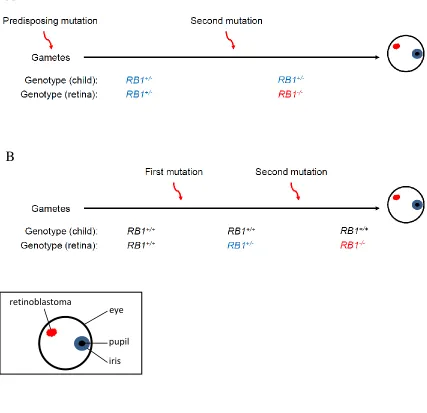
Figure 1.1: There are multiple ways to acquire retinoblastoma.
(A) Retinoblastoma can be inherited whereby one of the contributing gametes is RB1-.
All cells in the patient’s body are subsequently hemizygous for RB1. A second somatic
mutation in the retina results in retinoblastoma. (B) Retinoblastoma can occur
sporadically, with two somatic hits in the retina often before three years of age. Retinoblastoma can also arise from a hit early in development, resulting in a patient
wherein some cells have two wild type copies of RB1, and other cells have only one wild
type copy of RB1, including those in the retina. A second somatic hit in the retina leads
to retinoblastoma.
A
B
eye
pupil
acquired early enough during embryogenesis that patients can pass the mutation on to
their offspring.
In 1971, Knudson published a paper analyzing the mutation rate of RB1 in both
inherited and non-inherited retinoblastomas. He found that the mutation rate for loss of
the first RB1 gene in non-inherited cases, and the loss of the second RB1 gene in both
inherited and non-inherited cases was similar. From this study he proposed two key
findings. The first is that retinoblastoma resulted from just two genetic events (germinal
and somatic, or both somatic) resulting in loss of both copies of RB1. The second is that
loss of the first RB1 allele did not accelerate loss of the second RB1 allele. Therefore
hemizygosity of RB1 did not create a haploinsufficiency, rather it recapitulated the wild
type condition (Knudson 1971). As a result of this study, the retinoblastoma gene
became known as the “prototypic tumor suppressor”, in that its loss in retinoblastoma
was the prototype to which other proposed tumor suppressor genes were compared
(Murphree and Benedict 1984). To date, modeling Rb1 loss in mouse models of cancer
has supported Knudson’s “two-hit” hypothesis (Jacks et al 1992, Vooijs et al 1998).
However, data presented in this thesis suggest that certain aspects of pRB function
exhibit haploinsufficiency when disrupted. In chapter three, I will discuss the
applicability of extrapolating Knudson’s hypothesis to other contexts of tumorigenesis.
1.1.2
The retinoblastoma protein is a regulator of the cell division
cycle
Because loss of the retinoblastoma gene was found to be important for only a few
tissues in hereditary cancer development (retina, bone), it was expected to have only a
specialized role in these tissues. However, studies of the transforming activity of viral
oncoproteins including the adenovirus E1A, simian virus 40 TAg and the human
papilloma virus E7 oncoproteins, revealed that inactivation of the retinoblastoma protein
(pRB) was required for transformation, suggesting that pRB might have a more broad
tumor suppressive application (Whyte et al 1988, DeCaprio et al 1988, Dyson et al
Around the time of this discovery, loss of pRB function was found to be
associated with loss of cellular proliferation control (Takahashi et al 1991, Bookstein et
al 1990a, Huang et al 1988). For example, several studies demonstrated that adding back
pRB to human cancer cells (bladder, prostate, osteosarcoma) significantly reduced their
proliferative rate, their ability to form colonies in soft agar and reduced tumorigenicity in
nude mice (Takahashi et al 1991, Bookstein et al 1990b, Huang et al 1988). Two
regions, the “pocket” domain (res. 379-792) and the C-terminus of pRB (res. 793 – 928),
were found to be responsible for its growth suppression properties (Fig. 1.2A; Qin et al
1992). These regions were also demonstrated to be required for pRB binding to the
family of E2F transcription factors (Fig. 1.2B; Chellappan et al 1991, Bagchi et al 1991,
Chittenden et al 1991, Bandara and La Thangue 1991).
The first human genes demonstrated to be regulated in an E2F dependent manner
were c-myc and dihydrofolate reductase (DHFR), whose protein products are involved in
cellular proliferation and DNA synthesis respectively (Hiebert et al 1989, Thalmeier et al
1989, Blake and Azizkhan 1989). Furthermore, these genes were shown to be regulated
in a cell cycle-dependent manner (Hiebert et al 1989, Thalmeier et al 1989, Blake and
Azizkhan 1989). In this way, a paradigm was formed in which the growth suppressive
role of the retinoblastoma protein was mediated through inhibition of E2F target gene
transcription, which in turn could be deregulated by viral oncoproteins (Nevins 1992).
These and subsequent studies led to the understanding that pRB regulates the G1 to
S-phase transition of the cell division cycle by binding to E2Fs and inhibiting E2F target
gene transcription, and that this universal role for pRB in proliferation transcends all cell
types (studies outlining this principle mechanism of cell cycle control are reviewed in
Dyson 1998).
1.1.3
The pocket protein family
Based on sequence similarity and their ability to also be bound by viral
oncoproteins, two other members of the pRB family were identified, RBL1 and RBL2,
producing the p107 and p130 proteins respectively (Fig. 1.2A; Ewen et al 1991, Zhu et al
1993, Mayol et al 1993, Cobrinik et al 1993, Li et al 1993, Hannon et al 1993). These
Figure 1.2: Structures of the pocket protein and E2F families of proteins.
(A) The pocket protein family is defined by the “small pocket” they all possess, into
which viral oncoproteins bind. p107 and p130 are structurally more similar to each other
than to pRB. CDK- cyclin-dependent kinase. NLS- nuclear localization signal. (B) The
E2F family of transcription factors (E2Fs 1-8) mediate cell cycle advancement. E2Fs 1-6 heterodimerize with DP protein to bind to DNA. Activator E2Fs are bound by pRB, which prevents the transcription of genes required for S-phase progression. Repressor E2Fs either bind pRB, p107 or p130, or dimerize with atypical E2Fs (E2Fs 7 and 8) and recruit repressive complexes to DNA. NLS- nuclear localization signal. DBD- DNA binding domain. NES- nuclear export signal. DMZ- dimerization domain. MB- marked box. TA- transactivation domain. DP- Differentiation Related Transcription Factor-1 polypeptide-1.
A
is the most highly conserved region among the pocket protein family and across several
species of pRB (Lee et al 1998). As these proteins all contain this “pocket domain”, they
are commonly called “pocket proteins”. The small pocket contains an A and B domain,
as well as a flexible linker that separates the two; each A and B domain contains a single
cyclin fold, which interact to form the small pocket domain (Fig. 1.2A; Lee et al 1998).
This small pocket is sufficient to repress transcription and interact with viral oncoproteins
(Hu et al 1990, Sellers et al 1995, Chow and Dean 1996, Chow et al 1996).
Crystallography has demonstrated that the LXCXE motif contained in viral oncoproteins
makes contact with a shallow groove in the B domain of the small pocket (Fig. 1.2A; Lee
et al 1998, Ewen et al 1989, Munger et al 1989, Whyte et al 1989, Dyson et al 1992).
Indeed, several cellular proteins contain an LXCXE-like motif, or are otherwise able to
bind in the LXCXE binding cleft, and thereby interact with the pocket protein family
(summarized in Dick 2007). Many of the proteins that bind in the LXCXE binding cleft
are chromatin remodeling proteins or components of complexes that have chromatin
remodeling capabilities in order to effect transcriptional repression.
Pocket proteins also contain a C-terminal domain which, in conjunction with the
small pocket, comprises the large pocket (Fig. 1.2A). As described above, this large
pocket is the minimal growth suppressing domain and is sufficient to interact with the
E2F family of transcription factors and inhibit E2F target gene transcription (Hiebert et al
1992, Qin et al 1992, Bremner et al 1995). Thus recruitment of pocket proteins by E2Fs
to E2F target genes not only inhibits transactivation, but recruits transcriptionally
repressive complexes to introduce histone modifications and further prevent E2F target
gene transcription.
1.1.4
The current model of proliferative control by pocket proteins
pRB, p107 and p130 work in a concerted effort to mediate proliferative control
(Fig. 1.3). This is aided by the differential expression and sub-cellular localization of
pocket proteins during the cell division cycle, and their preference for binding to specific
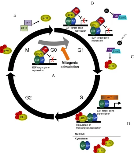
Figure 1.3: The current model of proliferative control by pocket proteins. (A-B) Briefly, upon mitogenic stimulation, p107 replaces p130 at E2F target gene
promoters and pRB-E2F complexes increase. CKIs inhibit the activity of cdks. (C)
Upon a feed forward loop of increasing activity of cyclin-cdk complexes, pocket proteins
become extensively hyperphosphorylated. (D) E2F target gene transcription occurs, and
cells are committed to divide. p107 and p130 are exported from the nucleus and pRB can
regulate the S-phase DNA damage checkpoint and DNA replication as needed. (E) At
the end of mitosis, phosphatases PP1 and PP2A dephosphorylate pocket proteins.
A
C
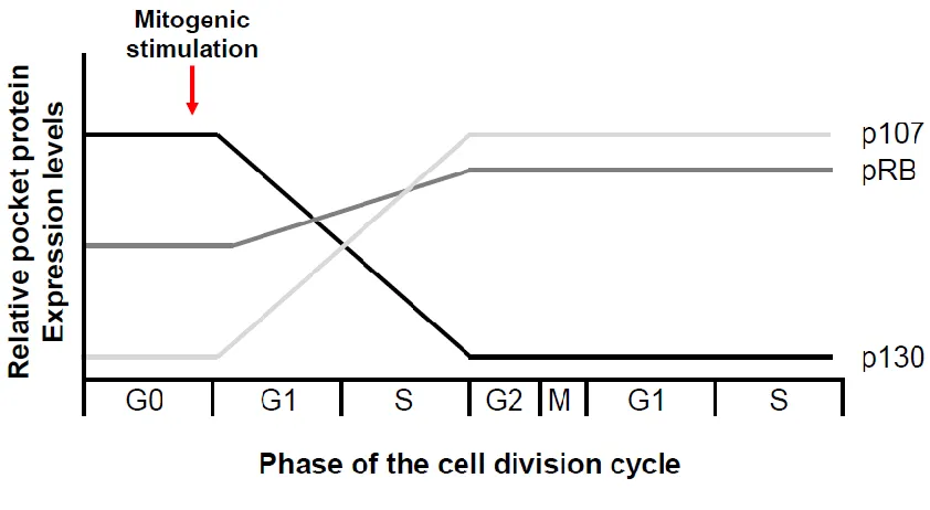
Figure 1.4: Pocket proteins are differentially expressed during the cell division cycle.
In brief, p130 binds to inhibitor E2Fs in G0 and recruits a transcriptionally
repressive DREAM complex, mediating a reversible cell division cycle arrest called
quiescence (Fig. 1.3A; Moburg et al 1996, Hurford et al 1997, Litovchick et al 2007).
Upon mitogenic stimulation, cells enter G1 and p107 replaces p130 at E2F target genes
(Takahashi et al 2000, Wells et al 2000). Expression of the retinoblastoma protein
increases and pRB localizes to E2F target genes, inhibiting the transcriptional activity of
activator E2Fs (Fig. 1.3B). This is mediated both by physically masking their
transactivation domain, and by recruiting repressive chromatin remodeling complexes via
the LXCXE binding cleft (Hiebert et al 1992, Schwarz et al 1993, Shirodkar et al 1992,
Helin et al 1993, Flemington et al 1993, Zamanian and La Thangue1993).
E2F target genes that are regulated by pocket proteins include positive regulators
of the cell division cycle and replication machinery, therefore E2F target gene
transcription must be activated to progress from G1 into S-phase of the cell division cycle
(Hurford et al 1997, Lavia and Jansen-Durr 1999, Takahashi et al 2000, Williams et al
2006). Activation of E2Fs is mediated by phosphorylation of pocket proteins in a cell
cycle-dependent manner by cyclin/cyclin-dependent kinase (cdk) complexes (DeCaprio
et al 1989, Sherr and Roberts 1999). This releases pocket proteins from binding to E2Fs
allowing them to transactivate their target genes (Fig. 1.3C; Mudryj et al 1991,
Chittenden et al 1993). The activation of cyclin/cdk complexes occurs in a feed-forward
loop that is antagonized by cdk inhibitors (CKIs) (Fig. 1.3B; Sherr and Roberts 1999,
Mittnacht 1998, Besson et al 2008). As a result of the cyclin-cdk feed forward loop that
maximizes phosphorylation of pRB and inhibition of CKIs, cells are committed to
irreversibly advance into S-phase.
At the end of mitosis, pocket proteins are dephosphorylated and once again bind
to E2Fs and negatively regulate entry into the next cell division cycle. Protein
phosphatases 1 and 2 are proposed to dephosphorylate either pRB or all pocket proteins
respectively (Fig. 1.3E; Nelson et al 1997, Yan et al 1999, Dunaief et al 2002, Garriga et
al 2004). Alternatively, pocket proteins are responsible for mediating the permanent cell
cycle exit paradigms of both senescence and differentiation (Bruce et al 2000, Campisi
2000, Dannenberg et al 2000, Peeper et al 2001, Gutierrez et al 2008, Berman et al 2008,
Corbeil et al 1995, Zaksenhaus et al 1996, De Falco et al 2006, Korenjak and Brehm
2005, Herwig and Strauss 1997, Sellers et al 1998, Lee et al 1999, Thomas et al 2001
Nguyen and McCance 2005). The above discussion of the cell division cycle by pocket
proteins is summarized in Figure 1.5.
It is clear that together, the pocket proteins are integral in mediating cell division
cycle control, and permanent cell cycle arrest. These functions are tumor suppressive as
in many human cancers, the activity of pocket proteins are disrupted (Sherr 1996). While
deregulated cdks and CKIs lead to the inactivation of all pocket proteins, selective
mutation of only the retinoblastoma protein in many human cancers distinguishes it from
p107 and p130 (Harbour et al 1988, Horowitz et al 1989, Lee et al 1988, Malumbres and
Barbacid 2001, Sherr 1996).
1.1.5
pRB is unique among pocket proteins
During S-phase, the retinoblastoma protein remains in the nucleus and has been
proposed to be important for mediating cell cycle arrest and facilitating repair upon DNA
damage by repressing E2F target gene transcription and suppressing further DNA
synthesis (Fig. 1.3D; Knudsen et al 2000, Sever-Chroneos et al 2001, Wells et al 2003,
Avni et al 2003). It is able to do so as its dephosphorylation can be induced upon DNA
damage, followed by acetylation to inhibit subsequent hyperphosphorylation. The
retinoblastoma protein can therefore remain active despite high levels of cdk activity to
mediate this S-phase DNA damage checkpoint (Fig. 1.3D; Avni et al 2003, Markham et
al 2006, Chan et al 2001).
pRB has also been indirectly implicated in events during mitosis. Certain E2F
target genes including Emi, Mad2 and BubR1 are required for maintaining the mitotic
checkpoint, preventing progression through mitosis if chromosomes are not properly
attached to the mitotic spindle and lined up at the metaphase plate. If pRB is unable to
repress transcription of these genes, their overexpression compromises mitosis, delaying
its progression and resulting in aneuploidy (Hernando et al 2004, Margottin-Goguet et al
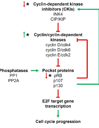
Figure 1.5: The pocket protein regulatory pathway.
Pocket proteins inhibit E2F target gene transcription, which in turn inhibits cell cycle progression. Pocket proteins are negatively regulated by cyclin-cdk complexes, and positively regulated by phosphatases. Activated CKIs inhibit cyclin-cdk complexes, restoring pocket protein activity. Pocket proteins can inhibit cyclin-cdk complexes and activate CKIs to maintain themselves in a hypophosphorylated state. (*) Indicates nodes in this regulatory pathway that are commonly mutated in cancer. (↑) Indicates
fail to progress through mitosis (Niculescu et al 1998, Srinivasan et al 2007). These cells
persist in culture and can resume proliferating with aneuploidy.
In addition to pRB’s role in the regulation of the cell division cycle, pRB plays
regulatory rolls in both apoptosis and the DNA damage response (Dick and Dyson 2003,
Julian et al 2008, Seifried et al 2008, Carnevale et al 2012, Cecchini and Dick 2011). It
does this by binding to E2F1 specifically using the C-terminus of pRB (Fig. 1.2A; Dick
and Dyson 2003). This specific interaction allows pRB to regulate E2F1-induced
apoptosis, even when pRB is hyperphosphorylated and presumed inactive (Dick and
Dyson 2003, Julian et al 2008, Seifried et al 2008, Carnevale et al 2012, Cecchini and
Dick 2011). Moreover, pRB-E2F1 interactions are particularly important in mediating
the DNA damage response, though this role appears to be context-dependent (Carnevale
et al 2012). In addition to being unique from the other E2F transcription factors, the
interaction between pRB and E2F1 is also unique amongst pocket proteins (Dick and
Dyson 2003, Cecchini and Dick 2011, Julian et al 2008).
More recent data such as that presented in Chapters 2 and 3 of this thesis has
begun to outline a unique role for pRB during mitosis that is independent of its regulation
of E2F target gene transcription (Coschi et al 2010, Longworth et al 2008, Manning et al
2010, van Harn et al 2010). Importantly, this novel function contributes to
pRB-mediated tumor suppression (Coschi et al 2010). Interestingly p107 and p130, whose
sequences are more similar to each other than to pRB, contain structural features that are
not found in pRB (Classon and Dyson 2001; Fig. 1.2A). This allows them to be
differentially regulated and to accomplish different tasks (Lacy and Whyte 1997, Zhu et
al 1995, Woo et al 1997, Moberg et al 1996). Conversely, structural features exclusive
to pRB have yet to be discovered to explain the unique roles described above. Thus it has
fallen to mouse models of the pocket proteins to describe the relative contributions of
1.2 Mouse models of Rb1
1.2.1
Rb1
-/-cells contribute to ‘normal’ tissues
The first models knocking out Rb1 in mice were reported in 1992 from three
different groups (Jacks et al 1992, Clarke et al 1992, Lee et al 1992). Each lab, using a
different genetic strategy, generated homozygous mutant mice (Rb1-/-) that died between
embryonic days (E) 13 and 15, indicating that Rb1 was required for embryogenesis,
though surprisingly it was indespensible up to at least E13.
Mice heterozygous for Rb1 were viable and fertile. Though humans hemizygous
for RB1 have a 90% likelihood of developing retinoblastoma by the age of three, Rb1
+/-mice did not develop retinoblastomas or exhibit any precursor lesions (retinomas)
(Matsunaga et al 1990; Jacks et al 1992, Lee et al 1992, Clarke et al 1992). One lab
followed Rb1+/- mice long enough to discover a tumor phenotype (Jacks et al 1992).
These mice succumbed to adenocarcinoma of the pituitary by ten months of age, and the
majority of these tumors had lost the remaining wild type Rb1 allele (LOH), seemingly
consistent with Knudson’s proposal that heterozygosity of the retinoblastoma gene was
not functionally relevant to tumorigenesis (Jacks et al 1992).
A decade later it was reported that the most apparent defect in Rb1-/- mice was
over-contribution of trophoblast cells to the placenta, creating an hypoxic environment
for Rb1-/- embryos (Wu et al 2003). In fact, if the mutant placenta were replaced with
wild type placenta, Rb1-/- embryos survived to birth with none of the neurological or
erythropoietic defects previously reported (Jacks et al 1992, Clarke et al 1992, Lee et al
1992, Wu et al 2003). Instead, these mice died at birth with severe skeletal muscle
dysplasia resulting in an inability of the diaphragm to work properly, preventing
breathing (Wu et al 2003). The fact that the major defect in Rb1-/- mice was now
reported to be in the trophoblast cells of the placenta, and that most other tissues appeared
to develop normally, helped to explain why Rb1-/- cells in chimeric mice were found to
contribute to a wide variety of what were described as ‘normal’ tissues (Williams et al
Several conditional knockout models of the Rb1 gene have improved upon our
understanding of the tissue-specific requirements of pRB. In general, knockout in certain
tissues leads to increased proliferation and/or lack of proper differentiation though, with
the exception of the pituitary, no spontaneous tumor development (Mayhew et al 2005,
Ruiz et al 2004, Classon et al 2000, Hansen et al 2004, Vooijs et al 1998). Instead, this
has been shown to require other complimentary mutations, such as disabling the p53
pathway (Wikenheiser-Brokamp 2004, Meuwissen et al 2003).
1.2.2
A structure-function approach to investigating pRB’s
contribution to tumor suppression
Generally, Rb1- alleles in mice have advanced our understanding of the role for
pRB in different tissues and stages of development. Furthermore, lack of a tumor, or any
overt phenotype, in both p107-/- and p130-/- mouse models has solidified pRB as unique
among pocket proteins for mediating tumor suppression (Lee et al 1996, Cobrinik et al
1996). As described previously, there are currently three known binding surfaces on pRB
that are distinct from one another and mediate binding to specific proteins. Knocking out
pRB in mouse models disrupts all of its functions and therefore we are unable to attribute
its tumor suppressive properties to one function or another. To this end, targeted
mutation of certain binding sites on pRB would reveal their functional contribution to
pRB-mediated tumor suppression. In the data chapters to follow, I will demonstrate how
targeted mutation of the LXCXE binding cleft of pRB contributes to pRB-mediated
tumor suppression, independently of E2F target gene transcription.
The advantage of a structure-function approach to investigating pRB function is
supported by phenotypes observed in the Rb1R654W mouse. The arginine to tryptophan
substitution at amino acid 654 (R654W) in the Rb1 gene inhibits the ability of pRB to
interact with E2Fs 1, 2 and 3, and thereby modulate their activity in a cell
cycle-dependent manner (Sun et al 2006). This mutation has been modeled after the R661W
mutation in the human RB1 gene (Sun et al 2006, Lohmann et al 1994, Onadim et al
1992). Rb1 R654W/R654W mice exhibit the same cell cycle defects as Rb1-/- embryos,
however they survive two days longer than Rb1-/- mice in utero, and defects in
This suggests that pRB can have important contributions to differentiation that are
unrelated to their ability to regulate E2F target gene transcription.
The idea that discrete interactions differentially contribute to pRB-mediated
tumor suppression is supported by cancer mutation data in humans, suggesting that
inactivation of the entire retinoblastoma gene is required to compromise pRB-mediated
tumor suppression, and not just one discrete interaction surface. For example, tumor
derived mutations of the RB1 gene are inconsistent with the disruption of a single
function of the retinoblastoma protein as there are thirty five known cancer-causing
missense mutations in RB1 from retinoblastoma patients, and most of these alleles have
been reported only once (Fig. 1.6; Retinoblastoma genetics website). Additionally, the
majority of these alleles are deletions or nonsense changes, abrogating complete pRB
function (Lohmann 1999, Retinoblastoma genetics website). This is contrary to
mutations in other oncogenes or tumor suppressors that cluster in regions based on
functional importance as in Ras or p53 (Schubbert et al 2007, Joerger and Fersht 2007).
With evidence indicating that disrupting discrete pRB interactions with cellular
proteins could help illuminate the contribution of that particular interaction to tumor
suppression, the concept of a “structure-function” approach to study the retinoblastoma
protein has been adopted. This was greatly aided by the publication of crystal structures
of pRB’s pocket showing which residues are on the surface of the protein, which are
buried, and which are important for interacting with viral oncoproteins and E2Fs (Lee et
al 1998, Rubin et al 2005, Xiao et al 2003). In this way, key residues mediating
interactions between pRB and its binding partners were identified and could be used for
targeted disruption. Indeed, substitution of amino acids required for pRB-E2F
interactions in the small pocket of pRB eliminated pRB-mediated transcriptional
repression of E2F target genes, and revealed a novel role for pRB in the regulation of
apoptosis (Dick and Dyson 2003, Chau et al 2006).
Though there may be other, as yet undefined binding sites, at the current time,
there are three well defined binding interfaces on the retinoblastoma protein that mediate
Figure 1.6: Spectrum of mutations of RB1 in retinoblastoma patients.
Mutations of RB1 in patients do not cluster within certain functional domains of the
sequence required for binding to E2F transcription factors, ii) the C-terminus, which
binds specifically to E2F1, and iii) the LXCXE binding cleft, which is located in the B
domain of the small pocket and mediates interactions with cellular proteins such as
chromatin regulatory factors (Fig. 1.2A). The next several sub-sections of this
introductory chapter will discuss the LXCXE binding cleft and the rationale for
investigating the contribution of the LXCXE binding cleft to pRB-mediated tumor
suppression.
1.3 The Rb1
ΔL/ΔLmouse
1.3.1
The LXCXE binding cleft
As previously described, the LXCXE binding cleft is located in the B domain of
the small pocket of pocket proteins (Lee et al 1998). Numerous cellular proteins are
reported to bind pRB using this site (reviewed in Dick 2007). Many of these proteins can
modify chromatin structure to induce heterochromatinization and thereby repression of
gene transcription including HDAC1/2, Suv39h1, BRG1, Brm, DNMT1 and hSWI/SNF
(Brehm et al 1998, Magnaghi et al 1998, Luo et al 1998, Lai et al 1999, Nielsen et al
2001, Dunaief et al 1994, Singh et al 1995, Robertson et al 2000, Zhang et al 2000).
Other proteins function in transcriptional repression or DNA replication, or have no as
yet known function. As previously discussed, binding of these proteins to the LXCXE
binding cleft of pRB facilitates inhibition of E2F target gene transcription, thereby
contributing to the inhibition of cell cycle progression.
Alternatively, using an LXCXE motif, viral oncoproteins bind in the LXCXE
binding cleft to inactivate pRB, p107 and p130 (Lee et al 1998, Ewen et al 1989, Munger
et al 1989, Whyte et al 1989, Dyson et al 1992). A study of amino acid sequence
conservation has revealed that the LXCXE binding cleft is the most highly conserved
region among pocket proteins and across several species of pRB (Lee et al 1998). As the
LXCXE binding cleft on pRB renders it susceptible to viral oncoproteins, it raises the
question as to why this site has remained so conserved over evolutionary time.
must perform an essential function, though this is not reflected by its seemingly modest
contribution to cell division cycle regulation.
Early efforts to address this conundrum began with the targeted disruption of
LXCXE binding cleft interactions without compromising binding to E2Fs in the large
pocket, or E2F1 in the C-terminus. These studies have revealed that while proliferative
control remains intact, the ability of such a mutant pRB to mediate permanent cell cycle
arrest is compromised (Dick et al 2000, Chan et al 2001, Chen and Wang 2000, Dahiya
et al 2000). In order to further investigate the role of the LXCXE binding cleft of pRB in
isolation, our lab has generated a mouse (Rb1ΔL/ΔL) with alanine substitutions at three key
amino acids in the LXCXE binding cleft that mediate important contacts with cellular
proteins (I746A, N750A and M754A) (Fig. 1.7; Isaac et al 2006). The Rb1ΔL/ΔL mouse is
viable, fertile and born at Mendelian ratios (Isaac et al 2006). Additionally, these mice
do not acquire spontaneous tumors (Isaac et al 2006). The Rb1ΔL/ΔL mouse is described in
detail below.
1.3.2
Rb1
ΔL/ΔLMEFs exhibit phenotypes independent of G1 to
S-phase transition regulation
Mouse embryonic fibroblasts from Rb1ΔL/ΔL mice reflect previous experiments in
Saos2 and C33A cells in that they maintain proliferative control (E2F target genes are not
upregulated), but fail to remain permanently arrested in senescence (Isaac et al 2006,
Talluri et al 2010). In cycling Rb1ΔL/ΔL MEFs, there is an accumulation of a >4N DNA
content peak, which is indicative of aneuploidy (Isaac et al 2006). They also exhibit a
reduction in histone H4-K20 trimethylation, a mark of heterochromatin. Centromeres
characterized by loss of this histone mark exhibit fusions leading to errors in mitosis
(Isaac 2006). Finally, these cells also exhibit hypocondensation of chromatin. Taken
together, genomic instability appears to be a hallmark of Rb1ΔL/ΔL MEFs.
In MEFs induced to senesce using oncogenic Ras, there is a reduction in the
histone H3-K9 trimethylation mark at E2F target genes, which correlates with their
upregulation. These cells also exhibit a striking inability to inactivate cell division cycle
Figure 1.7: Disruption of protein-interaction sites on the retinoblastoma protein.
The LXCXE binding cleft of pRB contains three important contact residues with LXCXE
motif-containing proteins, residues I746, N750 and M754 (Rb1+). Mutation of these
residues to alanine abrogates the ability of proteins to bind in the LXCXE binding cleft of
pRB. This mutation is called the Rb1ΔL mutation. The Rb1ΔL/ΔL mouse is defective for
increased >4N DNA content compared to wild type. DNA content was often 8N or
higher, indicating endoreduplication (Talluri et al 2010). Intriguingly, this is not
observed in differentiation, a separate paradigm of permanent cell cycle arrest, suggesting
that this is unique to pRB-mediated G1 arrest in response to oncogenic stress (Talluri et
al 2010).
1.3.3
The
Rb1
ΔL/ΔLmouse exhibits no overt tumor-suppressive
phenotype
As mentioned above, Rb1ΔL/ΔL mice are viable, fertile and born at the expected
Mendelian ratios (Isaac et al 2006). These mice do not acquire spontaneous tumors,
though they exhibit hyperplasia of the mammary gland ductal epithelium (Francis et al
2009). This hyperplasia was found to be a result of insensitivity to transforming growth
factor β (TGF-β) growth inhibition (Francis et al 2009). Studies in MEFs were used to
determine the mechanisms for this insensitivity and it was found that pRB-mediated
repression of E2F target genes was compromised in response to TGF-β signalling,
allowing for ectopic proliferation as measured by BrdU incorporation (Francis et al
2009).
In order to investigate the mechanism(s) by which the LXCXE binding cleft itself
contributes to pRB-mediated tumor suppression, we crossed Rb1ΔL/ΔL mice into a Trp53
-/-tumor prone mouse model. Trp53-/- mice reliably acquire thymic lymphomas that are
karyotypically normal, and so this is an appropriate tumor phenotype for us to modify
with the Rb1-ΔL mutation to observe whether its addition leads to increased genomic
instability. Additionally, as mentioned previously, Rb1ΔL/ΔL MEFs are compromised for
maintaining a G1 arrest in response to DNA damage and oncogene-induced senescence
(Talluri et al 2010). Because p53 acts upstream of pRB in mediating such a G1 arrest,
modification of the Trp53-/- tumor phenotype by pRB will be separate from its regulation
of this G1 arrest mechanism. The results of this large tumor study are reported in
1.3.4
A non-canonical role for the LXCXE binding cleft of pRB
The data described in this introductory chapter on the retinoblastoma protein have
focused on the various ways in which it regulates proliferation, with the exception of its
role in mediating E2F1-induced apoptosis, and the role of the LXCXE binding cleft in
mediating genome stability. Recently, a paper published by Longworth et al described a
role linking Drosophila pRB (RBF1) to the regulation of chromatin structure. This was
mediated by an interaction between RBF1 and a subunit of the Condensin II complex, a
member of the structural maintenance of chromosome (SMC) family of complexes that
facilitates mitotic chromosome condensation (Longworth et al 2008).
Longworth et al report that this subunit, dCAP-D3, partially colocalizes with
RBF1 on chromatin and that RBF1 is required for dCAP-D3 localization to chromatin
(Longworth et al 2008). Mutants of dCAP-D3 suppressed phenotypes induced by RBF1
overexpression and rbf1 mutants exhibited significant defects in chromatin condensation
during mitosis. Moreover in humans, adding wild type pRB to pRB-deficient cells
increased hCAP-D3 loading on chromatin in a manner dependent on pRB’s LXCXE
binding cleft (Longworth et al 2008). This report provides a hint at the mechanism by
which the LXCXE binding cleft was reported to mediate genome stability and contribute
to pRB-mediated tumor suppression (Isaac et al 2006). In subsequent chapters I will
describe the interaction of pRB with the Condensin II complex and will expand upon the
mechanism by which this occurs in the context of pRB-mediated genome stability and
tumor suppression.
1.4 Condensins
1.4.1
The structural maintenance of chromosomes (SMC) family of
complexes are integral for chromosome dynamics
The structural maintenance of chromosome (SMC) family of complexes consists
of the SMC5/6 complex, Cohesin (mitotic and meiotic), Condensin I and Condensin II
chromosome organization is evolutionarily conserved (Cobbe and Heck 2004). SMC
proteins always form a dimer and associate with regulatory subunits into a large
holocomplex. SMC proteins have a “hinge” domain where the protein self-folds, and an
ATP-binding cassette-like “head” domain separated by an antiparallel coiled-coil “arm”
(Fig. 1.8A). Two of these SMC proteins associate via their hinge to produce a V-shaped
dimer (Melby et al 1998, Anderson et al 2002, Haering et al 2002, Hirano and Hirano
2002) (Fig. 1.8A). It is predicted that ATP binding to, and hydrolysis at, the “head”
domain facilitates engagement and disengagement respectively (Haering et al 2004,
Lammens et al 2004, Hirano et al 2001, Hirano and Hirano 2004). Non-SMC regulatory
subunits of SMC complexes include kleisins, HEAT-repeat containing proteins or other
subunits (Fig. 1.8B). Kleisin subunits interact with the head domain of two SMC
proteins through their N- and C-terminal domains to form a ring-like structure whereas
HEAT-repeat containing proteins act as protein scaffolds to assemble cellular
components and are proposed to direct SMC proteins to specific loci on DNA (Schleiffer
et al 2003, Neuwald and Hirano 2000). HEAT-repeat regulatory subunits therefore
greatly influence chromosome dynamics.
The SMC5/6 complex is sequentially divergent from the other SMC complexes
and an understanding of its functions is reviewed elsewhere (Lehmann 2005, De Piccoli
et al 2009). The mitotic Cohesin holocomplex is comprised of SMC1 and SMC3, the
kleisin subunit hRAD21, and other subunits including a HEAT-repeat containing subunit
Scc2 (Losada and Hirano 2005) (Fig. 1.8C). Condensins I and II both have SMC2 and
SMC4 proteins, but differ in their regulatory subunits. Whereas Condensin I is composed
of the kleisin subunit hCAP-H and two HEAT-repeat containing subunits hCAP-D2 and
hCAP-G, Condensin II is comprised of the kleisin subunit hCAP-H2 and two
HEAT-repeat containing subunits hCAP-D3 and hCAP-G2 (Fig. 1.8D, E respectively).
In general, Cohesin ensures proper resolution of sister chromatids while
Condensins I and II facilitate sequential mitotic chromosome condensation (Fig. 1.9).
Briefly, the Cohesin complex forms a ring-like structure around DNA that maintains
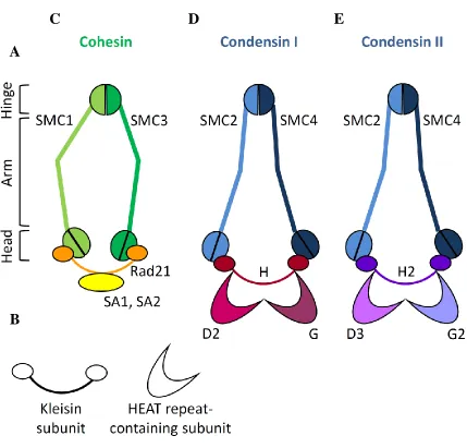
Figure 1.8: Cohesin, Condensin I and Condensin II are complexes of the structural maintenance of chromosome (SMC) family.
(A) Depiction of the domain organization of SMC proteins. Domains are organized by
the folding back upon itself of the SMC protein. The hinge domain is globular and binds to the hinge domain of another SMC protein. The arms are coiled coils, and the head
domain contains the N and C-terminus of the protein. (B) Depiction of the regulatory
subunits of SMC proteins. (C) Cohesin facilitates sister chromatid resolution and is
comprised of SMC1, SMC3, Rad21 and SA1 or SA2. (D) Condensin I is comprised of
SMC2, SMC4, CAP-H, CAP-G and CAP-D2. (E) Condensin II is comprised of SMC2,
SMC4, CAP-H2, CAP-G2 and CAP-D3. Adapted from Losada and Hirano, 2005.
A
B
Losada et al 2002). In prophase, mitotic Condensin II is loaded onto chromatin and
facilitates condensation; this is accompanied by release of some Cohesin from
chromosome arms (Losada et al 2002, Sumara et al 2002, Giménez-Abián et al 2004)
(Fig. 1.9B). After nuclear envelop breakdown Condensin I, which is constitutively
cytoplasmic, gains access to chromosomes and facilitates further mitotic chromosome
condensation during prometaphase and metaphase (Fig. 1.9C). Again, this is
accompanied by further release of Cohesin from chromosome arms. At this point, a
small subset of Cohesin remains at pericentromeric chromatin and is protected from
release by the protein shugoshin (Losada et al 2000, Clift et al 2009, Katis et al 2004,
Kitajima et al 2004, Marston et al 2004). At anaphase onset, shugoshin is targeted for
degradation and the kleisin subunit hRAD21 is cleaved by the separase enzyme, breaking
its ring structure and triggering Cohesin dissociation from chromosomes (Fig. 1.9D).
Mitotic chromosomes are then pulled to opposite poles of the cell (Gruber et al 2003,
Losada and Hirano 2005). After chromosomes have reached opposite poles, Condensins
are unloaded from chromatin (reviewed in Losada and Hirano 2005) (Fig. 1.9E).
1.4.2
Condensin I and Condensin II differentially contribute to
mitotic chromosomes
Condensins are proposed to facilitate mitotic chromosome condensation by
introducing supercoiling to DNA, or by associating with each other to create larger DNA
loops, bringing distant sites on chromosomes together. While data in Chapters 2 and 3
report a unique interaction of pRB specifically with the Condensin II complex,
Condensin I was the first discovered Condensin. As such, it has been extensively studied
and what has been learned about Condensin I is inferred for the mechanisms of action of
Condensin II. Condensin I has been shown to introduce positive supercoiling to double
stranded DNA (dsDNA) in an ATP-dependent manner (Kimura and Hirano 1997, Strick
et al 2004). This activity is stimulated by phosphorylation of the non-SMC subunits by
Cyclin B-cdk1 (Kimura et al 1998, Kimura et al 1999, Strick et al 2004). Indeed,
non-SMC subunit phosphorylation steadily increases from prophase to anaphase of the cell
division cycle, and this correlates with increasing compaction of mitotic chromosomes
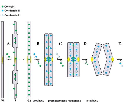
Figure 1.9: The SMC proteins are key chromosomal components for mitosis.
(A) As DNA is being replicated in S-phase, Cohesin is loaded onto chromosome arms to
facilitate sister chromatid resolution. (B) In prophase, Condensin II is loaded onto
chromosomes, facilitating partial chromosome condensation. Some cohesin is removed
from chromosome arms. (C) After nuclear envelope breakdown, Condensin I is loaded
onto chromosomes and further condenses mitotic chromosomes. The remaining Cohesin
is removed from chromosomes except at centromeres and near telomeres. (D) At
anaphase onset, the remaining Cohesin at centromeres is removed by separase-mediated cleavage of its kleisin subunit, and chromosomes are pulled apart by the mitotic spindle.
(E) After chromosomes are pulled apart, mitotic Condensins I and II are unloaded from
chromosomes. Adapted from Losada and Hirano, 2005.