Structural, Morphological and Optical Properties
of Pure and Mg Doped ZnO Thin Films Using
SILAR Method
S. Dineshraj1, M. Karunakaran2, K. Kasirajan3, V.Annalakshmi4, S. Maheswari5
1, 2, 3, 4
Department of Physics, Alagappa Government Arts College, Karaikudi – 630 003, India. 5
Department of Physics, Caussanel College of Arts and Science, Muthupettai – 623 523, India.
Abstract: In the present study, undoped and Mg doped ZnO thin films were prepared by modified SILAR method. The coated
films were annealed at 3500C. The structural, surface morphological and optical properties were studied by X-ray diffraction,
scanning electron microscopy, and UV– vis spectroscopy, respectively. The hexagonal wurtzite structure formation with preferential orientation along the (002) plane was confirmed from structural analysis. The surface morphology of the undoped and Mg doped ZnO thin films have the some nanorods and flower like shapes. The sizes of the grains are found to be in the range between 400 and 450 nm. The optical properties of the thin films were estimated using the transmission spectrum in the range of 200–1100 nm. The optical band gap energy of undoped and Mg doped ZnO thin films was found to be 3.2 and 3.4 eV. Keywords: Zinc Oxide, Thin Films, Structural Studies, Morphological Studies, Optical Properties, Optical band gap.
I. INTRODUCTION
Diluted magnetic semiconductors (DMS) have attracted much interest due to their potential application in Spintronics devices, such as spin-valve transistors, spin light-emitting diodes, non- volatile memory, logic devices, optical isolators and ultrafast optical switches [1]. In DMSs, transition metal doped II–VI and III–V semiconductors have been studied extensively [1]. One of the materials at the focus of much attention is the wide band gap wurtzite phase zinc oxide, a II–VI semiconductor, a well-known piezoelectric and electro-optic material with wide direct band gap (~3.37eV) and large exciton binding energy (60 meV) in which some of the zinc can be substituted by the manganese ions responsible for the ferromagnetic coupling [2,3].ZnO doped with Mn has also been considered as an ideal material for short wavelength magneto-optical applications due to its wide band gap and the thermal solubility of Mn in ZnO [4].Since the ionic radius of Mg2+ (0.57 Å) is similar to that of Zn2+(0.60 Å), the latter can be substituted by the Mg2+ ion resulting in a wide range of solid solution [5]. However, the thermo dynamic solubility limit of MgO in ZnO is only about 4% as suggested by the phase diagram of MgO–ZnO binary system [6]. There are a number of reports on the growth of MgxZn1xO thin films using various techniques such as pulsed laser deposition (PLD) [7,8], laser ablation-molecular beam epitaxy (LA-MBE) [9], and radio frequency(rf) magnetron sputtering [10-11]. The substitution limit was found to be different for different techniques which are about33% for PLD [12], 49% for molecular beam epitaxy (MBE) [13], and metal organic vapor phase epitaxy (MOVPE) [14]. Ohtomo et al. [15] have found that the thermodynamically MgO is soluble in MgxZn1xO up to a value of x = 0.15, while recently Ryoken et al. [16] reported the value to be in the composition range 0.12 < x < 0.18. There are very few studies on the sol–gel MgxZn1xO thin films where substitution up to 20%, 33%, and 36% were reported [17–19]. Most of the reported work mainly correlates with the ferromagnetic properties of Mn-doped ZnO nanostructures and its origin. Thus, much work is needed to address the structural and optical properties of Zn1-xMnxO owing to a growing interest of the magneto-optical effect. Pradhan et al. showed that Zn1−xMnxO films grown at a substrate temperature of 500 °C exhibited room temperature
ferromagnetism and beyond 500 °C the crystalline quality of the film increased at the expense of a decrease in the magnetization due to the formation of Mn related clusters [20]. Heo et al. studied the effect of post deposition annealing on the ferromagnetic properties of Mn implanted ZnO film deposited on sapphire substrate at 400 °C [21]. A significant enhancement in the magnetization of Zn1−xMnxO film with an increase in annealing temperature (<600 °C) was observed and attributed to the
II. EXPERIMENTAL PROCEDURE
Experimental details MZO thin films were grown on glass substrates employing SILAR method. Thin films of different thicknesses were deposited onto glass substrate. These substrates were precleaned well with distilled water, Iso-propyol alcohol and acetone before undertaking deposition. The deposition bath comprises of Zinc sulphate (ZnSO4) 1.5 M is dissolved in 20 ml of water and varied percent of (1.5 M) magnesium sulfate (MgSO4) and 3 M sodium hydroxide (NaOH). The pH was adjusted by using NaOH or ammonium hydroxide. The substrates were immersed in the experimental bath for less than 15 s. Initially, sodium zincate formed in the bath is adsorbed onto the substrate. Subsequently, it is converted to ZnO in the second immersion in hot water kept at 95 0C. The time of immersion in first and second dipping are maintained as 10 s. The intervals between the dips are also kept as 10 s. The films were annealed at 350 0C for 5 hours uniformly before characterization.
The reaction mechanism for the formation of Mg doped ZnO Thin films is given below Mg (So4.H2O) → Mn2+ +So4
Zn (So4.7H2O) → Zn2+ + So4
Na OH → Na+ + OH-
The formation of Mg doped ZnO thin films process could be expressed as Mg (So4.H2O) + Zn (So4.7H2O) + Na OH → Mg ZnO +2NaSo4 + H2O
Characterizations of the samples were performed at room temperature. X-ray diffractometer (XRD 6000, Shimadzu japan) with CuKa line wavelength 1.5406 A ° was used to analyze the structure. Surface morphological study was carried out using a scanning electron microscopy (Philips Model XL 30, USA). For optical characterization, the transmission spectra of Mg doped ZnO thin films annealed at different temperatures were recorded using a UV-Vis spectrophotometer (Varian Cary 500 Scan).
III. RESULT AND DISCUSSION A. Structural Studies
Figure 1 (b). XRD pattern of Mg doped ZnO
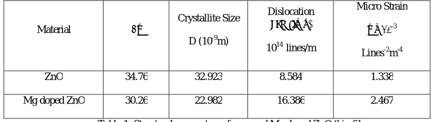
Figure.1.(b) shows the XRD pattern of Mg doped ZnO Thin films. The diffraction patterns reveal good crystalline quality without any appreciable changes from pure ZnO films and are genuinely polycrystalline with a hexagonal wurtzite structure. These results imply that there are no secondary phases such as a magnesium cluster or oxides. It also shows that the high intense peak is oriented along the c-direction and the corresponding peak is (110). Hence, this crystal ZnO film with stronger (002) preferred orientation will increase the Hall mobility [23]. Also Crystallized ZnO seed layers have a hexagonal crystal structure which was confirmed by the main diffracted peaks for the (002) (1 0 1) and (1 0 2) planes [24]. The observed‘d’ spacing values are in good agreement with the standard values of ZnO. Hence the overall structure of the doped films remains unchanged with the introduction of Mg. Also, no additional peaks of Mg or its phases are observed in the XRD spectrum and it suggests that the doped Mg atoms are incorporated into the ZnO thin film. The calculated values of crystallite size, Dislocation density and Micro structures are given in table1.
Material 2θ
Crystallite Size
D (10-9m)
Dislocation
density(δ)
1014 lines/m
Micro Strain
ε x 10-3
Lines-2m-4
ZnO 34.76 32.923 8.584 1.338
[image:4.612.150.496.88.277.2]Mg doped ZnO 30.26 22.982 16.386 2.467
Table 1: Structural parameters of pure and Mg doped ZnO thin films
B.Morphological studies
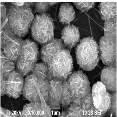
[image:4.612.89.524.442.564.2]Figure 2 (a). SEM image of pure doped ZnO
Figure 2 (b). SEM image of Mg doped ZnO
C. Optical studies UV-Visible Spectroscopy
[image:5.612.186.426.72.312.2]Figure 3. Transmission spectra of pure and Mg doped ZnO thin films
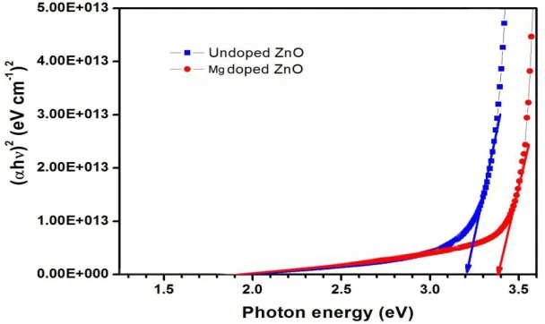
Figure 3. Taugh plot of pure and Mg doped ZnO thin films
Fig. 4 shows the variation of (αhν)2 with the photon energy for undoped and metals doped ZnO thin films deposited on glass
substrate. The absorption coefficient (α) and incident photon energy (hν) can be related as [25]
h E h
A( g)m
Where A is a constant and Eg is the band gap of the material. From the above equation, it is clear that the plot of (αhν)2 versus hν .
The band gaps are found to be 3.2 eV for ZnO thin films and it is decreased with doping metals in ZnO films. The optical band gap value increased to 3.4 eV as Mg atoms are incorporated in ZnO matrix. The Mg doped ZnO thin film band gap is about 3.4 the band gap increase may be due to increase of metal ion concentration in doped ZnO thin films.
Materials Direct Energy
Band Gap (eV)
Undoped ZnO 3.2
[image:6.612.164.468.338.519.2]IV. CONCLUSION
Mg doped ZnO thin films were prepared by employing SILAR technique. The films are annealed in air to improve the crystallinity and grain sizes. The observed structural, optical, electrical, morphological and compositional properties were plausibly explained in this work. The presence of the XRD pattern represents that the deposited films were found that polycrystalline in nature with hexagonal wurtzite structure. Also the micro structural parameters such as crystallite size, strain, dislocation density, were estimated. The SEM results represented that the film annealed at 5 h, have homogeneous surface with nanorods and flower like shaped grains. The optical band gap energy of undoped and doped ZnO thin films was found to be in the range between 3.2- 3.4 eV. This kind of micro structure is desirable for sensor applications.
REFERENCES
[1] Liu Y, Yang S-h, Zhang Y-l, Bao D-h. Influence of annealing temperature on structural, optical and magnetic properties of Mn-doped ZnO thin films prepared by sol–gel method. Journal of Magnetism and Magnetic Materials. 2009;321:3406-10
[2] Kim KJ, Park YR. Spectroscopic ellipsometry study of optical transitions in Zn[sub1 - x]Co[sub x]O alloys. Applied Physics Letters. 2002;81:1420-2. [3] Diaconu M, Schmidt H, Hochmuth H, Lorenz M, Benndorf G, Lenzner J, et al. UV optical properties of ferromagnetic Mn-doped ZnO thin films grown by
PLD. Thin Solid Films. 2005;486:117-21.
[4] Bates CH, White WB, Roy R. The solubility of transition metal oxides in zinc oxide and the reflectance spectra of Mn2+ and Fe2+ in tetrahedral fields.Journal of Inorganic and Nuclear Chemistry. 1966;28:397-405.
[5] Shannon R. Revised effective ionic radii and systematic studies of interatomic distances in halides and chalcogenides.ActaCrystallographica Section A. 1976;32:751-67
[6] Segnit ER, Holland AE. The System MgO-ZnO-SiO2.Journal of the American Ceramic Society. 1965;48:409-13.
[7] Sharma AK, Narayan J, Muth JF, Teng CW, Jin C, Kvit A, et al. Optical and structural properties of epitaxial Mg[sub x]Zn[sub 1 - x]O alloys. Applied Physics Letters. 1999;75:3327-9.
[8] Choopun S, Vispute RD, Yang W, Sharma RP, Venkatesan T, Shen H. Realization of band gap above 5.0 eV in metastable cubic-phase Mg[sub x]Zn[sub 1 - x]O alloy films. Applied Physics Letters. 2002;80:1529-31
[9] Hayashi I, Panish MB, Foy PW, Sumski S. JUNCTION LASERS WHICH OPERATE CONTINUOUSLY AT ROOM TEMPERATURE. Applied Physics Letters. 1970;17:109-11
[10] Minemoto T, Negami T, Nishiwaki S, Takakura H, Hamakawa Y. Preparation of Zn1−xMgxO films by radio frequency magnetron sputtering. Thin Solid Films. 2000;372:173-6.
[11] Jia C-L, Wang K-M, Wang X-L, Zhang X-J, Lu F. Formation of c-axis oriented ZnO optical waveguides by radio-frequency magnetron sputtering. Opt Express. 2005;13:5093-9
[12] Ohtomo A, Kawasaki M, Koida T, Masubuchi K, Koinuma H, Sakurai Y, et al. Mg[sub x]Zn[sub 1 - x]O as a II--VI widegap semiconductor alloy. Applied Physics Letters. 1998;72:2466-8.
[13] Ohtomo A, Kawasaki M, Sakurai Y, Ohkubo I, Shiroki R, Yoshida Y, et al. Fabrication of alloys and superlattices based on ZnO towards ultraviolet laser. Materials Science and Engineering: B. 1998;56:263-6.
[14] Park WI, Yi G-C, Jang HM. Metalorganicvapor-phase epitaxial growth and photoluminescent properties of Zn[sub 1 - x]Mg[sub x]O(0 <= x <= 0.49) thin films. Applied Physics Letters. 2001;79:2022-4.
[15] Ohtomo A, Shiroki R, Ohkubo I, Koinuma H, Kawasaki M. Thermal stability of supersaturated Mg[sub x]Zn[sub 1 - x]O alloy films and Mg[sub x]Zn[sub 1 - x]O/ZnO heterointerfaces. Applied Physics Letters. 1999;75:4088-90.
[16] Ryoken H, Ohashi N, Sakaguchi I, Adachi Y, Hishita S, Haneda H. Structures and properties of (Zn,Mg)O films studied from the aspect of phase equilibria. Journal of Crystal Growth. 2006;287:134-8.
[17] Murakawa T, Fukudome T, Hayashi T, Isshiki H, Kimura T. Structural and optical properties of MgxZn1–xO thin films formed by sol–gel method. physica status solidi (c). 2004; 1:2564-8.
[18] Ji Z, Song Y, Xiang Y, Liu K, Wang C, Ye Z. Characterization of MgxZn1−xO thin films prepared by sol–gel dip coating. Journal of Crystal Growth. 2004;265:5, 37-40.
[19] Zhao D, Liu Y, Shen D, Lu Y, Zhang J, Fan X. Photoluminescence properties of Mg[sub x]Zn[sub 1 - x]O alloy thin films fabricated by the sol-gel position method. Journal of Applied Physics. 2001; 90:5561-3.
[20] Pradhan AK, Zhang K, Mohanty S, Dadson JB, Hunter D, Zhang J, et al. High-temperature ferromagnetism in pulsed-laser deposited epitaxial (Zn,Mn)O thin films: Effects of substrate temperature. Applied Physics Letters. 2005;86:152511.
[21] Heo YW, Ivill MP, Ip K, Norton DP, Pearton SJ, Kelly JG, et al. Effects of high-dose Mn implantation into ZnO grown on sapphire. Applied Physics Letters. 2004;84:2292-4
[22] M. Girish, R. Sivakumar, and C. Sanjeeviraja “A Simple Approach to Deposit MnS Thin Films” Light and Its Interactions with Matter AIP conf.proc.1620, 235-239 (2014).
[23] P. Mitra and S. Mondal, progress in theoretical and applied physics, volume 1, 201 [24] S. Mondal, K.P Kanta and P. Mitra, Journal of Physical Science, vol. 12, 2008.