signalling
Transforming
growth factor
signalling
Critical
Oncogenic
Pathways in
Diffuse
Gastric
Cancer
Evolution
Clinical
Presentation
in Metastatic
Evolution
Left Ovary Metastasis
Krukenberg Tumor
Metastatic tumor evolution and organoid
modeling implicate
TGFBR2
as a cancer driver in
diffuse gastric cancer
Nadauld
et al.
R E S E A R C H
Open Access
Metastatic tumor evolution and organoid
modeling implicate
TGFBR2
as a cancer driver in
diffuse gastric cancer
Lincoln D Nadauld
1,2, Sarah Garcia
2, Georges Natsoulis
1, John M Bell
2, Laura Miotke
1, Erik S Hopmans
2, Hua Xu
2,
Reetesh K Pai
3, Curt Palm
2, John F Regan
4, Hao Chen
5, Patrick Flaherty
2, Akifumi Ootani
6, Nancy R Zhang
7,
James M Ford
1, Calvin J Kuo
6*and Hanlee P Ji
1,2*Abstract
Background:Gastric cancer is the second-leading cause of global cancer deaths, with metastatic disease representing the primary cause of mortality. To identify candidate drivers involved in oncogenesis and tumor evolution, we conduct an extensive genome sequencing analysis of metastatic progression in a diffuse gastric cancer. This involves a comparison between a primary tumor from a hereditary diffuse gastric cancer syndrome proband and its recurrence as an ovarian metastasis.
Results:Both the primary tumor and ovarian metastasis have common biallelic loss-of-function of both theCDH1 andTP53tumor suppressors, indicating a common genetic origin. While the primary tumor exhibits amplification of the Fibroblast growth factor receptor 2 (FGFR2) gene, the metastasis notably lacksFGFR2amplification but rather possesses unique biallelic alterations of Transforming growth factor-beta receptor 2 (TGFBR2), indicating the divergentin vivoevolution of aTGFBR2-mutant metastatic clonal population in this patient. AsTGFBR2mutations have not previously been functionally validated in gastric cancer, we modeled the metastatic potential of TGFBR2 loss in a murine three-dimensional primary gastric organoid culture. TheTgfbr2shRNA knockdown withinCdh1-/-; Tp53-/-organoids generates invasionin vitroand robust metastatic tumorigenicityin vivo, confirmingTgfbr2 metastasis suppressor activity.
Conclusions:We document the metastatic differentiation and genetic heterogeneity of diffuse gastric cancer and reveal the potential metastatic role ofTGFBR2loss-of-function. In support of this study, we apply a murine primary organoid culture method capable of recapitulatingin vivometastatic gastric cancer. Overall, we describe an integrated approach to identify and functionally validate putative cancer drivers involved in metastasis.
Background
Worldwide, gastric adenocarcinoma is the fourth most common malignancy and the second leading cause of can-cer deaths among men and women. Based on distinctive histopathologic features, gastric adenocarcinoma is cate-gorized into diffuse and intestinal subtypes [1]. In terms of histopathology, diffuse gastric cancers are generally
undifferentiated, frequently have signet cell ring features and invasively infiltrate normal stomach tissue. In con-trast, the intestinal subtype has epithelial features and forms discrete tumor masses similar to colon cancer. Diffuse gastric cancer has a higher incidence of metastatic disease and a generally worse prognosis compared to the intestinal subtype [2,3]. Currently, the genomic analyses of diffuse gastric cancer have involved a small number of samples including a recent study by the Cancer Genome Atlas Project (TCGA) and a whole genome sequencing survey of a set of diffuse gastric tumors [4]. However, there are few, if any, studies that detail the metastatic evolution of gastric cancer; metastatic tumors are typically absent from large-scale genomic cancer surveys such as * Correspondence:[email protected];[email protected]
6Division of Hematology, Department of Medicine, Stanford University
School of Medicine, CCSR 1155, 269 Campus Drive, Stanford, CA 94305-5151, USA
1
Division of Oncology, Department of Medicine, Stanford University School of Medicine, CCSR 1115, 269 Campus Drive, Stanford, CA 94305-5151, USA Full list of author information is available at the end of the article
© 2014 Nadauld et al.; licensee BioMed Central Ltd. This is an Open Access article distributed under the terms of the Creative Commons Attribution License (http://creativecommons.org/licenses/by/4.0), which permits unrestricted use, distribution, and reproduction in any medium, provided the original work is properly credited. The Creative Commons Public Domain Dedication waiver (http://creativecommons.org/publicdomain/zero/1.0/) applies to the data made available in this article, unless otherwise stated.
TCGA. Overall, little is known about the oncogenic process and tumor evolution of metastatic gastric cancer despite its paramount clinical importance [5].
In hereditary diffuse gastric cancer (HDGC), germline mutations inCDH1(that is, E-cadherin) confer a 70% life-time risk of developing diffuse gastric cancer [6,7]. The CDH1tumor suppressor gene encodes E-cadherin, a trans-membrane glycoprotein that mediates calcium-dependent cell-cell adhesion. Changes in CDH1 function affect the epithelial-mesenchymal transition (EMT) that has been implicated as playing a role in tumorigenesis. Studies of affected HDGC individuals’ tumors provide a unique op-portunity to determine the essential drivers of diffuse gas-tric cancer in the context of CDH1 loss of function. Supporting evidence of the role of CDH1in sporadic dif-fuse gastric cancers includes the observation that 50% con-tain CDH1 mutations or hypermethylation of the CDH1 promoter [8,9]. A recent whole genome sequencing survey of diffuse gastric cancer also identified frequentCDH1 mu-tations as the most common driver event [4]. The TCGA gastric cancer data also show a high frequency of somatic CDH1mutations [10]. Significantly less is known about the identity and role of co-occurring drivers that contribute to diffuse gastric metastasis.
Herein, we report a study of the metastatic evolution-ary process in diffuse gastric cancer. Our goal was to identify known and candidate drivers that delineate the tumor progression during metastasis. We performed an extensive genome sequencing analysis of a primary gastric tumor and metastasis from an individual with a germline CDH1 mutation (Figure 1) who presented with a gastric primary, followed after 3 years by metas-tasis in the left ovary. Given the existing germline muta-tion in CDH1, the cancer genome only requires a second allelic hit via a somatic genetic aberration, as is
demonstrated in the tumor from this individual. Because the initial cancer driver event is known, Mendelian cancer genomes provide a rare and highly informative‘ experi-ment of nature’ that provides an opportunity to delin-eate somatic genetics of metastasis. Genome sequencing analysis of both tumors revealed evidence of a common origin based on shared mutations but greater genomic diversity seen both at the level of mutations as well as extensive allelic imbalance and copy number aberra-tions for the metatasis.
We determined if the candidate drivers from this metastatic progression were sufficient to reproduce dif-fuse gastric cancer. Our cancer modeling methodology used in vitrogastric organoids and allows one to engin-eer the genetic driver context of these cancers and study the process of metastatic evolution and oncogenic path-way divergence. Integrating genetic analysis and bio-logical modeling, we determined the independent role of TGFBR2 (transforming growth factor-β receptor 2) in the oncogenesis of diffuse gastric cancer. Our experi-mental cancer modeling relies on an air-liquid interface for primary mouse intestinal culture that contains both epithelial and mesenchymal elements, accurately recapit-ulates long-term proliferation, multilineage differenti-ation, the Wnt/Notch-dependent stem cell niche, and peristalsis [11]. We reported an analogous primary gas-tric organoid culture system that accurately recapitulates multilineage epithelial differentiation and stromal ele-ments [12]. Recently, we achieved robust in vitro onco-genic transformation of primary gastric, colon, and pancreatic organoids via mutations in Kras and Trp53, which induce high-grade dysplasia and invasion in vitro with adenocarcinoma upon subcutaneous transplant-ation into mice [13]. We demonstrate the functional validation of candidate gastric cancer metastasis drivers
Birth Age 37 Age 40
Germline CDH1 mutation
Common drivers CDH1 and TP53
Metastasis genetic aberrations Primary
tumor genetic aberrations
[image:3.595.60.539.529.666.2]Divergence
from cancer genomic profiling studies, focusing on mod-eling theTGFBR2driver as proof of principle.
Results
Diffuse gastric cancer and metastatic progression
At the age of 37 years, the index patient (525) was diag-nosed with stage III (T3N1M0) poorly differentiated dif-fuse gastric adenocarcinoma (Figure 1). Her 42-year-old sister was diagnosed with diffuse gastric adenocarcinoma 2 months earlier. Based on the family history of gastric cancer and the unusually young age of onset, she under-went germline CDH1 mutation testing. The patient and her sister were found to have a germline splice site muta-tion in intron 10 (c.1565 + 2insT). This germline mutamuta-tion was subsequently reported in another family with heredi-tary diffuse gastric cancer (HDGC) [14]. The patient underwent a total gastrectomy to remove her primary tumor and was found to have a single lymph node metas-tasis. She received standard adjuvant treatment including combined chemotherapy (cisplatinum and 5-fluorouracil) and radiation. Three years after her initial presentation, the patient reported progressive lower abdominal full-ness. A computed tomography (CT) scan demonstrated a large pelvic mass consistent with a left ovarian metas-tasis (Figure 1). Subsequently, the patient underwent laparotomy with bilateral salpingo-oophorectomy and biopsy of the pelvic mass. Pathological studies demon-strated metastatic adenocarcinoma involving the ovary, otherwise referred to as a Krukenberg tumor, with the same histologic appearance as the primary tumor. One study reported that among diffuse gastric cancer with metastatic dissemination, the ovary was a metastatic site in 28.8% of cases [15]. Thus, the ovary is a common site for metastatic disease.
Cancer genome sequencing analysis
Both exome and whole genome paired-end sequencing were performed on the primary tumor, ovarian metastasis, and normal tissue which included blood and normal gas-tric tissue (Additional file 1: Table S1). Tissue from the lymph node metastasis was not available for analysis. Mul-tiple sequencing methods were employed to compensate for the extent of normal stromal mixture, a direct result of the infiltrative invasiveness of the diffuse gastric cancer subtype. We determined the extent of normal genome mixture and corrected for inclusion of the normal DNA (Additional file 1: Methods). Given the complexity of the tumor samples, we conducted an additional round of tar-geted sequencing to confirm the presence of mutations and other genetic aberrations that occurred in exons, near exon boundaries or promoters.
Overall, we obtained greater than 100× average cover-age for each exome and generally relied on exome data for the discovery of coding region mutations. For
whole genome sequencing, we had greater than 60× average coverage for the primary cancer whole genome sample and 30× for the metastatic genome. The whole genome sequencing was used for identifying larger scale genetic aberrations such as copy number variation (CNVs), allelic imbalances, rearrangements, and other classes of structural rearrangements. After alignment, we conducted variant calling to identify somatic muta-tions and other classes of genetic aberramuta-tions. This in-cluded somatic mutations, insertion-deletions (indels), CNVs, loss-of-heterozygosity regions (LOH), and cancer rearrangements (Additional file 1: Table S3 and Table S4). As a control for single nucleotide variant calling, we geno-typed the samples with Affymetrix 6.0 single nucleotide polymorphism (SNP) arrays; we compared the genotypes to the identified SNPS from the sequence data. The con-cordance of exome and whole genome SNP data to the array data was 99%.
Coding region mutations and validation with deep sequencing
We identified mutations that occurred in exons and in-tronic mutations within 100 bases of the exon boundary and the results are summarized in Additional file 1: Table S2. As noted previously, the tumor samples had complex composition that reduced the sequence cover-age of some mutations. We proceeded with an additional round of targeted sequencing to validate these mutations and determine their presence in both tumors. We designed an assay for deep targeted resequencing that covered approximately 300 bases around the specific mutation loci (Additional file 1: Table S5). The average targeted sequencing coverage for each putative mutation or loci was 278× for the normal, 251× for the primary tumor and 152× for the metastasis.
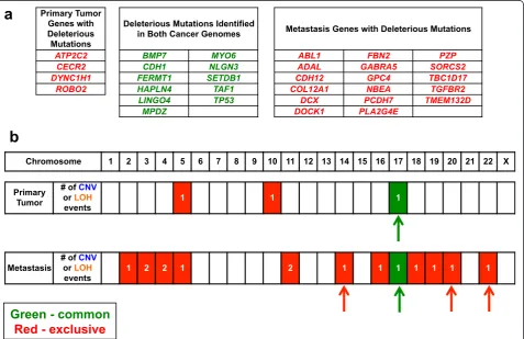
Between the two tumors, we independently validated a total of 77 mutations that occurred within or proximal to exons (Additional file 1: Methods and Table S5). Vali-dated genetic aberrations included: (1) non-synonymous mutations, (2) synonymous mutations, (3) insertions, or (4) deletions. With the targeted sequencing data, we de-termined the mutation allelic frequency (MAF) between the primary tumor and metastasis for each mutation. This involves determining the fraction of a sequence read with a mutation in comparison to the reference se-quence reads. We were able to identify which mutations were common or exclusive to the primary tumor versus the metastasis. Among the 77 validated mutations, the distribution was such that mutations were generally unique either to the primary tumor or metastatic site. For example, the primary tumor had eight mutations that were not present in the metastasis while the metas-tasis had 37 mutations not present in the primary tumor. Common to both cancers were 32 mutations.
Nadauldet al. Genome Biology2014,15:428 Page 3 of 17
Given the interval of three years prior to the detection of the metastasis, there is a possibility that the metastasis-specific mutations occurred independently from the pri-mary tumor. Mutations specific to the pripri-mary tumor that were not present in the ovarian metastasis may have been the result of random genetic drift. The mutations com-mon to both indicate a comcom-mon origin but the exact tim-ing of the differentiation between the two tumors is less clear as noted by those mutations with lower MAF. A sub-set of these genes had high MAF values, indicating a higher likelihood of being present in all clonal populations in the primary tumor or metastasis. As we describe later, these genes were prioritized for further experimental test-ing in gastric organoids.
Mutations affecting gene function
Among the mutations that were externally validated, we focused on the subset of mutations leading to amino acid substitutions, premature stop codons and indels that
altered the open reading frame. Subsequently, we deter-mined if these coding mutations were potentially deleteri-ous to gene function using a number of prediction algorithms such as Polyphen [16] and SIFT [17] among others. Based on the MAF information for each mutation, we determined whether these mutations with a possible deleterious impact on the gene products were common or exclusive to the primary tumor and metastasis (Figure 2).
[image:5.595.61.539.325.634.2]On the subset of deleterious mutations, we conducted additional biological pathway analysis, literature review and comparison against the Cancer Genome Atlas data available for diffuse gastric cancer. This identified a set of known cancer genes and likely cancer-related candi-dates with mutations that likely had an impact on pro-tein function. We focused on a number of candidate driver genes (Table 1) that had previously been demon-strated to have oncogenic potential or were known tumor suppressors with biallelic changes present in the cancer genomes.
Table 1 Cancer oncogenes with amplifications or cancer drivers with biallelic events
Origin Known or candidate
cancer driver
Biallelic event
Allelic alteration 1
Mutation or genomic aberration
Chr Chr position or interval Allelic alteration 2
Unique to the primary FGFR2* Amplification 6-fold amplification 10 117820033 - 119748751
Common to the primary tumor and metastasis
CDH1 Yes Deletion Partial deletion of exon 9 16 68847326 - 68847403 Germline mutation inCDH1
TP53 Yes 5’splice site mutation
Aberrant splicing 17 7578370 Hemizygous loss of 17p arm
Unique to the metastasis TGFBR2 Yes Frameshift indel Stop codon in exon 4 3 30691871 Hemizygous deletion of wild-type TGFBR2locus
PCDH7 Yes Missense S87R 4 30723305 Hemizygous deletion of wild-type 4 arm
FERMT1loci Yes Loss of heterozygosity
FERMT1 located in 20p12.3 20 FERMT1 mutation
BMP7loci Yes Loss of heterozygosity
BMP7located in 20q13.3 20 BMP7mutation
Chr: chromosome.
Nadauld
et
al.
Genome
Biology
2014,
15
:428
Page
5
o
f
1
7
http://geno
mebiology
.com/2014/
Copy number variations and allelic imbalances distinguishing the primary tumor from the metastasis We noted larger scale genomic aberrations that differenti-ated the primary from the metastasis (Figures 2b and 3a). This included copy number changes and LOH events. Unique to the primary tumor were two genomic amplifi-cations on chromosomes 5 and 10, and two inversions on chromosomes 15 and 16 (Additional file 1: Table S4). The chromosome 10 amplification covered a 1.66 Mb interval. When considering deletions or allelic imbalances, the only major event that was noted involved a loss of the p arm of chromosome 17.
In contrast to the primary tumor, the metastatic tumor had numerous chromosomal scale LOH events and gen-omic deletions affecting 12 different chromosomes, the
majority of which were unique to the metastatic tumor (Figure 2). This included multiple deletions and copy neutral LOH events that are detailed in Additional file 1: Table S3. There was a five-fold genomic amplification in chromosome 2 but no specific known genes existed in the affected interval. There were no detectable inter-chromosomal translocations in either the primary tumor or metastasis genomes. Other cancer rearrangements were identified but did not point towards aberrations in any candidate driver genes (Additional file 1: Table S4). There were indications of large scale genomic instability based upon allelic imbalance analysis; chromosomes 14, 17, 20, and 22 all involved the entire chromosome.
[image:7.595.60.539.290.634.2]For copy number aberrations and allelic imbalances, we identified exclusive versus common events between the
primary tumor and metastasis. The only common genetic aberration involved the p arm of chromosome 17. Overall, the lack of overlap was indicative of significant genetic di-vergence from the primary tumor and metastasis despite a common origin as denoted by shared mutations in critical tumor suppressors.
The genomic intervals of the LOH, copy number ab-erration and rearrangement events were compared with the position of validated gene mutations. This inte-grated analysis pointed to a number of genes that had biallelic alterations involving both a loss of the wild-type allele from a large interval genomic aberration and a mutant allele. The results for genes with biallelic hits were considered to be strong candidates for a loss-of-function involvement in cancer (Table 1).
Identification of cancer drivers common to the primary tumor and metastasis
Both the primary and the metastasis contained cancer driver events that were likely to be critical for tumori-genesis in the context of the initial CDH1 mutation (Table 1, Figure 2). In addition to the germline CDH1 intronic mutation, the second CDH1 allele had a som-atic 77 bp genomic deletion of a portion of exon 9 that affects the downstream coding regions as well. TheCDH1 somatic mutation was identical in both the primary and metastatic gastric cancer genomes, dem-onstrating a common genetic origin and providing strong genetic evidence that this driver had a critical role in diffuse gastric tumorigenesis. Mutations affect-ing CDH1 exon 9 that lead to loss of protein expres-sion have frequently been detected in diffuse gastric cancer [18-20]. This exon’s amino acid sequence is a putative calcium-binding site that is likely important for receptor function.
The primary and metastatic tumor also shared biallelic splice donor site mutation (c.559 + 1G > A) of the fifth in-tron ofTP53and a chromosome 17p LOH event encom-passing the TP53 locus (Additional file 1: Figure S1). The TP53 splicing mutation interrupts RNA splicing [21] and is a previously reported cancer mutation [22,23]. The analyses of sporadic and inherited gastric cancers have identifiedTP53mutations that occur concurrently withCDH1mutation [24,25].CDH1inactivation in gas-tric parietal cells does not induce gasgas-tric carcinoma, suggesting that loss of CDH1 is insufficient for tumor initiation [26]. However, double conditional knockout of CDH1andTP53induces development of diffuse gastric carcinoma [26]. Interestingly, the genomic interval of the LOH event affecting the TP53 locus was larger in the metastasis compared to the primary tumor. This could have occurred because of independent genomic instability events given the strong selection for biallelic loss of TP53 function.
FGFR2is an actionable cancer driver exclusive to the primary gastric tumor
In the primary tumor, there was a six-fold genomic amp-lification of a region of chromosome 10 q arm and cov-ered an interval of 1.66 Mb. Within this genomic regions was an oncogenic candidate driver FGFR2 also referred to as the fibroblast growth factor receptor 2 (Figure 3c). This was confirmed with multiple methods including sequencing, array analysis, and validation by quantitative PCR. FGFR2 is a transmembrane receptor that acts as part of a key signal transduction pathway regulating tissue repair and embryonic development among a host of other functions [26].
To validate the prevalence of FGFR2 amplification in diffuse versus intestinal gastric cancers, we analyzed 37 diffuse and 27 intestinal subtype primary gastric tumor samples with digital PCR [27]. Previously, we demon-strated that this method is profoundly sensitive for de-tecting copy number aberration even in the context of normal diploid DNA diluting tumor DNA. Our study demonstrated FGFR2 amplification in four of 37 (11%) diffuse tumor samples, which was absent in the intes-tinal subtype samples (Figure 4a).
In support of its role as a candidate driver, FGFR2 amplification is present in a number of gastric cancer cell lines [28,29] and subsequently reported in various gastrointestinal malignancies such as esophageal adeno-carcinoma [30]. In addition, treatment of cancer cell lines with FGFR2-specific small molecule inhibitors or shRNAs leads to potent growth inhibition [28] suggest-ing a functional role forFGFR2amplification in the dif-fuse subtype.
Functional analysis of theFGFR2driver in combination withCDH1andTP53
We identified two examples of a primary diffuse gastric cancer with co-occurrence of known and putative cancer drivers involvingCDH1,TP53, andFGFR2as seen in the index patient. The first example included a diffuse gastric cancer sample that was among the gastric adenocarcin-omas analyzed by TCGA. Using the cBio TCGA portal [10], we identified a patient (TCGA-BR-6803) who had a similar complement of genetic aberrations in CDH1, TP53, and FGFR2, all of which have been previously de-scribed in cancer as seen in the COSMIC cancer mutation repository. This included the following: a missense muta-tion in CDH1 (D254Y) that has been described in three other cancers; a missense mutation (L130F) in TP53 where mutations in this codon have been reported in 37 other cancers; the FGFR2 amplification which we and others have identified in diffuse gastric cancer.
As the second example, we identified a human diffuse gastric cancer cell line, KatoIII, which has a similar com-position of genetic aberrations affecting the same cancer
Nadauldet al. Genome Biology2014,15:428 Page 7 of 17
genes as the primary tumor of our index patient. KatoIII has a CDH1 mutation leading to an intronic sequence insertion in the mRNA [31,32], aTP53mutation leading to a complete gene deletion [33] and theFGFR2 amplifi-cation [29] (Figure 4b). This cell line allowed us to assess the potential oncogenic role of the FGFR2 amplification in the specific genetic context ofCDH1andTP53 muta-tions, similar to the index patient’s primary tumor.
To determine the contribution of FGFR signaling to neo-plastic growth, we treated KatoIII cells with several FGFR2 small molecule tyrosine-kinase inhibitors (TKIs), including Brivanib, TKI258, Ponatinib, and AZD4547 [34]. As a con-trol, we used the gastric cancer cell line AGS which is wild type for FGFR2, CDH1, and TP53, but has mutations in KRASandPIK3CA [35] (Figure 4b). All FGFR2 inhibitors induced cell death in KatoIII but not AGS cells (Figure 4c
and d). The most potent of these TKIs, AZD4547, has an IC50of approximately 2 nM in KatoIII cells and 39,580 nM
in AGS cells (Figure 4c and d). Each of the inhibitors dem-onstrated a statistically significant lower IC50 in
FGFR2-amplified KatoIII cells compared to non-FGFR2-FGFR2-amplified AGS cells at all concentrations tested (Figure 4c and d).
In contrast, treatment of KatoIII and AGS cells with cytotoxic chemotherapeutic agents such as paclitaxel, 5-fluorouracil and carboplatin did not have a significant effect on either KatoIII or AGS lines, with similar IC50identified
[image:9.595.60.538.88.425.2]in each (Additional file 1: Table S7). For AZD4547, the 20,000-fold difference in sensitivity to FGFR inhibitors suggests that FGF signaling is a critical driver toCDH1 -ini-tiated gastric cellular proliferation and this TKI represents a potential targeted therapy in diffuse subtype cancers har-boringFGFR2amplifications.
Figure 4Prevalence ofFGFR2in human gastric tumors and its contribution to cellular proliferation. (a)Sporadic gastric cancer samples were evaluated by quantitative digital PCR to determine FGFR2 genomic copy number. Black dots represent diffuse gastric cancers. Red dots indicate the intestinal subtype of gastric cancer.(b)Genetic characteristics of the AGS (FGFR2diploid) and KatoIII (FGFR2 amplified) gastric cancer cell lines are shown.(c)Percent survival for the AGS cancer cell line is shown with FGFR2 inhibitors of varying specificity.(d)The KatoIII diffuse gastric cancer cell line was treated with FGFR2 inhibitors of varying specificity. The Y-axis depicts percent survivalversusthe X-axis with log concentrations. In all panels, error bars represent standard error of the mean. The difference in percent cell survival between KatoIII and AGS cells was statistically significant (P<0.05) at the three highest concentrations of all drugs, except Brivanib which was only significant at the
Biallelic inactivation ofTGFBR2is exclusive to the ovarian metastasis
Genetic divergence was evident; the metastasis harbored its own unique subset of mutations and genomic aberra-tions. As we described, the metastasis had the same CDH1 and TP53 mutations as the primary tumor but lacked the FGFR2 amplification found in the primary cancer site (Figure 3c). To eliminate the possibility that the absence ofFGFR2amplification was related to a sub-population not present in our original metastatic section, we performed a highly sensitive quantitative digital PCR on a separate geographic region from the metastasis (data not shown). This method has been previously been demonstrated to identifyFGFR2copy number amplifica-tions with high sensitivity and specificity, even in the context of diluted mixtures [27]. This independent ana-lysis again confirmed that the FGFR2 locus was not amplified in a separate region of the metastatic tumor.
The most striking event uniquely defining the metastasis was aTGFBR2deletion in exon 3 (Table 1). We looked for the presence of this somatic mutation among the normal and primary tumor sequence from the independent data-sets (for example, whole genome, exome, and deep tar-geted resequencing). The MAF of the mutation among all of these sequencing datasets indicated exclusivity specific to the ovarian metastasis (Additional file 1: Table S5). The mutation was not found in any significant fraction among the primary and normal genomes.
TGFBR2 encodes a receptor for the transforming growth factorβ(TGF-β) pathway. WhileTGFBR2is mu-tated in numerous human cancers with particular preva-lence in mismatch repair-deficient colon cancer [36], its functional relevance in gastric cancer is unknown. This particular deletion markedly reduces mRNA levels, pre-sumably due to nonsense-mediated decay [37]. The me-tastasis also harbored a unique large genomic deletion of chromosome arm 3p encompassing the TGFBR2 locus as shown by both CNV and LOH events, resulting in biallelic events affecting the wildtype TGFBR2 alleles (Figure 3b).
TGFBR2 exon 3 deletions are typically associated with colorectal tumors displaying microsatellite instability (MSI), a molecular marker for the loss of DNA mismatch repair (MMR). We assessed the primary and metastatic tumor for DNA mismatch repair defects. The primary tumor had normal immunohistochemical staining for the major DNA mismatch repair proteins MLH1, MSH2, PMS2, and MSH6. Neither the primary tumor nor metas-tasis exhibited elevated MSI at any of the diagnostic gen-etic markers (Additional file 1: Table S7). In addition, the patient had no germline, primary tumor or metastatic somatic mutations in the MMR genes.
We examined the cBIO TCGA dataset for gastric can-cers classified by the Lauren histopathologic criteria as
diffuse. Among the TCGA set, three of 79 diffuse gastric tumor samples had mutations inTGFBR2. This included two cancers in which there was biallelic loss of the wild-type allele [10]. These samples were MSI stable. The dif-fuse subtype samples withTGFBR2mutations include: a homozygous deletion (TCGA-BR-A4QM); biallelic mu-tations involving F442S and A426V in (TCGA-D7-6522); Q418 splice site mutation (TCGA-CD-8531). The exam-ples of TGFBR2 mutations existing in diffuse gastric cancers are supportive evidence for the potential role of TGFBR2as a driver.
Other candidate cancer genes delineating the metastasis from the primary tumor
Additional candidate cancer genes were identified that distinguished the metastasis from the primary gastric tumor (Table 1). A novel predicted pathogenic mutation in BMP7 was identified in the primary and metastatic tumor but the metastatic tumor had a unique copy neu-tral loss of heterozygosity event encompassing the entire chromosome arm 20 q including theBMP7locus. BMP7 (that is, bone morphogenic protein) interacts with the TGF-βpathway and has a well-studied role in osteoclast differentiation and bone development [38]. In addition, BMP7 expression has been correlated with tumor recur-rence in gastric cancer [39].
Similarly, a novelDOCK1mutation was uniquely identi-fied in the metastatic genome. DOCK1 regulates cell mo-tility and migration and has been implicated in ovarian cancer tumorigenesis [40] (Additional file 1: Table S5). Another genomic amplification unique to the primary tumor occurred in the 5q22.3 locus (Additional file 1: Table S3). Among the 15 genes within the amplification locus, the major oncogenic-related cancer gene was TRIM36 that is overexpressed in prostate cancer. It has been hypothesized its overexpression leads to chromo-somal instability [41,42].
TGFBR2knockdown in the context ofCDH1andTP53is sufficient to induce metastatic diffuse gastric cancer in a primary gastric organoid murine model
Given the metastasis-specific, biallelic alteration of TGFBR2, we exploited our validated primary air-liquid interface murine gastric organoid system [12,13] to in-vestigate ifTGFBR2knockdown was sufficient to induce gastric cancer metastasis. Its consideration as a candi-date was also suggested by the TCGA data. Previously, we observed that Trp53 deletion and KrasG12D induced pronouncedin vitrodysplasia and invasion of gastric orga-noids within vivotumorigenicity upon subcutaneous im-plantation, but spontaneous metastasis was not seen by 50 days [13].
Since both the primary and metastasis shared common CDH1 and TP53 mutations, primary gastric organoids
Nadauldet al. Genome Biology2014,15:428 Page 9 of 17
were established from Cdh1fl/fl;Trp53fl/flneonatal mouse stomach. Gastric organoid infection with a control adeno-virus (Ad Fc) encoding an immunoglobulin Fc fragment [43] resulted in gastric organoids with wild-type Cdh1 and Tp53, while adenovirus Cre-green fluorescent protein (Ad Cre-GFP) induced deletion of the floxedCdh1andTrp53 alleles, with Cdh1 and Trp53 loss confirmed by immuno-fluorescence (Figure 5a and Additional file 1: Figure S2), accurately modeling theCdh1andTrp53loss common to both the primary and metastatic tumors. As we previously reported [12,13], Ad Fc-treated organoids with wild-type Cdh1/Trp53contained epithelial and mesenchymal com-ponents, accurately recapitulating in vivo stomach tissue architecture (Figure 5a and 5d).
[image:11.595.58.542.277.635.2]To model the effect of the TGFBR2 in metastatic oncogenesis, we infected the sameCdh1-/-;Trp53-/- gas-tric organoids with retrovirus expressing shRNA against Tgfbr2, confirmingTgfbr2knockdown by immunofluor-escence and Western blot analysis (Figure 5a and b). Likewise, gene expression of Tgfbr2 was also reduced as determined by real time PCR (Additional file 1: Figure S3). TheTgfbr2 shRNA did not grossly increase the growth rate of Cdh1-/-;Trp53-/- gastric organoids over a 20-day period, possibly because of dominant ef-fects of theCdh1andTrp53deletions (Figure 5c). How-ever, histologic analysis revealed that the resultant Cdh1-/-;Trp53-/-; Tgfbr2 shRNA gastric organoids but notCdh1-/-; Trp53-/- controls demonstrated features of
Figure 5Dysplastic epithelium in gastric organoids. (a)Gastric organoid cultures were made from gastric tissue of neonatal mice harboring
Cdh1andTrp53floxed alleles then subsequently infected with Fc-expressing adenovirus, or CreGFP-expressing adenovirus +/- retrovirus
expressing shRNA againstTgfbr2. Images indicate immunofluorescence with nuclear DAPI staining and antibodies against CDH1, TGFBR2, or PCNA. Intrinsic GFP fluorescence from adenovirus CreGFP was abrogated by tissue passaging and subsequent formaldehyde fixation, and is not visible in immunofixation experiments.(b)GSM-06 murine gastric epithelial cells were infected with scrambled shRNA or an shRNA against murineTgfbr2
diffuse subtype gastric cancer. Severe dysplasia along with focal areas of invasion, signet ring formation, and nuclear pleomorphism were found throughout the ana-lyzed organoids (Figure 5d).
To examine potentialTgfbr2effects on in vivo metasta-sis, the Cdh1-/-;Trp53-/-;Tgfbr2 shRNA organoids versus Cdh1-/-;Trp53-/- controls were disaggregated and injected subcutaneously into immunodeficient NOG mice.Cdh1-/-; Trp53-/-organoids produced extremely slow but detectable tumor growth by day 50 as we previously documented [13] (Figure 6a and b). In contrast,Cdh1-/-;Trp53-/-;Tgfbr2 shRNAgastric organoids exhibited robustin vivo tumori-genicity (Figure 6a and c). Notably,Cdh1-/-;Trp53-/-;Tgfbr2 shRNA primary tumors exhibited a poorly differentiated adenocarcinoma histology with signet ring features as oc-curs in diffuse gastric cancer (Figure 6e to g). Immuno-fluorescence analysis confirmed loss of Cdh1 and Tgfbr2 knockdown (Figure 5a).
Evaluation for distant disease confirmed the presence of pulmonary metastases in NOG mice harboring Cdh1-/-;Trp53-/-;Tgfbr2 shRNA tumors, comprised of poorly differentiated adenocarcinoma with signet ring features (Figure 6f, g). Metastatic tumors were located in the lungs bilaterally, were grossly observable upon
dissection and had similar histologic appearance to diffuse gastric cancer. Overall, these studies support the role of Tgfbr2 as a putative tumor suppressor gene in diffuse gastric cancer, demonstrate successful in vitro conversion of primary gastric tissue to metastatic adenocarcinoma, and reveal the utility of a primary gas-tric organoid system for functional validation of candi-date metastasis drivers.
Discussion
To address the question of identifying the genetic drivers of diffuse gastric cancer metastasis, we performed an ex-tensive genome sequencing analysis of the metastatic evo-lutionary process. This involved sequencing of a matched gastric primary and subsequent metastasis from the same patient. We leveraged the unique Mendelian genetics of a HDCG proband as an‘experiment of nature’to delineate essential cancer drivers in diffuse gastric cancer.
Our genomic analysis revealed FGFR2 amplification exclusive to the primary gastric tumor and not present in the metastasis. Our results fully confirm several de-scriptions of FGFR2 amplification, as well as increased sensitivity of FGFR2-amplification positive cell lines such as KatoIII to small molecule FGFR inhibitors [44-47],
b
c
Cdh1-/-; Trp53-/-
Cdh1-/-; Trp53-/- + Tgfbr2 shRNA
Cdh1-/-; Trp53-/-+ Tgfbr2 shRNA
a
T
umor volume (mm
3)
Days after transplantation
0 500 1000 1500 2000 2500
0 10 20 30 40 50
Cdh1-/-; Trp53-/-
Cdh1-/-; Trp53
-/-+ Tgfbr2 shRNA
Cdh1-/-; Trp53-/-+ Tgfbr2 shRNA
s.c. primary tumor Spontaneous lung metastasis
s.c. primary tumor *
*
e
f
g
[image:12.595.57.540.390.647.2]d
Figure 6Gastric organoid tumor explants.Gastric organoids with the indicated genotypes are shown.(a)Tumor volumes were measured over time post-injection and plotted according to genotype of the driver combinations being tested. This includesCdh1-/-;Trp53-/-as shown in blue and
Cdh1-/-;Trp53-/-;Tgfbr2 shRNAas shown in red. Error bars represent SEM. Asterisk (*) indicatesP<0.01 forCdh1-/-;Trp53-/-;Tgfbr2shRNA compared to
Cdh1-/-;Trp53-/-tumor volumes.(b, c)With different driver combinations, transformed gastric organoids were dissociated and subcutaneously (s.c.)
injected into the flanks of immunodeficient NOG mice. Images indicated tumor growth at 30 days post injection.(d, e)Histological analysis of tumors confirms the presence of poorly differentiated adenocarcinoma with signet ring features, as indicated by the yellow arrows, only in theCdh1-/-;Trp53-/-;
Tgfbr2 shRNAorganoids. After flank injections with dissociated organoids, histological analysis of murine lungs after 30 days revealed metastatic gastric adenocarcinoma with signet ring features at low(f)and high(g)magnification.
Nadauldet al. Genome Biology2014,15:428 Page 11 of 17
with accompanying implications for FGFR2-targeted treatment [48]. The striking absence of FGFR2 amplifi-cation in the metastasis in the context of the common somatic CDH1 and TP53 mutations argues strongly for a tumor evolutionary divergence.
The functional validation of the metastatic potential of Tgfbr2 knockdown in our well-validated air-liquid interface gastric organoid method [12,13] provides the first demonstration that TGFBR2 functions as a bona fide metastasis suppressor gene in diffuse gastric cancer. HomozygousTGFBR2deletion is also present in a sub-set of TCGA gastric cancers [10] which are largely com-prised of non-metastatic tumors. It will be interesting to evaluate whether TGFBR2alterations are more prevalent in gastric metastases, such as to ovary or other sites. Fur-thermore, the functional relevance of other potential loci undergoing alteration in these samples merits additional exploration.
Our study also describes the first successfulin vitro con-version of primary gastric tissue to metastatic gastric adenocarcinoma, suggesting the general applicability of the organoid method to the functional validation of gastric cancer loci involved in progression and/or metastasis. As shown here, such three-dimensional organoid-based functional validation strategies can potentially combine both the experimental tractability of two-dimensional culture of transformed cell lines with the accurate tissue ultrastructure and stromal components of transgenic mouse systems.
Conclusions
Exclusive FGFR2 and TGFBR2 genetic aberrations delin-eated the evolution of metastatic recurrence. Our finding may have implications for targeted cancer therapy. For ex-ample, the index patient in this study may have conceiv-ably responded to a FGFR2 inhibitor based on FGFR2 amplification in the primary tumor. However, the patient’s metastatic recurrence did not harbor this same FGFR2 amplification, suggesting that treatment with a therapeutic small molecule inhibitor may not have had a discernible biological effect on the patient’s metastatic disease. In the precision management of individuals with metastatic can-cer, one may need to account for the genetic heterogeneity and subsequent variation in cancer biology that differenti-ates metastatic sites from the primary tumor before the initiation of targeted therapy.
Although a comprehensive knowledge of the meta-static process is crucial for improved cancer treatment, the driver events underlying metastatic spread are unfor-tunately poorly understood [49]. Our paucity of know-ledge regarding metastasis is further compounded by the relative omission of metastatic samples in large-scale genomic cancer surveys such as TCGA. This study is an initial effort to further address these questions about the
genetics and biology of metastatic evolution by integrat-ing genomic sequencintegrat-ing analysis within vitro validation of clonal-specific candidate drivers.
Materials and methods Cancer samples
This study was conducted in compliance with the Helsinki Declaration. The institutional review board (IRB) at Stanford University School of Medicine approved the study protocols (11886 and 19071). For all patients cited in this study, we obtained informed consent to conduct research and publish the results. Samples were obtained from the Stanford Cancer Institute Tissue Bank. Frozen tissue sec-tions were prepared from each tumor and hematoxylin-eosin (H&E) staining was performed on a single section. We estimated overall tumor composition that generally was approximately 50% or greater for most samples. Tumors were macro-dissected to increase tumor cellular-ity and processed for genomic DNA. Full details are in the Additional file 1: Methods.
Sample preparation for whole genome, exome, and targeted resequencing analysis
Genomic DNA was extracted from blood, normal gastric tissue and tumor samples using the E.Z.N.A. SQ DNA/ RNA Protein Kit (Omega Bio-Tek, Norcross, GA, USA). Concentrations of genomic DNA were determined with a Nanodrop instrument (Thermo Scientific, Wilmington, DE, USA). Genomic DNA from matched normal and cancer tissue were then used for creating sequencing li-braries. DNA from peripheral leukoctyes was used for the Affymetrix SNP array.
Cancer genome sequencing
See Additional file 1 for complete details regarding the whole genome, exome, and targeted resequencing data analysis. This includes information about the targeted resequencing process, variant calling, allelic frequency determination, and mutation interpretation The oligo-nucleotide sequences for deep targeted resequencing are listed in Additional file 2.
FGFR2amplification analysis from diffuse and intestinal gastric cancers
Quantitative PCR was performed using the Bio-Rad QX100 droplet digital PCR (ddPCR) system (Bio-Rad, Pleasanton, CA, USA). We used a standard set ofFGFR2 -specific TaqMan primers and probes (Life Technology, Foster City, CA, USA) compared with standard references using an ultra-conserved region on chromosome 1. Briefly, TaqMan PCR reaction mixtures were assembled using 2× ddPCR Supermix for probes, 20× assays (18μM primers and 5 μM probe) and restriction digested DNA samples (Biorad). To assessFGFR2copy number, 125 ng of each tumor DNA sample was digested with 1.25 units of BsaJI (NEB) in 15μL for 1 h at 60°C. The digests were diluted 1.67-fold to 25 μL with nuclease free water then 25 ng (5 μL) was assayed per 20 μL ddPCR reaction. FGFR2 assay sequences were (forward primer) 5’-GGCT GGCTGCTGAAGTCT-3’, (reverse primer) 5’-CTTAATC GCCTGTATGGTGGTAACA-3’, and (probe) 5’-FAM-TC
TTGGTCGTGTTCTTCATTCGGCACAG-BHQ1-3’. The
FGFR2 assay was duplexed with a standard reference se-quence on Chromosome 1. This standard reference assay used the following primers: (forward primer) 5’-TGAGG GATTCGGCAGATGTTG-3’, (reverse primer) 5’-CTGAA AGGCTGGACTTGACAGA-3’, and (probe) 5’-VIC-ACT GTGTGCTGGACCT-MGB-3’. All assay primers were ordered from Integrated DNA Technologies. Thermal cycling conditions were 95°C 10 min (1 cycle), 94°C 30 s and 60°C 60 s (40 cycles), 98°C 10 min (1 cycle), and a 12°C hold. FGFR2copy number per cell was estimated as the ratio of the FGFR2 and RPP30 concentrations multiplied by two to account for the two copies of RPP30 that are expected per diploid genome. Analysis of the ddPCR data was performed using the CNV mode of the QX100 analysis software (version 1.2.9.0). Quad-ruplicate ddPCR wells were analyzed for each sample.
FGFR2 inhibitor sensitivity assay
KatoIII cells (HTB-103, ATCC) and AGS cells (CRL-1739, ATCC) were grown in Dulbecco’s Modified Eagle Medium (DMEM), supplemented with 10% fetal bovine serum and 100 U/mL of Pen Strep Glutamine (Gibco). All cells were cultured at 37°C in a humidified atmosphere and 5% CO2. Survival of KatoIII and AGS cells was determined
using the WST-1 Proliferation Assay (Roche). We tested
multiple FGFR inhibitors including TKI-258, Brivanib (BMS-540215), Ponatnib (AP24534), and AZD4547 (Sell-eck Chemical). Cells were seeded at a density of 2 × 10⁴ cells/well in 96-well microtiter plates, 100μL medium/well and maintained 18 h for attachment. Afterwards, we treated the cultures with varying concentrations of each drug diluted in DMSO. After 30 h incubation, 10 μL of WST-1 reagent was added followed by 1 h at 37°C. The cleavage of tetrazolium salt (WST-1) into a visible forma-zan by viable cells was spectrophotometrically measured using a reference wavelength of 450 nm. Each test was per-formed in triplicate. Percentages of cell survival were cal-culated as follows:% cell survival = (absorbance of treated cells/ absorbance of cells with vehicle solvent) × 100. The half inhibitory concentration (IC50) was calculated with a
non-linear regression from the dose–response curve.
Mismatch repair protein immunohistochemistry
Mismatch repair protein immunohistochemistry was performed on the primary diffuse gastric tumor using the standard streptavidin-biotin-peroxidase procedure. Primary monoclonal antibodies against MLH1 (clone G168-728, 1:200, BD PharMingen, San Diego, CA, USA 1:200), MSH2 (clone FE11, 1:100, Oncogene Research Products, Cambridge, MA, USA), MSH6 (clone 44, 1:200, BD Transduction, San Jose, CA, USA) and PMS2 (clone MRQ-28, 1:10, Cell Marque, Rocklin, CA, USA) were ap-plied to formalin-fixed, paraffin embedded sections four microns thick. The sections were deparaffinized in xylene, and rehydrated through graded alcohols to distilled water before undergoing antigen retrieval by heat treatment in either citrate solution pH 6.0 (MLH1, PMS2, and MSH2) or EDTA solution pH 9.0 (MSH6). An automated de-tection using a Leica Bond Autostainer (Leica, Buffalo Groove, IL, USA) was employed. Normal expression was defined as nuclear staining within tumor cells, using infil-trating lymphocytes as positive internal control. Negative protein expression was defined as complete absence of nu-clear staining within tumor cells in the face of concurrent positive labeling in internal non-neoplastic tissues.
Gastric organoid cancer modeling in mice and functional analysis
All procedure involving animal were approved the Stanford University Administrative Panel on Laboratory Animal Care and was fully compliant with the USDA Animal Wel-fare Act, and our Assurance of Compliance with the PHS Policy on Human Care and Use of Laboratory Animals. Air-liquid interface organoid culture was performed as de-scribed [11,12].
Cdh1flox/flox;Trp53flox/flox mice were generated by
crossing Cdh1flox/flox mice, obtained from Jackson La-boratory, andTrp53flox/floxmice, kindly provided by Dr. Anton Berns [50] NOD.Cg-Prkdcscid Il2rgtm1Sug/JicTac
Nadauldet al. Genome Biology2014,15:428 Page 13 of 17
mice were obtained from Taconic Farms, Inc. We dis-sected stomachs from neonatal mice (age P1-10) and washed them in cold PBS to remove all luminal con-tents. We extensively minced either large 25% sections or any entire neonatal stomach per dish and embedded the minced tissues in a 3D collagen gel using a double-dish air-liquid interface culture system as previously described [11]. To maintain the organoids, we applied fresh medium (F12, 20%FCS, gentamicin 50 ug/mL) every week.
Tgfbr2shRNAs were obtained from Origene (catalog TG516186). Retroviral plasmids were cotransfected with pCL-Eco into 293 T cells by Lipofectamine2000 (Invi-trogen). Retroviral supernatants were collected 48 and 72 h post-transfection and concentrated by PEG-it virus precipitation solution (5×, System Biosciences). Virus titer was determined by infection of NIH3T3 cells and FACS analysis of GFP positive cells 48 h post infection.
Cdh1flox/flox;Trp53flox/flox gastric organoids were
in-fected at day 0 with adenovirus Ad Cre-GFP (University of Iowa Vector Core) or control adenovirus Ad Fc [43] encoding a mouse immunoglobulin IgG2α Fc fragment by layering viral particles (109pfu) suspended in 500μL culture media over the top of the collagen matrix con-taining primary tissue. For retrovirus infection of sec-ondary organoids, primary organoids at 14 to 20 days of growth were recovered from collagen gel by collagenase IV (Worthington) incubation followed by 0.05% trypsin/ EDTA incubation to dissociate organoids into a single cell suspension. Following extensive washing with 10% FBS to inactivate collagenase/trypsin, cells were pelleted by centrifugation and incubated with retroviral particles (2 μL of 108 pfu/mL) encoding Tgfbr2-shRNA in the presence of growth medium and TransDux (System Biosciences) at room temperature for 60 min before serial replating into 3D collagen gel air-liquid interface culture.
Samples were fixed with 4% paraformaldehyde overnight, paraffin-embedded, sectioned, and sections stained by H&E for initial histology analysis. Further immunohistochemistry analysis, used the following antibodies: PCNA (1:300; Invitrogen), CDH1 (1:300; BD Biosciences Pharmagen), TGFBR2 (1:250; Abbiotec), p53 (1:100; Santa Cruz). Cell ly-sates of mouse gastric culture cell or GSM-06 cells trans-fected withTgfbr2-shRNA-GFP were immunoblotted with TGFBR2 (1:2,000, Abbiotec) andβ-actin (1:2,000, Abcam).
Cells from gastric organoids were collected from the air-liquid interface collagen gel by disaggregation with collagenase IV (Worthington). For transplantation, 400,000 cells per mouse flank were mixed with matrigel (50% Matrigel, 10%FCS, 40% F12, 100 μL of Matrigel mixture per = mouse) and injected into NOD.Cg-Prkdcscid Ilr2rgtm1Sug/JicTac mice. Mice were sacrificed after day 50, after which tumors were dissected and exam-ined by H&E staining.P values were determined using a
two-tailed Student’s t-test assuming unequal variances. A Pvalue of 0.05 was considered significant.
Data availability
The data from this study have been submitted to the NCBI Sequence Read Archive under the accession num-ber SRP044347.
Additional files
Additional file 1:Supplementary Methods and Data. Table S1.
Summary of normal, tumor, and metastasis exome and whole genome sequencing coverage.Table S2.Single nucleotide variant and small indel calling from genome sequencing datasets.Table S3.Summary of the validated cancer copy number variations and loss-of-heterozygosity (LOH) events.Table S4.Summary of the cancer rearrangement events.
Table S5.Validated cancer mutations and analysis of mutant allelic frequency.Table S6.Microsatelllite genetic markers assessed for microsatellite instability.Table S7.Gastric cancer cell line IC50s for different gastric cancer cell lines. The IC50s of cytotoxic chemotherapies (paclitaxel, carboplatin, 5-fluorouracil) or an FGFR2 small molecule tyrosine kinase inhibitor (AZD4547) are listed.Figure S1.Biallelic events in theTP53loci and gene.Figure S2.Deletion ofCdh1andTrp53
expression in gastric organoids.Figure S3.shRNA-mediated knockdown ofTgfbr2demonstrate by real time PCR [16,17,23,51-74].
Additional file 2: Table S8.Mutation targeted sequencing study.
Abbreviations
Ad:Adenovirus; BMP: Bone morphogenic protein; CNV: Copy number variation; ddPCR: Droplet digital PCR; EMT: Epithelial mesenchymal transition;
FGFR2: Fibroblast growth factor receptor 2; GFP: Green fluorescent protein; H&E: Hematoxylin-eosin; HDGC: Hereditary diffuse gastric cancer; indel: Insertion-deletions; IRB: Institutional review board; LOH: Loss of heterozygosity; MAF: Minor allele frequency; Mb: Megabase; SNP: Single nucleotide polymorphism; TCGA: The Cancer Genome Atlas;TGFBR2: Transforming growth factor-βreceptor 2; TKI: tyrosine kinase inhibitor.
Competing interests
Dr. John Regan is an employee of Bio-Rad that manufactures the droplet digital PCR system used in this study.
Authors’contributions
HPJ, LDN, and JMF designed the analysis of the HDGC patient. HX and ESH conducted the sequencing assays. LDN, LM, and JR conducted molecular assays for validation. SG, GN, JMB, PF, CP, HC, NRZ, and HPJ conducted the sequencing data and genetic analysis. RKP provided pathologic review of the gastric cancers and organoids. LDN and LM conducted the cell line and organoid experiments. LDN, AO, and CJK developed the organoid system. CJK supervised the development of cancer organoid modeling. HPJ supervised and coordinated all aspects of the analysis and experiments. All authors revised, read, and approved the final manuscript.
Acknowledgements
Author details 1
Division of Oncology, Department of Medicine, Stanford University School of Medicine, CCSR 1115, 269 Campus Drive, Stanford, CA 94305-5151, USA.
2
Stanford Genome Technology Center, Stanford University, Palo Alto, CA 94304, USA.3Department of Pathology, University of Pittsburgh Medical
Center, Pittsburgh, PA 15213, USA.4Bio-Rad, Inc, Pleasanton, CA 94566, USA.
5Department of Statistics, Stanford University, Stanford, CA 94305, USA. 6
Division of Hematology, Department of Medicine, Stanford University School of Medicine, CCSR 1155, 269 Campus Drive, Stanford, CA 94305-5151, USA.7Department of Statistics, The Wharton School, University of
Pennsylvania, Philadelphia, PA 19104, USA.
Received: 29 April 2014 Accepted: 27 August 2014 Published: 27 August 2014
References
1. Lauren P:Histogenesis of intestinal and diffuse types of gastric carcinoma.Scand J Gastroenterol Suppl1991,180:160–164.
2. Cunningham D, Allum WH, Stenning SP, Thompson JN, Van de Velde CJ, Nicolson M, Scarffe JH, Lofts FJ, Falk SJ, Iveson TJ, Smith DB, Langley RE, Verma M, Weeden S, Chua YJ, MAGIC Trial Participants:Perioperative chemotherapy versus surgery alone for resectable gastroesophageal cancer.N Engl J Med2006,355:11–20.
3. Lim L, Michael M, Mann GB, Leong T:Adjuvant therapy in gastric cancer.
J Clin Oncol2005,23:6220–6232.
4. Lee YS, Cho YS, Lee GK, Lee S, Kim YW, Jho S, Kim HM, Hong SH, Hwang JA, Kim SY, Hong D, Choi IJ, Kim BC, Kim BC, Kim CH, Choi H, Kim Y, Kim KW, Kong G, Kim HL, Bhak J, Lee SH, Lee JS:Genomic profile analysis of diffuse-type gastric cancers.Genome Biol2014,15:R55.
5. Yamashita K, Sakuramoto S, Watanabe M:Genomic and epigenetic profiles of gastric cancer: potential diagnostic and therapeutic applications.
Surg Today2011,41:24–38.
6. Cisco RM, Ford JM, Norton JA:Hereditary diffuse gastric cancer: implications of genetic testing for screening and prophylactic surgery.
Cancer2008,113:1850–1856.
7. Chen Y, Kingham K, Ford JM, Rosing J, Van Dam J, Jeffrey RB, Longacre TA, Chun N, Kurian A, Norton JA:A prospective study of total gastrectomy for CDH1-positive hereditary diffuse gastric cancer.Ann Surg Oncol2011,
18:2594–2598.
8. Becker KF, Atkinson MJ, Reich U, Becker I, Nekarda H, Siewert JR, Hofler H:
E-cadherin gene mutations provide clues to diffuse type gastric carcinomas.Cancer Res1994,54:3845–3852.
9. Machado JC, Oliveira C, Carvalho R, Soares P, Berx G, Caldas C, Seruca R, Carneiro F, Sobrinho-Simoes M:E-cadherin gene (CDH1) promoter methylation as the second hit in sporadic diffuse gastric carcinoma.
Oncogene2001,20:1525–1528.
10. Cerami E, Gao J, Dogrusoz U, Gross BE, Sumer SO, Aksoy BA, Jacobsen A, Byrne CJ, Heuer ML, Larsson E, Antipin Y, Reva B, Goldberg AP, Sander C, Schultz N:The cBio cancer genomics portal: an open platform for exploring multidimensional cancer genomics data.Cancer Discov2012,
2:401–404.
11. Ootani A, Li X, Sangiorgi E, Ho QT, Ueno H, Toda S, Sugihara H, Fujimoto K, Weissman IL, Capecchi MR, Kuo CJ:Sustained in vitro intestinal epithelial culture within a Wnt-dependent stem cell niche.Nat Med2009,
15:701–706.
12. Katano T, Ootani A, Mizoshita T, Tanida S, Tsukamoto H, Ozeki K, Ebi M, Mori Y, Kataoka H, Kamiya T, Toda S, Joh T:Establishment of a long-term three-dimensional primary culture of mouse glandular stomach epithelial cells within the stem cell niche.Biochem Biophys Res Commun2013,
432:558–563.
13. Li X, Nadauld L, Ootani A, Corney DC, Pai RK, Gevaert O, Cantrell MA, Rack PG, Neal JT, Chan CW, Yeung T, Gong X, Yuan J, Wilhelmy J, Robine S, Attardi LD, Plevritis SK, Hung KE, Chen CZ, Ji HP, Kuo JC:Oncogenic transformation of diverse gastrointestinal tissues in primary organoid culture.Nat Med2014,20:769–777.
14. Kluijt I, Siemerink EJ, Ausems MG, van Os TA, de Jong D, Simoes-Correia J, van Krieken JH, Ligtenberg MJ, Figueiredo J, van Riel E, Sijmons RH, Plukker JT, van Hillegersberg R, Dekker E, Oliveira C, Cats A, Hoogerbrugge N, Dutch Working Group on Hereditary Gastric Cancer:CDH1-related hereditary diffuse gastric cancer syndrome: Clinical variations and implications for counseling.Int J Cancer2012,131:367–376.
15. Hass HG, Smith U, Jager C, Schaffer M, Wellhausser U, Hehr T, Markmann HU, Nehls O, Denzlinger C:Signet ring cell carcinoma of the stomach is significantly associated with poor prognosis and diffuse gastric cancer (Lauren's): single-center experience of 160 cases.Onkologie2011,
34:682–686.
16. Adzhubei IA, Schmidt S, Peshkin L, Ramensky VE, Gerasimova A, Bork P, Kondrashov AS, Sunyaev SR:A method and server for predicting damaging missense mutations.Nat Methods2010,7:248–249. 17. Kumar P, Henikoff S, Ng PC:Predicting the effects of coding
non-synonymous variants on protein function using the SIFT algorithm.
Nat Protoc2009,4:1073–1081.
18. Berx G, Becker KF, Hofler H, van Roy F:Mutations of the human E-cadherin (CDH1) gene.Hum Mutat1998,12:226–237.
19. Gamboa-Dominguez A, Dominguez-Fonseca C, Chavarri-Guerra Y, Vargas R, Reyes-Gutierrez E, Green D, Quintanilla-Martinez L, Luber B, Busch R, Becker KF, Becker I, Hofler H, Fend F:E-cadherin expression in sporadic gastric cancer from Mexico: exon 8 and 9 deletions are infrequent events associated with poor survival.Hum Pathol2005,
36:29–35.
20. Hiraguri S, Godfrey T, Nakamura H, Graff J, Collins C, Shayesteh L, Doggett N, Johnson K, Wheelock M, Herman J, Baylin S, Pinkel D, Gray J:
Mechanisms of inactivation of E-cadherin in breast cancer cell lines.
Cancer Res1998,58:1972–1977.
21. Foti A, Bar-Eli M, Ahuja HG, Cline MJ:A splicing mutation accounts for the lack of p53 gene expression in a CML blast crisis cell line: a novel mechanism of p53 gene inactivation.Br J Haematol1990,
76:143–145.
22. Petitjean A, Achatz MI, Borresen-Dale AL, Hainaut P, Olivier M:TP53 mutations in human cancers: functional selection and impact on cancer prognosis and outcomes.Oncogene2007,26:2157–2165.
23. Forbes SA, Bindal N, Bamford S, Cole C, Kok CY, Beare D, Jia M, Shepherd R, Leung K, Menzies A, Teague JW, Campbell PJ, Stratton MR, Futreal PA:
COSMIC: mining complete cancer genomes in the Catalogue of Somatic Mutations in Cancer.Nucleic Acids Res2011,39:D945–D950.
24. Strickler JG, Zheng J, Shu Q, Burgart LJ, Alberts SR, Shibata D:p53 mutations and microsatellite instability in sporadic gastric cancer: when guardians fail.Cancer Res1994,54:4750–4755.
25. Tamura G, Sakata K, Nishizuka S, Maesawa C, Suzuki Y, Iwaya T, Terashima M, Saito K, Satodate R:Inactivation of the E-cadherin gene in primary gastric carcinomas and gastric carcinoma cell lines.Jpn J Cancer Res1996,
87:1153–1159.
26. Shimada S, Mimata A, Sekine M, Mogushi K, Akiyama Y, Fukamachi H, Jonkers J, Tanaka H, Eishi Y, Yuasa Y:Synergistic tumour suppressor activity of E-cadherin and p53 in a conditional mouse model for metastatic diffuse-type gastric cancer.Gut2012,61:344–353. 27. Nadauld LD, Regan JF, Miotke L, Pai RK, Longacre TA, Kwok SS,
Saxonov S, Ford JM, Ji HP:Quantitative and sensitive detection of cancer genome amplifications from formalin fixed paraffin embedded tumors with droplet digital PCR.Transl Med (Sunnyvale)
2012,2:1–5.
28. Kunii K, Davis L, Gorenstein J, Hatch H, Yashiro M, Di Bacco A, Elbi C, Lutterbach B:FGFR2-amplified gastric cancer cell lines require FGFR2 and Erbb3 signaling for growth and survival.Cancer Res2008,
68:2340–2348.
29. Tsujimoto H, Sugihara H, Hagiwara A, Hattori T:Amplification of growth factor receptor genes and DNA ploidy pattern in the progression of gastric cancer.Virchows Arch1997,431:383–389.
30. Dulak AM, Schumacher SE, van Lieshout J, Imamura Y, Fox C, Shim B, Ramos AH, Saksena G, Baca SC, Baselga J, Tabernero J, Barretina J, Enzinger PC, Corso G, Roviello F, Lin L, Bandla S, Luketich JD, Pennathur A, Meyerson M, Ogino S, Shivdasani RA, Beer DG, Godfrey TE, Beroukhim R, Bass AJ:
Gastrointestinal adenocarcinomas of the esophagus, stomach, and colon exhibit distinct patterns of genome instability and oncogenesis.
Cancer Res2012,72:4383–4393.
31. Bass AJ, Lawrence MS, Brace LE, Ramos AH, Drier Y, Cibulskis K, Sougnez C, Voet D, Saksena G, Sivachenko A, Jing R, Parkin M, Pugh T, Verhakk RG, Stransky N, Boutin AT, Barretina J, Solit DB, Vakiani E, Shao W, Mishina Y, Warmuth M, Jimenez J, Chiang DY, Signoretti S, Kaelin WG, Spardy N, Hahn WC, Hoshida Y, Ogino S,et al:Genomic sequencing of colorectal adenocarcinomas identifies a recurrent VTI1A-TCF7L2 fusion.Nat Genet
2011,43:964–968.
Nadauldet al. Genome Biology2014,15:428 Page 15 of 17
32. Oda T, Kanai Y, Oyama T, Yoshiura K, Shimoyama Y, Birchmeier W, Sugimura T, Hirohashi S:E-cadherin gene mutations in human gastric carcinoma cell lines.Proc Natl Acad Sci U S A1994,91:1858–1862.
33. Yamada Y, Yoshida T, Hayashi K, Sekiya T, Yokota J, Hirohashi S, Nakatani K, Nakano H, Sugimura T, Terada M:p53 gene mutations in gastric cancer metastases and in gastric cancer cell lines derived from metastases.
Cancer Res1991,51:5800–5805.
34. Gavine PR, Mooney L, Kilgour E, Thomas AP, Al-Kadhimi K, Beck S, Rooney C, Coleman T, Baker D, Mellor MJ, Brooks AN, Klinowska T:AZD4547: an orally bioavailable, potent, and selective inhibitor of the fibroblast growth factor receptor tyrosine kinase family.Cancer Res2012,72:2045–2056. 35. Yoon YK, Kim HP, Han SW, Hur HS, Oh do Y, Im SA, Bang YJ, Kim TY:
Combination of EGFR and MEK1/2 inhibitor shows synergistic effects by suppressing EGFR/HER3-dependent AKT activation in human gastric cancer cells.Mol Cancer Ther2009,8:2526–2536.
36. Bellam N, Pasche B:Tgf-beta signaling alterations and colon cancer.
Cancer Treat Res2010,155:85–103.
37. Markowitz S, Wang J, Myeroff L, Parsons R, Sun L, Lutterbaugh J, Fan RS, Zborowska E, Kinzler KW, Vogelstein B:Inactivation of the type II TGF-beta receptor in colon cancer cells with microsatellite instability.Science1995,
268:1336–1338.
38. Itoh F, Asao H, Sugamura K, Heldin CH, ten Dijke P, Itoh S:Promoting bone morphogenetic protein signaling through negative regulation of inhibitory Smads.EMBO J2001,20:4132–4142.
39. Aoki M, Ishigami S, Uenosono Y, Arigami T, Uchikado Y, Kita Y, Kurahara H, Matsumoto M, Ueno S, Natsugoe S:Expression of BMP-7 in human gastric cancer and its clinical significance.Br J Cancer2011,104:714–718. 40. Wang H, Linghu H, Wang J, Che YL, Xiang TX, Tang WX, Yao ZW:The role
of Crk/Dock180/Rac1 pathway in the malignant behavior of human ovarian cancer cell SKOV3.Tumour Biol2010,31:59–67.
41. Balint I, Muller A, Nagy A, Kovacs G:Cloning and characterisation of the RBCC728/TRIM36 zinc-binding protein from the tumor suppressor gene region at chromosome 5q22.3.Gene2004,332:45–50.
42. Miyajima N, Maruyama S, Nonomura K, Hatakeyama S:TRIM36 interacts with the kinetochore protein CENP-H and delays cell cycle progression.
Biochem Biophys Res Commun2009,381:383–387.
43. Wei K, Piecewicz SM, McGinnis LM, Taniguchi CM, Wiegand SJ, Anderson K, Chan CW, Mulligan KX, Kuo D, Yuan J, Vallon M, Morton L, Lefai E, Simon MC, Maher JJ, Mithieux G, Rajas F, Annes J, McGuinness OP, Thurston G, Giaccia AJ, Kuo CJ:A liver Hif-2alpha-Irs2 pathway sensitizes hepatic insulin signaling and is modulated by Vegf inhibition.Nat Med2013,
19:1331–1337.
44. Nakatani H, Sakamoto H, Yoshida T, Yokota J, Tahara E, Sugimura T, Terada M:Isolation of an amplified DNA sequence in stomach cancer.Jpn J Cancer Res1990,81:707–710.
45. Su X, Zhan P, Gavine PR, Morgan S, Womack C, Ni X, Shen D, Bang YJ, Im SA, Ho Kim W, Jung E-J, Grabsch HI, Kilgour E:FGFR2 amplification has prognostic significance in gastric cancer: results from a large international multicentre study.Br J Cancer2014,110:967–975. 46. Xie L, Su X, Zhang L, Yin X, Tang L, Zhang X, Xu Y, Gao Z, Liu K, Zhou
M, Gao B, Shen D, Zhang L, Ji J, Gavine PR, Zhang J, Kilgour E, Zhang X, Ji Q:FGFR2 gene amplification in gastric cancer predicts sensitivity to the selective FGFR inhibitor AZD4547.Clin Cancer Res2013,
19:2572–2583.
47. Lee J, Ou SH:Towards the goal of personalized medicine in gastric cancer–time to move beyond HER2 inhibition. Part I: Targeting receptor tyrosine kinase gene amplification.Discov Med2013,15:333–341. 48. Roychowdhury S, Iyer MK, Robinson DR, Lonigro RJ, Wu YM, Cao X,
Kalyana-Sundaram S, Sam L, Balbin OA, Quist MJ, Barrette T, Everett J, Siddiqui J, Kunju LP, Navone N, Araujo JC, Troncoso P, Logothetis CJ, Innis JW, Smith DC, Lao CD, Kim SY, Roberts JS, Gruber SB, Pienta KJ, Talpaz M, Chinnaiyan AM:Personalized oncology through integrative high-throughput sequencing: a pilot study.Sci Transl Med2011,3:111ra121. 49. Nguyen DX, Massague J:Genetic determinants of cancer metastasis.
Nat Rev Genet2007,8:341–352.
50. Meuwissen R, Linn SC, Linnoila RI, Zevenhoven J, Mooi WJ, Berns A:
Induction of small cell lung cancer by somatic inactivation of both Trp53 and Rb1 in a conditional mouse model.Cancer Cell2003,4:181–189. 51. Fitzgerald RC, Hardwick R, Huntsman D, Carneiro F, Guilford P, Blair V,
Chung DC, Norton J, Ragunath K, Van Krieken JH, Dwerryhouse S, Caldas C, International Gastric Cancer Linkage Consortium:Hereditary diffuse gastric
cancer: updated consensus guidelines for clinical management and directions for future research.J Med Genet2010,47:436–444.
52. Igartua C, Turner EH, Ng SB, Hodges E, Hannon GJ, Bhattacharjee A, Rieder MJ, Nickerson DA, Shendure J:Targeted enrichment of specific regions in the human genome by array hybridization.Curr Protoc Hum Genet2010,
Chapter 18:Unit 18 13.
53. Li H, Durbin R:Fast and accurate long-read alignment with Burrows-Wheeler transform.Bioinformatics2010,26:589–595.
54. McKenna A, Hanna M, Banks E, Sivachenko A, Cibulskis K, Kernytsky A, Garimella K, Altshuler D, Gabriel S, Daly M, DePristo MA:The Genome Analysis Toolkit: a MapReduce framework for analyzing next-generation DNA sequencing data.Genome Res2010,20:1297–1303.
55. Koboldt DC, Chen K, Wylie T, Larson DE, McLellan MD, Mardis ER, Weinstock GM, Wilson RK, Ding L:VarScan: variant detection in massively parallel sequencing of individual and pooled samples.Bioinformatics2009,
25:2283–2285.
56. DePristo MA, Banks E, Poplin R, Garimella KV, Maguire JR, Hartl C, Philippakis AA, del Angel G, Rivas MA, Hanna M, McKenna A, Fennell TJ, Kernytsky AM, Sivachenko AY, Cibulskis K, Gabriel SB, Altshuler D, Daly MJ:A framework for variation discovery and genotyping using next-generation DNA sequencing data.Nat Genet2011,43:491–498.
57. Bhagwat M:Searching NCBI’s dbSNP database.Curr Protoc Bioinformatics
2010,Chapter 1:Unit 1 19.
58. Sherry ST, Ward MH, Kholodov M, Baker J, Phan L, Smigielski EM, Sirotkin K:
dbSNP: the NCBI database of genetic variation.Nucleic Acids Res2001,
29:308–311.
59. A map of human genome variation from population-scale sequencing.
Nature2010,467:1061–1073.
60. Solomon E, Voss R, Hall V, Bodmer WF, Jass JR, Jeffreys AJ, Lucibello FC, Patel I, Rider SH:Chromosome 5 allele loss in human colorectal carcinomas.Nature1987,328:616–619.
61. Bettegowda C, Agrawal N, Jiao Y, Sausen M, Wood LD, Hruban RH, Rodriguez FJ, Cahill DP, McLendon R, Riggins G, Velculescu VE, Oba-Shinjo SM, Marie SK, Vogelstein B, Bigner D, Yan H, Papadopoulos N, Kinzler KW:
Mutations in CIC and FUBP1 contribute to human oligodendroglioma.
Science2011,333:1453–1455.
62. Chen H, Xing H, Zhang NR:Estimation of parent specific DNA copy number in tumors using high-density genotyping arrays.PLoS Comput Biol2011,7:e1001060.
63. Shen JJ, Zhang NR:Change-point model on non-homogeneous Poisson processes with application in copy number profiling by next-generation DNA sequencing.Ann Appl Stat2012,6:476–496.
64. Chen K, Wallis JW, McLellan MD, Larson DE, Kalicki JM, Pohl CS, McGrath SD, Wendl MC, Zhang Q, Locke DP, Shi X, Fulton RS, Ley TJ, Wilson RK, Ding L, Mardis ER:BreakDancer: an algorithm for high-resolution mapping of genomic structural variation.Nat Methods2009,6:677–681. 65. Lam HY, Pan C, Clark MJ, Lacroute P, Chen R, Haraksingh R, O’Huallachain M,
Gerstein MB, Kidd JM, Bustamante CD, Snyder M:Detecting and annotating genetic variations using the HugeSeq pipeline.Nat Biotechnol2012,30:226–229. 66. Ye K, Schulz MH, Long Q, Apweiler R, Ning Z:Pindel: a pattern growth
approach to detect break points of large deletions and medium sized insertions from pairedend short reads.Bioinformatics2009,25:2865–2871. 67. Abyzov A, Urban AE, Snyder M, Gerstein M:CNVnator: an approach to
discover, genotype, and characterize typical and atypical CNVs from family and population genome sequencing.Genome Res2011,21:974–984. 68. Lam HY, Mu XJ, Stutz AM, Tanzer A, Cayting PD, Snyder M, Kim PM, Korbel
JO, Gerstein MB:Nucleotide-resolution analysis of structural variants using BreakSeq and a breakpoint library.Nat Biotechnol2010,28:47–55. 69. Myllykangas S, Buenrostro JD, Natsoulis G, Ji HP, Bell JM:Efficient targeted
resequencing of human germline and cancer genomes by
oligonucleotideselective sequencing.Nat Biotechnol2011,29:1024–1027. 70. Robinson JT, Thorvaldsdottir H, Winckler W, Guttman M, Lander ES, Getz G,
Mesirov JP:Integrative genomics viewer.Nat Biotechnol2011,29:24–26. 71. Zerbino DR, Birney E:Velvet: algorithms for de novo short read assembly
using de Bruijn graphs.Genome Res2008,18:821–829.
72. Reimand J, Kull M, Peterson H, Hansen J:Vilo J: g:Profiler–a web-based toolset for functional profiling of gene lists from large-scale experiments.
Nucleic Acids Res2007,35:W193–W200.
73. Kanehisa M, Goto S, Sato Y, Furumichi M, Tanabe M:KEGG for integration and interpretation of large-scale molecular data sets.Nucleic Acids Res
74. Ding L, Ley TJ, Larson DE, Miller CA, Koboldt DC, Welch JS, Ritchey JK, Young MA, Lamprecht T, McLellan MD, McMichael JF, Wallis JW, Lu C, Shen D, Harris CC, Dooling DJ, Fulton RS, Fulton LL, Chen K, Schmidt H, Kalicki-Veizer J, Magrini VJ, Cook L, McGrath SD, Vickery TL, Wendl MC, Heath S, Watson MA, Link DC, Tomasson MH,et al:Clonal evolution in relapsed acute myeloid leukaemia revealed by whole-genome sequencing.Nature2012,481:506–510.
doi:10.1186/s13059-014-0428-9
Cite this article as:Nadauldet al.:Metastatic tumor evolution and organoid modeling implicateTGFBR2as a cancer driver in diffuse gastric cancer.Genome Biology201415:428.
Submit your next manuscript to BioMed Central and take full advantage of:
• Convenient online submission
• Thorough peer review
• No space constraints or color figure charges
• Immediate publication on acceptance
• Inclusion in PubMed, CAS, Scopus and Google Scholar
• Research which is freely available for redistribution
Submit your manuscript at www.biomedcentral.com/submit
Nadauldet al. Genome Biology2014,15:428 Page 17 of 17