Addresses: *Laboratory for Molecular Neurobiology, RIKEN Brain Science Institute, 2-1 Hirosawa, Wako, Saitama 351-0198, Japan. †Department of Anatomy and Neurobiology, Nagasaki University School of Medicine, 1-12-4 Sakamoto, Nagasaki, Nagasaki 852-8523, Japan.
Correspondence: Teiichi Furuichi. Email: [email protected]
Summary
The Homer family of adaptor proteins consists of three members in mammals, and homologs are also known in other animals but not elsewhere. They are predominantly localized at the post-synaptic density in mammalian neurons and act as adaptor proteins for many postpost-synaptic density proteins. As a result of alternative splicing each member has several variants, which are classified primarily into the long and short forms. The long Homer forms are constitutively expressed and consist of two major domains: the amino-terminal target-binding domain, which includes an Enabled/vasodilator-stimulated phosphoprotein (Ena/VASP) homology 1 (EVH1) domain, and the carboxy-terminal self-assembly domain containing a coiled-coil structure and leucine zipper motif. Multimers of long Homer proteins, coupled through their carboxy-terminal domains, are thought to form protein clusters with other postsynaptic density proteins, which are bound through the amino-terminal domains. Such Homer-mediated clustering probably regulates or facilitates signal transduction or cross-talk between target proteins. The short Homer forms lack the carboxy-terminal domain; they are expressed in an activity-dependent manner as immediate-early gene products, possibly disrupting Homer clusters by competitive binding to target proteins. Homer proteins are also involved in diverse non-neural physiological functions.
Published: 21 February 2007
GenomeBiology2007, 8:206 (doi:10.1186/gb-2007-8-2-206) The electronic version of this article is the complete one and can be found online at http://genomebiology.com/2007/8/2/206
© 2007 BioMed Central Ltd
Gene organization and evolutionary history
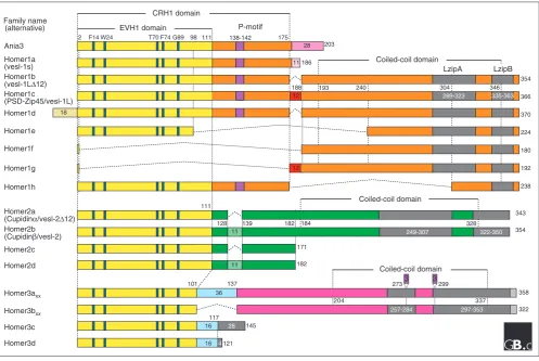
The Homer family of adaptor proteins consists in mammals of three members, Homer1, Homer2, and Homer3, all of which have several isoforms as a result of alternative splicing (Figure 1). A short murine Homer, Homer1a (also called vesl-1s, 186 amino acids in length), was the first to be iso-lated; it is encoded by an immediate-early gene induced in the hippocampus by neuronal activation such as electro-convulsive seizure and long-term potentiation [1,2]. A carboxy-terminal splicing variant very similar to Homer1a, Ania3, was also found as an immediate-early gene induced by dopaminergic stimulation [3]; it has 28 different carboxy-terminal residues in place of 11 residues at the Homer1a carboxyl terminus [4]. By screening for sequence similarity, another class of alternative splicing variants called the long Homer forms were cloned that have longer carboxy-terminal
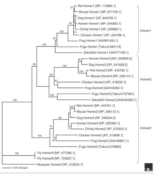
Orthologs of the Homer proteins have been identified in other animal species: Drosophila[12], Xenopus[13,14], and zebrafish [15]. Homer proteins have also been predicted from the genome sequences of the chimpanzee, the dog, Fugu, and the mosquito. A phylogenetic tree depicting the evolution of the Homer family (Figure 2) suggests that Homer1, Homer2, and Homer3 are found in vertebrates and separated when the fishes first evolved (Figure 2). No homologs have been found from eukaryotes outside the animals.
Characteristic structural features
Homer family proteins share two main structural features: the conserved amino-terminal domain, which is very similar to the Enabled/vasodilator-stimulated phosphoprotein (Ena/VASP) homology 1 (EVH1) domain, and the long Homer-specific carboxy-terminal domain, which consists of a coiled-coil
structure and two leucine zipper motifs [5,6,8,16] (Figure 1). Short Homer forms, such as Homer1a and Ania3, completely lack this carboxy-terminal domain [1-3].
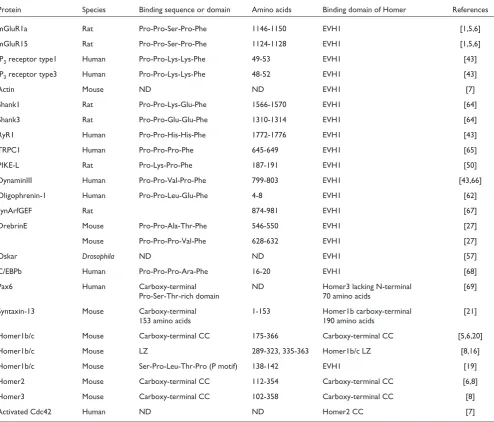
[image:2.595.56.554.86.419.2]The amino-terminal EVH1-like domain (also called the target-binding or ligand-binding domain) interacts with the proline-rich sequences of the form Pro-Pro-x-x-Phe (where x is any amino acid) that are found in many target or ligand proteins, as listed in Table 1. The carboxy-terminal domain (also called the self-assembly or multimerization domain) of long Homer forms mediates homophilic interactions or hetero-philic interactions with different members of the family. Multimers of long Homer proteins, which bind through their carboxy-terminal domains, are thought to act as a protein signaling complex that enables the linkage of various kinds of target proteins in close proximity and thereby facilitates signal transduction among these target proteins.
Figure 1
Primary structures of Homer family proteins. The conserved amino-terminal EVH1-like domain (which shows 80% sequence similarity between family members) is in yellow. The conserved region of Homer1 (CRH1) [19] and a proline motif (P-motif, 138-Ser-Pro-Leu-Thr-Pro-142) is specific to the mammalian Homer1 subfamily. The carboxy-terminal regions contain coiled-coil and leucine zipper structures and show only 30% sequence similarity among the family members. The coiled-coil regions are in orange, green and pink for the Homer1, Homer2, and Homer3 alternatively spliced forms, respectively. The leucine zipper structures, as predicted by Sun et al.[16], are shown as LzipA and LzipB in gray. The nomenclature is from Soloviev et al. [9], Saito et al.[10], Bottai et al.[4] and Klugmann et al.[11]. Homer3axxand Homer3bxxrepresent the products of four alternative splicing variants, where xx can be 00, 01, 10, or 11 to show the combination of two three-amino-acid insertions (purple) in the coiled-coil domain, as has been suggested for the human forms [9]. Residues involved in ligand contacts are colored light blue.
184
Coiled-coil domain
98
2 111 175
111
128 139 182
354
188 240 304
366
101 273 299
358 137
117
145 121
203 186
354
370 224 180 192 238
343
171 182
322 18
11 28
12
12
11
11
36
16 16
28 4
F14W24 T70F74G89 138-142
LzipA
289-323 335-363
249-307 322-350
257-284 297-353
EVH1 domain P-motif
CRH1 domain
Coiled-coil domain
Coiled-coil domain
193 346
328
3 3
7 3 3 4
0 2
Ania3
Homer1a (vesl-1s) Homer1b (vesl-1L∆12) Homer1c
(PSD-Zip45/vesl-1L) Homer1d
Homer1e
Homer1f
Homer1g
Homer1h
Homer2a
(Cupidinα/vesl-2∆12) Homer2b
(Cupidinβ/vesl-2)
Homer2c
Homer2d
Homer3axx
Homer3bxx
Homer3c
Homer3d Family name
(alternative)
Figure 2
A phylogenic tree of Homer family proteins. Whole protein sequences of the longest isoform of each family member from human (Homo sapiens), chimp (Pan troglodytes), dog (Canis familiaris), rat (Rattus norvegicus), mouse (Mus musculus), chicken (Gallus gallus), frog (Xenopus laevis), Fugu (Takifugu rubripes), zebrafish (Danio rerio), fly (Drosophila melanogaster) and mosquito (Anopheles gambiae) were aligned. The accession numbers of the proteins are indicated in brackets. Multiple sequence alignment was performed using CLUSTAL X [70]. Phylogenetic analysis was constructed using the neighbor-joining method [71] using PAUP* version 4.0 beta [72], and the reliability of the tree was estimated by bootstrapping. The tree was rooted with proteins from invertebrates (fly and mosquito). The branch lengths are proportional to the amount of inferred evolutionary change, and numbers between internal nodes indicate bootstrap values as percentages of 1,000 replications.
Rat Homer1 [NP_113895.1]
Mouse Homer1 [NP_671705.1]
Dog Homer1 [XP_849709.1]
Human Homer1 [NP_004263.1]
Chimp Homer1 [XP_526882.1]
Chicken Homer1 [XP_424768.1]
Frog Homer1 [AAW51455.1]
Fugu Home1 [Takru4:584113]
Zebrafish Homer1 [AAH77128.1]
Human Homer3 [NP_004829.2]
Dog Homer3 [XP_541929.2]
Rat Homer3 [NP_445762.1]
Mouse Homer3 [NP_036114.1]
Chicken Homer3 [XP_418233.1]
Frog Homer3 [AAH45262.1]
Fugu Homer3 [Takru4:737481]
Zebrafish Homer3 [AAH45383.1]
Rat Homer2 [NP_445761.1]
Mouse Homer2 [NP_036113.1]
Dog Homer2 [XP_536204.2]
Human Homer2 [NP_955362.1]
Chimp Homer2 [XP_510553.1]
Chicken Homer2 [XP_413836.1]
Frog Homer2 [AAH46847.1]
Fugu Homer2 [Takru4:578894]
Fly HomerA [NP_477396.1]
Fly HomerB [NP_723207.1]
Mosquito Homer2 [XP_319234.1]
0.05 changes100
100 100
89
100
84
100 93
100
96
100
100
68
74
85
72 67
100 92
61
51 65
Homer1
Homer3
The amino-terminal EVH1-like domain
The tertiary structure of the amino-terminal EVH1-like domain has been predicted using X-ray crystallographic analysis (Figure 3) [17,18]. The Homer EVH1-like domain forms a small globular structure that consists of a seven-stranded antiparallel βbarrel with a carboxy-terminal αhelix packed alongside it. No significant topological differences from the EVH1 domains of mammalian Enabled (Mena) or Ena/VASP can be seen. Interestingly, the consensus motif (Pro-Pro-x-x-Phe) found in proteins that bind to the Homer EVH1 domain has the opposite sequence orientation to the motif found in proteins that bind to the Ena/VASP EVH1 domain (Phe-Pro-Pro-Pro-Pro). Both of these proline-rich
consensus peptides seem to form a type II polyproline helix and bind at a distinct binding site on the corresponding EVH1 domain oriented in the same way [17,18]. This distinctive mode of Homer target binding minimizes the potential for cross-reaction with the many other available proline-rich target sequences, although the Homer EVH1 (class II) and other EVH1 domains (class I) seem to be derived from an ancestral polyproline-binding protein [18].
[image:4.595.58.553.119.541.2]The amino-terminal region containing 1-175 amino acids of mammalian Homer1 proteins is highly conserved and has been called the conserved region of Homer1 (CRH1) [19]. The CRH1 includes the EVH1-like domain and a second
Table 1
Interaction partners of Homer family proteins
Protein Species Binding sequence or domain Amino acids Binding domain of Homer References
mGluR1a Rat Pro-Pro-Ser-Pro-Phe 1146-1150 EVH1 [1,5,6]
mGluR15 Rat Pro-Pro-Ser-Pro-Phe 1124-1128 EVH1 [1,5,6]
IP3receptor type1 Human Pro-Pro-Lys-Lys-Phe 49-53 EVH1 [43]
IP3receptor type3 Human Pro-Pro-Lys-Lys-Phe 48-52 EVH1 [43]
Actin Mouse ND ND EVH1 [7]
Shank1 Rat Pro-Pro-Lys-Glu-Phe 1566-1570 EVH1 [64]
Shank3 Rat Pro-Pro-Glu-Glu-Phe 1310-1314 EVH1 [64]
RyR1 Human Pro-Pro-His-His-Phe 1772-1776 EVH1 [43]
TRPC1 Human Pro-Pro-Pro-Phe 645-649 EVH1 [65]
PIKE-L Rat Pro-Lys-Pro-Phe 187-191 EVH1 [50]
DynaminIII Human Pro-Pro-Val-Pro-Phe 799-803 EVH1 [43,66]
Oligophrenin-1 Human Pro-Pro-Leu-Glu-Phe 4-8 EVH1 [62]
synArfGEF Rat 874-981 EVH1 [67]
DrebrinE Mouse Pro-Pro-Ala-Thr-Phe 546-550 EVH1 [27]
Mouse Pro-Pro-Pro-Val-Phe 628-632 EVH1 [27]
Oskar Drosophila ND ND EVH1 [57]
C/EBPb Human Pro-Pro-Pro-Ara-Phe 16-20 EVH1 [68]
Pax6 Human Carboxy-terminal ND Homer3 lacking N-terminal [69]
Pro-Ser-Thr-rich domain 70 amino acids
Syntaxin-13 Mouse Carboxy-terminal 1-153 Homer1b carboxy-terminal [21]
153 amino acids 190 amino acids
Homer1b/c Mouse Carboxy-terminal CC 175-366 Carboxy-terminal CC [5,6,20]
Homer1b/c Mouse LZ 289-323, 335-363 Homer1b/c LZ [8,16]
Homer1b/c Mouse Ser-Pro-Leu-Thr-Pro (P motif) 138-142 EVH1 [19]
Homer2 Mouse Carboxy-terminal CC 112-354 Carboxy-terminal CC [6,8]
Homer3 Mouse Carboxy-terminal CC 102-358 Carboxy-terminal CC [8]
Activated Cdc42 Human ND ND Homer2 CC [7]
proline-containing motif (the P-motif, 138-Ser-Pro-Leu-Thr-Pro-142 in mouse Homer1), which is specific to the mamma-lian Homer1 proteins. The CRH1 interacts with the neigh-boring CRH1 in the crystal by intermolecular binding of the P-motif to the EVH1 domain at a site that partly overlaps that used for Pro-Pro-x-x-Phe binding [19]. Given that its binding to the metabotropic glutamate receptor (mGluR) induces the homo-multimerization of Homer1c [8], it is assumed that there are two types of binding of the Homer1 EVH1 domain: binding to the Pro-Pro-x-x-Phe of the target protein and binding to the P-motif of Homer1; the two types of binding induce and arrest this homo-multimerization, respectively.
The carboxy-terminal self-assembly and multimerization domain
Long Homer forms have a characteristic carboxy-terminal region comprising a coiled-coil structure followed by two leucine zipper motifs; this domain can mediate homomeric or heteromeric interactions between long Homer forms [5,6]. Although coiled coils are often implicated in protein-protein interactions, the Homer coiled-coil domain does not interact directly with other coiled-coil proteins, such as dynein [5]. In Homer1c, it is the leucine zipper motifs (ZipA and ZipB) that are involved in the multimerization, and the carboxy-terminal one, ZipB, is crucial [8,16]. Homer1b and Homer3 have been shown to form a tetramer via the
carboxy-terminal domain, including coiled coils and leucine zipper motifs, with no significant involvement of the amino terminal EVH-1 domain and P-motif [20].
An interaction of the carboxy-terminal domains of long Homer forms with other signaling proteins has been indicated. The carboxy-terminal region of Homer1b has been shown to be slightly similar to a mutated in colorectal cancer (MCC)-like domain [6] and to interact with syntaxin-13 [21]. The region of Homer2 from Ser90 to the carboxyl terminus has a weak and fragmentary identity (22%) with a part of Citron, a Rho-effector kinase, including the Rho/Rac-binding site and a part of the leucine zipper motif, and this region interacts with the small GTPase Cdc42 in a GTP-dependent manner [7].
Localization and function
Tissue, cellular and subcellular distribution
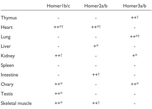
[image:5.595.58.556.88.340.2]Members of the Homer family are predominantly expressed in the nervous system, and they are also expressed in peripheral tissues at low levels. The tissue distribution of the long Homer forms is summarized in Tables 2 and 3. (Most studies have detected the very similar long forms together, so we refer here to ‘Homer1b/c’, ‘Homer2a/b and ‘Homer3a/b’ to indicate expression of one or the other or both isoforms of each Homer protein.) In the postnatal developing mouse
Figure 3
The crystal structure of the Homer EVH1 domain [17]. (a)Ribbon diagrams of the Homer1 EVH1 domain, with five residues of mGluR1 (residues 1,146-1,150) as a representative bound ligand in red. The side chains of the adjacent serine and proline in the bound ligand are oriented away from the Homer surface, and the phenylalanine residue forms a unique contact that is not shared by other EVH1 domains (not shown). (b)A surface
representation of the Homer EVH1 domain, showing the location of residues that are essential (red) and nonessential (light blue) for binding to ligand (dark blue). Reproduced with permission from [17].
(a) (b)
C
N αC
β1
β3 β2
β4 β5
β6 β7
βi
Gly89
Thr70
Gln76
Phe74
Trp24
brain, three long Homer forms are differentially expressed in various regions [22] (Table 2). The retina and spinal cord also express Homer1 [23,24]. The expression level of the long Homer forms in non-neural tissues is very low in comparison with that in the nervous system: the Homer1 and Homer2 long forms are expressed in skeletal and cardiac muscle [5,22,25,26], Homer1b/c in the ovary and testis, Homer2a/b in the liver and spleen, and Homer3a/b in the lung, spleen, kidney and ovary [5,22].
The long Homer family members have distinct cellular distributions in time and space during postnatal develop-ment of mouse brain [22]. In the cerebellum, Homer1b/c
and Homer2a/b are predominantly localized at the post-synapses of developing granule cells connecting the mossy fibers in the glomeruli. Homer3a/b is concentrated in the dendritic spines of Purkinje cells connecting the parallel fibers and is also present in their axons. The expression of Homer1b/c and Homer2a/b is regulated reciprocally to that of Homer3a/b in the hippocampus and the developing olfactory bulb; in general, where they are upregulated Homer3a/b is downregulated, and vice versa. In the hippo-campus, Homer1b/c and Homer2a/b are predominantly localized in the CA1 region and CA1-CA2 region, respec-tively, whereas Homer3a/b is concentrated in the CA2-CA3 regions.
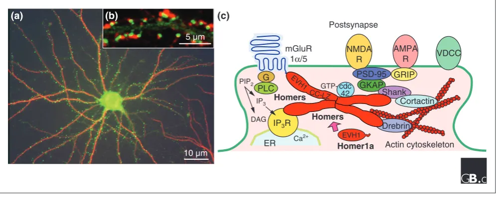
In fractionation studies on the rodent brain, the long Homer proteins are mainly found in subcellular fractions that are enriched with PSD proteins or postsynaptic membrane proteins [5,7] and in the PSD area of glutamatergic synapses [5,22,27-29] (Figure 4). An axonal distribution of Homer proteins has also been reported, however, with Homer2a/b found in cerebellar Purkinje cells and olfactory neurons [21] and Homer1b/c in Xenopus optic tectal neurons [13,14]. Homer1a, a short form, is found at very low levels in hippocampal cells [30,31]. Homer1a is targeted to synapses and regulated in the hippocampus by inducing the inhibition of the ubiquitin-proteasome system [32].
Dynamics of synaptic distribution
[image:6.595.57.559.118.315.2]The synaptic localization of long Homer forms is not static and becomes dynamic in response to synaptic activity. Post-synaptic clusters of exogenously expressed Homer1c fused to green fluorescent protein (GFP) in cultured hippocampal neurons showed a rapid redistribution and a higher steady-state turnover rate, in response to an increase in intracellular
Table 2
Homer protein expression in postnatal mouse brain development
Homer1b/c Homer2a/b Homer3a/b
Postnatal week 1w 2w 3w 8w 1w 2w 3w 8w 1w 2w 3w 8w
Cerebral cortex +++ +++ +++ +++ +++ +++ +++ +++ - - -
-Olfactory bulb ++ ++ ++ ++ +++ +++ +++ +++ + - -
-Hippocampus +++ +++ +++ +++ +++ +++ +++ +++ + ++ ++ +
Thalamus ++ ++ ++ ++ +++ +++ +++ +++ - - -
-Midbrain ++ ++ ++ + +++ ++ ++ ++ - - -
-Inferior colliculus ++ ++ ++ + +++ ++ ++ ++ - - -
-Medulla oblongata ++ ++ + + +++ +++ + + - - -
-Corpus striatum ND ++ ++ ++ ND ++ ++ ++ ND - -
-Cerebellum ++ + + - ++ ++ + + ++ +++ +++ +++
Pons ++ ++ + + +++ +++ ++ ++ - - -
-Levels of expression are indicated as follows: +++, high; ++, intermediate; +, low; -, not detected; ND, no data.
Table 3
Distribution of Homer protein in mouse peripheral tissues at 2 weeks after birth
Homer1b/c Homer2a/b Homer3a/b
Thymus - - ++†
Heart ++*† ++*†
-Lung - - ++*†
Liver - +*
-Kidney ++† - +*
Spleen - -
-Intestine - ++†
-Ovary ++* - ++*
Testis ++* -
-Skeletal muscle ++* ++†
[image:6.595.54.297.389.563.2]Ca2+ mediated through the activation of N-methyl-D
-aspartate receptor (NMDA) receptors or voltage-dependent Ca2+ channels [33]. By analysis of GFP fluorescence
intensity, it was estimated that the typical single post-synaptic site of cultured hippocampal cells contains 343 ± 57 Homer family proteins [34]. Homer1a, induced by brain-derived neurotrophic factor or a proteasome inhibitor, also causes the redistribution of Homer1c [35]. In cultured cerebellar granule cells, endogenous Homer2 and exoge-nously expressed GFP-fused Homer2 showed a reversible declustering induced by an NMDA receptor-mediated Ca2+
influx followed by activation of a downstream pathway including mitogen-activated protein (MAP) kinases, extra-cellular signal-regulated kinases (ERKs) and protein tyro-sine kinases [28]. This Homer2 declustering is induced before declustering of filamentous (F-)actin and Drebrin (a dendritic actin-binding protein), suggesting that it is inde-pendent of the actin cytoskeletal reorganization. The synaptic-activity-dependent dynamics of synaptic long Homer seems to be involved in remodeling the molecular constitution of the PSD protein complex at mature and/or differentiating glutamatergic synapses [28,33].
Targeting of long Homer forms to PSDs seems to be correlated with synaptic contact formation [27] or F-actin
accumulation in PSDs [36]. Newly extended dendrites of cultured hippocampal neurons rapidly acquire Homer clusters, whose density reaches the level of parental dendrites within a few days [37]. Synaptic targeting of Homer is independent of the molecular motor protein myosin Va [29]. Throughout synaptic differentiation of cultured hippocampal neurons, all three long Homer proteins not only form heteromeric co-clusters, but also localize close to NMDA receptor clusters, including those containing the NMDA receptor 2B subunit and PSD-95 [27]. Synaptic Homer clustering is enhanced by simultaneous blockade of NMDA receptor and cAMP phosphodiesterase activities, as is clustering of NMDA receptors [27]. These data suggest a coincidence in developmental and activity-regulated synaptic targeting between Homer and the NMDA receptor complex. This suggests that synaptic targeting of both Homer and the NMDA receptor complex is coincidently regulated during the development of hippocampal neurons and their activity-dependent synapse formation.
Mechanism
[image:7.595.57.555.89.290.2]Many of the proteins that bind to Homer proteins are func-tionally related to one another: for example, mGluR1α/5 and mGluR5 (mGluR1α/5), inositol 1,4,5-trisphosphate (IP3) receptors (IP3R), ryanodine receptors, Shank (an adaptor for
Figure 4
Homer proteins form a physical tether linking signaling molecules in postsynaptic densities. (a)Cultured hippocampal neurons co-immunostained for Homer2a/b (green) and the neuronal dendritic marker MAP2 (red). (b)Dendritic spines of cultured hippocampal neurons expressing exogenous GFP-Homer2 (green) and immunostained for synaptophysin (a presynapse marker; red). (c)A model of the long Homer multimer-mediated postsynaptic protein complex and Homer1a. Long Homer forms (Homers) bind to each other through their carboxy-terminal domains (probably forming a tetramer [20]) and to the target proteins, such as mGluR1α/5, IP3 receptor, NMDA receptor and Drebrin, to the actin cytoskeleton through their amino-terminal domains, forming a cluster at the postsynaptic density area. Homer1a, which lacks the carboxy-terminal domain, is thought to compete with long Homer forms for binding to target proteins, thus disrupting the cluster. For instance, Homers modulate mGluR-induced intracellular calcium release by linking mGluRs and IP3R: activation of mGluRs results in the phospholipase C (PLC)-mediated hydrolysis of membrane phosphatidylinositol diphosphate (PIP2) to diacylglycerol (DAG) and IP3, which activates the IP3R to release intracellular calcium. Abbreviations: AMPAR, 5-methyl-4-isoxazolepropionate receptor; CC-LZ, carboxy-terminal coiled-coil structure and leucine zipper motifs of long Homer forms; ER, endoplasmic reticulum; EVH1, the amino-terminal EVH1 domain of Homer; G, G protein; GKAP, guanylate kinase-associated protein; GRIP, glutamate receptor interacting protein; mGluR, metabotropic glutamate receptor; NMDAR, N-methyl-D-aspartate receptor; VDCC, voltage-dependent Ca2+channel.
(c)
10 µm
(a)
(b)
5 µm
Actin cytoskeleton Postsynapse
AMPA
R VDCC NMDA
R
Drebrin
ER
mGluR 1α/5
IP3
Ca2+
DAG
GKAP
PLC
GTP-PSD-95 GRIP
EVH1
G
PIP2
cdc 42
IP3R
Shank Cortactin
Homer1a Homers
Homers EVH1
the NMDA receptor complex), and transient receptor poten-tial channels are all involved in Ca2+ signaling pathways at
the PSD. Clusters of long Homer proteins seem to act as PSD signaling complexes through which signal transduction or cross-talk among target proteins is facilitated (Figure 4). Activity-dependent, reversible declustering of long Homer-mediated target protein complexes may be involved in remodeling the target composition of the complex. Homers also associate with the actin cytoskeleton, through which the Homer complex is probably anchored to proper post-synaptic sites.
The short Homer proteins Homer1a and Ania3 are transcrip-tionally induced only upon neuronal stimulation. They consequently disrupt the scaffolding capability of long Homer forms by sequestering their binding partners. Thus, Homer1a and Ania3 function as natural activity-dependent dominant-negative forms that regulate the scaffolding and signaling capabilities of the long forms. This property seems to be related to synapse and circuit regulation. Indeed, there are several reports demonstrating that Homer1a or Ania3 are upregulated by various physiological treatments that induce synaptic activities: seizure and kindling [2,4], stimu-lation by light [1], dopaminergic stimustimu-lation [4], exploration of a novel environment [38], learning or long-term potentia-tion [39,40], and administrapotentia-tion of psychoactive stimulants or drugs. The signaling cascades involved in the induction of Homer1a expression include the MAP kinase cascade in cerebellar granule cells [41] and the ERK1/2 cascade in hippocampal dentate gyrus cells [42].
Functions in the synapse
Many reports of Homer functions have recently been published; here, we can describe only a fraction of them. Postsynaptic Homer can regulate the synaptic localization of target proteins or the cross-talk signaling among these proteins at the PSD. Homer proteins regulate the activity of mGluR1α/5 in various ways, including attenuation of its effects by Homer1a, probably as a result of Homer1a’s dominant-negative binding [43]; modulation of its linkage to MAP kinase cascades [44]; regulation of its coupling ion channels [45,46]. Homer proteins are thought to act syner-gistically with Shank, another scaffold protein for the NMDA receptor/PSD-95 complex, via GKAP (guanylate kinase associated protein) in the functional linking of mGluR1α/5 and IP3R in the PSD [47]. There seems to be a difference in target-binding affinity or specificity of the EVH1 domain, or other functional properties, among different Homer family proteins [48].
Homer1a expressed in response to neuronal activity regulates synapse function. In cerebellar granule cells, Homer1a induced by NMDA or kainate stimulation triggered the constitutive activity of mGluR1α/5 independent of binding of an agonist (for example glutamate), but long Homer3 did not show the same activity [48]. Overexpression of Homer1a,
but not of Homer1c, enhanced synaptic transmission in cultured rat hippocampal slices, probably as a result of an increase in synaptic targeting of 5-methyl-4-isoxazole-propionate (AMPA) receptors [40]. In addition, Homer1a enhanced spike-induced Ca2+ influx in rat visual cortex
pyramidal cells [49]. A recent study showed a role for Homer1a in the attenuation of inflammatory hyper-sensitivity in spinal cord neurons [24]. Also, the long form of phosphoinositide 3-kinase (PI 3-kinase) enhancer (PIKE-L), a nuclear GTPase that activates nuclear PI 3-kinase, inter-acts with Homer1c and Homer2a (Table 1), and activation of mGluR1α/5 enhances formation of an mGluR1α /5-Homer-PIKE-L complex, leading to activation of PI 3-kinase activity and the prevention of neuronal apoptosis [50].
Cell-surface clustering of mGluR1αα/5
Homer modulates the trafficking of mGluR1α/5 and its targeting to the membrane. Following heterologous expres-sion in HeLa cells, Homer1b inhibited cell-surface targeting of mGluR5 and induced its retention in the endoplasmic reticulum, whereas Homer1a increased cell-surface mGluR5 [51]. In cerebellar granule cells, exogenously expressed Homer1b also induced intracellular retention of mGluR5 in the endoplasmic reticulum, whereas exogenously expressed Homer1a induced surface clustering of mGluR5 [52]. By contrast, exogenously expressed Homer1b, but not Homer1a, increased cell-surface clustering of mGluR5 and confined its movement within the membrane of cultured hippocampal neurons [53]. Depolarization induced endogenous Homer1a expression through the MAP kinase pathway in cerebellar Purkinje cells, which enhanced cell-surface targeting of mGluR1α, leading to the increment in mGluR1 responsive-ness [54]. These results indicate that the long and short Homer proteins both regulate cell-surface targeting and clustering of mGluR1α/5, probably by the opposite actions. There are a few differences that seem to be caused by the expression levels of Homer proteins or the cell-type analyzed, however. Because Homer proteins interact with various target proteins, which seem to differ in their number and identity from cell to cell, including membrane proteins and actin-binding proteins, these proteins probably contribute to anchoring the Homer complex at the appropriate intracellular compartments.
Functions in neuronal development
increased by coexpression with Shank1B, resulting in the enlargement of spine heads (mature mushroom-type spines) and an increase in synaptic currents [55], whereas exogenously expressed Homer1a affects spine morphology (causing a decrease in mature spines but increased elongated processes) [31]. This indicates that Homer1b together with Shank induces spine maturation, and that activity-dependent Homer1a operates in a negative feedback loop to regulate the structure and function of synapses. In addition to the postsynaptic regulation, Homer1b/c regulates axonal path finding of Xenopusoptic tectal neurons [13].
Homer also has roles in the functions of non-neural tissues. During the differentiation of muscle, Homer2b is up-regulated and seems to increase RyR-mediated Ca2+release,
which is necessary for traffic of the transcription factor nuclear factor of activated T cells (NFAT) into the nuclei of the myotube [56]. In pattern formation in Drosophila
embryos, Homer and another F-actin-binding protein, Bif, show asymmetric localization to the apical cortex of embryonic neuroblasts and may be involved in neuroblast asymmetric divisions by promoting posterior localization of
oskarmRNA and of proteins that are essential for posterior patterning [57].
Functions in behavior
Several lines of evidence obtained by the disruption or virus-vector-mediated expression of Homer genes demonstrate the involvement of Homer family proteins in animal behavior. Mutation of DrosophilaHomer caused defects in behavioral plasticity and in the control of locomotor activity, but not in basic neurotransmission. This suggests that Homer regulates the development and function of neural networks underlying locomotor control and behavioral plasticity in Drosophila[12]. In an adult rat hippocampus in which exogenous Homer proteins were overexpressed using a recombinant adeno-associated virus gene delivery system, increased levels of Homer1a led to impaired hippocampus-dependent memory, whereas increased levels of Homer1g (which lacks the amino-terminal target binding domain; Figure 1) and Homer1c slightly enhanced memory perfor-mance, suggesting that Homer1 splice variants have an active role in behavioral plasticity [11]. Transgenic mice over-expressing Homer1a in striatal medium spiny neurons, in which mGluR1α/5 is important in synaptic transmission, showed impairments in motor performance and coordination in behavioral tasks and showed repetitive, compulsive behavior (stereotypy) induced by the psycho-motor stimulant amphetamine, thereby suggesting a critical role for Homer1 in the modulation of striatal circuits [58].
Homer has been shown to be relevant in drug addiction (reviewed in [59]). Homer2 knockout mice studies showed that Homer proteins are involved in the sensitization of behavior produced by repeated cocaine treatment. Further-more, the loss of Homer2 induces similar behavioral and
neurochemical phenotypes to those produced by repeated cocaine administration. In addition, both chronic and acute overexpression of constitutive Homer1b and Homer2a/b, but not of short Homer forms, by adeno-associated virus abolished cocaine-induced sensitization of locomotor hyper-activity and prevented the development of glutamate abnor-malities in the accumbens. In alcoholism, Homer2 knockout and rescue with adeno-associated virus-expressed Homer2b indicated a necessary and active role for Homer2 in the accumbens in regulating alcohol-induced behavioral and cellular neuroplasticity.
Homer proteins and brain disease
An abnormality of glutamate receptor signaling has been implicated in many different brain diseases, including learning and memory disability, epilepsy, schizophrenia and affective disorder. The genes encoding those Homer family proteins that interact directly with mGluR1α/5 and in-directly with NMDA receptors at their glutamatergic synapses seem to be candidate genes for involvement in neuro-psychiatric phenotypes. Analysis of single-nucleotide poly-morphisms in the Homer gene family identified seven, including three in exons, but failed to implicate any of the Homer genes in schizophrenia; these variants remain plausible candidates for other neuropsychiatric phenotypes [60]. A recent study of Homer1 knockout mice, however, indicated that the loss of Homer1 function causes behavioral abnormalities (motivational, emotional, cognitive and sensorimotor processing) that are consistent with animal models of schizophrenia, and blunts a cocaine-stimulated increase in extracellular glutamate levels in the prefrontal cortex, suggesting reduced activation in this region as the hypo-frontality that is commonly observed in schizophrenia [61].
Frontiers
Homer interacts with many different target proteins carrying the Pro-Pro-x-x-Phe consensus motif. There are many other candidate proteins with this motif that have not yet been analyzed and thus should be investigated. In addition, it should be determined whether the Homer family proteins, including alternative splicing variants, have functional differences, for example in their target-binding affinity or preference. Moreover, the structural features of each Homer form, including various protein phosphorylation consensus sites and other putative functional sites (Figure 1), suggest differences in the modulation of these functions.
Long Homer forms are characterized by multimerization through the carboxy-terminal domain. To understand the molecular complexity of target proteins in a long Homer-linked complex, the number of Homer subunits that can assemble in a multimer needs to be determined. Synaptic clustering of long Homer proteins seems to be brought about by preferential anchoring of a few multimers at a specific site or subcellular compartment. The actin cytoskeleton is a candidate for such Homer clustering through actin-associated Pro-Pro-x-x-Phe-containing proteins, such as Drebrin. Moreover, the molecular mechanism underlying the activity-dependent dynamics of long Homer-target clustering is unknown. Declustering of Homer is induced by an increase in intracellular Ca2+ concentration through
NMDA receptors or voltage-dependent Ca2+ channels.
Signaling by protein phosphorylation is likely to be involved downstream of this Ca2+signaling. In response to synaptic
activity, reversible clustering and declustering of the Homer complex is probably an important mechanism used to alter the molecular composition within the complex, so that cross-talk signaling among cross-linked target proteins can be regulated (Figure 4).
There are still only a few lines of evidence to support the hypothesis that short Homer1a induced in an activity-dependent manner behaves as a natural dominant negative to compete with the target binding of long Homer proteins and to disrupt the long Homer-target protein complex in in vivoneurons and within the brain. Other roles for Homer1a can be anticipated (for example, a conformational change of target proteins conferred by the protein-protein interaction), as Homer1a binding induces constitutive activation of mGluR1α/5 independently of agonist binding [49].
One of the striking features of the expression of the Homer family is that short Homer1a is expressed in an activity-dependent manner and seems to act at the active synaptic site. The mechanism underlying the activity-dependent induction of short Homer1a, in both promoter regulation and alternative splicing, remains to be elucidated. In addition, how the expressed Homer1a is transported to the active synaptic site and what its tag to that site is needs to be investigated.
Homer proteins link many critical synaptic proteins, including those involved in glutamate signaling, which implicates them in many different brain functions and diseases. Genetically engineered Homer mouse models are an important tool with which to clarify the in vivo functions of Homer family proteins. As Homer knockout mice show neuropsychologically altered phenotypes, further study of these model mice will shed light on the roles of this interesting protein family in higher brain functions.
Acknowledgements
We thank Dr A. Mizutani (The university of Tokyo) for comments. This work was supported by grant number 18053025 (T.F.) from the Ministry of Education, Culture, Sports, Science and Technology, Japan.
References
1. Brakeman PR, Lanahan AA, O’Brien R, Roche K, Barnes CA, Huganir RL, Worley PF: Homer: a protein that selectively binds metabotropic glutamate receptors. Nature 1997, 386:284-288. 2. Kato A, Ozawa F, Saitoh Y, Hirai K, Inokuchi K: vesl, a gene
encoding VASP/Ena family related protein, is upregulated during seizure, long-term potentiation and synaptogenesis. FEBS Lett 1997, 412:183-189.
3. Berke JD, Paletzki RF, Aronson GJ, Hyman SE, Gerfen CR: A complex program of striatal gene expression induced by dopaminergic stimulation. J Neurosci 1998, 18:5301-5310. 4. Bottai D, Guzowski JF, Schwarz MK, Kang SH, Xiao B, Lanahan A,
Worley PF, Seeburg PH: Synaptic activity-induced conversion of intronic to exonic sequence in Homer 1 immediate early gene expression.J Neurosci 2002, 22:167-175.
5. Xiao B, Tu JC, Petralia RS, Yuan JP, Doan A, Breder CD, Ruggiero A, Lanahan AA, Wenthold RJ, Worley PF: Homer regulates the association of group 1 metabotropic glutamate receptors with multivalent complexes of homer-related, synaptic pro-teins.Neuron 1998, 21:707-716.
6. Kato A, Ozawa F, Saitoh Y, Fukazawa Y, Sugiyama H, Inokuchi K: Novel members of the Vesl/Homer family of PDZ proteins that bind metabotropic glutamate receptors. J Biol Chem 1998, 273:23969-23975.
7. Shiraishi Y, Mizutani A, Bito H, Fujisawa K, Narumiya S, Mikoshiba K, Furuichi T: Cupidin, an isoform of Homer/Vesl, interacts with the actin cytoskeleton and activated rho family small GTPases and is expressed in developing mouse cerebellar granule cells.J Neurosci 1999, 19:8389-8400.
8. Tadokoro S, Tachibana T, Imanaka T, Nishida W, Sobue K: Involve-ment of unique leucine-zipper motif of PSD-Zip45 (Homer 1c/vesl-1L) in group 1 metabotropic glutamate receptor clustering. Proc Natl Acad Sci USA 1999, 96:13801-13806.
9. Soloviev MM, Ciruela F, Chan WY, McIlhinney RA: Molecular char-acterisation of two structurally distinct groups of human homers, generated by extensive alternative splicing. J Mol Biol 2000, 295:1185-1200.
10. Saito H, Kimura M, Inanobe A, Ohe T, Kurachi Y: An N-terminal sequence specific for a novel Homer1 isoform controls traf-ficking of group I metabotropic glutamate receptor in mammalian cells. Biochem Biophys Res Commun 2002, 296:523-529.
11. Klugmann M, Wymond Symes C, Leichtlein CB, Klaussner BK, Dunning J, Fong D, Young D, During MJ: AAV-mediated hip-pocampal expression of short and long Homer 1 proteins differentially affect cognition and seizure activity in adult rats. Mol Cell Neurosci 2005, 28:347-360.
12. Diagana TT, Thomas U, Prokopenko SN, Xiao B, Worley PF, Thomas JB: Mutation of Drosophilahomer disrupts control of locomotor activity and behavioral plasticity. J Neurosci 2002, 22:428-436.
Cline HT: Homer expression in the Xenopustadpole nervous system.J Comp Neurol 2005, 487:42-53.
15. Gasperini R, Foa L: Homer 1b/c expression correlates with zebrafish olfactory system development. J Neurocytol 2004, 33:671-680.
16. Sun J, Tadokoro S, Imanaka T, Murakami SD, Nakamura M, Kashi-wada K, Ko J, Nishida W, Sobue K: Isolation of PSD-Zip45, a novel Homer/vesl family protein containing leucine zipper motifs, from rat brain.FEBS Lett 1998, 437:304-308.
17. Beneken J, Tu JC, Xiao B, Nuriya M, Yuan JP, Worley PF, Leahy DJ: Structure of the Homer EVH1 domain-peptide complex reveals a new twist in polyproline recognition.Neuron 2000, 26:143-154.
18. Barzik M, Carl UD, Schubert WD, Frank R, Wehland J, Heinz DW: The N-terminal domain of Homer/Vesl is a new class II EVH1 domain. J Mol Biol 2001, 309:155-169.
19. Irie K, Nakatsu T, Mitsuoka K, Miyazawa A, Sobue K, Hiroaki Y, Doi T, Fujiyoshi Y, Kato H: Crystal structure of the Homer 1 family conserved region reveals the interaction between the EVH1 domain and own proline-rich motif. J Mol Biol 2002, 318:1117-1126.
20. Hayashi MK, Ames HM, Hayashi Y: Tetrameric hub structure of postsynaptic scaffolding protein homer. J Neurosci 2006, 26: 8492-8501.
21. Minakami R, Kato A, Sugiyama H: Interaction of Vesl-1L/Homer 1c with syntaxin 13. Biochem Biophys Res Commun 2000, 272:466-471. 22. Shiraishi Y, Mizutani A, Yuasa S, Mikoshiba K, Furuichi T:
Differen-tial expression of Homer family proteins in the developing mouse brain. J Comp Neurol 2004, 473:582-599.
23. Brandstatter JH, Dick O, Boeckers TM: The postsynaptic scaffold proteins ProSAP1/Shank2 and Homer1 are associated with glutamate receptor complexes at rat retinal synapses. J Comp Neurol 2004, 475:551-563.
24. Tappe A, Klugmann M, Luo C, Hirlinger D, Agarwal N, Benrath J, Ehrengruber MU, During MJ, Kuner R: Synaptic scaffolding protein Homer1a protects against chronic inflammatory pain.Nat Med 2006, 12:677-681.
25. Soloviev MM, Ciruela F, Chan WY, McIlhinney RA: Mouse brain and muscle tissues constitutively express high levels of Homer proteins. Eur J Biochem 2000, 267:634-639.
26. Volpe P, Sandri C, Bortoloso E, Valle G, Nori A: Topology of Homer 1c and Homer 1a in C2C12 myotubes and trans-genic skeletal muscle fibers.Biochem Biophys Res Commun 2004, 316:884-892.
27. Shiraishi Y, Mizutani A, Mikoshiba K, Furuichi T: Coincidence in dendritic clustering and synaptic targeting of homer pro-teins and NMDA receptor complex propro-teins NR2B and PSD95 during development of cultured hippocampal neurons. Mol Cell Neurosci 2003, 22:188-201.
28. Shiraishi Y, Mizutani A, Yuasa S, Mikoshiba K, Furuichi T: Gluta-mate-induced declustering of post-synaptic adaptor protein Cupidin (Homer 2/vesl-2) in cultured cerebellar granule cells.J Neurochem 2003, 87:364-376.
29. Petralia RS, Wang YX, Sans N, Worley PF, Hammer JA, 3rd, Wen-thold RJ: Glutamate receptor targeting in the postsynaptic spine involves mechanisms that are independent of myosin Va.Eur J Neurosci 2001, 13:1722-1732.
30. Kato A, Fukazawa Y, Ozawa F, Inokuchi K, Sugiyama H: Activation of ERK cascade promotes accumulation of Vesl-1S/Homer-1a immunoreactivity at synapses. Brain Res Mol Brain Res 2003, 118:33-44.
31. Sala C, Futai K, Yamamoto K, Worley PF, Hayashi Y, Sheng M: Inhi-bition of dendritic spine morphogenesis and synaptic trans-mission by activity-inducible protein Homer1a. J Neurosci 2003, 23:6327-6337.
32. Ageta H, Kato A, Hatakeyama S, Nakayama K, Isojima Y, Sugiyama H: Regulation of the level of Vesl-1S/Homer-1a proteins by ubiquitin-proteasome proteolytic systems. J Biol Chem 2001, 276:15893-15897.
33. Okabe S, Urushido T, Konno D, Okado H, Sobue K: Rapid redis-tribution of the postsynaptic density protein PSD-Zip45 (Homer 1c) and its differential regulation by NMDA recep-tors and calcium channels. J Neurosci 2001, 21:9561-9571. 34. Sugiyama Y, Kawabata I, Sobue K, Okabe S: Determination of
absolute protein numbers in single synapses by a GFP-based calibration technique.Nat Methods 2005, 2:677-684.
H: Activity-inducible protein Homer1a/Vesl-1S promotes redistribution of postsynaptic protein Homer1c/Vesl-1L in cultured rat hippocampal neurons. Neurosci Lett 2004, 354:143-147.
36. Usui S, Konno D, Hori K, Maruoka H, Okabe S, Fujikado T, Tano Y, Sobue K: Synaptic targeting of PSD-Zip45 (Homer 1c) and its involvement in the synaptic accumulation of F-actin. J Biol Chem 2003, 278:10619-10628.
37. Ebihara T, Kawabata I, Usui S, Sobue K, Okabe S: Synchronized formation and remodeling of postsynaptic densities: long-term visualization of hippocampal neurons expressing post-synaptic density proteins tagged with green fluorescent protein.J Neurosci 2003, 23:2170-2181.
38. Vazdarjanova A, McNaughton BL, Barnes CA, Worley PF, Guzowski JF: Experience-dependent coincident expression of the effec-tor immediate-early genes arc and Homer 1a in hippocam-pal and neocortical neuronal networks. J Neurosci 2002, 22: 10067-10071.
39. Matsuo R, Murayama A, Saitoh Y, Sakaki Y, Inokuchi K: Identifica-tion and cataloging of genes induced by lasting long-term potentiation in awake rats. J Neurochem 2000, 74: 2239-2249.
40. Hennou S, Kato A, Schneider EM, Lundstrom K, Gahwiler BH, Inokuchi K, Gerber U, Ehrengruber MU: Homer-1a/Vesl-1S enhances hippocampal synaptic transmission. Eur J Neurosci 2003, 18:811-819.
41. Sato M, Suzuki K, Nakanishi S: NMDA receptor stimulation and brain-derived neurotrophic factor upregulate homer 1a mRNA via the mitogen-activated protein kinase cascade in cultured cerebellar granule cells.J Neurosci 2001, 21:3797-3805. 42. Rosenblum K, Futter M, Voss K, Erent M, Skehel PA, French P, Obosi L, Jones MW, Bliss TV: The role of extracellular regu-lated kinases I/II in late-phase long-term potentiation. J Neu-rosci 2002, 22:5432-5441.
43. Tu JC, Xiao B, Yuan JP, Lanahan AA, Leoffert K, Li M, Linden DJ, Worley PF: Homer binds a novel proline-rich motif and links group 1 metabotropic glutamate receptors with IP3 recep-tors.Neuron 1998, 21:717-726.
44. Mao L, Yang L, Tang Q, Samdani S, Zhang G, Wang JQ: The scaf-fold protein Homer1b/c links metabotropic glutamate receptor 5 to extracellular signal-regulated protein kinase cascades in neurons. J Neurosci 2005, 25:2741-2752.
45. Kammermeier PJ, Xiao B, Tu JC, Worley PF, Ikeda SR: Homer pro-teins regulate coupling of group I metabotropic glutamate receptors to N-type calcium and M-type potassium chan-nels. J Neurosci 2000, 20:7238-7245.
46. Yamamoto K, Sakagami Y, Sugiura S, Inokuchi K, Shimohama S, Kato N: Homer 1a enhances spike-induced calcium influx via L-type calcium channels in neocortex pyramidal cells. Eur J Neurosci 2005, 22:1338-1348.
47. Sala C, Roussignol G, Meldolesi J, Fagni L: Key role of the postsy-naptic density scaffold proteins Shank and Homer in the functional architecture of Ca2+ homeostasis at dendritic spines in hippocampal neurons.J Neurosci 2005, 25:4587-4592. 48. Shin DM, Dehoff M, Luo X, Kang SH, Tu J, Nayak SK, Ross EM,
Worley PF, Muallem S: Homer 2 tunes G protein-coupled receptors stimulus intensity by regulating RGS proteins and PLCbeta GAP activities.J Cell Biol 2003, 162:293-303.
49. Ango F, Prezeau L, Muller T, Tu JC, Xiao B, Worley PF, Pin JP, Bock-aert J, Fagni L: Agonist-independent activation of metabo-tropic glutamate receptors by the intracellular protein Homer.Nature 2001, 411:962-965.
50. Rong R, Ahn JY, Huang H, Nagata E, Kalman D, Kapp JA, Tu J, Worley PF, Snyder SH, Ye K: PI3 kinase enhancer-Homer complex couples mGluRI to PI3 kinase, preventing neuronal apoptosis.Nat Neurosci 2003, 6:1153-1161.
51. Roche KW, Tu JC, Petralia RS, Xiao B, Wenthold RJ, Worley PF: Homer 1b regulates the trafficking of group I metabotropic glutamate receptors. J Biol Chem 1999, 274:25953-25957. 52. Ango F, Robbe D, Tu JC, Xiao B, Worley PF, Pin JP, Bockaert J, Fagni
L: Homer-dependent cell surface expression of metabo-tropic glutamate receptor type 5 in neurons.Mol Cell Neurosci 2002, 20:323-329.
54. Minami I, Kengaku M, Smitt PS, Shigemoto R, Hirano T: Long-term potentiation of mGluR1 activity by depolarization-induced Homer1a in mouse cerebellar Purkinje neurons. Eur J Neu-rosci 2003, 17:1023-1032.
55. Sala C, Piech V, Wilson NR, Passafaro M, Liu G, Sheng M: Regula-tion of dendritic spine morphology and synaptic funcRegula-tion by Shank and Homer. Neuron 2001, 31:115-130.
56. Stiber JA, Tabatabaei N, Hawkins AF, Hawke T, Worley PF, Williams RS, Rosenberg P: Homer modulates NFAT-dependent signal-ing dursignal-ing muscle differentiation. Dev Biol 2005, 287:213-224. 57. Babu K, Cai Y, Bahri S, Yang X, Chia W: Roles of Bifocal, Homer,
and F-actin in anchoring Oskar to the posterior cortex of Drosophilaoocytes. Genes Dev 2004, 18:138-143.
58. Tappe A, Kuner R: Regulation of motor performance and stri-atal function by synaptic scaffolding proteins of the Homer1 family.Proc Natl Acad Sci USA 2006, 103:774-779.
59. Szumlinski KK, Kalivas PW, Worley PF: Homer proteins: implica-tions for neuropsychiatric disorders.Curr Opin Neurobiol 2006, 16:251-257.
60. Norton N, Williams HJ, Williams NM, Spurlock G, Zammit S, Jones G, Jones S, Owen R, O’Donovan MC, Owen MJ: Mutation screen-ing of the Homer gene family and association analysis in schizophrenia.Am J Med Genet B Neuropsychiatr Genet 2003, 120: 18-21
61. Szumlinski KK, Lominac KD, Kleschen MJ, Oleson EB, Dehoff MH, Schwarz MK, Seeburg PH, Worley PF, Kalivas PW: Behavioral and neurochemical phenotyping of Homer1 mutant mice: possi-ble relevance to schizophrenia. Genes Brain Behav 2005, 4:273-288.
62. Govek EE, Newey SE, Akerman CJ, Cross JR, Van der Veken L, Van Aelst L: The X-linked mental retardation protein oligo-phrenin-1 is required for dendritic spine morphogenesis.Nat Neurosci 2004, 7:364-372.
63. Giuffrida R, Musumeci S, D’Antoni S, Bonaccorso CM, Giuffrida-Stella AM, Oostra BA, Catania MV: A reduced number of metabotropic glutamate subtype 5 receptors are associated with constitutive homer proteins in a mouse model of fragile X syndrome.J Neurosci 2005, 25:8908-8916.
64. Tu JC, Xiao B, Naisbitt S, Yuan JP, Petralia RS, Brakeman P, Doan A, Aakalu VK, Lanahan AA, Sheng M, et al.: Coupling of mGluR/Homer and PSD-95 complexes by the Shank family of postsynaptic density proteins.Neuron 1999, 23:583-592 65. Yuan JP, Kiselyov K, Shin DM, Chen J, Shcheynikov N, Kang SH,
Dehoff MH, Schwarz MK, Seeburg PH, Muallem S, et al.: Homer binds TRPC family channels and is required for gating of TRPC1 by IP3 receptors.Cell 2003, 114:777-789.
66. Gray NW, Fourgeaud L, Huang B, Chen J, Cao H, Oswald BJ, Hemar A, McNiven MA: Dynamin 3 is a component of the postsy-napse, where it interacts with mGluR5 and Homer. Curr Biol 2003, 13:510-515.
67. Inaba Y, Tian QB, Okano A, Zhang JP, Sakagami H, Miyazawa S, Li W, Komiyama A, Inokuchi K, Kondo H,et al.: Brain-specific potential guanine nucleotide exchange factor for Arf, synArfGEF (Po), is localized to postsynaptic density. J Neurochem 2004, 89:1347-1357.
68. Ishiguro K, Xavier R: Homer-3 regulates activation of serum response element in T cells via its EVH1 domain. Blood 2004, 103:2248-2256.
69. Cooper ST, Hanson IM: A screen for proteins that interact with PAX6: C-terminal mutations disrupt interaction with HOMER3, DNCL1 and TRIM11.BMC Genet 2005, 6:43. 70. Thompson JD, Gibson TJ, Plewniak F, Jeanmougin F, Higgins DG:
The CLUSTAL_X windows interface: flexible strategies for multiple sequence alignment aided by quality analysis tools. Nucleic Acids Res 1997, 25:4876-4882.
71. Saitou N, Nei M: The neighbor-joining method: a new method for reconstructing phylogenetic trees. Mol Biol Evol 1987, 4: 406-425.
![Figure 3The crystal structure of the Homer EVH1 domain [17]. (a) Ribbon diagrams of the Homer1 EVH1 domain, with five residues of mGluR1 (residues1,146-1,150) as a representative bound ligand in red](https://thumb-us.123doks.com/thumbv2/123dok_us/8673378.872998/5.595.58.556.88.340/figure-crystal-structure-ribbon-diagrams-residues-residues-representative.webp)