Comparative Evaluation of PCR with Commercial
Multiplex
M. tuberculosis
Detection Kit
Paras Singh
1,*, Mradula Singh
1, Puncham Adlakha
1, Pusphendra Verma
2,Vithal Prasad Myneedu
3,
RohitSarin
21 Specialist-II & Head of Dept. of Molecular Medicine,LRS Institute of Tuberculosis & Respiratory Diseases, New Delhi-110030, India
2Department of tuberculosis & Respiratory Diseases and Director, Lala Ram Sarup Institute of Tuberculosis and Respiratory Diseases, Sri Aurobindo Marg, New Delhi-110030, India
3Department of Microbiology, Lala Ram Sarup Institute of Tuberculosis and Respiratory Diseases, Sri Aurobindo Marg, New Delhi-110030, India
*Corresponding Author: [email protected]
Copyright © 2013 Horizon Research Publishing All rights reserved.
Abstract
Background & Objectives: Undiagnosed and mismanaged tuberculosis (TB) continues to fuel the global epidemic. Rapid, accurate and early diagnosis of tuberculosis is a major health concern especially in developing country like India. It is important to develop rapid, sensitive and specific test (i.e. Polymerase chain reaction; PCR) for early diagnosis of tuberculosis because the conventional methods like Ziehl-Neelsen (ZN) smear, lack the sensitivity & specificity, cultures on Lowenstein-Jensen (LJ) media is time consuming and cumbersome techniques. Current study evaluates the significance of In house PCR with commercially available multiplex PCR kit (IS6110 and MPB64 gene targets) in clinical M. tuberculosis samples. Methods: The performance of In-House PCR for the detection of M. tuberculosis (IS6110 & MPB 64 gene target) was compared with multiplex M.tuberculosis PCR kit (SeeplexRMTB ACE Detection Kit; www. Seegene.com). Fifty samples were processed for ZN smear, culture on LJ media and for PCR.Results: Overall combined PCR positivity for both PCR (In House PCR and multiplex M. tuberculosis PCR kit) was observed 76% (38/50) and 70.7% (29/50) in smear negative (S-ve), culture negative (C-ve) extra-pulmonary tuberculosis samples. No significant difference was observed between the positivity rate of both PCR (p = 0.832; χ2 =0.045). However the smear ZN (6%) and culture positivity (14%) in LJ media was observed very low in extra-pulmonary paucibacillary samples.Interpretation & Conclusion: Current study evaluated the significance of In-House PCR (IS6110 & MPB 64 gene target) with multiplex M. tuberculosis PCR kit in rapid diagnosis of tuberculosis in clinical tuberculosis samples particularly in extra-pulmonary smear negatives.
Keywords
Culture, IS6110, MPB64, M.tuberculosis, PCR, multiplex, EPTB1. Introduction
Tuberculosis (TB), caused by Mycobacterium tuberculosis (MTB), has been a major public health threat worldwide since last several decades. Tuberculosis infects 8.7 million people every year, most of them children (especially in endemic areas), and it leads to approximately 1.4 million deaths annually [1, 2, 3]. These numbers are expected to increase in the coming years because of (a) the AIDS epidemic high percentage of the patients with human immunodeficiency virus (HIV) are co-infected with M. tuberculosis, and (2) the emergence of drug-resistant strains of the tuberculosis organisms [1, 4]. India is a high tuberculosis burden country contributing to 26% (or one quarter) of global tuberculosis burden [5]. This alarming increase in morbidity and mortality highlights the need to strengthen control measures. Early diagnosis of tuberculosis is one of the primary challenges in curtailing the spread of Tuberculosis. Almost 20% of all reported cases of tuberculosis are of extra pulmonary forms of disease. Accurate and rapid diagnosis is essential for controlling the disease, yet the traditional tests for tuberculosis produce results that are either inaccurate or take too long to be definitive. A fast and reliable diagnostic method that could differentiate between active and latent tuberculosis infection is lacking as well.
sensitivity.
Diagnosis of extra-pulmonary tuberculosis (EPTB) is not always possible by conventional methods like ZN smear & culture which either less sensitive or time consuming (4-8 weeks) and have very limited role in diagnosis of paucibacillary extra-pulmonary tuberculosis or ZN smear negative cases; hence the need of rapid molecular methods or tools [6] are the current diagnostic requirements. Molecular applications methods present many advantages compared to conventional diagnostics. Results are quick, fast reliable and reproducible, and even mixed cultures can be analyzed. DNA probes/primers are extensively used by clinical laboratories for identification of the most commonly encountered mycobacterial species [1, 7, 8].
The system must be operationally simple for use at the point of care in the developing world and must have rapidity, in addition to diagnostic accuracy in terms of sensitivity and specificity. Several reports showed that In house polymerase chain reaction (PCR) has lack the analytical sensitivity and specificity as compared to real-time PCR technology [7, 9]. But several literatures have been described failures of real-time PCR assays to detect M.tuberculosis complex (MTBC) due to slight changes in primer and probe sequences [8]. Several groups have previously validated PCR assays for the identification of M. tuberculosis directly in clinical samples [10-14].
Numerous studies have been done to evaluate the performance of PCR in rapid diagnosis of tuberculosis. However, due to variability of the PCR sensitivity by using various gene targets in the diagnosis of extra-pulmonary cases. Dual-priming oligonucleotide technology (DPO) method has been reported for the M.tuberculosis detection that increases both the sensitivity and specificity of endpoint PCR reactions without the requiring real-time PCR or extensive processing of PCR products for use in sequencing reactions or restriction enzyme analysis [8,15,16]. The aim of our study is to evaluate the accuracy of In House PCR with commercial multiplex M. tuberculosis PCR kit in samples particularly extra-pulmonary paucibacillary smear negatives.
2. Material and Methods
2.1 Study Population
From January 2012 to April 2013 we conducted this study in Lala Ram Sarup Institute of Tuberculosis and Respiratory Diseases, New Delhi, India. Different categories of samples (sputum for pulmonary tuberculosis; and body fluids for extra-pulmonary cases were collected from all study population. Current study was approved by LRS Institute research & ethics committee in 2012.
2.2. Laboratory methods:
2.2.1 Collection of the Clinical samples
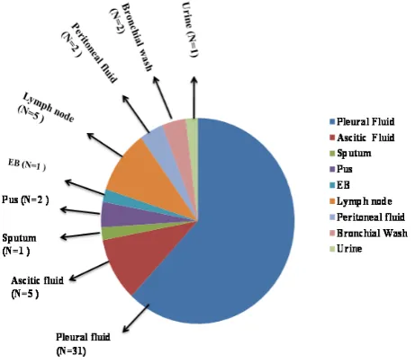
[image:2.595.320.551.355.557.2]Fifty (N=50) clinical tuberculosis patients (Figure 1) attending outdoor patient department (OPD) of Lala Ram Sarup Institute of Tuberculosis and Respiratory Diseases, New Delhi, India, for their illness during the period of 2012-2013 year were recruited for this current study. All the samples (extra-pulmonary and pulmonary tuberculosis samples) were collected in sterile containers from patients. Among 50 patients 35 (70%) were men and 15 (30%) women with age ranged from 16-68 year. Majority of patients were observed between their reproductive ages 20-45 years which represented that overall increase rate of infection in younger one in endemic areas. Collected samples were further categorized into sputum (N=1), pleural fluid (N=31), ascitic fluid (N=5), pus (N=2), endometrial blood (N=1), lymph node (N=5), peritoneal blood (N=2), bronchial washing (N=2) and urine (N=1). Data of clinically presentations, radiological and other laboratory examinations for diagnosis of extra-pulmonary tuberculosis (included smears and culture and other cyto-pathological reports wherever available etc) were also collected from OPD and ward for these patients.
Figure 1. Graphical representation of clinical tuberculosis samples (N=50) recruited for the study during 2012-2013 period. Further extra pulmonary samples were categorized as pleural fluid (N=31), ascitic fluid (N=5), lymph node (N=5), peritoneal fluid (N=2), pus (N=2), endometrial blood (EB, N=1), urine (N=1). PTB samples were sputum (N=1) and bronchial wash (N=2).
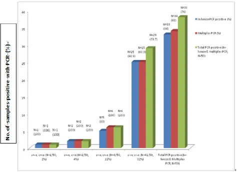
Abbreviations:S+ve = smear positive, S-ve= smear negative, C+ve=culture positive, C-ve=culture negative, N=number
Figure 2. In house (IS6110 gene target) PCR compared with multiplex PCR results in tuberculosis samples.
2.2.2 Processing of the samples
All samples were decontaminated by standard protocol i.e.
N-acetyl-L-cysteine (NALC)–sodium hydroxide (NaOH) procedure which included 2% NaOH, 2.9% trisodium citrate, 0.5% NALC [17]. The processed samples were used for ZN smears, culture on LJ medium (bacteriological identification as a gold standard) and for DNA extraction for molecular methods. DNA was extracted from all body fluids as well as sputum by QIAamp DNA Mini Kit (Qiagen, Germany) according to manufacturer’s instructions and protocols. Eluted DNA was stored at -20oC. To avoid contamination during DNA extraction and amplification, strict precautions were taken, including separate areas for DNA extraction, reagent preparation, amplification and product detection and regular meticulous cleaning of surface with 10% hypochlorite were also applied to maintain the standard molecular laboratory procedures 2.2.3. DNA Amplification of Mycobacterium Detection
Amplification of bacterial DNA was performed by using In-House PCR (IS6110 and MPB64 gene targets) and through multiplex M.tuberculosis PCR kit (SeeplexRMTB
ACE Detection Kit) in extracted DNA of all pulmonary and
extra-pulmonary samples as per the below mentioned protocols and procedures.
2.2.3.1. In-house PCR:
Total 25µl of PCR reaction volume was containing 1x PCR buffer, 0.2 mM dNTPs, 50ŋg of each primer (IS6110: Forward 5’- CCT GCG AGC GTA GGC GTC GG-3’ and Reverse 5’-CTC GTC CAG CGC CGC TTC GG-3’) & MPB64: Forward 5’-TCC GCT GCC AGT CGT CTT CC-3’ & Reverse 5’-GTC CTC GCG AGT CTA GGC CA-3’ [19, 31], 1 unit of Taq polymerase (Banglore Genei, India) and 10µl of tested sample DNA along with positive control (strain of M.tuberculosis H37Rv) and negative control (nuclease free water).
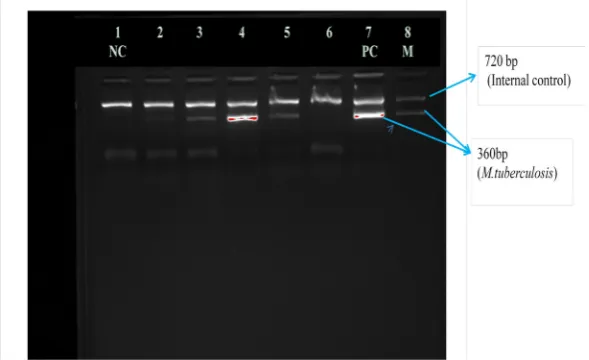
Figure 3A.
IS6110 gene targeted In-house PCR for detection of Mycobacteriumtuberculosis. Electrophoretic separation of the amplicon into 2% agarose gel is documented across Lanes 1-5. Lanes-1 represents the negative control (nuclease free water), Lane-2 and 3 represent clinical extra-pulmonary samples, Lanes-4 was positive control (Mycobacterium tuberculosis culture H37Rv). Lane-5 was 100bp marker. The presence of a 123 bp amplicon in the lanes 2-4 indicated the presence of the target.
Figure 3B. MPB64gene targeted In-house PCR for detection of Mycobacterium tuberculosis. Electrophoretic separation of the amplicon into 2% agarose gel is documented across Lanes 1-10. Lanes-1 represents the negative control (nuclease free water), lane-2 to 8 represents clinical extra pulmonary samples, Lanes-9 was positive control H37Rv (Mycobacterium tuberculosis culture). Lane-10 was 50bp marker. The presence of a 240bp. amplicon in the lanes 2 to 4, 9 indicated the presence of the target.
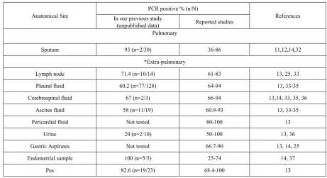
Figure 4. Commercial multiplex M. tuberculosis PCR kit(multitarget IS6110 and MPB64) for M.tuberculosis detection. Lane 1: negative control representing only internal control band (720bp) and absence of M.tuberculosis gene targets (360bp). Lane 2-6: clinical extra pulmonary samples, Lane 7: positive controlrepresenting both internal control band (720bp) and M.tuberculosis gene targets (360bp). As per the manufactures protocols internal control
[image:4.595.159.454.511.691.2]2.2.3.2. Multiplex M. tuberculosis PCR kit (SeeplexRMTB ACE Detection Kit)
Simultaneously, similar samples (pulmonary and extra pulmonary cases) were also tested with commercially available multiplex M. tuberculosis PCR kit (SeeplexRMTB ACE Detection Kit; www. Seegene.com).Amplification was performed according to the manufacturer instruction and protocols. Manufacturer has mentioned the principal of Dual Priming Oligonucleotide (DPOTM) technology, which provides freedom in primer design and PCR optimization and maximizes PCR specificity and sensitivity by fundamentally blocking of non specific priming. It is a multiplex assay that permits the amplification of target DNA of M. tuberculosis. The PCR kit designed on multi-targets (IS6110 and MPB64) PCR instead of a single target PCR for specific detection of M. tuberculosis in a single tube. To prevent false negative results caused by lack of this insertion sequence this kit carries out both (IS6110 and MPB64 genes) PCR. The kit contained following reagent for amplification. The 2X Multiplex Master Mix contains dNTP and enzyme for the specific amplification of the pathogens genome and 10XMTB ACE PM is primer mixture for initiation of PCR. The internal control (DNA plasmid) was provided within the primers mixtures in kit for each amplification reactions to identify processed samples containing substances that may interfere with PCR amplification. These features were selected to ensure equivalent amplification of internal control and M. tuberculosis target DNA. The internal control is introduced into each amplification reaction and is co-amplified with target DNA from the clinical samples. Each set of amplification protocols also have one positive (M.tuberculosis H37Rv strain culture) and negative (nuclease free water) controls. In addition, the 8- methoxypsoralen (8-MOP) system is used to extinguish the template activity of contaminated DNAs. It is known to intercalate into double stranded nucleic acids and of contaminated DNAs.
The amplification protocol involved the following amplifications steps (1 cycle at 94°C for 15 min; 40 cycles at 94°C for 30s, 62°C for 1.30min, 72°C for 1.30min; 1 cycle at 72 °C for 10 min). The gel electrophoresis was performed on 2% agarose gel (Fermentas, Lithuania),at 80 V for 60 min
and further visualized under Gel DOCTM XR+ system (Bio-Rad, USA). The product sizes for internal control & M. tuberculosis amplicons were 720 bp & 360bp, respectively (Figure 4).
2.3. Statistical Analysis
The statistical calculations were performed using MSTAT software by Chi square (χ2 )test. The p values <0.05 were considered to be significant.
3. Results
The study compared the efficacy of In House PCR with commercial multiplex M. tuberculosis PCR kit in extra-pulmonary paucibacillary smear negatives samples for the detection of M.tuberculosis. The conventional culture on LJ media was used as gold standard for the detection of mycobacterium growth. Out of 50 cases, 3(6%) were smear positive (S+ve), and 7 (14%) were culture positive (C+ve) (Table-1 and Figure 2). Overall bacteriological results which include ZN smear & LJ culture showed 9 (18%) positive tuberculosis samples. The total PCR positivity (N=50) by In House PCR was found to be 66% and 68% with multiplex M. tuberculosis PCR kit. Further overall combined PCR positivity by both PCR (In House PCR with multiplex M. tuberculosis PCR kit) was observed 76% (38/50) which increased approximately 10%. However the positivity was remained 70.7% (29/50) in smear negative (S-ve), culture negative (C-ve) samples (Table 1). The statistical analysis using MSTAT software by Chi square (χ2) test indicated no significant difference between the positivity of both PCR (p = 0.832; χ2 =0.045).
However PCR positivity was found to be 100% in smear positive, culture negative samples (S+ve, C-ve) (2/2) & smear positive, culture positive ( S+ve, C+ve) (1/1) samples through both PCR amplification assays.
PCR positivity in smear negative and culture positive samples (S-ve, C+ve) was 83.3% (5/6) and 100% (6/6) by In House PCR and multiplex M. tuberculosis PCR kit, respectively.
Table 1.
Comparative analysis of In House PCR with multiplex M. tuberculosis PCR kit
Smear/ culture (N, %)
smear +ve, culture +ve (a=1/50; 2%)
smear +ve, culture -ve (b=2/50; 4%)
smear -ve, culture +ve (c=6/50; 12%)
smear -ve, culture -ve (d=41/50; 82%)
Total PCR positive (N=50)
In House PCR positive(%) 1 (100) 2 (100) 5(83.3) 25 (60.9) 33 (66)
Multiplex M. tuberculosisPCR kit(%) 1 (100) 2 (100) 6 (100) 25 (60.9) 34 (68)
Total PCR positive
(N=50)(Both PCR) 1 (100) 2 (100) 6 (100) 29 (70.7) 38 (76)
Total Smear positives: n= 3(6%) Total Culture positives: n=7(14%)
Total Smear and Culture positives: n=9(18%)
Table 2. Comparison of the In House PCR positivity with other reported studies
Anatomical Site In our previous study PCR positive % (n/N) References
(unpublished data) Reported studies Pulmonary
Sputum 93 (n=2/30) 36-86 11,12,14,32
*Extra-pulmonary
Lymph node 71.4 (n=10/14) 61-83 13, 25, 33
Pleural fluid 60.2 (n=77/128) 64-94 13, 33-35
Cerebrospinal fluid 67 (n=2/3) 66-94 13,14, 33, 35, 36
Ascites fluid 58 (n=11/19) 60.9-93 13, 33-35
Pericardial fluid Not tested 80-100 13
Urine 20 (n=2/10) 50-100 13, 36
Gastric Aspirates Not tested 66.7-90 13, 14, 25
Endometrial sample 100 (n=5/5) 25-74 14, 37
Pus 82.6 (n=19/23) 68.4-100 13
Majority of the extra pulmonary samples were smear negative
N=Total number of samples
n= Positive PCR for IS6110 gene
4. Discussion
Tuberculosis i.e. pulmonary and extra-pulmonary tuberculosis is a significant health problem, in both developing and developed countries. The reported proportions of extra-pulmonary tuberculosis in developed countries ranged from 12% to 28.5% while in India, 10 to 20% extra-pulmonary tuberculosis cases were reported which affects mainly the lymph nodes, pleural area, kidney, spine, and growing ends of the bones with a 25% to 50% case mortality rate within months [18]. Many forms of extra-pulmonary tuberculosis are difficult to diagnose by less sensitive & time consuming conventional methods (microscopy and culture) because of the paucibacillary nature of these specimens. M.tuberculosis detection by PCR was found to be significantly more sensitive and rapid compared to culture and microscopy with sensitivities ranging between 36% and 100% and specificities ranging between 85% and 100% using various PCR targets such as IS6110, MPB64, TRC4, GCRS, hup–B, Pab gene etc. [13,19-23]. In addition, we further compared the results of PCR positivity (our previous unpublished data in smear negative extra-pulmonary samples) as represented in Table 2. Our finding has similar concordance with reported published studies with variable positivity rates of PCR in different categories of extra-pulmonary samples. However, due to variability in the sensitivity rates in different studies, the role of PCR remains controversial [13,19-23].
In this study, In House PCR and commercial multiplex M.
tuberculosis PCR kit were used for detection of
M.tuberculosis in clinical samples particularly
extra-pulmonary paucibacillary smear negatives. The multiplex M.tuberculosis PCR kit allows the amplification of target DNA of M.tuberculsois complex which utilized multi-targets (IS6110 and MPB 64) instead of a single-target in PCR amplification assays for specific detection of
M.tuberculosis. IS6110 sequence is the most widely used target gene for detection of M.tuberculosis. However, several M.tuberculosis strains that lack this insertion sequence have been isolated and reported [24]. To prevent false-negative results caused by lack/zero copy of this insertion sequence, multiplex M. tuberculosis PCR kit carries out both IS6110 and MPB64 genes.
Further data analysis, showed that M.tuberculosis
detection rate (PCR positivity) by single gene target In house- PCR was found to be similar 66% (33/50) with 68% (34/50) in multiplex M. tuberculosis PCR kit with no significant difference in their positivity rate which further suggested the utility of in house PCR protocols in early diagnosis of extra-pulmonary tuberculosis in poor setting or developing country like India having higher burden of disease. The overall positivity rates were increased up to 10% (76%; 38/50) further in addition to multigene targeting primers for detection of M. tuberculosis in clinical samples. It has been earlier reported that the sensitivity of PCR can be increased by using one more sets of primers in extra-pulmonary tuberculosis cases i.e 77% [25].
Several other studies reported that conventional method culture (4% to 43.3%) and smear (0.4% to 28.5%) has low
M.tuberculosis detection rate in extra-pulmonary
M.tuberculosis detection rate by LJ culture (14%; 7/50) and by ZN smear (6%; 3/50) because of paucibacillary specimens, use of the solid LJ medium in culture method, and presence of non viable bacterial cells in the samples.
On the other side, the PCR positivity was found 100% in smear positive samples by utilizing In House PCR as well as commercial multiplex M. tuberculosis PCR kit. One culture positive & smear negative sample could not detect by In house PCR. It may be due to the presence of zero copy (0-19 copies of IS6110) [24]. However, one false negative result was observed through In-house PCR. This could be explained or resulted from (a) The presence of inhibitors not detected by the control amplification; (b) Non-homogenous distribution of bacteria or that the fraction tested does not contain mycobacteria; and (c) low number of bacilli in the sample. Also, a number of studies in India have shown that, nucleotide sequence in IS6110 is not always present within strains of M. tuberculosis causing disease in India. Primers with different nucleotide sequence are now being used in India by a few laboratories to ensure that all cases are detected by PCR amplification [6,19, 21, 31].
Further data interpretation, revealed the equal positivity detection rate of M.tuberculosis by utilizing in house PCR and commercial multiplex M. tuberculosis PCR kit in the cases of smear negatives and culture negative (S-veC-ve) (25/41, 60/9%). We further analyzed the clinical finding, radiological features and other laboratory observations in these 25 smear negatives and culture negative cases (S-ve, C-ve, PCR+ve) which were found to be positive by PCR for the diagnosis of tuberculosis. Out of 25 cases, total six cases (pleural fluid-2, ascitic fluid-1, lymph node-1, sputum-1, bronchial wash-1) were reported on ATT with good response to treatment, 12 pleural effusion/ pleurisy tuberculosis cases (unilateral, N=11 & bilateral, N=1) further out of eleven unilateral cases 2 cases were with high level of ADA in pleural effusion which indicated pleurisy tuberculosis in these suspects. One unilateral case was left venrticular failure (LVF) with massive pleural thickening further confirmed the pleurisy tuberculosis. Two lymphadenopathy cases were cervical lymph nodes with manutoux positive. One case was bronchial wash with cavitations on ATT, four ascites cases with lymphocytic predominance and negative bacterial cultures. One urine cases was suspected to genital urinary tuberculosis. One endometrial blood was suspected to have female genital tuberculosis. Two peritoneal cases were suspected to have abdominal koch’s.
5. Conclusion
This study reveals that In House PCR and multiplex M. tuberculosis PCR kit both have significant utility in the early diagnosis (by decreasing time duration) of extra pulmonary smear negatives tuberculosis which is not diagnosed by conventional test or low detection rate by ZN smear and LJ culture. In conclusion, multiplex M. tuberculosis PCR kit
showed almost equivalent sensitivity and specificity to a more labor intensive endpoint In House PCR. The overall positivity rate increased while combining the results of both PCR assays. The criteria of diagnosis of tuberculosis in smear and culture negative extra-pulmonary tuberculosis suspect’s may constitute and the epidemiological, clinical, radiological, tuberculin testing, and response to treatment, along with M. tuberculosis PCR. To conclude, more work is needed to evaluate the utility of PCR for diagnosing tuberculosis in extra-pulmonary cases in Indian scenario.
Acknowledgements
The study was funded by LRS Institute of Tuberculosis & Respiratory Diseases. We thank the staff members of the Department of Microbiology. We appreciate the participants for their good will to give the necessary information.
REFERENCES
[1] World Health Organization. "The global tuberculosis report 2012".
http://www.who.int/tb/publications/global_report/en/index.ht ml.2012.
[2] S. K. Kabra, R. Lodha, V. Seth. Some current concepts on childhood tuberculosis. Indian J Med Res, 120,387-397, 2004.
[3] B. J. Marais, M. Pai. Recent advances in the diagnosis of childhood tuberculosis. Arch Dis Child, 92, 446-452, 2007. [4] E. L. Corbett, C. J. Watt,N. Walker, D. Maher,B. G. Williams,
M. C. Raviglione, C. Dye. The Growing Burden of Tuberculosis : Global Trends and Interactions With the HIV Epidemic. Arch Intern Med, 163, 1009-1021, 2003.
[5] S. K. Sharma, A. Mohan. Tuberculosis: From an incurable scourge to a curable disease - journey over a millennium. Indian J Med Res, 137, 455-493. 2013.
[6] K. Sharma, M. Modi, K. Goyal, A. Sharma, P. Ray, S. K. Sharma, S. Prabhakar, S. Varma, M. Sharma. Evaluation of PCR using MPB64 primers for rapid diagnosis of tuberculosis Meningitis. 1: 204. doi:10.4172/scientificreports.204, 2012. [7] D. Rimek, S. Tyagi, R. Kappe. Performance of an
IS6110-based PCR assay and the COBASamplicor MTB PCR system for detection ofMycobacterium tuberculosis complex DNAin human lymph node samples.J ClinMicrobiol, 40,3089–3092, 2002.
[8] S. J. Drews, A.Eshaghi,D. Pyskir, P.Chedore, E. Lombos, G. Broukhanski, R. Higgins, D. N. Fisman, J. Blair,F. Jamieson. The relative test performance characteristics of two commercial assays for the detection of Mycobacterium tuberculosis complex in paraffin-fixed human biopsy s Diagnostic Pathology,3, 37-40, 2008.
[10] S. V. Balasingham, T. Davidsen, I. Szpinda, S. A. Frye, T. Tonjum. Molecular diagnostics in tuberculosis: Basis and implications for therapy. MolDiognTher, 13, 137-151, 2009. [11] S. Kulkarni, P. Singh, A.Memon, G. Nataraj, S. Kanade, R. Kelkar. An in-house multiplex PCR test for the detection of Mycobacterium tuberculosis, its validation &comparision with a single target TB-PCR kit. Indian J Med Res, 135, 788-794, 2012.
[12] A. Mashta, P. Mishra, S.Philipose, S. Tamilzhalagan, H. Mahmud, S. Bhaskar, P.Upadhyay. Diagnosis of tuberculosis: the experience at a specialized diagnostic laboratory.JNegat Results Biomed, 10, 16-22, 2011.
[13] A. K. Maurya, S. Kant, V. I. Nag, R. Kushwaha, T. N. Dhole. Detection of 123bp fragment of insertion element IS6110 Mycobacterium tuberculosis for diagnosis of extra pulmonary tuberculosis. Ind J Med Microbiol, 30, 182-186, 2012. [14] I. Shukla, S. Varshney, Sarfraz, A. Malik, Z. Ahmad.
Evaluation of nested PCR targeting IS6110 of Mycobacterium tuberculosis for the diagnosis of pulmonary and extra-pulmonary tuberculosis. Biology and Medicine, 3, 171-175, 2011.
[15] J. Y. Chun, K. J. Kim, I. T. Hwang, Y. J. Kim, D. H. Lee, I. K. Lee, J. K. Kim.Dual priming oligonucleotide system for the multiplex detection of respiratory viruses and SNP genotyping of CYP2C19 gene. Nucleic Acids Res, 35, e40, 2007.
[16] R. Srivastava, R. P.Punde, H. Pandey, R. M. Samarth, K. K. Maudar. Evolutionary development of molecular tools in the diagnosis of Mycobacterium tuberculosis: A Review. J Med Sci, 10, 124-129, 2010.
[17] P. T. Kent, G. P. Kubica. Public Health Mycobacteriology: A guide for the level III laboratory. Washington, DC: U.S. Department of Health and Human Services, 1985.
[18] S. Gunal, Z. Yang, M. Agarwal, M. Koroglu, Z. K. Arıcı, R. Durmaz. Demographic and microbial characteristics of extra pulmonary tuberculosis cases diagnosed in Malatya, Turkey, 2001-2007. BMC Public Health, 11,154-161, 2011. [19] N. V. Bhanu, U. B. Singh, M. Chakravorty, N. Suresh, J.
Arora, T. Rana, D. Takkar, P. Seth. Improved diagnostic value of PCR in the diagnosis of female genital tuberculosis leading to infertility. J Med Microbiol, 54, 927–931, 2005. [20] S. Chakravorty, M. K. Sen, J. S. Tyagi. Diagnosis of extra
pulmonary tuberculosis by smear, culture, and PCR using universal sample processing technology. J Clin Microbiol, 43, 4357–4362, 2005.
[21] S. Sankar, S.Kuppanan, B. Balakrishnan, B. Nandagopal. Analysis of sequence diversity among IS6110 sequence of Mycobacterium tuberculosis: possible implications for PCR based detection. Bioinformation, 6, 283-5, 2011.
[22] V. M. Katoch. Newer diagnostic techniques for tuberculosis. Ind J Med Res, 120, 418-428, 2004.
[23] A. Jain. Extra pulmonary tuberculosis: A diagnostic dilemma. Ind J ClinBiochem, 26, 269–273, 2011.
[24] D. S. Chauhan, V. D. Sharma, D. Parashar, A. Chauhan, D. Singh, H. B. Singh, R. Das, B.M. Aggarwal, B. Malhotra, A. Jain, M. Sharma, V.K. Kataria, J.K. Aggarwal, M.Hanif, A.Shahani, V.M. Katoch. Molecular typing of Mycobacterium tuberculosis isolates from different parts of India based on IS6110 element polymorphism using RFLP analysis. Ind J Med Res, 125, 577-581, 2007.
[25] H. B. Singh, P. Singh, G. P. S. Jadaun, Srivastava K, Sharma VD, Chauhan DS, Sharma SK, Katoch VM. Simultaneous use of two PCR systems targeting IS6110 and MPB64 for confirmation of diagnosis of tuberculous lymphadenitis. J Commun Dis, 38, 274-279, 2006.
[26] A.N. Agarwal, D. Gupta, S. K. Jindal. Diagnosis of tubercular pleural effusion. Ind J Chest Dis Allied Sci, 41, 89–100, 1999. [27] S. Iqbal, R. Ahmed, S. Z. Adhami, A. Mumtaz. Importance of polymerase chain reaction in diagnosis of pulmonary and extra-pulmonary tuberculosis. J Ayub Med Coll Abbottabad, 23, 73-76, 2011.
[28] R. Misra, S. P. Sharma, R. Jina, N. Pant, D. K. Srivastava. Female genital tract tuberculosis with special reference to sterility in Eastern UP. J ObstetGynaecol India, 104–109, 1996.
[29] K. L. Therese, U. Jayanthi, H. N. Madhavan. Application of nested polymerase chain reaction (nPCR) using MPB64 gene primers to detect Mycobacterium tuberculosis DNAin clinical s from extra pulmonary tuberculosis patients. Indian J Med Res, 122, 165-170, 2005.
[30] S. H. Bouakline, J. P. Vincensini, V.Giacuzzo, P. H. Lagrange, J. L. Herrmann. Rapid diagnosis of extra pulmonary tuberculosis by PCR: Impact of sample preparation and DNA extraction. J ClinMicrobiol, 41, 2323–2329, 2003.
[31] D. Kumar, R. Srivastava, S. B. Srivastava. Epidemiology of tuberculosis by molecular tools. Proc Nat AcadSci India, 65, 113-121, 1995.
[32] J. Cordova, R. Shiloh, R. H. Gilman, P. Sheen, L. Martin, F. Arenas, L. Caviedes, V. Kawai, G. Soto, D. L. Williams, M. Zimic, A. R. Escombe, C. A. Evans. Evaluation of molecular tools for detection and drug susceptibility testing of Mycobacterium tuberculosis in stool s from patients with pulmonary tuberculosis. J Clin Microbiol, 48, 1820–1826, 2010.
[33] B. Sekar, L. Selvaraj, A. Alexis, S. Ravi, K. Arunagiri, L. Rathinavel. The utility of IS6110 sequence based polymerase chain reaction in comparision to conventional methods in the diagnosis of extra-pulmonary tuberculosis. Indian J Med Microbiol, 26, 352-355, 2008.
[34] R. L. C. Orallo, M. T. Mendoza, M. A. D. Lansang, C. F. Ang. Evaluation of the usefulness of PCR in the diagnosis of Mycobacterium tuberculosis in tissues and body fluids in UP-Philippine General Hospital. Philippine Microbiol Infect Dis, 37, 20-32, 2008.
[35] L. Portillo-Gómez, S. L. Morris, A. Panduro. Rapid and efficient detection of extra-pulmonary Mycobacterium tuberculosis by PCR analysis. Int J Tuberc Lung Dis, 4, 361–70, 2000.
[36] G. Garcia-Elorriaga, C. Gracida-Osorno, G. Carrillo-Montes, C. Gonzalez-Bonilla. Clinical usefulness of the nested polymerase chain reaction in the diagnosis of extra pulmonary tuberculosis. SaludPublicaMex, 51:240-245, 2009.