Human major group rhinoviruses
downmodulate the accessory function of
monocytes by inducing IL-10
Johannes Stöckl, … , Ernst Kuechler, Walter Knapp
J Clin Invest.
1999;
104(7)
:957-965.
https://doi.org/10.1172/JCI7255
.
Human rhinoviruses (HRVs) are the predominant cause of the common cold. Although this
disease is per se rather harmless, HRV infection is considered to set the stage for more
dangerous pathogens in vivo. Here we demonstrate that HRV-14, a member of the major
group HRV family, can efficiently inhibit antigen-induced T-cell proliferation and T-cell
responses to allogeneic monocytes. HRV-14 triggered a significant downregulation of MHC
class II molecules on monocytes. Moreover, supernatants from monocytes cultured in the
presence of HRV-14 strongly reduced the allogeneic T-cell stimulatory property of untreated
monocytes and monocyte-derived dendritic cells (md-DCs), whereas Epstein Barr virus–
transformed B-lymphoblastoid cells were not sensitive. Analysis of the supernatant revealed
that HRV-14 induced the production of significant amounts of the immunosuppressive
cytokine IL-10. The important T-cell stimulatory cytokine IL-12 or the proinflammatory
cytokines IL-1
b
or TNF-
a
were not detected or were only minimally detected. Finally,
monocytes pretreated with HRV-14 were greatly inhibited in their production of IL-12 upon
stimulation with IFN-
g
/LPS. These observations suggest that altered cytokine production in
mononuclear phagocytes upon interaction with HRV downmodulates appropriate immune
responses during the viral infection.
Article
Introduction
Common colds induced by the human rhinovirus (HRV) occur worldwide (1). The frequent appearance of HRV, and its economic importance in terms of employee absenteeism, physician visits, and medication costs in industrial countries, make it a subject of pri-mary importance (2). Despite this, the pathogenesis of HRV infection remains poorly understood (1, 3).
HRV primarily infects the ciliated epithelial cells of the upper respiratory tract (4). However, histological exam-inations of HRV-infected nasal epithelium demonstrat-ed no obvious changes in the morphology or integrity of the nasal epithelium (4, 5). Instead, HRV infection is accompanied by a release of inflammatory mediators (3). In particular, proinflammatory cytokines including IL-1β, TNF-α, IL-8, IL-6, and IL-11 (6–8) and the vasoactive peptides bradykinin and lysyl-bradykinin (9, 10) were found in nasal secretions of patients with colds. In addi-tion, cytokine production was detectable in HRV-infect-ed epithelial cells in vitro (6–8, 10–13). As a result, it is now considered that common cold symptoms result from an inflammatory “cytokine disease” (3).
Attracted by these mediators, inflammatory leuko-cytes, preferentially granulocytes and monoleuko-cytes, are found to migrate to the site of HRV infection (9, 14, 15). However, despite recruitment of invading leukocytes, appropriate immune responses appear to be hindered or dysregulated in the respiratory tract upon HRV
infec-tion. This causes a well-documented disposition to bac-terial infections leading to sinusitis, otitis media, bron-chitis, pneumonia, and asthmatic exacerbations (16–24). Here we demonstrate that interaction of immune cells with HRV-14, a member of the major group HRV fami-ly, efficiently inhibits antigen-specific T-cell responses by inducing IL-10 production in mononuclear phagocytes. IL-10 is a prominent immunosuppressive cytokine, which is critically involved in terminating inflammato-ry reactions. Owing to its anti-inflammatoinflammato-ry properties, release of IL-10 might be responsible for the disturbed cellular immune responses during HRV infection.
Methods
Media, reagents, and chemicals. The cell culture medium RPMI-1640 (GIBCO BRL, Grand Island, New York, USA) supplemented with 2 mM L-glutamine, 10% FCS, 100 U/mL penicillin, and 100 µg/mL streptomycin was used in this study. The HRV-blocking reagent WIN 52035-2 (25) was a kind gift from the Sterling-Winthrop Research Institute (Rensselaer, New York) and was used at a final concentration of 5 µg/mL. The superantigens staphylococcal enterotoxin A (SEA) and B (SEB) from
Staphylococcus aureuswere obtained from Serva (Heidel-berg, Germany) and were used at a concentration of 10 ng/mL. Tetanus toxoid and purified protein derivative of Mycobacterium tuberculosis(PPD) were purchased from Connaught Laboratories (Willowdale, Ontario,
Cana-Human major group rhinoviruses downmodulate the
accessory function of monocytes by inducing IL-10
Johannes Stöckl,
1Helga Vetr,
2Otto Majdic,
1Gerhard Zlabinger,
1Ernst Kuechler,
2and Walter Knapp
11Institute of Immunology, and
2Institute of Biochemistry, University of Vienna, A-1090 Vienna, Austria
Address correspondence to: Johannes Stöckl, Institute of Immunology, University of Vienna, Borschkegasse 8a, A-1090 Vienna, Austria. Phone: 431-4277-64935; Fax: 431-4277-9649; E-mail: [email protected].
Received for publication May 4, 1999, and accepted in revised form August 10, 1999.
Human rhinoviruses (HRVs) are the predominant cause of the common cold. Although this disease is per se rather harmless, HRV infection is considered to set the stage for more dangerous pathogens in vivo. Here we demonstrate that HRV-14, a member of the major group HRV family, can efficient-ly inhibit antigen-induced T-cell proliferation and T-cell responses to allogeneic monocytes. HRV-14 triggered a significant downregulation of MHC class II molecules on monocytes. Moreover, super-natants from monocytes cultured in the presence of HRV-14 strongly reduced the allogeneic T-cell stimulatory property of untreated monocytes and monocyte-derived dendritic cells (md-DCs), where-as Epstein Barr virus–transformed B-lymphoblwhere-astoid cells were not sensitive. Analysis of the super-natant revealed that HRV-14 induced the production of significant amounts of the immunosup-pressive cytokine IL-10. The important T-cell stimulatory cytokine IL-12 or the proinflammatory cytokines IL-1βor TNF-αwere not detected or were only minimally detected. Finally, monocytes pre-treated with HRV-14 were greatly inhibited in their production of IL-12 upon stimulation with
IFN-γ/LPS. These observations suggest that altered cytokine production in mononuclear phagocytes upon interaction with HRV downmodulates appropriate immune responses during the viral infection.
da) and used at a concentration of 1 µg/mL. LPS from
Escherichia coli(serotype 0127-B8) and polymyxin B were obtained from Sigma Chemie GmbH (Deisenhofen, Germany). Recombinant human GM-CSF and IL-4 were kindly provided by Novartis Research Institute (Vienna, Austria). IFN-γwas a gift from G.R. Adolf (Ernst Boehringer Institut für Arzneimittelforschung, Vienna, Austria). IL-10 was purchased from R&D Sys-tems Inc. (Minneapolis, Minnesota, USA).
Antibodies used in this study. The following murine mAb’s were generated in our laboratory: negative con-trol mAb VIAP (calf intestine alkaline phosphatase–spe-cific), 6B6 (CD11a), VIM13 (CD14), 4D3 (CD33), and 1/47 (MHC class II). Hybridomas producing mAb
W6/32 (MHC class I), G28-5 (CD40), and TS2/9 (CD58) were obtained from American Tissue Culture Collection (ATCC; Rockville, Maryland, USA). UCHT-1 (CD3), MEM18 (CD14), and UCHL1 (CD45R0) were kindly provided by An der Grub (Bio Forschungs GmbH, Kaumberg, Austria). OKT3 (CD3) was obtained from Ortho Diagnostics (Raritan, New Jersey, USA). The mAb HD37 (CD19) was provided by G. Moldenhauer (Hei-delberg, Germany). MAb RR1/1 (CD54) was a gift from Bender AG (Vienna, Austria). The mAb L307 (CD80) was from Becton Dickinson Immunocytometry Sys-tems (San Jose, California, USA). IT2.2 (CD86) and the PE-labeled mAb C11.5 (anti–IL-12) were purchased from PharMingen (San Diego, California, USA). MAb 3G8 (CD16), the FITC-labeled mAb MP9-20A4 (anti–TNF-α), and PE-labeled mAb JES3-9D7 (anti–IL-10) for cytoplasmic staining of IL-10 were obtained from Caltag Laboratories Inc. (Burlingame, California, USA). FITC-labeled FIB-3 mAb (anti–IL-1β) was from Dianova (Hamburg, Germany). The neutralizing poly-clonal anti–IL-10 antibody (PAL-hIL10) was obtained from Strahtmann Biotech (Hannover, Germany).
Rhinovirus preparation. HRV-14 was obtained from ATCC and routinely grown in suspension cultures of HeLa cells (strain Ohio; Flow Laboratories, McLean, Vir-ginia, USA). Cells were cultivated in S-MEM medium (Joklik modification; catalog no. 22300-107; Life Tech-nologies Inc., Rockville, Maryland, USA) supplemented with 7% heat-inactivated horse serum (catalog number 01051-M; Diagnostic Products GesmbLT, Wiener Neu-dorf, Austria), 1% penicillin/streptomycin, 1% glutamine, 1% pluronic F68 (catalog no. P1300; Sigma Chemical Co., St. Louis, Missouri, USA), and 1% nonessential amino acids. The same medium was used during infec-tions, except that serum was reduced to 2% and 1 mM MgCl2was added. Cells were infected at a ratio of ten 50% tissue culture infectious doses (TCID)50per cell. HRV-14 was harvested 40 hours after infection. Cell debris was pelleted by low-speed centrifugation. HRV was prepared from cell culture supernatant by poly-ethanolglycol (PEG) precipitation (7% PEG 6000, 3% NaCl). Virus was resuspended from the PEG pellet in PBS, aliquoted, and stored at –70°C. Working dilutions of used HRV-14 stock preparations contained less than 10 pg LPS/mL. No biologic effects of these very low LPS concentrations were detectable. The preparations did not induce significant TNF-αor IL-6 production in monocytes (see Figure 5). Moreover, the capacities of HRV-14 preparations to induce IL-10 production, inhib-it T-cell proliferation, and downregulate MHC class II expression could not be inhibited by the LPS inhibitor polymyxin B (10 µg/mL) (data not shown).
[image:3.612.80.284.49.404.2]Preparation of purified HRV. Virus stock preparations were further purified using sucrose gradients. Virus was pelleted by centrifugation at 100,000 gfor 2 hours and resuspended in a small volume of “virus buffer” (50 mM Tris [pH 7.5], 2 mM MgCl2). It was then treated with DNase I (5 mg/mL; catalog no. 104169; Boehringer Mannheim, Mannheim, Germany) and RNase A (5
Figure 1
HRV-14 inhibits antigen-specific T-cell proliferation. PBMCs (105)
were stimulated with mAb OKT3 (1 µg/mL), SEA or SEB (10 ng/mL), and PPD or tetanus toxoid (1 µg/mL) in the presence (filled bars) or absence (gray bars) of HRV-14 (10 TCID50 per cell). T-cell
prolifera-tion was measured on day 3 (for OKT3, SEA, SEB stimulaprolifera-tion) and day 5 (for PPD, tetanus toxoid stimulation) of culture by adding (methyl-3H)-TdR followed by measuring thymidine incorporation 18
mg/mL; catalog no. 104159; Boehringer Mannheim) for 10 minutes at room temperature and further digested with trypsin (0.5 mg/mL; catalog no. 01104-H; Dipro) for 5 minutes at 37°C. After addition of N -laurylsar-cosin (0.1%; catalog no. LA083; Schuchardt, Munich, Germany) digestion was continued at 4°C overnight. After removal of unsoluble material by low-speed cen-trifugation, the sample was centrifuged on a sucrose gradient (7.5–45% sucrose in virus buffer) for 2 hours at 155,000 g. Virus-containing fractions are visible as a tur-bid band in the middle of the gradient and were col-lected with a syringe. Virus was concentrated by pellet-ing and resuspension in virus buffer.
Ultraviolet inactivation of virus. Ultraviolet-inactivated (UV-inactivated) virus was prepared by irradiating virus suspensions in a 24-well tissue culture dish (200
µL/well) on ice for 10 minutes with a 75 W UV source (254 nm) at a distance of 5 cm. Treatment resulted in complete loss of infectious titer.
Cell preparation. PBMCs were isolated from heparinized whole blood of normal healthy donors by standard den-sity gradient centrifugation with Ficoll-Paque (Pharma-cia, Uppsala, Sweden). Subsequently, T cells and mono-cytes were separated by magnetic sorting using the MACS technique (Miltenyi Biotec GmbH, Bergisch Gladbach, Germany) as described previously (26). Puri-fied T cells were obtained through negative depletion of CD11b, CD14, CD16, CD19, CD33, and MHC class II–positive cells with the respective mAb’s. Monocytes were enriched by using the biotinylated CD14 mAb VIM13 (purity >95%) as described previously (26).
Cultivation of monocytes.Monocytes (1 ×10⁶/mL) were cultured in 24-well plates (Corning-Costar Europe, Bad-hoevedorp, the Netherlands) in the presence of HRV-14
(10 TCID50 per cell) or aliquot amounts of supernatants from uninfected HeLa cells at 37°C. After 2 days, cells were harvested, washed in PBS, and analyzed by flow cytometry. Monocyte-derived dendritic cells (md-DCs) were generated by culturing purified blood monocytes for 8 days with a combination of GM-CSF (50 ng/mL) and IL-4 (100 U/mL) as described previously (26).
T-cell proliferation assays. For the allogeneic mixed lym-phocyte reaction (MLR), allogeneic, purified T cells (105) were incubated with graded numbers of irradiated (30 Gy, 137Cs source) freshly isolated monocytes, md-DCs, or Epstein Barr virus–transformed (EBV-trans-formed) B-lymphoblastoid cells (B-LCLs) for the indi-cated periods. HRV (10 TCID50 per cell) or respective aliquots of mock-cultured HeLa supernatants were added at the beginning of the MLR. In some experi-ments, supernatants (100 µL) of monocytes cultured in the presence of HRV-14 or HeLa supernatants were added. Experiments were performed in 96-well cell cul-ture plates in RPMI-1640 medium supplemented with 5% human AB-serum (PAA Laboratories, Munich, Ger-many). Proliferation of T cells was monitored by meas-uring (methyl-3H)-TdR (ICN Pharmaceuticals Inc., Irvine, California, USA) incorporation on day 5 of cul-ture. Cells were harvested 18 hours later, and radioac-tivity was determined on a microplate scintillation counter (Packard, Meriden, Connecticut).
Antigen stimulation assays were performed with fresh PBMCs (105per well). The cultures were set up in the presence of tetanus toxoid (1 µg/mL), PPD (1
[image:4.612.60.527.52.231.2]µg/mL), SEA or SEB (10 ng/mL), or OKT3 (1 µg/mL). Proliferation was measured by (methyl-3H)-TdR incor-poration on day 3 (SEA, SEB, OKT3) and day 5 (tetanus toxoid, PPD) of culture.
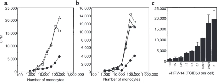
Figure 2
Inhibition of allogeneic T-cell proliferation by HRV-14. (a) Purified T cells (105) were incubated with graded numbers of allogeneic, purified
monocytes in the presence of HRV-14 (10 TCID50 per cell; filled circles), control HeLa cell culture supernatants (open triangles), or medium
alone (open circles). Proliferation of T cells was monitored on day 5 of culture by adding (methyl-3H)-TdR followed by measuring thymidine
Immunofluorescence analysis. For membrane staining, cells (5 ×10⁵) were incubated for 30 minutes at 0–4°C with unconjugated mAb. After washing twice with PBS, Oregon Green–conjugated goat anti-mouse antibody from Molecular Probes Inc. (Eugene, Oregon, USA) was used as a second-step reagent. Flow cytometric analysis was performed using a FACScan flow cytometer (Bec-ton Dickinson).
Determination of cytokine production. Monocytes (1 × 10⁶/mL) were cultured either mock treated or stimulat-ed with LPS (100 ng/mL), with or without pretreatment for 24 hours with IFN-γ(300 U/mL), or in the presence of HRV (10 TCID50/monocyte) in 24-well plates (Corn-ing-Costar Europe). After 24 hours, the supernatants were harvested and analyzed by ELISA or used in T-cell proliferation assays. For cytoplasmic staining, mon-ensin (5 µM) was added during the last 12 hours. The cells were harvested and fixed for 20 minutes at room temperature by adding 100 µL of FIX solution (An der Grub). Subsequently, cells were washed once with 4 mL of PBS, resuspended in 100 µL of PBS, and permeabi-lized by the addition of 100 µL of PERM solution (An der Grub). Immediately, the indicated PE-conjugated anti-cytokine mAb’s were added and incubated for 20 minutes at room temperature. The cells were then washed twice, resuspended in PBS (200 µL), and ana-lyzed by flow cytometry.
Cytokines were measured by sandwich ELISAs using matched-pair antibodies. Capture and detection anti-bodies for human IL-1βwere obtained from Genzyme Pharmaceuticals (Cambridge, Massachusetts, USA); for
IL-10 and IL-12 p70, from R&D Systems Inc.; and for TNF-α, from PharMingen. Standards consisted of human recombinant material from R&D Systems Inc. Assays were performed in duplicate according to the recommendations of the manufacturers. The lower limit of detection was 10 pg/mL for IL-1β and 20 pg/mL for IL-10, IL-12, and TNF-α.
Results
HRV-14 reduces antigen-induced T-cell proliferation. To ana-lyze the influence of HRV on immune responses, we investigated its effect on antigen-induced T-cell prolif-eration. Results shown in Figure 1 demonstrate that addition of major group HRV-14 to PBMCs strongly reduced the proliferative T-cell response induced by recall antigens (tetanus toxoid, PPD), superantigens (SEA, SEB), or CD3 mAb OKT3. In addition, T-cell pro-liferation induced by allogeneic monocytes was also strongly inhibited (Figure 2a). These effects were observed with purified HRV-14 and with UV-inactivat-ed HRV-14 (data not shown) but were not inducUV-inactivat-ed with supernatants from uninfected HeLa cells used for con-trol (Figure 2a). Preincubation of HRV-14 with WIN 52035-2, which sterically blocks the binding sites on HRV for its cellular receptor ICAM-1 (CD54) (25), reversed the inhibitory effect of HRV-14 (Figure 2b). Thus, binding of HRV-14 to cells was essential for inhi-bition of T-cell proliferation. This observation also demonstrates that direct viral effects and not contam-inations are responsible. Titration of HRV-14 revealed that significant inhibitory effects were detectable down to an HRV-14 (TCID50)/cell ratio of 0.025:1 (Figure 2c).
Downregulation of MHC class II expression on monocytes upon culture with HRV-14. Stimulation of T cells by anti-gen-presenting cells is based on a complex molecular interaction that involves MHC-, adhesion-, and costim-ulatory molecules as well as cytokines. To elucidate which of these molecules might be affected by HRV-14, we cultured monocytes in the presence of HRV-14 or supernatants from uninfected HeLa cells for 2 days and analyzed the immunophenotype of the cells.
[image:5.612.60.293.54.243.2]As can be seen in Figure 3, interaction of monocytes with HRV-14 had pronounced effects on their marker profile. The spontaneous downregulation of CD14 expression due to cultivation of monocytes was prevent-ed in the presence of HRV-14. Surprisingly, the costim-ulatory molecules CD80 and CD40 were found to be neoexpressed or upregulated, respectively, on monocytes cultured in the presence of HRV-14 compared with mock-treated cells. Expression of the costimulatory mol-ecule CD86 and the adhesion molmol-ecule CD54 was down-regulated on monocytes upon HRV-14 interaction. Other important adhesion molecules such as CD11a and CD58 were only minimally affected. Most striking-ly, however, cultivation of monocytes in the presence of HRV-14 strongly downregulated cell-surface expression of MHC class II molecules (Figure 3), in some instances down to 20% of control cells. In contrast, MHC class I expression was found to be slightly upregulated.
Figure 3
Expression of cell-surface molecules on HRV-14–treated monocytes. Purified monocytes were cultured in the presence of HRV-14 (10 TCID50 per cell; gray histograms) or control HeLa cell culture
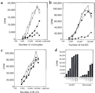
Monocytes produce an inhibitory factor upon interaction with HRV-14. The observations described here raised the question of how HRV-14 can downregulate the T-cell stimulatory capacity of monocytes. A first clue was obtained when the supernatant of monocytes cultured in the presence of HRV-14 for 2 days was transferred to untreated monocytes. Addition of this supernatant, but not of the supernatant of mock-treated monocytes, strongly reduced the proliferative T-cell response induced by allogeneic monocytes (Figure 4a). Remark-ably, inhibition by the supernatant of monocytes cul-tured in the presence of HRV-14 was always much stronger than the effect produced by addition of the virus alone and worked even when diluted down to 1:512 (Figure 4, a and d). Adding this supernatant to md-DCs, we found that the allostimulatory capacity of md-DCs was also strongly reduced, whereas HRV-14 alone showed no such effect (Figure 4, b and d). In con-trast to monocytes and md-DCs, neither the super-natant of HRV-cultured monocytes nor the virus alone was able to diminish the allogeneic T-cell response induced by EBV-transformed B-LCLs (Figure 4c).
HRV-14 induces IL-10 production in monocytes. A promi-nent immunosuppressive factor that strongly inhibits the accessory function of monocytes and of DCs (27) and downregulates MHC class II cell-surface expression (28) but does not inhibit B cell–mediated responses (27, 28) is the cytokine IL-10. Therefore, we analyzed the super-natant of HRV-14–treated monocytes after 2 days of cul-ture for the presence of IL-10. The results of this analysis revealed that large amounts of IL-10 were present in the
supernatant of HRV-14–treated monocytes (Figure 5a). The amounts of IL-10 and the number of monocytes pro-ducing this cytokine (Figure 5b) upon stimulation with HRV-14 were comparable with those seen after LPS stim-ulation. HRV-14 samples used in this study contained no detectable IL-10 (<20 pg/mL) and low LPS contamina-tion (<10 pg/mL). Moreover, in contrast to LPS stimula-tion, HRV-14 did not induce production of the proin-flammatory cytokine TNF-αand induced only small amounts of IL-1βin monocytes (Figure 5a).
[image:6.612.57.369.51.355.2]HRV-14 inhibits IL-12 production in monocytes. An important immunoregulatory property of IL-10 is its ability to inhibit the production of IL-12 (29), the crit-ical cytokine that drives T-cell responses toward type 1 effector cells (30). This prompted us to examine the influence of HRV-14 on IL-12 production of mono-cytes. Monocytes are known to produce IL-12 upon stimulation by IFN-γ/LPS (31). In contrast, pretreat-ment of monocytes with IFN-γand subsequent addi-tion of HRV-14 did not result in IL-12 producaddi-tion (Figure 6a). We therefore asked whether HRV-14 might inhibit the induction of IL-12. For this pur-pose, monocytes were cultured with HRV-14 or con-trol HeLa supernatants before IFN-γ/LPS was added to induce IL-12 production. Pretreatment of mono-cytes with HRV-14 strongly inhibited the amounts of IL-12 (Figure 6a) and the number of cells producing IL-12 as a result of IFN-γ/LPS stimulation, whereas IL-6 production was not reduced (Figure 6b). Inhibi-tion of IL-12 producInhibi-tion induced by HRV-14 was only partially reversed in the presence of neutralizing
Figure 4
HRV-14–treated monocytes release an inhibitory factor. Monocytes were cultured for 2 days in the absence or presence of HRV-14 (10 TCID50 per cell). The cell
anti–IL-10 antibodies (Figure 6b). This observation demonstrates that an additional IL-10–independent mechanism is critically involved in the selective inhi-bition of IL-12 production by HRV-14.
Neutralization of IL-10 abrogates the inhibitory effect of HRV-14. To ascertain that IL-10 was the responsible
sup-pressive factor present in the supernatant of HRV-14–treated monocytes, the supernatant was pretreated with neutralizing IL-10 antibodies and subsequently tested in the allo-MLR assay. Results in Figure 7 demon-strate that the addition of anti–IL-10 antibodies reversed the inhibitory effect of the supernatant obtained from HRV-14–treated monocytes and that monocytes stimulated T cells in the presence of this supernatant nearly as well as untreated cells. Because intact HRV-14 is still present in the supernatant obtained from HRV-14 treated monocytes, neutralizing anti–IL-10 antibodies did obviously inhibit both trans-ferred IL-10 and IL-10 induced by transtrans-ferred HRV-14.
Discussion
In this study, we demonstrate that HRV-14, a member of the major group HRV family, can efficiently inhibit antigen-specific T-cell responses. These inhibitory effects were found to result from a particular spectrum of cytokines released by mononuclear phagocytes upon HRV-14 interaction. Most prominent among them is IL-10, a well-established immunosuppressive cytokine (32), which is strongly induced by HRV-14. In contrast, major proinflammatory cytokines such as TNF-αand IL-1βand the Th1-activator IL-12 (30) are not induced or are only minimally induced, and IL-12 production induced by IFN-γ/LPS was significantly inhibited in monocytes after HRV-14 interaction. On the basis of these observations, it is tempting to speculate that the particular cytokine profile induced by HRV in mononuclear phagocytes might have adverse effects on local immunity in areas of HRV infection. Such reduced local immunocompetence may predispose affected individuals to secondary infections and could possibly explain the frequently observed occurrence of sinusitis, otitis media, bronchitis, and pneumonia in HRV-infected persons (23, 24).
Inhibition of antigen-specific T-cell responses by major group HRV have been reported previously (33). This property cannot be attributed to cytopathic effects because HRV does not infect or damage leuko-cytes (34). Inhibition was explained by the capacity of major group HRV to bind the T-cell ligand ICAM-1 on accessory cells (monocytes) and thereby prevent acces-sory-cell to T-cell contact and, consequently, T-cell acti-vation. Such a mechanism seems to be plausible, as other studies have shown that binding of HRV to ICAM-1 blocks ICAM-1/LFA-1 pair formation (35, 36). Although direct interference may contribute to a reduc-tion of T-cell reactivity, it cannot account for the inhibitory effects of HRV observed by us. In particular, HRV-14 did not inhibit md-DCs or B-LCL–induced allogeneic T-cell responses, in which ICAM-1/LFA-1 interactions are also critically involved (37–39). Rather, the induction of a soluble factor by HRV-14 in mono-cytes seems to be primarily responsible for the inhibi-tion of the antigen-specific T-cell response. Data pre-sented here demonstrate that this factor is IL-10. First, addition of supernatant from HRV-14–treated
mono-Figure 5
Induction of IL-10 production in monocytes by HRV-14. (a) Super-natants of monocytes (1 ×10⁶/mL) cultured in the presence of
HRV-14 (10 TCID50 per cell), LPS (100 ng/mL), or medium for 2 days were
analyzed for IL-10, IL-1β, and TNF-α production by ELISA. The figure shows mean values ± SEM of 3 experiments. (b) In separate experi-ments, detection of cytokine production in monocytes was performed by cytoplasmic staining with specific mAb’s. Monocytes (1 ×10⁶/mL)
were cultured for 24 hours in the presence of HRV-14 (10 TCID50 per
[image:7.612.57.292.48.440.2]cytes only inhibited the allostimulatory capacity of monocytes and md-DCs, whereas B-LCLs were not affected, which is typical for IL-10 (27, 28). Second, monocytes stimulated with HRV-14 were found to pro-duce biologically relevant amounts of IL-10 (27). Third, neutralization of IL-10 with a specific antibody revert-ed the blocking effects of HRV on T-cell stimulation. The mechanism of inhibition of antigen-specific, cel-lular immune responses by IL-10 is well defined (32). Most relevant in this respect is the massive downregu-lation of MHC class II molecule expression in the pres-ence of IL-10 (28). Accordingly, HRV-14–induced down-regulation of MHC class II molecules was inhibited in the presence of anti–IL-10 antibodies (data not shown). In contrast to MHC class II, MHC class I expression on monocytes is only slightly affected by IL-10, and respon-siveness of activated CD8+T cells is even enhanced by IL-10 (40, 41). It has also been demonstrated that IL-10 reduces CD80 and CD86 costimulatory molecule expression (41, 42) and inhibits IL-12 production (29). Upon HRV-14 treatment of monocytes, we have also observed downregulation of CD86 expression and an inhibition of IL-12 production. However, inhibition of IL-12 production induced by HRV-14 was only partial-ly reversed in the presence of neutralizing anti–IL-10 antibodies, indicating that an IL-10–independent mech-anism is also critically involved, which is reminiscent of what has been found to be the case with measles virus-es (31). Also in contrast to IL-10 effects, CD80 and CD40 expression was clearly upregulated in HRV-treat-ed monocytes. Another marker reproducibly found to be upregulated on monocytes upon HRV-14 stimula-tion was CD14, a cell-surface molecule that is not sig-nificantly influenced by IL-10 (40, 42). Thus, the immunophenotype of HRV-14–treated monocytes dif-fers in some aspects from that reported for IL-10 stim-ulation, and not all functional effects of HRV-14 may be due to induction of IL-10 production.
The classical cellular receptor for major group HRV is ICAM-1 (43, 44). Here we provide 3 pieces of evidence that engagement of ICAM-1 is required to elicit the inhibitory effects of HRV. First, cell-surface expression of ICAM-1 on monocytes was reduced upon HRV-14 interaction. This is likely to be primarily due to recep-tor usage of HRV-14 (45) and only marginally reversed in the presence of anti–IL-10 antibodies (data not shown). Second, the specific inhibitor WIN 52035-2, which blocks HRV binding to ICAM-1 (25), abolished the HRV-induced inhibitory capacity. Third, inhibition was not observed with the minor group HRV-2, which interacts with cells via the receptor and/or LDL-receptor–related proteins (46) (data not shown).
[image:8.612.315.532.51.591.2]The mechanism of IL-10 induction in monocytes by HRV-14 and the role of ICAM-1 are not yet under-stood. Signal transduction via ICAM-1 is possible. It can be induced by specific antibodies (47, 48) or by its ligand fibrinogen (49, 50). We observed that engage-ment of ICAM-1 with CD54 mAb RR1/1 did not stim-ulate IL-10 production in monocytes (data not
Figure 6
Inhibition of IL-12 production by HRV-14. Purified monocytes (1 × 10⁶/mL) were cultured for 2 days in the absence or presence of
HRV-14 (10 TCID50 per cell) or LPS (100 ng/mL) with or without
shown). However, because 1 HRV capsid has 60 recep-tor binding sites and binds to ICAM-1 with high avid-ity (51), rhinovirus particles might trigger pronounced cross-linking of ICAM-1 and subsequent signal trans-duction, leading to IL-10 production. It is conceivable that this may not be achieved by mAb’s or by other lig-ands of ICAM-1. Gern and coworkers have recently reported that binding of HRV-16, another major group HRV, to airway macrophages, and induction of TNF-α, were not inhibited with antibodies against ICAM-1 (34). These observations suggest that HRV may also interact with mononuclear phagocytes via yet undefined signal transducing receptor structures. Therefore, future experiments are necessary to clarify whether such cellular receptors for HRV are expressed on mononuclear phagocytes.
IL-10 is 1 of the key factors that regulate inflamma-tory responses (32, 52). The anti-inflammainflamma-tory action of IL-10 is primarily achieved by inhibition of the pro-duction of proinflammatory cytokines such as IL-1, IL-6, and TNF-α(53–55). The crucial role of IL-10 in controlling inflammatory responses is best illustrat-ed in IL-10 knockout mice that suffer from chronic inflammatory bowel disease (56) and in studies in which injection of IL-10 rescued mice from LPS-induced toxic shock (57). Common cold symptoms are considered to be caused by proinflammatory cytokines produced in the nasal mucosa (3). In the course of the infection, however, major group HRV may induce the production of IL-10 in infiltrating monocytes. This may result in a decrease of the local inflammatory reaction and in the inhibition of cellu-lar immune responses.
Acknowledgments
The authors thank S. Künig, L. Gschwantler, I. Gösler, and K. Wenhardt for expert technical assistance, and M. Epstein, W. Pickl, and M. Waclavicek for critical reading of the manuscript. This work was supported by the Fonds zur Förderung der Wissenschaftlichen Forschung in Österreich (SFB 005).
1. Couch, R. 1996. Rhinoviruses. In Fields virology. B. Fields et al., editors. Lippincott-Raven Publishers. Philadelphia, PA. 713–734.
2. Garibaldi, R. 1985. Epidemiology of community-acquired respiratory tract infections in adults. Am. J. Med. 78:32–37.
3. Pitkäranta, A., and Hayden, F.G. 1998. What’s new with common colds? Pathogenesis and diagnosis. Infect. Med. 15:50–59.
4. Turner, R.B., Hendley, J.O., and Gwaltney, J.M., Jr. 1982. Shedding of infect-ed ciliatinfect-ed epithelial cells in rhinovirus colds. J. Infect. Dis. 145:849–853. 5. Winther, B., Brofeldt, S., Christensen, B., and Mygind, N. 1984. Light and
scanning electron microscopy of nasal biopsy material from patients with naturally acquired common colds. Acta Otolaryngol. (Stockh.)
97:309–318.
6. Noah, T.L., et al. 1995. Nasal cytokine production in viral acute upper respiratory infection of childhood. J. Infect. Dis. 171:584–592. 7. Einarsson, O., Geba, G.P., Zhu, Z., Landry, M., and Elias, J.A. 1996.
Inter-leukin 11: stimulation in vivo and in vitro by respiratory viruses and induction of airways hyperresponsiveness. J. Clin. Invest. 97:915–924. 8. Zhu, Z., et al. 1996. Rhinovirus stimulation of interleukin-6 in vivo and
in vitro. Evidence of nuclear factor κB–dependent transcriptional acti-vation. J. Clin. Invest. 97:421–430.
9. Naclerio, R.M., et al. 1987. Kinins are generated during experimental rhi-novirus colds. J. Infect. Dis. 157:133–142.
10. Proud, D., Naclerio, R.M., Gwaltney, J.M., and Hendley, J.O. 1990. Kinins are generated in nasal secretions during natural rhinovirus colds. J. Infect. Dis. 161:120–123.
11. Proud, D., et al. 1994. Increased levels of interleukin-1 are detected in nasal secretions of volunteers during experimental rhinovirus colds. J. Infect. Dis. 169:1007–1013.
12. Subauste, M.C., Jacoby, D.B., Richards, S.M., and Proud, D. 1995. Infec-tion of a human respiratory epithelial cell line with rhinovirus. Induc-tion of cytokine release and modulaInduc-tion of susceptibility to infecInduc-tion by cytokine exposure. J. Clin. Invest. 96:549–557.
13. Johnstone, S.L., et al. 1998. Low grade rhinovirus infection induces a pro-longed release of IL-8 in pulmonary epithelium. J. Immunol. 160:6172–6181. 14. Winther, B., et al. 1984. Histopathologic examination and enumeration of polymorphonuclear leukocytes in the nasal mucosa during experi-mental rhinovirus colds. Acta Otolaryngol. (Stockh.) 413:19–24. 15. Levandowski, R.A., Weaver, C.W., and Jackson, G.G. 1988.
Nasal-secre-tion leukocyte populaNasal-secre-tions determined by flow cytometry during acute rhinovirus infection. J. Med. Virol. 25:423–432.
16. Evans, F.O., Jr., et al. 1975. Sinusitis of the maxillary antrum. N. Engl. J. Med.293:735–739.
17. Elkhatieb, A., Hipskind, G., Woerner, D., and Hayden, F.G. 1993. Middle ear abnormalities during natural rhinovirus colds in adults. J. Infect. Dis.
168:618–621.
18. Buchman, C.A., Doyle, W.J., Skoner, D., Fireman, P., and Gwaltney, J.M., Jr. 1994. Otologic manifestations of experimental rhinovirus infection.
Laryngoscope. 104:1295–1299.
19. Pitkäranta, A., Arruda, E., Malmberg, H., and Hayden, F.G. 1997. Detec-tion of rhinovirus in sinus brushings of patients with acute communi-ty-acquired sinusitis by reverse transcription-PCR. J. Clin. Microbiol.
35:1791–1793.
20. Pitkäranta, A., Virolainen, A., Jero, J., Arruda, E., and Hayden, F.G. 1998. Detection of rhinovirus, respiratory syncytial virus and coronavirus infections in acute otitis media by reverse transcriptase polymerase chain reaction. Pediatrics.102:291–295.
21. Pitkäranta, A., Jero, J., Arruda, E., Virolainen, A., and Hayden, F.G. 1998. Polymerase chain reaction-based detection of rhinovirus, respiratory syncytial virus and coronavirus in otitis media with effusion. J. Pediatr.
133:390–394.
22. Pitkäranta, A., and Hayden, F.G. 1998. Rhinoviruses: important respira-tory pathogens. Ann. Med. 30:529–537.
23. Pitkäranta, A., and Hayden, F.G. 1998. What’s new with common colds? Complications and management. Infect. Med. 15:117–128.
24. Gern, J.E., and Busse, W.W. 1999. Association of rhinovirus infections with asthma. Clin. Microbiol. Rev. 12:9–18.
25. Shepard, D.A., Heinz, B.A., and Rueckert, R.R. 1993. WIN 52032-2 inhibits both attachment and eclipse of human rhinovirus 14. J. Virol.
67:2245–2254.
[image:9.612.88.277.52.250.2]26. Pickl, W.F., et al. 1996. Molecular and functional characteristics of den-dritic cells generated from highly purified CD14+peripheral blood
Figure 7
Neutralization of IL-10 abolishes the inhibitory effect of HRV-14. Purified T cells (105) were incubated with graded numbers of
monocytes. J. Immunol. 157:3850–3859.
27. Caux, C., et al. 1994. Interleukin 10 inhibits T cell alloreaction induced by human dendritic cells. Int. Immunol. 6:1177–1185.
28. de Waal Malefyt, R., et al. 1991. Interleukin 10 (IL-10) and viral IL-10 strongly reduce antigen-specific human T cell proliferation by dimin-ishing the antigen presenting capacity of monocytes via downregulation of class II major histocompatibility expression. J. Exp. Med. 174:915–924. 29. D’Andrea, A., et al. 1993. Interleukin 10 inhibits human lymphocyte interferon gamma-production by suppressing natural killer cell stimula-tory factor/IL-12 synthesis in accessory cells. J. Exp. Med. 178:1041–1048. 30. Trinchieri, G. 1993. Interleukin-12 and its role in the generation of Th1
cells. Immunol. Today. 14:335–338.
31. Karp, C.L., et al. 1996. Mechanism of suppression of cell-mediated immunity by measles virus. Science. 273:228–231.
32. de Waal Malefyt, R. 1998. Interleukin-10. In Cytokines.A. Mire-Sluis and R. Thorpe, editors. Academic Press. London, United Kingdom. 151–167. 33. Gern, J.E., Joseph, B., Galagan, D.M., Borcherding, W.R., and Dick, E.C. 1996. Rhinovirus inhibits antigen-specific T cell proliferation through an intercellular adhesion molecule-1-dependent mechanism. J. Infect. Dis.
174:1143–1150.
34. Gern, J.E., et al. 1996. Rhinovirus enters but does not replicate inside monocytes and airway macrophages. J. Immunol. 156:621–627. 35. Staunton, D.E., Dustin, M.L., Erickson, H.P., and Springer, T.A. 1990.
The arrangement of the immunoglobulin-like domains of ICAM-1 and the binding sites for LFA-1 and rhinovirus. Cell. 61:243–254. 36. Piela-Smith, T.H., Aneiro, L., and Korn, J.H. 1991. Binding of human
rhi-novirus and T cells to intercellular adhesion molecule-1 on human fibroblasts. Discordance between effects of IL-1 and IFN-γ. J. Immunol.
147:1831–1836.
37. Sanders, V.M., Snyder, J.M., Uhr, J.W., and Vitetta, E.S. 1986. Character-ization of the physical interaction between antigen-specific B and T cells.
J. Immunol. 137:2395–2404.
38. King, P.D., and Katz, D.R. 1989. Human tonsillar dendritic cell-induced T cell responses: analysis of molecular mechanisms using monoclonal antibodies. Eur. J. Immunol. 19:581–587.
39. Metlay, J.P., Pure, E., and Steinman, R.M. 1989. Distinct features of den-dritic cells and anti-Ig activated B cells as stimulators of the primary mixed leukocyte reaction. J. Exp. Med. 169:239–254.
40. Spittler, A., et al. 1995. IL-10 augments CD23 expression on U937 cells and down-regulates IL-4-driven CD23 expression on cultured human blood monocytes: effects of IL-10 and other cytokines on cell phenotype and phagocytosis. Immunology. 85:311–317.
41. Groux, H., Bigler, M., de Vries, J.E., and Roncarolo, M.G. 1998. Inhibito-ry and stimulatoInhibito-ry effects of IL-10 on human CD8+T cells. J. Immunol. 160:3188–3193.
42. Kubin, M., Kamoun, M., and Trinchieri, G. 1994. Interleukin 12
syner-gizes with B7/CD28 interaction in inducing efficient proliferation and cytokine production of human T cells. J. Exp. Med. 180:211–222. 43. Staunton, D.E., et al. 1989. A cell adhesion molecule, ICAM-1, is the
major surface receptor for rhinoviruses. Cell. 56:849–853.
44. Greve, J.M., et al. 1989. The major human rhinovirus receptor is ICAM-1. Cell. 56:839–847.
45. Gern, J.E., Vrtis, R., Kelly, E.A., Dick, E.C., and Busse, W.W. 1996. Rhi-novirus produces nonspecific activation of lymphocytes through a monocyte-dependent mechanism. J. Immunol. 157:1605–1612. 46. Hofer, F., et al. 1994. Members of the low density lipoprotein receptor
family mediate cell entry of a minor group common cold virus. Proc. Natl. Acad. Sci. USA. 91:1839–1842.
47. Durieu-Trautmann, O., Chaverot, N., Cazaubon, S., Strosberg, A.D., and Couraud, P.-O. 1994. Intercellular adhesion molecule 1 activation induces tyrosine phosphorylation of the cytoskeleton-associated protein cortactin in brain microvessel endothelial cells. J. Biol. Chem. 269:12536–12540. 48. Holland, J., and Owens, T. 1997. Signaling through intercellular
adhe-sion molecule 1 (ICAM-1) in a B cell lymphoma line. The activation of lyn tyrosine kinase and the mitogen-activated protein kinase pathway. J. Biol. Chem. 272:9108–9112.
49. Duperray, A., et al. 1997. Molecular identification of novel fibrinogen binding site on the first domain of ICAM-1 regulation leukocyte-endothelium bridging. J. Biol. Chem. 272:435–441.
50. Gardiner, E.E., and D’Souza, S.E. 1997. A mitogenic action for fibrino-gen mediated through intercellular adhesion molecule-1. J. Biol. Chem.
272:15474–15480.
51. Casasnovas, J.M., and Springer, T.A. 1995. Kinetics and thermodynam-ics of virus binding to receptor. Studies with rhinovirus, intercellular adhesion molecule-1 (ICAM-1) and surface plasmon resonance. J. Biol. Chem. 270:13216–13224.
52. Howard, M., and O’Garra, A. 1992. Biological properties of interleukin 10. Immunol. Today.13:198–200.
53. Moore, K.W., et al. 1990. Homology of cytokine synthesis inhibitory fac-tor (IL-10) to the Epstein Barr virus gene BCRF1. Science. 248:1230–1234. 54. de Waal Malefyt, R., Abrams, J., Bennett, B., Figdor, C.G., and de Vries, J.E. 1991. Interleukin 10 (IL-10) inhibits cytokine synthesis by human monocytes: an autoregulatory role of IL-10 produced by monocytes. J. Exp. Med.174:1209–1220.
55. Fiorentino, D.F., Zlotnik, A., Mosmann, T.R., Howard, M., and O’Garra, A. 1991. IL-10 inhibits cytokine production by activated macrophages.
J. Immunol. 147:3815–3822.