RESEARCH ARTICLE
Multiple modes of Lrp4 function in modulation of Wnt/
β
-catenin
signaling during tooth development
Youngwook Ahn1,2,*, Carrie Sims1, Megan J. Murray1, Paige K. Kuhlmann1, Jesús Fuentes-Antrás1, Scott D. Weatherbee3and Robb Krumlauf1,4,*
ABSTRACT
During development and homeostasis, precise control of Wnt/β -catenin signaling is in part achieved by secreted and membrane proteins that negatively control activity of the Wnt co-receptors Lrp5 and Lrp6. Lrp4 is related to Lrp5/6 and is implicated in modulation of Wnt/β-catenin signaling, presumably through its ability to bind to the Wise (Sostdc1)/sclerostin (Sost) family of Wnt antagonists. To gain insights into the molecular mechanisms of Lrp4 function in modulating Wnt signaling, we performed an array of genetic analyses in murine tooth development, where Lrp4 and Wise play important roles. We provide genetic evidence that Lrp4 mediates the Wnt inhibitory function of Wise and also modulates Wnt/β-catenin signaling independently of Wise. Chimeric receptor analyses raise the possibility that the Lrp4 extracellular domain interacts with Wnt ligands, as well as the Wnt antagonists. Diverse modes of Lrp4 function are supported by severe tooth phenotypes of mice carrying a human mutation known to abolish Lrp4 binding to Sost. Our data suggest a model whereby Lrp4 modulates Wnt/β-catenin signaling via interaction with Wnt ligands and antagonists in a context-dependent manner.
KEY WORDS: Lrp4, Sostdc1, Mouse, Tooth, Wnt/β-catenin signaling
INTRODUCTION
Wnt/β-catenin signaling plays a pivotal role in the patterning, morphogenesis and growth of a variety of tissues and organs during development and in homeostasis in the adult. Aberrant Wnt signaling activity is causally linked to congenital defects, degenerative diseases and cancers (Clevers and Nusse, 2012). Therefore, understanding the molecular mechanisms that regulate the outputs of this signaling pathway in differentin vivocontexts and expanding our knowledge of how this is governed through dynamic crosstalk among different tissues and cell types are fundamentally important.
In the Wnt/β-catenin signaling pathway, initiation of signaling requires interaction between Wnt ligands, their frizzled (Fz) receptors and Wnt co-receptors low-density lipoprotein receptor-related proteins 5 and 6 (Lrp5/6) (MacDonald and He, 2012). These interactions on the cell membrane trigger a cascade of intracellular
events leading to stabilization and nuclear localization ofβ-catenin, which together with TCF/LEF transcription factors activates the expression of target genes (MacDonald and He, 2012; MacDonald et al., 2009).
A variety of secreted Wnt antagonists have been shown to inhibit Wnt/β-catenin signaling at the earliest step, presumably by altering or blocking the formation of Wnt/Fz/co-receptor complexes (Cruciat and Niehrs, 2013). In vitro binding studies have suggested that, among the Wnt antagonists, sclerostin (Sost) and Wise (also known as Sostdc1) can inhibit Wnt/β-catenin signaling via their ability to bind to the extracellular domains of Lrp5/6 (Ellies and Krumlauf, 2006; Itasaki et al., 2003; Li et al., 2005; Semenov et al., 2005). Sost and Wiseare closely related, as they emerged through genome-wide duplication and divergence, but they display mostly non-overlapping expression patterns (Collette et al., 2013). The function of Sost and Wise in Wnt regulation via direct binding to Lrp5/6 has been further supported by genetic interaction studies in multiples tissues where they play a crucial role in development and homeostasis (Ahn et al., 2010, 2013; Chang et al., 2014b).
Lrp4 has emerged as an important component of the Wnt/β -catenin signaling pathway. The sequence and structure of its extracellular domain are similar to those of Lrp5 and Lrp6. Since the Lrp4 intracellular domain lacks some of the motifs in Lrp5 and Lrp6 known to be essential for Wnt co-receptor function, Lrp4 was proposed to be a negative regulator of Wnt signaling (Herz and Bock, 2002; Johnson et al., 2005; Weatherbee et al., 2006; Willnow et al., 2012). Supporting this idea, overexpression ofLrp4results in decreased Wnt/β-catenin signaling activity in cultured cells (Johnson et al., 2005; Li et al., 2010; Ohazama et al., 2008). Inin vitrobinding assays, the extracellular domain of Lrp4 can directly interact with Sost and Wise, suggesting that the Wnt inhibitory function of Lrp4 may depend on its interaction with the Wnt antagonists (Choi et al., 2009; Karner et al., 2010; Ohazama et al., 2008).
In support of interaction between Lrp4 and Wise, mice deficient for Lrp4 or Wise share similar developmental defects in the ectodermal tissues, e.g. teeth, hair and mammary glands (Ahn et al., 2013; Narhi et al., 2012; Ohazama et al., 2008). Early development of these tissues requires reciprocal interactions between the epithelium and underlying mesenchyme, and Wnt signaling along with other major signaling pathways has diverse roles in the control of patterning and morphogenesis at different stages (Ahn, 2015; Balic and Thesleff, 2015; Biggs and Mikkola, 2014). In the tooth germ, Lrp4is expressed in the epithelial signaling centers, while
Wiseis expressed in the surrounding epithelial and mesenchymal cells (Ahn et al., 2010; Laurikkala et al., 2003; Ohazama et al., 2008). Mice homozygous for a hypomorphicLrp4allele phenocopy
Wise-null mice and display various tooth defects, such as supernumerary teeth and molar fusion (Ahn et al., 2010; Ohazama et al., 2008). Since the tooth defects inWise-null mice Received 22 February 2017; Accepted 30 June 2017
1Stowers Institute for Medical Research, Kansas City, MO 64110, USA.
2Department of Molecular Biology, Cell Biology and Biochemistry, Brown
University, Providence, RI 02912, USA.3Department of Genetics, Yale School of
Medicine, New Haven, CT 06520, USA.4Department of Anatomy and Cell Biology,
University of Kansas Medical Center, Kansas City, KS 66160, USA.
*Authors for correspondence ([email protected]; [email protected])
Y.A., 5988-7388; M.J.M., 6335-0444; J.F.-A., 0000-0001-5805-2362; S.D.W., 0000-0002-0915-1329
DEVEL
O
are caused by elevated Wnt/β-catenin signaling (Ahn et al., 2010), it is possible that Lrp4 functions through its interplay with Wise as part of an important molecular mechanism for modulating Wnt/β -catenin signaling in teeth and other contexts.
To address this question, we utilized gain- and loss-of-function mouse models andin vitroreporter assays to investigate howLrp4
interacts with Lrp5/6and Wise. Our genetic interaction analyses focused on tooth development indicate that Lrp4 negatively regulates Wnt/β-catenin signaling to control tooth number, morphology and growth through potentiation of the Wnt inhibitory function of Wise. In addition, our study provides evidence suggesting a Wise-independent role for Lrp4 through its interaction with Wnt ligands and Fz receptors. This work has uncovered novel and diverse mechanisms by which Lrp4 contributes to the modulation of Wnt/β-catenin signaling during development.
RESULTS
Lrp4deficiency results in survival of R2 vestigial buds and delayed development of the first molar
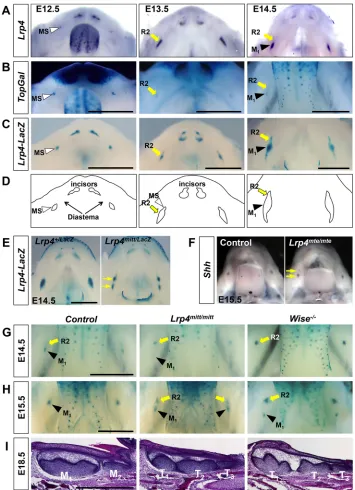
We investigated the spatiotemporal expression pattern ofLrp4in the diastema and molar region of the mandible during early tooth development. In mice, two tooth vestigial buds, namely MS and R2, develop sequentially in the toothless diastema region, but they undergo degeneration without advancing to the cap stage of tooth development (Ahn, 2015; Peterkova et al., 2006) (Fig. 1D). Consistent with a previous report (Ohazama et al., 2008), Lrp4
[image:2.612.47.402.246.736.2]transcripts were detected in MS and R2 at E12.5 and E13.5, respectively, similar to the expression pattern of theTopGalWnt activity reporter (Fig. 1A,B). At E14.5, Lrp4 expression is diminished in degenerating R2, while strong expression is observed in the more proximal region of the dental epithelium where the first molar (M1) develops (Fig. 1A). Compared with
Fig. 1.Lrp4deficiency results in survival of the R2 diastema tooth bud in mice.(A)Lrp4
is expressed in the diastema tooth buds, MS and R2, during normal embryogenesis. Dorsal views of a dissected mandible afterin situ
hybridization forLrp4at three embryonic stages. (B,C) X-Gal-stained mandibles of
TopGal(B) andLrp4lacZ(C) mice.
(D) Schematic of normal early tooth
development in the mandible from A-C. MS and R2 develop in the diastema region, but undergo degeneration. (E,F) X-Gal staining (Lrp4lacZ)
andin situhybridization (Shh) reveal two domains (arrows) of expression indicating survival of R2 inLrp4-null mice.
(G-I) Comparison of R2 and M1development
betweenLrp4-null andWise-null mice.TopGal
expression at E14.5 (G) and E15.5 (H) and Hematoxylin and Eosin staining at E18.5 (I) indicate that R2 gives rise to a supernumerary tooth (T1) in both mutants. However,Lrp4-null
mice display a shorter distance between R2 and M1and a smaller T1compared withWise
-null mice. Representative images are shown for each genotype (n≥3). Scale bars: 1 mm.
DEVEL
O
TopGal, theLrp4expression domain at E13.5 and E14.5 is broader and extends more proximally to reach the proximal end of the tooth germ (Fig. 1A,B). The Lrp4 expression pattern was further examined in a reporter knock-in model, Lrp4lacZ, and found to mimic thein situhybridization pattern ofLrp4in the developing incisor and molar regions (Fig. 1A,C). At E14.5, residual β-galactosidase activity marks degenerating R2, which is likely to be due to the stability of the lacZ transcript and/or β-galactosidase protein itself. These expression patterns are consistent with roles for Lrp4 in determining the fate of the diastema buds and M1.
Supernumerary cheek teeth have been reported with a relatively low frequency in the mandible of mice homozygous for a hypomorphic allele, Lrp4ECD (Ohazama et al., 2008). To clarify whether the supernumerary teeth arise from R2, we analyzed multiple stages of tooth development in mice homozygous for the
Lrp4-null allelesLrp4mteorLrp4mitt(Weatherbee et al., 2006). First,
Lrp4mittmice were crossed withLrp4lacZmice and the presence of two domains of reporter expression at E14.5, as compared with controls, suggests that R2 continues to develop inLrp4-null mice (Fig. 1E). Furthermore, an additional Shh expression domain is observed distal to M1at E15.5, suggesting continuous development
of R2 in Lrp4-null mice (Fig. 1F). Lastly, narrower and weaker expression domains of enamel knot markers and their downstream target genes indicate abnormal tooth development inLrp4-null mice (Fig. S1).
The progressive development of R2 and M1facilitated by loss of Lrp4was further monitored utilizing theTopGalreporter line and histological sections (Fig. 1G-I). Consistent with the idea of survival of R2 inLrp4-null mice,TopGalexpression is sustained in R2 at E14.5, whereas M1development is delayed as it arises in the
more proximal region (Fig. 1G). Survival and continued development of R2 has also been observed in Wise-null mice (Ahn et al., 2010). However, there are significant differences in the temporal and spatial expression patterns ofTopGalin theWiseand
Lrp4 mutants (Fig. 1G,H). In Wise-null mice, the continued development of R2 is greatly enhanced, as evidenced by a strong and enlargedTopGalexpression domain at E14.5. Conversely, M1
development is greatly delayed inWise-null mice, as theTopGal
expression domain marking M1 is not detected until E15.5. R2
continues to develop to form a prominent supernumerary tooth, T1
at E18.5 (Fig. 1I). By contrast, inLrp4-null mice R2 is maintained with a relatively modest level ofTopGalexpression at E14.5-15.5 and the delay in M1 development in Lrp4-null mice is not as
significant as inWise-null mice (Fig. 1G,H). The distance between R2 and M1is variable and generally shorter inLrp4-null mice than
inWise-null mice at E15.5. Consistent with these differences, T1,
which originates from R2, is smaller in Lrp4-null mice at E18.5 (Fig. 1I). Together with reduced expression of the enamel knot markers and their targets, this suggests that R2 is not undergoing accelerated development inLrp4-null mice and this is associated with a weak inhibition of M1.
Lrp4deficiency amelioratesWise-null tooth defects
The comparative analyses above highlight differences as well as similarities in tooth phenotypes of Lrp4 or Wise mutant mice, suggesting the possibility of overlapping and independent mechanisms by which Lrp4 and Wise control tooth development. With respect to overlapping mechanisms, it is possible that similar effects of deficiency ofLrp4orWiseon tooth development arise through their involvement in the regulation of a common signaling pathway. Our primary focus was the Wnt/β-catenin signaling
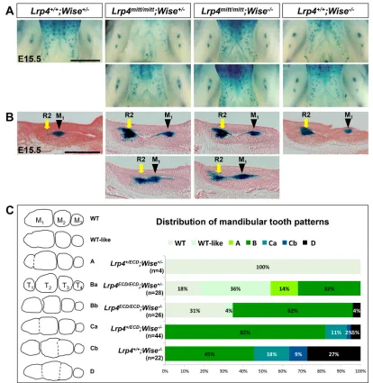
pathway, as mutants of these two genes show altered Wnt signaling activity (Fig. 1G). We performed genetic interaction studies in order to explore this possibility and gain a deeper mechanistic understanding of their roles in tooth germs. We first investigated how Lrp4 and Wise impact each other’s function during tooth development by crossing Lrp4mitt mice with Wise-null mice and monitoring TopGal expression patterns in double mutants. This enabled us to determine whether inactivation of both genes results in significant changes in Wnt signaling and tooth defects compared with the individual mutants (Fig. 2A,B).Lrp4mitt/mitt;Wise−/−mice display no sign of exacerbated tooth defects and instead show variable distance between R2 and M1, similar toLrp4mitt/mittmice.
This indicates that tooth defects ofLrp4mitt/mitt;Wise−/− mice are milder than those of Wise-null mice, in which a greater distance between R2 and M1is invariably maintained.
SinceLrp4mitt/mittmice die immediately after birth (Weatherbee et al., 2006), we also usedLrp4ECD/ECDmice to evaluate how these changes during embryogenesis are translated into the number, size and shape of adult teeth. We observed that embryonic tooth development is similarly disrupted inLrp4ECD/ECDmice as inLrp4 -null mice (Fig. S2). After obtaining Lrp4ECD;Wise compound mutants, we categorized mandibular and maxillary tooth patterns into distinct groups based on the severity of fusion and presence of the supernumerary cheek teeth (T1) to aid our comparative analyses
of these tooth phenotypes (Fig. 2C, Fig. S3). In the mandible,
Lrp4ECD/ECD mice display tooth defects similar to, but generally milder than, those ofWise-null mice (Fig. 2C). In contrast to the full penetrance of the T1 phenotype in Wise-null mice, only 32% of Lrp4ECD/ECD;Wise+/− mice display T
1. This indicates that in the Lrp4-deficient mice, even though R2 initially escapes degeneration, the majority of R2 buds fail to develop into T1and instead merge
into M1during later development. In addition to T1,Wise-null mice
frequently develop lateral supernumerary teeth and fusions between neighboring cheek teeth due to overgrowth, and these defects are rarely observed in Lrp4ECD/ECD or Lrp4ECD/ECD;Wise+/− mice (Fig. 2C; data not shown). In the maxilla, both mutants display fusions of distal molars, with a higher penetrance observed inWise -null mice (Fig. S3).
Comparison of mandibular tooth phenotypes amongLrp4ECD;
Wise compound mutants indicated that Lrp4+/+;Wise−/− mice display the most severe defects. Removing one allele ofLrp4in a
Wise−/−background (Lrp4+/ECD;Wise−/−) and then the secondLrp4 allele (Lrp4ECD/ECD;Wise−/−) progressively reduces the severity of the phenotypes (Fig. 2C). For example, fully or partially separated T1was observed with 100%, 66% and 46% penetrance inLrp4+/+; Wise−/−, Lrp4ECD/ECD;Wise−/− and Lrp4ECD/ECD;Wise+/− mice, respectively. A similar trend was observed in the maxilla of the compound mutants (Fig. S3). In order to rule out the possibility that the released extracellular domain of Lrp4 expressed from the
Lrp4ECD allele (Dietrich et al., 2010) has an effect on tooth development and complicates our analyses, we performed the same analyses with another hypomorphic mutant, Lrp4mdig, combined with Lrp4mitt mice and obtained similar results (Fig. S4). These genetic analyses indicate that Lrp4 makes an independent contribution to the generation of the severe tooth defects inWise -null mice.
Reduced dosages ofLrp5andLrp6rescue the tooth defects ofLrp4mutant mice
The more mild phenotypes observed upon loss of Lrp4in Wise
mutants suggests that Lrp4 might exert a stimulatory effect on Wnt/ β-catenin signaling in a Wise-independent manner during tooth
DEVEL
O
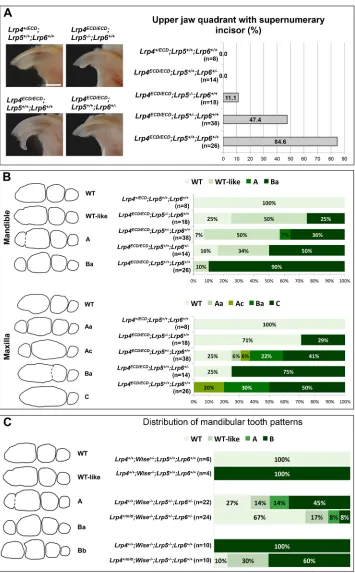
development. To explore whether elevated Wnt/β-catenin signaling is causally associated with the tooth defects inLrp4mutants, we crossedLrp4ECDmice withLrp5- andLrp6-null mice and examined the effect of reducedLrp5/6gene dosages on the tooth phenotypes among littermates. A supernumerary incisor is observed with∼85% penetrance in the maxilla of Lrp4ECD/ECD and this phenotype is rescued by reduced dosages ofLrp5/6(Fig. 3A). In the mandible, a supernumerary cheek tooth (T1) is observed with high frequency
amongLrp4ECD/ECDmice (90%, n=26) in this strain background, and reduced dosages of Lrp5/6 lead to a lower frequency of T1
(Fig. 3B, top). In the maxilla, molar fusions common inLrp4ECD/ECD mice are rescued by reduced dosages ofLrp5/6(Fig. 3B, bottom). This dosage-dependent rescue ofLrp4tooth defects by deficiency inLrp5/6implies that elevated Wnt/β-catenin signaling is primarily responsible for the abnormalities in incisor and molar development inLrp4mutant mice. Similar to the results from our previous study with Wise-null mice (Ahn et al., 2010), we observed differences
betweenLrp5andLrp6in their ability to rescue different aspects of the tooth defects with reduced gene dosages.
Although genetic analyses in teeth (Fig. 3A,B) and other tissues (Ahn et al., 2013) indicate that Lrp4 and Lrp5/6 generally have opposite roles in Wnt signaling, our data lead us to hypothesize that this relationship might be altered in the absence of Wise, revealing a potential Wise-independent role for Lrp4 in positively modulating Wnt/β-catenin signaling. To genetically test this idea, we investigated how theLrp4-null alleleLrp4mittinteracts withLrp5 and Lrp6in Wise-null mice. Interestingly, inactivating a copy of
Lrp4enhances the effect of reducedLrp5andLrp6gene dosage on
[image:4.612.99.513.56.481.2]Wise-null tooth defects and results in further rescue in the mandible (Fig. 3C). These data suggest that, in the absence of Wise, Lrp4 can positively regulate Wnt/β-catenin signaling, mimicking the normal roles of Lrp5/6. Together, these genetic analyses have uncovered dual roles for Lrp4. In the presence of Wise, Lrp4 negatively regulates Wnt/β-catenin signaling as its major mechanism of action Fig. 2.Lrp4deficiency amelioratesWise-null molar defects.(A,B)Lrp4mitt/mitt;Wise−/−mice display variable, but generally shorter, distance between R2 and M
1,
mimickingLrp4mitt/mittmice. Whole-mount images (A) and histological sections (B) ofTopGalstained E15.5 mandibles are shown. Scale bars: 1 mm in A; 0.5 mm
in B. (C)Lrp4deficiency ameliorates tooth defects ofWise-null mice in a dosage-dependent manner. Mandibular tooth patterns are categorized based on the number, size and fusion of cheek teeth (left). Distribution of different tooth patterns among littermates of theLrp4ECDandWise-null combinatorial mutants
(right).
DEVEL
O
to control tooth number and growth. In the absence of Wise, Lrp4 can play a stimulatory role in Wnt/β-catenin signaling through a different mechanism. This illustrates that Wise can alter the impact of Lrp4 on Wnt signaling in a context-dependent manner.
Lrp4is necessary forWiseto inhibit tooth development
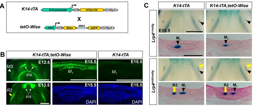
Overexpression ofWisein the dental epithelium leads to delay and hypoplasia in molar development (Ahn et al., 2010). We investigated whether this Wise gain-of-function phenotype
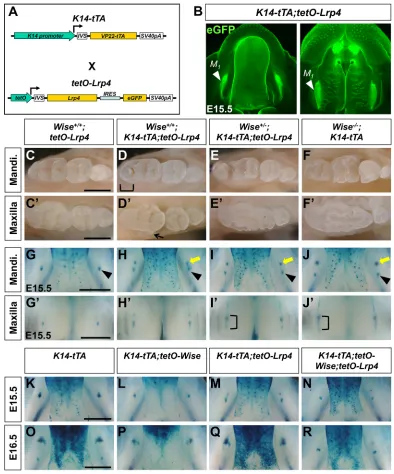
requires Lrp4. Since K14-Wise mice were not able to breed, we instead utilized the Tet-off system, in which the keratin 14 (K14) promoter drives expression of a transactivator (tTA) in the first transgene (K14-tTA), and in the absence of doxycycline tTA in turn activates the expression ofWiseandeGFPin the second transgene (tetO-Wise) (Ahn et al., 2013) (Fig. 4A). InK14-tTA;tetO-Wise
[image:5.612.50.405.54.627.2]mice, eGFP, which serves as an indicator of Wise expression, is detected specifically in the dental epithelium (Fig. 4B). After birth, molars appear to be smaller and the third maxillary molar is
Fig. 3. Genetic interaction ofLrp4with Lrp5andLrp6in tooth development.
(A) Reduced dosages ofLrp5andLrp6
rescue the supernumerary incisor phenotype ofLrp4ECD/ECDmice. Scale bar: 1 mm.
(B) Reduced dosages ofLrp5andLrp6
ameliorate molar abnormalities of
Lrp4ECD/ECDmice. (C) Reduced dosages of Lrp4,Lrp5andLrp6ameliorateWise-null tooth defects in an additive manner.
DEVEL
O
frequently missing, reminiscent of the phenotypes of theK14-Wise
mice (Fig. S5).
These two transgenic lines were then crossed withLrp4mutant lines to generate Lrp4-deficient mice overexpressing Wise. In
Lrp4+/mdigmice, overexpression ofWiseresults in a reduced domain of the dental epithelial cells expressingTopGaland in hypoplasia of tooth germs at E15.5 (Fig. 4C). InLrp4mitt/mdigmice, R2 continues to develop, similar to our observation in otherLrp4mutants, and this phenotype is not altered by overexpression ofWise(Fig. 4C). The lack of a Wisegain-of-function phenotype in Lrp4-deficient tooth germs suggests that Wise depends on Lrp4 to exert its Wnt inhibitory activity in tooth development.
Ectopically expressedLrp4disrupts tooth development in a Wisedosage-dependent manner
With its highly restricted expression pattern in the dental epithelium,
Lrp4might provide a spatial cue for the action of Wise, which is broadly expressed in the tooth germ (Ahn et al., 2010; Laurikkala et al., 2003; Ohazama et al., 2008). To test this idea, we performed gain-of-function analyses of Lrp4 by generating a tetO-Lrp4
expression line (Fig. 5A). The transgenic line was first tested for its ability to rescue the limb defects ofLrp4mutants. For this rescue experiment, we also generated Lrp4BAC-tTA driver lines, which expresstTAin theLrp4expression domains, including the apical ectodermal ridge (AER) of limb buds (Fig. S6A). Severe patterning defects in the distal limb of Lrp4 mutants are fully rescued in
Lrp4BAC-tTA;tetO-Lrp4 mice, indicating that functional Lrp4 protein is expressed from the transgene (Fig. S6B).
ThetetO-Lrp4line was then crossed with theK14-tTAline to test the effect of ectopic Lrp4 expression in the dental epithelium (Fig. 5B). Intriguingly, inK14-tTA;tetO-Lrp4mice, overexpression ofLrp4results in temporary survival of R2 (Fig. 5G,H), which fails to form a supernumerary tooth and instead merges into M1at later
stages, leading to abnormal cusp patterning in the distal region of M1(Fig. 5C,D). We hypothesized that ectopic expression ofLrp4
disrupts the normal distribution and/or function of Wise and hence causes elevation in Wnt/β-catenin signaling in the epithelial signaling center of R2. This idea was supported by the observation that a half dose ofWiseexacerbates theLrp4 gain-of-function tooth phenotypes. Wise+/−;K14-tTA;tetO-Lrp4 mice display more sustained survival of R2 (Fig. 5I,J), which gives rise to a supernumerary cheek tooth in the mandible (Fig. 5E,F). In the maxilla, overgrowth/fusion of distal molars was frequently observed inWise+/−;K14-tTA;tetO-Lrp4mice reminiscent ofWise-null tooth phenotypes (Fig. 5C′-J′). The idea was also tested by simultaneously overexpressing Wise and Lrp4 in the dental epithelium. Tooth development is further delayed in K14-tTA; tetO-Wise;tetO-Lrp4 compared with K14-tTA;tetO-Wise mice, as evidenced by much smaller domains of TopGal expression at E15.5-E16.5 (Fig. 5K-R). Together, these data support a model whereby Lrp4 directs Wise function in the epithelial signaling center of developing teeth via direct interactions.
Lrp4-Lrp6 fusions uncover domain-specific roles for Lrp4
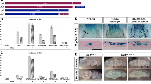
To gain insights into the molecular interactions between Lrp4, Wise and Lrp5/6 uncovered from ourin vivo studies, we utilized anin vitro reporter system in which the activity of Wnt/β-catenin signaling is measured in a human cell line expressing different combinations of the proteins (Fig. S7). As expected, expression of Wnt1 or human WNT3A leads to a dramatic increase in Wnt reporter activity, indicating that the amount of Wnt ligand is a limiting factor for signaling activation in the cultured cells. Lrp4 and Wise antagonize the activity of WNT3A as well as Wnt1, consistent with the Wnt inhibitory potential of Lrp4 and Wise over these two classes of Wnt ligands. Co-expression ofLrp4and Wiseleads to further reduction in Wnt reporter activity.
[image:6.612.52.561.56.271.2]The extracellular domain (ECD) of Lrp4 is similar to those of Lrp5 and Lrp6, whereas its intracellular domain (ICD) is distinct. To investigate whether this difference in the ICD underlies the Wnt inhibitory role of Lrp4, we generated chimeric proteins of Lrp4 and Fig. 4.Lrp4is necessary forWiseto inhibit tooth development.(A) The Tet-off binary transgenic system that overexpressesWisein the dental epithelium. (B) InK14-tTA;tetO-Wisemice, eGFP fluorescence is seen in the incisor (ins) and the MS and R2 buds, as shown in dorsal views of the dissected mandible (left). A frozen section of M1fromK14-tTA;tetO-Wisemice (top, middle) indicates that eGFP is restricted to the epithelial cells at E15.5, whereas no eGFP is
detectable in the control (top, right). Scale bars: 0.5 mm. (C)Lrp4is required for theWisegain-of-function tooth phenotypes. Normally,Wiseoverexpression suppresses tooth development (Lrp4+/mdig, compare right with left). InLrp4-deficient mice,Wiseoverexpression has no significant effect on tooth development
(Lrp4mitt/mdig, compare right with left). Scale bars: 1 mm (whole mount) and 0.5 mm (section).
DEVEL
O
Lrp6 and tested their activity in the cultured cells (Fig. 6A-C). In general, the level of the Wnt co-receptor appeared to be another limiting factor, as co-expression of Lrp6 with the Wnt ligands results in further elevation of reporter activity. When Wnt ligands and Lrp6 are co-expressed, Wise is ineffective in suppressing the reporter activity. Lrp4ECD-Lrp6ICD (L4L6) mimics Lrp6, facilitating activation of Wnt/β-catenin signaling by Wnt1 or WNT3A, although not as efficiently as Lrp6 itself. Conversely, Lrp6ECD-Lrp4ICD (L6L4) mimics Lrp4, leading to a reduction in Wnt reporter
[image:7.612.108.502.56.530.2]activity. These results suggest that the respective ICD determines whether these two Lrp receptors play a stimulatory or inhibitory role in Wnt/β-catenin signaling. Furthermore, the Wnt stimulatory activity of L4L6 implies that Lrp4ECD can act like Lrp6ECD and interact with the Wnt ligands. L4L6 is more responsive to Wise co-expression, resulting in a more dramatic decrease in reporter activity compared with Lrp6. By contrast, L6L4 alone is more potent than Lrp4 in Wnt inhibition and this inhibitory activity is not enhanced by Wise co-expression. This Fig. 5.Lrp4overexpression disrupts tooth development in aWise-dependent manner.(A) The Tet-off binary transgenic system that overexpressesLrp4in the dental epithelium. (B) eGFP fluorescence is seen in M1of the mandible (left) and maxilla (right) inK14-tTA;tetO-Lrp4mice. (C-F′)Lrp4overexpression in
otherwise wild-type mice results in abnormal cusp patterns in the distal part of the mandibular M1(D, bracket) and a lateral supernumerary tooth in the maxilla (D′,
arrow). InWise+/−mice,Lrp4overexpression results in a supernumerary cheek tooth in the mandible (E) and fusion of distal teeth in the maxilla (E′). (G-J′) X-Gal-stained mandibles (G-J) and maxilla (G′-J′) withTopGal. In the mandible,Lrp4overexpression results in survival of R2 (yellow arrow), which is close to M1
(arrowhead) (H). InWise+/−;K14-tTA;tetO-Lrp4mice, R2 maintains distance from M
1after survival in the mandible (I), and the diastema bud and M1are often fused
in the maxilla (I′) as inWise-null mice (J′, bracket). (K-R) X-Gal-stained mandibles withTopGalat E15.5 indicate that simultaneous overexpression ofWiseand
Lrp4strongly inhibits tooth development (K-N). At E16.5,TopGalexpression domains remain much smaller in mice overexpressing bothWiseandLrp4as compared with mice overexpressingWiseonly (O-R). Scale bars: 1 mm.
DEVEL
O
suggests that Wise interacts with Lrp4ECD more efficiently than with Lrp6ECD to impact receptor activity.
Gain- and loss-of-functionin vivostudies support domain-specific roles for Lrp4
We tested the in vivo relevance of the above chimeric receptor findings by expressingLrp6and L4L6 in transgenic mice utilizing the Tet-off system. As predicted, expression of Lrp6 in the dental epithelium results in the ectopic formation of tooth bud-like structures accompanied byTopGalexpression, indicating increased activation of Wnt/β-catenin signaling (Fig. 6D). A similar phenotype was observed when L4L6 is expressed in the dental epithelium, consistent with its in vitro activity of stimulating Wnt signaling (Fig. 6D). Together, ourin vitroandin vivodata indicate that the ECDs of Lrp4 and Lrp6 share an ability to interact with both Wnt ligands and Wise. It has been shown that the ICD is dispensable for Lrp4 function in the neuromuscular junction as a receptor for agrin (Choi et al., 2013; Gomez and Burden, 2011). In this regard, it is unclear whether the tooth defects ofLrp4ECD/ECDmice result from lack of anchorage to the cell membrane or from lack of ICD. Therefore, we utilized CRISPR/Cas9 technology to generate a newLrp4allele,Lrp4ΔICD, which produces an Lrp4 protein lacking the ICD. Mice homozygous for this allele are viable and display tooth defects comparable to those ofLrp4ECD/ECDmice, indicating that the ICD is essential for the normal function of Lrp4 in tooth development (Fig. 6E). The majority (77.8%,n=36) ofLrp4ΔICD/ΔICDmice show a typical pattern of T1, T2-T3and T4, with rare occurrence of later supernumerary teeth
(6.7%) in the mandible. Some (16.7%) show signs of incomplete separation of T1and T2teeth, resulting in the pattern T1-T2-T3and T4.
Overall, the tooth defects ofLrp4ΔICD/ΔICDmice are milder than those ofWise-null mice based on criteria such as the relative size of T1and
the frequency of lateral supernumerary teeth. This highlights the important role of the Lrp4 ICD in tooth development.
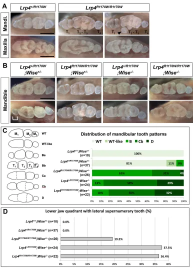
Lrp4R1170Wmutation mimicsWise-null tooth phenotypes
The relatively mild tooth phenotypes of variousLrp4mutants and the genetic interaction of Lrp4 with Wise and Lrp5/6 together suggest the existence of Wise-independent roles for Lrp4 in tooth development. Validation of this hypothesis requires a context in which the Wise-dependent roles are blocked while other aspects of Lrp4 function are preserved. Since no such mouse models were available, we created mice carrying the human R1170W mutation (G to T), which has been shown to abolish SOST binding to LRP4 (Leupin et al., 2011). This is likely to abolish Wise binding to Lrp4, without affecting the normal cell membrane localization of Lrp4 (Leupin et al., 2011). Mice homozygous for theLrp4R1170Wallele are viable, indicating that the Lrp4 function in the neuromuscular junction is not significantly affected. To our surprise,Lrp4R1170W/ R1170Wmice display tooth phenotypes more severe than those of
Lrp4ECD/ECDandLrp4ΔICD/ΔICDmice and comparable to those of
Wise-null mice (Fig. 7A). For example, T1is typically longer with
multiple cusps and lateral supernumerary teeth are frequently observed inLrp4R1170W/R1170Wmice. This suggests that the single amino acid substitution is sufficient to prevent Lrp4 from inhibiting Wnt/β-catenin signaling during tooth development. It also implies that the substitution efficiently blocks the Lrp4-Wise interaction.
Severe syndactyly and/or oligodactyly are common to all known
[image:8.612.53.560.402.685.2]Lrp4 mutant models and are attributed to disruption in AER
Fig. 6. Domain-specific roles for Lrp4.(A) Lrp4-Lrp6 fusion proteins. Numbers within the boxes indicate amino acid residues (Lrp4, NP_766256.3; Lrp6, NP_032540.2) encoded by each construct. TM, transmembrane domain. LV and S are linker peptides. (B,C) Relative luciferase activity fromTOPflashreporter after transfecting HEK 293T cells with constructs driving expression ofWnt1(B) orWNT3A(C) in combination with other proteins. An empty vector ( pCS2) was used to show the basal level of reporter activity. (D) Overexpression ofLrp6andLrp4ECD-Lrp6ICDresults in forced activation of Wnt/β-catenin signaling in the dental epithelium. (E) Removal of the intracellular domain of Lrp4 results in abnormal tooth development. Scale bars: 1 mm (whole mount) and 0.5 mm (section).
DEVEL
O
patterning (Li et al., 2010; Pohlkamp et al., 2015; Simon-Chazottes et al., 2006; Weatherbee et al., 2006). Similarly,Lrp4ΔICD/ΔICDmice display severe defects in distal limbs (Fig. S8). Surprisingly,
Lrp4R1170W/R1170Wmice display no apparent limb defects, indicating that the arginine residue is not essential for the Lrp4 function in patterning of the AER (Fig. S8). Since the limb defects are causally associated with elevated Wnt/β-catenin signaling (Ahn et al., 2013), our data suggest that Lrp4 negatively regulates Wnt/β-catenin
signaling largely independently of Wise/Sost during early limb patterning. Supporting this idea, single and double mutants forWise
and Sost display no, or very subtle, limb defects (Collette et al., 2013).
[image:9.612.112.498.54.590.2]Lrp4R1170W mice were then crossed withWise-null mice to test whether the Lrp4R1170W allele interacts with the Wise-null allele differently, as compared with otherLrp4alleles. Whereas no tooth defects are present inLrp4+/R1170WandWise+/−mice, a small portion Fig. 7. Tooth defects of mice with the R1170W mutation.(A)Lrp4R1170W/R1170Wmice display strong tooth defects such as a larger T
1and lateral supernumerary
teeth (arrowhead). (B) Genetic interaction between theLrp4R1170WandWise-null alleles. Some transheterozygotes develop a supernumerary tooth (bracket)
distal to M1. Two representative samples are shown for each genotype. Scale bars: 1 mm. (C,D) Distribution of mandibular tooth patterns (C) and penetrance of
the lateral supernumerary tooth phenotype (D) among mice carrying theLrp4R1170WandWise-null alleles.
DEVEL
O
(8.1%, n=37) of transheterozygotes display T1, which has
presumably developed from R2 (Fig. 7B,C). In contrast to
Lrp4ECD/ECD;Wise−/− andLrp4mitt/mdig;Wise−/− mice, in which the
Wise-null tooth phenotypes are ameliorated, there is no significant difference in the severity of tooth defects between Lrp4+/R1170W;
Wise−/−andLrp4R1170W/R1170W;Wise−/−mice (Fig. 7B-D). Together, these genetic data support a model whereby Lrp4 mediates the Wnt inhibitory function of Wise in tooth development, and the R1170 residue is essential for the Lrp4-Wise interaction. Since
Lrp4R1170W/R1170Wmice phenocopyWise-null mice with severe tooth defects, we conclude that Lrp4 has a Wise-independent Wnt stimulatory activity that is not disrupted by the missense mutation.
DISCUSSION
In this study, an extensive series of genetic analyses in teeth provide strong experimental evidence for a hypothesis that Lrp4 and Wise physically interact with each other to negatively regulate Wnt/β -catenin signaling. The findings also provide new insights into molecular mechanisms associated with interactions between Lrp4, Wise and the Wnt co-receptors Lrp5/6. We demonstrate that Lrp4 acts as both inhibitor and activator of the Wnt/β-catenin pathway dependent upon its interactions with Wise. In vitro and in vivo
chimeric receptor analyses unveil roles played by the extracellular and intracellular domains of Lrp4 in its interplay with Wnt ligands, antagonists and Lrp5/6 co-receptors. By generating a mouse model of a human missense mutation, we demonstrate that Wise-dependent and Wise-inWise-dependent roles for Lrp4 can be separated. These findings broaden our understanding of mechanisms that incorporate different inputs to achieve precise spatiotemporal control of signaling activity during development.
Similarities and differences in tooth defects ofLrp4 -deficient andWise-deficient mice
In this study, we uncovered not only similarities, but also significant qualitative and quantitative differences in tooth phenotypes between
Wise-deficient andLrp4-deficient mice. We utilized aWise-null allele and a series of Lrp4-null and hypomorphic alleles and carefully analyzed tooth phenotypes to rule out the possibility that these differences result from partial inactivation of the gene or differences in strain backgrounds. In mice deficient for either of the genes, the diastema bud R2 escapes from degeneration and gives rise to a supernumerary cheek tooth distal to M1in the mandible. Whereas this
phenotype is ∼100% penetrant in Wise-null mice regardless of strain background,Lrp4mutants display a wide-range of penetrance (30-90%) in different strain backgrounds. One possible explanation for this difference is that the degree of alteration in signaling activity caused byLrp4deficiency is relatively low so that penetrance of the phenotype is affected by genetic modifiers. We discovered that, unlike inWise-null mice, R2 fails to maintain distance from M1and
often becomes merged into M1 in Lrp4-null mice. In the lateral
inhibition model, an existing tooth delays development of the next tooth (Kavanagh et al., 2007). This suggests that a lack of accelerated development of R2, and hence reduced lateral inhibition on M1,
results in relatively early development of M1inLrp4mutants. As R2
loses its inhibitory advantage over M1, it is more likely to be merged
into M1, mimicking the outcome in normal tooth development. We
also discovered that other aspects of tooth defects are milder inLrp4
mutants. For example, lateral supernumerary teeth are rarely observed and overgrowth is not common in the mandible ofLrp4 mutants compared withWise-null mice.
We observed that molar defects are more severe in the maxilla than in the mandible in bothWise-deficient andLrp4-deficient mice
leading to overgrowth and fusion of the distal teeth, consistent with published observations (Ahn et al., 2010; Ohazama et al., 2008). This less variable fusion phenotype is attributed to elevation of Wnt/ β-catenin signaling to a much higher level in the maxilla of the mutants, as significant rescue of the defect requires removal of at least two copies ofLrp5andLrp6(Ahn et al., 2010) (this study).
Wise-dependent and Wise-independent roles for Lrp4 in tooth development
That there are significant differences in the severity of tooth defects caused by deficiency ofLrp4orWiseis in line with the two proteins also having independent roles. In this regard, our previous study in embryonic mammary gland development revealed stage-specific roles for Lrp4 and Wise (Ahn et al., 2013). The earlier and more severe patterning defects inLrp4mutant mice compared withWise -null mice may be attributed to a Wise-independent role for Lrp4 in the mammary placodes. Unexpectedly, in Lrp4;Wise double-homozygous mutants the tooth defects are ameliorated compared with those ofWise-null mice. Furthermore, removing a copy ofLrp4
on top of reduced dosages of Lrp5/6results in further rescue of
Wise-null tooth phenotypes. Since both Lrp4 and Wise single mutants display tooth defects associated with elevated Wnt signaling and these defects are rescued by reduced dosages of
Lrp5/6, inhibition of Wnt/β-catenin signaling is likely to be the major mechanism of action of Lrp4 in tooth development. However, our data point to the presence of an additional Wnt stimulatory role for Lrp4 that is independent of Wise. In this scenario, the net effect of Lrp4 deficiency would still be an increase in Wnt/β-catenin signaling, as the inhibitory activity of Lrp4 in the presence of Wise is greater than its stimulatory activity. Consequently, overall tooth phenotypes ofLrp4mutants would be milder than those of
Wise-null mice, in which Lrp4 maintains its stimulatory activity. Lack of the stimulatory activity would explain the amelioration of
Wise-null phenotypes in Lrp4ECD/ECD;Wise−/− and Lrp4mitt/mdig;
Wise−/−mice. It is possible that this stimulatory activity is enhanced in the absence of Wise, contributing to the severe tooth defects of
Wise-null mice.
Our gain-of-function analyses provided further insights into the interplay betweenWiseandLrp4in tooth development. Whereas overexpression of Wisein the dental epithelium suppresses tooth development, excessWisefails to exert any significant effect on the dynamics of R2 and M1development inLrp4-deficient mice. This
dependence of Wise on Lrp4 suggests that Lrp4 is required for most, if not all, aspects of Wise function in the inhibition of Wnt/β-catenin signaling. Lrp4 gain-of-function tooth phenotypes are more intriguing as they mimic, to a certain degree, Lrp4 loss-of-function phenotypes. Since Lrp4 expression is temporally dynamic and spatially restricted, it is likely that ectopic overexpression disrupts Lrp4 function in its normal expression domain, which overlaps with the epithelial signaling centers. It is possible that this represents the Wise-independent Wnt stimulatory activity of Lrp4 mentioned above. We speculated that secreted Wise proteins become a limiting factor when Lrp4 is in excess, which can alter the distribution and function of Wise. This idea was supported by the observation that a reduced dosage of Wise exacerbatesLrp4
gain-of-function phenotypes and that simultaneous overexpression of Wise and Lrp4 results in stronger suppression of tooth development.
Dissecting domain-specific roles for Lrp4
Our chimeric receptor analyses indicate that the ICD determines whether Lrp4 and Lrp5/6 play a stimulatory or inhibitory role in
DEVEL
O
Wnt/β-catenin signaling. The observation that the ECDs of Lrp4 and Lrp6 are somewhat interchangeable suggests that the ECDs of Lrp4 and Lrp5/6 can interact with a similar set of signaling molecules, such as Wnt ligands and antagonists. This might be linked to Wise-independent roles for Lrp4 in certain contexts. Interestingly, Lrp4ECD appeared to be more responsive to co-expression of Wise, and Lrp6ECD more responsive to co-expression of Wnt ligands. This difference might reflect a higher affinity of Lrp4 to Wise and a lower affinity to Wnt ligands. Our analyses of Lrp4ΔICD/ΔICD mice indicate that the ICD is essential for Lrp4 function in limb and tooth development, but dispensable in the neuromuscular junction. Since the mutant mice display tooth defects comparable to those observed in mice with other
Lrp4mutations that presumably disrupt most aspects of Lrp4 function, it is likely that removal of the ICD leads to loss of both Wise-dependent and -inWise-dependent functions of Lrp4. Anchorage to the cell membrane alone appears insufficient for Lrp4 function in the modulation of Wnt/β-catenin signaling. It remains to be investigated whether the ICD is required for proper trafficking to the cell membrane or is directly involved in the modulation of Wnt/β-catenin signaling. In postnatal bone, loss ofLrp4in osteoblasts leads to increased bone mass reminiscent of bone phenotypes observed in mice deficient forSost(Chang et al., 2014a; Collette et al., 2012; Li et al., 2008; Xiong et al., 2015). Human patients with theLRP4mutation R1170W display bone overgrowth and the mutation results in reduced binding to SOST and abolishes the LRP4 function as a facilitator of SOST in cultured cells (Leupin et al., 2011). The severe tooth phenotypes ofLrp4R1170Wmice and the lack of amelioration of
Wise-null tooth defects inLrp4R1170W/R1170W;Wise−/−mice together suggest that Wise and Sost bind to the same domain of Lrp4 to inhibit Wnt/β-catenin signaling. Sost and Wise originate from a common ancestral gene (Collette et al., 2013), but have since
diverged from each other. This study provides evidence that despite significant differences (less than 40% amino acid identity) between the two antagonists, Wise interacts similarly with Lrp4 to regulate Wnt/β-catenin signaling during tooth development. Lrp4R1170W mice provide a valuable in vivo model in which the antagonist-dependent Wnt inhibitory role of Lrp4 is abolished while its antagonist-independent roles in the modulation of Wnt and other signaling pathways are retained.
Multiple modes of Lrp4 function in the modulation of Wnt/β -catenin signaling
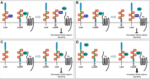
With a large ECD possessing multiple protein-protein interaction motifs, Lrp4 can potentially interact with an array of signaling molecules to control Wnt/β-catenin signaling. Based on findings from the current and earlier studies, we propose a model involving multiple modes of action deployed by Lrp4 to modulate Wnt/β -catenin signaling (Fig. 8).
Modes I and II describe two mechanisms for Wise-dependent Wnt inhibitory activity of Lrp4. Lrp4 might act as an anchor/ presenter molecule for Wise. During tooth development, Lrp4 recruits Wise to the epithelial signaling centers to inhibit Wnt/β -catenin signaling. Wise might be presented to Lrp5/6 via Lrp4, resulting in displacement of Wnt ligands and inhibition of signaling (mode I, Fig. 8A). Alternatively, Lrp4 together with Wise might compete with Lrp5/6 for binding to Fz receptors (mode II, Fig. 8B). In some developmental contexts, Lrp4 might inhibit Wnt/β -catenin signaling independently of Wise. In this scenario, Lrp4 might compete with Lrp5/6, interfering with the formation of Wnt/ Fz/co-receptor complexes (mode III, Fig. 8C).
[image:11.612.52.561.427.694.2]The current study also discovered a Wise-independent Wnt stimulatory role for Lrp4. It remains to be investigated whether this role involves a direct interaction between Lrp4 and Wnt/β-catenin
Fig. 8. Proposed modes of Lrp4 function in different contexts.(A) Mode I: Lrp4 presents Wise to Lrp5/6 resulting in displacement of Wnt ligands and inhibition of Wnt/β-catenin signaling. (B) Mode II: Lrp4 in the presence of Wise competes with Lrp5/6 for binding to Fz receptors. (C) Mode III: in the absence of Wise, Lrp4 inhibits Wnt/β-catenin signaling by competing with Lrp5/6 for Wnt ligands and/or Fz receptors. (D) Mode IV: in the absence of Wise, Lrp4 binds to Wnt ligands and presents them to Lrp5/6 for activation of Wnt/β-catenin signaling.
DEVEL
O
signaling or is mediated via another signaling pathway. Lrp4 might bind to Wnt ligands and present them to Lrp5/6 for activation of Wnt/β-catenin signaling (mode IV, Fig. 8D). This mode might be advantageous when the signaling needs to be activated rapidly in a defined domain and then turned off by expression of the antagonists. Our findings suggest that Lrp4 acts as a modulator of Wnt/β -catenin signaling, integrating multiple inputs, and that its mode of action is determined by the presence and relative concentration of signaling molecules such as Wnt ligands, antagonists and receptors. Although it is unknown how Lrp4 regulates different signaling pathways, our study demonstrates that different domains of Lrp4 can be linked to pathway-specific roles, opening a door to more effective and safer therapeutics to treat disease conditions caused by abnormal LRP4 function (Shen et al., 2015).
MATERIALS AND METHODS Mouse strains
TopGal,Lrp4mitt,Lrp4mte,Lrp4mdig,Lrp4ECD,Wise-null,Lrp5-null,Lrp6
-null,K14-tTAandtetO-Wisemice were described previously (Ahn et al.,
2013; DasGupta and Fuchs, 1999; Johnson et al., 2005; Kato et al., 2002; Pinson et al., 2000; Simon-Chazottes et al., 2006; Weatherbee et al., 2006) (Table S1). All experiments involving mice were performed under approved protocols issued to R.K. as the principal investigator by the Institutional Animal Care and Use Committee of the Stowers Institute for Medical Research (Protocol ID: 2016-0164).
Generation ofLrp4BAC-tTA,tetO-Lrp4,tetO-Lrp6and
tetO-Lrp4ECD-Lrp6ICDtransgenic mice
The Lrp4BAC-tTA was constructed by inserting VP22-tTA-SV40pA
(Gossen and Bujard, 1992) in-frame into the first coding exon ofLrp4
in a 134 kb Lrp4 BAC clone (Ahn et al., 2013) using bacterial
recombination technology (Lee et al., 2001). ThetetOconstructs were
generated by replacing theWiseORF in thetetO-Wiseconstruct (Ahn
et al., 2010) with coding sequences of Lrp4, Lrp6 and
Lrp4ECD-Lrp6ICD. Transgenic founders were generated by pronuclear injection of linearized constructs into (C57BL/10JxCBA)F2 embryos. F0 founder or
N1 mice carrying individualtetOtransgenes were crossed withK14-tTA
mice to identify expression lines that drive eGFP expression in the
presence of tTA.
Generation ofLrp4lacZ,Lrp4ΔICDandLrp4R1170Wmice using CRISPR/Cas9 technology
Gene editing was achieved by pronuclear injection of pX330 plasmids
expressing Cas9 and single guide RNA (sgRNA) (Cong et al., 2013;
Mashiko et al., 2013; Wang et al., 2013). Twenty base pair seed sequences preceding the PAM sequence (NGG) at the target loci were cloned into the
pX330 BbsI site.Lrp4lacZmice carrying an in-frame insertion oflacZinto
the first exon were generated in (C57BL/10JxCBA)F2 embryos by
co-injecting a donor plasmid (15 ng/µl) andpX330plasmid (3 ng/µl) with
5′-GGCGCCCTGCTCTGCGCACA-3′as a seed sequence (supplementary
Materials and Methods).
Lrp4ΔICD mice were generated by introducing two stop codons after
Lys1751 using an oligo donor (5′
-ATACCTATAAAGTTCTCAACTGA- TTTCAGCCCGATTTTTCCTCTTGAAGACACAGAAAATGATAACA-
GACGGATCCTGGAATGGGAAACCTGACCTATAGCAACCCCTCC-TACCGAACTTCCACTCAGGA-3′; mismatched bases against the wild-type
allele are underlined) andpX330with 5′
-ACACAGAAAATCCAAGTTCA-3′as a seed sequence. The R1170W (C to T) mutation was introduced into
Lrp4by co-injecting an oligo donor (5′
-TTGGCAACCTGGATGGGTC- TATGCGGAAAGTGTTGGTGTGGCAGAACCTTGACAGTCCCTG-
GGCCATTGTATTATACCATGAAATGGGGTGAGAGCTGGCTTT-ATCACTCTGAGTGGAC-3′) and pX330 with 5′
-TGGTATAATA-CAATGGCCCG-3′as a seed sequence. BothLrp4ΔICDandLrp4R1170W
mice were generated and maintained on the FVB/N strain (supplementary Materials and Methods).
X-Gal staining andin situhybridization
To detect β-galactosidase activity from lacZ reporters, embryos were
dissected and fixed in either 0.1% paraformaldehyde (PFA)/0.2% glutaraldehyde (E11.5-E13.5) or 4% PFA (E14.0 or older) for 30-60 min on ice. After washes in PBS, fixed samples were stained in X-Gal for 4-20 h
at 4°C or at room temperature. Whole-mountin situ hybridization was
performed with dissected jaws fixed in 4% PFA overnight according to standard protocols using DIG-labeled antisense riboprobes (Roche) against Lrp4 (Weatherbee et al., 2006) and Shh (Dassule and McMahon, 1998). For histological sections, stained samples were paraffin embedded after post-fixation in 4% PFA, sectioned at 8 µm and counterstained with Nuclear Fast Red.
Dual luciferase assay
HEK 293T cells were grown in DMEM (Dulbecco’s Modified Eagle
Medium with 4 mM glutamine, Gibco) supplemented with 10% FBS. Cells
were plated on 24-well plates at ∼50% confluence, transfected with
Lipofectamine 2000 (Thermo Fisher Scientific) the following day according
to the manufacturer’s protocol and harvested 24 h after transfection.
Expression constructs were generated by inserting a full-length cDNA of each gene into the multicloning sites of pCS2+ (Turner and Weintraub, 1994). The amounts of individual DNA constructs used per well were
100 ngTOPflash(EMD Millipore), 5 ngRenillaluciferase, 100 ngWnt1or
humanWNT3A, 100 ngLrp4orLrp6or chimeric receptors, and 200 ng
WiseorSost. The empty vector pCS2+ was added to the DNA mix to keep
the total amount of DNA at∼700 ng/well. Dual-luciferase reporter assays
(Promega) were performed according to the manufacturer’s protocol. Each
experiment was performed in duplicate and representative results are from one of three independent experiments.
Acknowledgements
We thank Jenny Reynolds, Heidi Monnin and the Stowers Institute Histology Facility for technical assistance and members of the R.K. laboratory for valuable discussion.
Competing interests
The authors declare no competing or financial interests.
Author contributions
Conceptualization: Y.A., R.K.; Methodology: Y.A., R.K.; Validation: Y.A.; Formal analysis: Y.A., M.J.M., P.K.K., J.F.-A., R.K.; Investigation: Y.A., C.S., M.J.M., P.K.K., J.F.-A., S.D.W., R.K.; Resources: Y.A., C.S., S.D.W., R.K.; Data curation: Y.A., C.S.; Writing - original draft: Y.A., R.K.; Writing - review & editing: Y.A., S.D.W., R.K.; Visualization: Y.A., R.K.; Supervision: Y.A., R.K.; Project administration: C.S., R.K.; Funding acquisition: R.K.
Funding
Y.A., C.S., M.J.M., P.K.K., J.F.-A. and R.K. were supported by funds from Stowers Institute for Medical Research, and S.D.W. was supported by a grant from the National Institutes of Health (R01AR059687). Deposited in PMC for release after 12 months.
Data availability
Original data underlying this manuscript can be accessed from the Stowers Original Data Repository at http://www.stowers.org/research/publications/libpb-1185.
Supplementary information
Supplementary information available online at
http://dev.biologists.org/lookup/doi/10.1242/dev.150680.supplemental
References
Ahn, Y.(2015). Signaling in tooth, hair, and mammary placodes.Curr. Top. Dev. Biol.111, 421-459.
Ahn, Y., Sanderson, B. W., Klein, O. D. and Krumlauf, R.(2010). Inhibition of Wnt signaling by Wise (Sostdc1) and negative feedback from Shh controls tooth number and patterning.Development137, 3221-3231.
Ahn, Y., Sims, C., Logue, J. M., Weatherbee, S. D. and Krumlauf, R.(2013). Lrp4 and Wise interplay controls the formation and patterning of mammary and other skin appendage placodes by modulating Wnt signaling.Development 140, 583-593.
Balic, A. and Thesleff, I.(2015). Tissue interactions regulating tooth development and renewal.Curr. Top. Dev. Biol.115, 157-186.
DEVEL
O
Biggs, L. C. and Mikkola, M. L.(2014). Early inductive events in ectodermal appendage morphogenesis.Semin. Cell Dev. Biol.25-26, 11-21.
Chang, M.-K., Kramer, I., Huber, T., Kinzel, B., Guth-Gundel, S., Leupin, O. and Kneissel, M. (2014a). Disruption of Lrp4 function by genetic deletion or pharmacological blockade increases bone mass and serum sclerostin levels.
Proc. Natl. Acad. Sci. USA111, E5187-E5195.
Chang, M.-K., Kramer, I., Keller, H., Gooi, J. H., Collett, C., Jenkins, D., Ettenberg, S. A., Cong, F., Halleux, C. and Kneissel, M.(2014b). Reversing LRP5-dependent osteoporosis and SOST deficiency-induced sclerosing bone disorders by altering WNT signaling activity.J. Bone Miner. Res.29, 29-42.
Choi, H. Y., Dieckmann, M., Herz, J. and Niemeier, A.(2009). Lrp4, a novel receptor for Dickkopf 1 and sclerostin, is expressed by osteoblasts and regulates bone growth and turnover in vivo.PLoS ONE4, e7930.
Choi, H. Y., Liu, Y., Tennert, C., Sugiura, Y., Karakatsani, A., Kroger, S., Johnson, E. B., Hammer, R. E., Lin, W. and Herz, J.(2013). APP interacts with LRP4 and agrin to coordinate the development of the neuromuscular junction in mice.Elife2, e00220.
Clevers, H. and Nusse, R.(2012). Wnt/beta-catenin signaling and disease.Cell
149, 1192-1205.
Collette, N. M., Genetos, D. C., Economides, A. N., Xie, L., Shahnazari, M., Yao, W., Lane, N. E., Harland, R. M. and Loots, G. G.(2012). Targeted deletion of Sost distal enhancer increases bone formation and bone mass.Proc. Natl. Acad. Sci. USA109, 14092-14097.
Collette, N. M., Yee, C. S., Murugesh, D., Sebastian, A., Taher, L., Gale, N. W., Economides, A. N., Harland, R. M. and Loots, G. G.(2013). Sost and its paralog Sostdc1 coordinate digit number in a Gli3-dependent manner.Dev. Biol.383, 90-105.
Cong, L., Ran, F. A., Cox, D., Lin, S., Barretto, R., Habib, N., Hsu, P. D., Wu, X., Jiang, W., Marraffini, L. A. et al.(2013). Multiplex genome engineering using CRISPR/Cas systems.Science339, 819-823.
Cruciat, C.-M. and Niehrs, C.(2013). Secreted and transmembrane wnt inhibitors and activators.Cold Spring Harb. Perspect. Biol.5, a015081.
DasGupta, R. and Fuchs, E. (1999). Multiple roles for activated LEF/TCF transcription complexes during hair follicle development and differentiation.
Development126, 4557-4568.
Dassule, H. R. and McMahon, A. P.(1998). Analysis of epithelial-mesenchymal interactions in the initial morphogenesis of the mammalian tooth.Dev. Biol.202, 215–227.
Dietrich, M. F., van der Weyden, L., Prosser, H. M., Bradley, A., Herz, J. and Adams, D. J.(2010). Ectodomains of the LDL receptor-related proteins LRP1b and LRP4 have anchorage independent functions in vivo.PLoS ONE5, e9960.
Ellies, D. L. and Krumlauf, R.(2006). Bone formation: the nuclear matrix reloaded.
Cell125, 840-842.
Gomez, A. M. and Burden, S. J.(2011). The extracellular region of Lrp4 is sufficient to mediate neuromuscular synapse formation.Dev. Dyn.240, 2626-2633.
Gossen, M. and Bujard, H.(1992). Tight control of gene expression in mammalian cells by tetracycline-responsive promoters. Proc. Natl. Acad. Sci. USA 89, 5547-5551.
Herz, J. and Bock, H. H.(2002). Lipoprotein receptors in the nervous system.Annu. Rev. Biochem.71, 405-434.
Itasaki, N., Jones, C. M., Mercurio, S., Rowe, A., Domingos, P. M., Smith, J. C. and Krumlauf, R.(2003). Wise, a context-dependent activator and inhibitor of Wnt signalling.Development130, 4295-4305.
Johnson, E. B., Hammer, R. E. and Herz, J.(2005). Abnormal development of the apical ectodermal ridge and polysyndactyly in Megf7-deficient mice.Hum. Mol. Genet.14, 3523-3538.
Karner, C. M., Dietrich, M. F., Johnson, E. B., Kappesser, N., Tennert, C., Percin, F., Wollnik, B., Carroll, T. J. and Herz, J.(2010). Lrp4 regulates initiation of ureteric budding and is crucial for kidney formation–a mouse model for Cenani-Lenz syndrome.PLoS ONE5, e10418.
Kato, M., Patel, M. S., Levasseur, R., Lobov, I., Chang, B. H.-J., Glass, D. A., II, Hartmann, C., Li, L., Hwang, T.-H., Brayton, C. F. et al. (2002). Cbfa1-independent decrease in osteoblast proliferation, osteopenia, and persistent embryonic eye vascularization in mice deficient in Lrp5, a Wnt coreceptor.J. Cell Biol.157, 303-314.
Kavanagh, K. D., Evans, A. R. and Jernvall, J.(2007). Predicting evolutionary patterns of mammalian teeth from development.Nature449, 427-432.
Laurikkala, J., Kassai, Y., Pakkasjarvi, L., Thesleff, I. and Itoh, N.(2003). Identification of a secreted BMP antagonist, ectodin, integrating BMP, FGF, and SHH signals from the tooth enamel knot.Dev. Biol.264, 91-105.
Lee, E.-C., Yu, D., Martinez de Velasco, J., Tessarollo, L., Swing, D. A., Court, D. L., Jenkins, N. A. and Copeland, N. G.(2001). A highly efficient Escherichia coli-based chromosome engineering system adapted for recombinogenic targeting and subcloning of BAC DNA.Genomics73, 56-65.
Leupin, O., Piters, E., Halleux, C., Hu, S., Kramer, I., Morvan, F., Bouwmeester, T., Schirle, M., Bueno-Lozano, M., Ramos Fuentes, F. J. et al.(2011). Bone overgrowth-associated mutations in the LRP4 gene impair sclerostin facilitator function.J. Biol. Chem.286, 19489-19500.
Li, X., Zhang, Y., Kang, H., Liu, W., Liu, P., Zhang, J., Harris, S. E. and Wu, D.
(2005). Sclerostin binds to LRP5/6 and antagonizes canonical Wnt signaling.
J. Biol. Chem.280, 19883-19887.
Li, X., Ominsky, M. S., Niu, Q.-T., Sun, N., Daugherty, B., D’Agostin, D., Kurahara, C., Gao, Y., Cao, J., Gong, J. et al.(2008). Targeted deletion of the sclerostin gene in mice results in increased bone formation and bone strength.
J. Bone Miner. Res.23, 860-869.
Li, Y., Pawlik, B., Elcioglu, N., Aglan, M., Kayserili, H., Yigit, G., Percin, F., Goodman, F., Nürnberg, G., Cenani, A. et al.(2010). LRP4 mutations alter Wnt/
β-catenin signaling and cause limb and kidney malformations in Cenani-Lenz syndrome.Am. J. Hum. Genet.86, 696-706.
MacDonald, B. T. and He, X.(2012). Frizzled and LRP5/6 receptors for Wnt/beta-catenin signaling.Cold Spring Harb. Perspect. Biol.4, a007880.
MacDonald, B. T., Tamai, K. and He, X. (2009). Wnt/beta-catenin signaling: components, mechanisms, and diseases.Dev. Cell17, 9-26.
Mashiko, D., Fujihara, Y., Satouh, Y., Miyata, H., Isotani, A. and Ikawa, M.(2013). Generation of mutant mice by pronuclear injection of circular plasmid expressing Cas9 and single guided RNA.Sci. Rep.3, 3355.
Narhi, K., Tummers, M., Ahtiainen, L., Itoh, N., Thesleff, I. and Mikkola, M. L.
(2012). Sostdc1 defines the size and number of skin appendage placodes.Dev. Biol.364, 149-161.
Ohazama, A., Johnson, E. B., Ota, M. S., Choi, H. J., Porntaveetus, T., Oommen, S., Itoh, N., Eto, K., Gritli-Linde, A., Herz, J. et al.(2008). Lrp4 modulates extracellular integration of cell signaling pathways in development.PLoS ONE3, e4092.
Peterkova, R., Lesot, H. and Peterka, M. (2006). Phylogenetic memory of developing mammalian dentition.J. Exp. Zoolog. B Mol. Dev. Evol. 306B, 234-250.
Pinson, K. I., Brennan, J., Monkley, S., Avery, B. J. and Skarnes, W. C.(2000). An LDL-receptor-related protein mediates Wnt signalling in mice.Nature407, 535-538.
Pohlkamp, T., Durakoglugil, M., Lane-Donovan, C., Xian, X., Johnson, E. B., Hammer, R. E. and Herz, J.(2015). Lrp4 domains differentially regulate limb/ brain development and synaptic plasticity.PLoS ONE10, e0116701.
Semenov, M., Tamai, K. and He, X.(2005). SOST is a ligand for LRP5/LRP6 and a Wnt signaling inhibitor.J. Biol. Chem.280, 26770-26775.
Shen, C., Xiong, W.-C. and Mei, L.(2015). LRP4 in neuromuscular junction and bone development and diseases.Bone80, 101-108.
Simon-Chazottes, D., Tutois, S., Kuehn, M., Evans, M., Bourgade, F., Cook, S., Davisson, M. T. and Guenet, J.-L.(2006). Mutations in the gene encoding the low-density lipoprotein receptor LRP4 cause abnormal limb development in the mouse.Genomics87, 673-677.
Turner, D. L. and Weintraub, H.(1994). Expression of achaete-scute homolog 3 in Xenopus embryos converts ectodermal cells to a neural fate.Genes Dev.8, 1434-1447.
Wang, H., Yang, H., Shivalila, C. S., Dawlaty, M. M., Cheng, A. W., Zhang, F. and Jaenisch, R.(2013). One-step generation of mice carrying mutations in multiple genes by CRISPR/Cas-mediated genome engineering.Cell153, 910-918.
Weatherbee, S. D., Anderson, K. V. and Niswander, L. A.(2006). LDL-receptor-related protein 4 is crucial for formation of the neuromuscular junction.
Development133, 4993-5000.
Willnow, T. E., Christ, A. and Hammes, A.(2012). Endocytic receptor-mediated control of morphogen signaling.Development139, 4311-4319.
Xiong, L., Jung, J.-U., Wu, H., Xia, W.-F., Pan, J.-X., Shen, C., Mei, L. and Xiong, W.-C.(2015). Lrp4 in osteoblasts suppresses bone formation and promotes osteoclastogenesis and bone resorption. Proc. Natl. Acad. Sci. USA 112, 3487-3492.