0095-1137/11/$12.00 doi:10.1128/JCM.01254-11
Copyright © 2011, American Society for Microbiology. All Rights Reserved.
Comparative Performance of Human Papillomavirus DNA
Testing Using Novel Sample Collection Methods
䌤
Julia C. Gage,
1* Edward E. Partridge,
2Alfio Rausa,
3Patti E. Gravitt,
4Sholom Wacholder,
1Mark Schiffman,
1Isabel Scarinci,
2and Philip E. Castle
5Division of Cancer Epidemiology and Genetics, National Cancer Institute, National Institutes of Health, DHHS, Bethesda,
Maryland1; University of Alabama at Birmingham, Birmingham, Alabama2; Mississippi State Department of Health,
Jackson, Mississippi3; Department of Epidemiology, Johns Hopkins School of Public Health, Baltimore,
Maryland4; and American Society for Clinical Pathology, Washington, DC5
Received 24 June 2011/Returned for modification 23 July 2011/Accepted 3 October 2011
To explore alternative cervical cancer screening approaches in an underserved population, we compared the performance of human papillomavirus (HPV) DNA assays in combination with different sample collection methods for primary cervical screening in the Mississippi Delta region. Three specimens were collected from women aged 26 to 65 years who were either routinely undergoing screening (n ⴝ 252) or not (n ⴝ 191): clinician-collected cervical specimens, clinician-collected cervicovaginal specimens, and self-collected cervico-vaginal specimens taken at home. A novel collection device and medium were used for cervicocervico-vaginal sampling. Specimens were tested by three HPV DNA assays: hybrid capture 2 (HC2; Qiagen Corp., Gaithersburg, MD), Linear Array (LA; Roche Molecular Systems, Pleasanton, CA), and Amplicor (Roche Molecular Systems, Pleasanton, CA). Liquid-based cytology was performed on cervical specimens. We compared the overall positivity (a proxy for clinical specificity) for any carcinogenic HPV genotype and calculated the agreement across assay and specimen type using McNemar’s test for differences in test positivity. Across all three assays there were no significant differences between clinician-collected and self-collected cervicovaginal specimens (P> 0.01 for all comparisons). For both cervicovaginal specimens (clinician collected and self-collected), fewer women tested positive by HC2 than by LA or Amplicor (P < 0.01 for all comparisons). HC2 had the best agreement between specimens for all assays. HC2 is likely more clinically specific, although possibly less sensitive, than either PCR test. Thus, use of HC2 on cervicovaginal specimens for screening could result in fewer referrals compared to LA and Amplicor.
In the United States, annual cervical cancer incidence and related mortality have fallen to⬃10,000 and⬃4,000 per year, respectively (18). However, these reductions have not been uniformly achieved since more than half of all cervical cancer occurs in medically underserved populations (http://www.cdc .gov/cancer/cervical/), of which the Mississippi Delta region ranks highest (13). The Mississippi Delta region is one of the poorest areas in the United States, and it has been referred to as a “Third World country in the heart of America” (23). Overall, the rates of cervical cancer incidence and mortality in this region are some of the highest in the country and compa-rable to rates in some low- and middle-income countries (13). Cervical cancers in the United States arise from both lack of screening and lack of appropriate follow-up of abnormal re-sults (10). Unfortunately, even in the United States, formida-ble barriers remain for cytology programs to successfully pre-vent cancer in underserved populations. Women must repeat screening through clinic visits throughout their adult life be-cause cervical cytology is insensitive (21), and women who screen positive are often lost to follow-up (2, 4, 12). In order to reduce the excessive burden of cervical cancer in these medi-cally underserved populations, novel approaches to overcome
these barriers might be useful to reducing cancer health dis-parities.
There is now convincing evidence that carcinogenic human papillomavirus (HPV) DNA testing is cost-effective and sensi-tive for the detection of precancerous lesions (5, 28), albeit less specific than cytology for primary cervical cancer screening (20, 22). In the United States, carcinogenic HPV DNA testing in conjunction with cervical cytology has been accepted for cer-vical cancer screening in women 30 and older, with those who test negative for both not recommended for screening again for 3 years (33).
One possible method to expand cervical cancer screening in underserved populations is through HPV testing of self-col-lected cervicovaginal specimens among older women (ageⱖ30 years), after the initial peak of HPV prevalence observed at younger ages. Several studies have now evaluated self-collec-tion in combinaself-collec-tion with the U.S. Food and Drug Administra-tion-approved HPV DNA test, Hybrid Capture 2 (HC2; Qia-gen, Gaithersburg, MD), as a potential alternative to cytology (24, 32). In the absence of participation in cytology-based pro-grams, HPV DNA testing in self-collected samples might broaden population coverage of cervical cancer screening (6, 16). Women can self-sample in their home and attend the clinic only if their HPV result is positive (ca. 5 to 15% of women over age 30 [6, 9]).
The optimal method to self-sample, particularly outside of the clinic setting, has not been identified. It is possible that observed differences in test performance of self-sampled ver-* Corresponding author. Mailing address: Clinical Genetics Branch
Division of Cancer Epidemiology and Genetics, National Cancer In-stitute, 6120 Executive Blvd., MSC 7231, Rockville, MD 20852. Phone: (301) 594-7296. Fax: (301) 496-1854. E-mail: [email protected].
䌤Published ahead of print on 12 October 2011.
4185
on May 16, 2020 by guest
http://jcm.asm.org/
sus the traditional clinician-collected method (3, 16, 24, 32) might simply result from the location of sampling (cervicovag-inal being less optimal than cervical) or the self-collection method itself (as opposed to a clinician-collected cervicovagi-nal sample).
Also, HC2 is known to cross-react with noncarcinogenic HPV genotypes that are phylogenetically related to carcino-genic HPV genotypes, whereas PCR HPV DNA tests have greater fidelity for genotyping (8). Therefore, it is possible that HC2 might have higher test positivity and therefore lower clinical specificity than other tests. However, studies among women with cytological abnormalities have shown HC2 to have lower HPV positivity (sometimes translating into lower clinical sensitivity and greater clinical specificity) compared to PCR assays such as the Linear Array (LA; Roche Molecular Sys-tems, Pleasanton, CA) (17, 30) and Amplicor (Roche Molec-ular Systems, Pleasanton, CA) (29–31).
In this study of women attending screening in the Mississippi Delta, we sought to compare HC2 analytic performance with two PCR assays in three types of collections: clinician-collected cervical specimens, clinician-collected cervicovaginal speci-mens, and self-collected cervicovaginal specimens. Because colposcopically guided biopsy diagnosis was not possible among women who screened HPV negative and cytology nor-mal, we are considering, as a proxy for clinical specificity, test positivity. Previous studies suggest that HPV test positivity is a reasonable proxy for clinical specificity (17, 30, 31). Although higher specificity can result in corresponding lower sensitivity, the present study presents data from a general screening pop-ulation and is not sufficiently powered to examine clinical sen-sitivity. We therefore only present HPV positivity (clinical specificity) results, as opposed to sensitivity.
MATERIALS AND METHODS
Study population. We recruited nonpregnant, nonhysterectomized women aged 26 to 65 years without history of treatment and attending a Mississippi State Department of Health screening clinic in Tallahatchie, Leflore, Sunflower, or Washington Counties. Our goal was to reach 250 women attending a standard screening visit and 250 women who had not been screened within the past 3 years in accordance with screening guidelines (“underscreened”). Additional exclusion criteria included inability to speak English, perceived mental incompetence, and visualization of an overt cancerous lesion at the clinical exam.
Clinical visit and specimen collection.After study staff described the study and showed a video explaining proper use of the self-collection device (Fournier Self-Sampler; Arthur Fournier, University of Miami) (19), women provided written consent. Before undergoing a pelvic exam, clinicians (physicians and nurse practitioners) collected a cervicovaginal specimen using a Fournier sam-pling device from seated participants, to serve as a reference standard (“mock self-sample”) compared to self-collected specimens. The mock self-sample was then placed in a vial of safe collection medium (Scope mouthwash; Proctor & Gamble) (7). Clinicians then collected a cervical specimen using direct sampling with a Dacron swab into PreservCyt liquid-based cytology medium for standard of care screening.
Finally, women were asked to take a “kit” home to collect a second cervico-vaginal specimen 7 to 14 days after the clinic visit. The “kit” was composed of a Fournier self-sampler, a vial of Scope transport medium, an instruction pam-phlet, a brochure on HPV and cervical cancer, and return packaging. Participants received a phone call from the study staff to remind them to perform the self-collection. Women were asked to mail specimens back to the clinic using a postage-paid, preaddressed package or return the kit directly to the clinic where they were enrolled on a prespecified day and time. As compensation for return of the cervical specimen, U.S. $20 was provided upon return of the specimens.
Testing.Liquid-based cytology (LBC) specimens were processed and read by local cytopathologists at the reference laboratory at the University of Mississippi in Jackson, MS. Residual PreservCyt and two study cervicovaginal specimens
were sent to Johns Hopkins School of Public Health for HPV DNA testing using HC2 (25), Amplicor (using a 1.0-relative-light-unit cutoff) (26), and Linear Array (LA; Roche Molecular Systems) (27). HC2 is a clinical pooled-probe, signal amplification DNA test for detection of 13 carcinogenic HPV types. LA is a type-specific PGMY09/11 L1 primer PCR assay for 37 HPV types, and Amplicor is a pooled-probe, DNA amplification (PCR) test that targets the same HPV types as HC2. Specimens testing positive for any of 13 carcinogenic HPV geno-types (HPV geno-types 16, 18, 31, 33, 35, 39, 45, 51, 52, 56, 58, 59, and 68) or HPV66 (because HC2 strongly cross-reacts with this HPV genotype [8]) by LA were considered carcinogenic HPV positive.
Statistical analysis. We first calculated the age-specific carcinogenic HPV prevalence, with 95% confidence intervals (95% CI), to permit comparison with other U.S. populations. We then calculated overall test positivity. Kappa statis-tics and paired McNemar’s tests were used to test agreement between HPV assays and sampling methods.
All aspects of this study were reviewed and approved by National Cancer Institute, Mississippi State Board of Health, and University of Alabama institu-tional review boards for human subject research.
RESULTS
Between 2007 and 2009, we recruited 252 women for routine screening and 191 women who had not been screened in the previous 3 years achieving 100.8 and 76.4% of recruitment goals, respectively. Overall, 92.6% of participants provided self-samples, a proportion that did not differ between groups (chi-square test,P⫽0.42).
The mean and median age differed significantly by screening group: 34.7 and 33 in the screened group and 40.3 and 40 in the underscreened group (Pⱕ0.01). Using HC2 test results as the reference for carcinogenic HPV detection, the age-group-spe-cific prevalence in this population is shown in Fig. 1A. Among women age 40 and older, HC2 positivity was higher in under-screened compared to previously under-screened women (23.0% ver-sus 13.2%, respectively; chi-square test,P⫽ 0.02). A similar association was observed among women with normal cytology, although it did not reach statistical significance (16.2% versus 11.8%, respectively; chi-square test,P⫽0.26) (Fig. 1B).
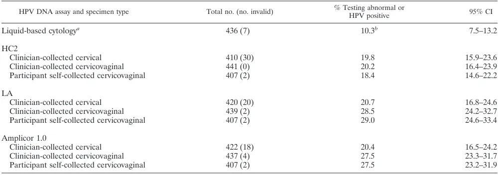
The percentages of abnormal cytology and carcinogenic HPV positivity for all three tests are shown in Table 1 for both screened and underscreened groups combined, as the mea-sures did not differ between groups. Overall, 10.3% of women had an abnormal cytology result (atypical squamous cells of undetermined significance or worse [ASCUS⫹]). By compar-ison, the percent HC2 positive was 19.2% for the cervical specimens, 20.2% for the clinician-collected cervicovaginal specimens, and 18.4% for the self-collected specimens. The percent LA positive for carcinogenic HPV genotypes was 20.7% for the cervical specimens, 28.5% for the clinician-col-lected cervicovaginal specimens, and 29.0% for the self-col-lected specimens. The percent Amplicor positive was 20.4% for the cervical specimens, 27.5% for the clinician-collected cervicovaginal specimens, and 27.0% for the self-collected specimens.
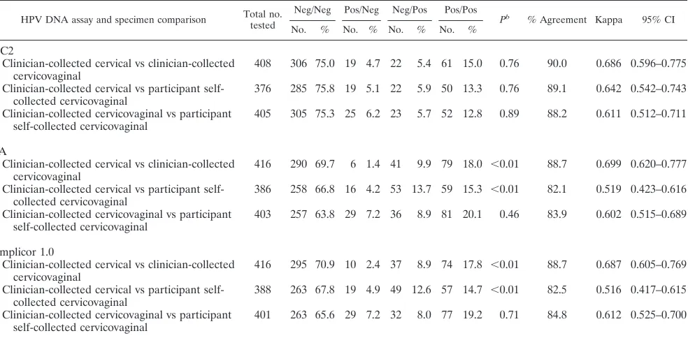
In the subset of specimens that had paired test results by the same assay (Table 2), there were (i) no significant differences between clinician-collected and self-collected cervicovaginal specimens across all three assays (P⬎0.01 for all comparisons) and (ii) no significant differences between the percent HC2 positive by specimen type. In addition, both cervicovaginal specimens were more likely to test positive by LA than the cervical specimen (P⬍0.01 for both comparisons), and both cervicovaginal specimens were more likely to test positive by
on May 16, 2020 by guest
http://jcm.asm.org/
Amplicor than the cervical specimen (P⬍0.01 for both com-parisons). For all three assays the agreement between speci-men types was best for the cervical and clinician-collected cervicovaginal specimen (⬃90% agreement and kappa⬃0.70) compared to the other pairwise comparisons.
Comparisons of assays within specimens showed some sig-nificant differences as concordance between HPV DNA assays differed by specimen type (Table 3). The best test agreement was for LA and Amplicor on any specimen type, whereas there was poorer agreement between each of these assays and HC2. In both clinician- and participant-collected cervicovaginal specimens, LA and Amplicor tended to call more women pos-itive for carcinogenic HPV than HC2 (P⬍0.01 for all com-parisons), explaining the poorer agreement between HC2 and LA or Amplicor than between these two assays for cervicovag-inal specimens.
Conclusions. We examined the performance of different tests and specimens on detection of carcinogenic HPV, as a proxy for clinical specificity. Both PCR assays, LA and Ampli-cor, had higher positivity (and probably lower clinical specific-ity) in clinician-collected and participant self-collected cervi-covaginal specimens compared to (i) cervical specimens using the same test and (ii) HC2 from the same cervicovaginal spec-imen. We believe a likely explanation is that the PCR assays such as LA and Amplicor detected lower viral load of carci-nogenic HPV types in cervicovaginal swabs resulting in higher HPV positivity. However, it is also possible that the difference between PCR positivity in cervicovaginal versus cervical spec-imens was not due to the sampling location, but the possibility that the novel Fournier device used for cervicovaginal sam-pling did not effectively shield against irrelevant vaginal infec-tions in the context of analytically sensitive HPV tests.
With the exception of clinician-collected cervical samples, HC2 positivity was lower than those of LA and Amplicor in both clinician- and participant-collected cervicovaginal speci-mens. Since similar positivity was observed across both trans-port mediums (clinician-collected in PreservCyt and partici-pant self-collected in Scope), it is unlikely that the lower positivity was due to utilizing the novel transport medium, FIG. 1. (A) Percentage of all women testing HC2 positive by
[image:3.585.41.541.534.710.2]screening history and age group (years). Among all women ofⱖ40 years, HC2 positivity was higher in underscreened versus screened women (23.0% versus 13.2%, respectively; chi-square test,P⫽0.02). (B) Percentage of women with a concurrently normal cytology who tested HC2 positive by screening history and age group (years). Among women with normal cytology and over age 40, women underscreened had higher HC2 positivity than screened women, although it did not reach statistical significance (16.2% versus 11.8%, respectively; chi-square test,P⫽0.26).
TABLE 1. Overall positivity of HPV DNA tests and liquid-based cytology
HPV DNA assay and specimen type Total no. (no. invalid) % Testing abnormal or
HPV positive 95% CI
Liquid-based cytologya 436 (7) 10.3b 7.5–13.2
HC2
Clinician-collected cervical 410 (30) 19.8 15.9–23.6
Clinician-collected cervicovaginal 441 (0) 20.2 16.4–23.9
Participant self-collected cervicovaginal 407 (2) 18.4 14.6–22.2
LA
Clinician-collected cervical 420 (20) 20.7 16.8–24.6
Clinician-collected cervicovaginal 439 (2) 28.5 24.2–32.7
Participant self-collected cervicovaginal 407 (2) 29.0 24.6–33.4
Amplicor 1.0
Clinician-collected cervical 422 (18) 20.4 16.5–24.2
Clinician-collected cervicovaginal 437 (4) 27.5 23.3–31.7
Participant self-collected cervicovaginal 407 (2) 27.5 23.2–31.9
a
Positive threshold ASCUS or worse. b
LBC had significantly lower positivity than all HPV DNA assays from clinician-collected cervical specimens (P⬍0.01 for all comparisons).
on May 16, 2020 by guest
http://jcm.asm.org/
Scope mouthwash. It is possible that HC2 is less clinically sensitive than LA and Amplicor in cervicovaginal specimens, as previously reported (3). However, among clinician-collected and participant-collected cervicovaginal specimens that tested negative by HC2 but positive by LA and/or Amplicor, only 51.4 and 54.3%, respectively, were positive by both LA and Ampli-cor, not more than to be expected by chance alone (P⫽0.99). Anecdotally, among the three cases of CIN3 and one case of CIN2 detected at follow-up colposcopy for 149 women (61.1% of 244 women referred because of an abnormal HPV or cytol-ogy result had colposcopy), all HPV assays in all specimens tested HPV positive, with the exception of three clinician-collected cervicovaginal HC2 tests that tested HPV negative
(one CIN2 and two CIN3). All but one CIN3 were detected in the underscreened group.
[image:4.585.49.541.82.324.2]HC2 is known to cross-react with noncarcinogenic HPV genotypes that are phylogenetically related to carcinogenic HPV genotypes (8). We investigated whether PCR tests’ greater fidelity for carcinogenic HPV genotypes would trans-late to lower positivity and therefore better clinical specificity. In the present study, HC2 was likely more clinically specific than the PCR tests. Often greater specificity corresponds to reduced sensitivity. Unfortunately, our study was not ade-quately powered to measure clinical sensitivity of HPV assays, but it is possible that lower HC2 positivity in cervicovaginal TABLE 2. Interspecimen agreement by HPV DNA assaya
HPV DNA assay and specimen comparison Total no. tested
Neg/Neg Pos/Neg Neg/Pos Pos/Pos
Pb % Agreement Kappa 95% CI No. % No. % No. % No. %
HC2
Clinician-collected cervical vs clinician-collected cervicovaginal
408 306 75.0 19 4.7 22 5.4 61 15.0 0.76 90.0 0.686 0.596–0.775
Clinician-collected cervical vs participant self-collected cervicovaginal
376 285 75.8 19 5.1 22 5.9 50 13.3 0.76 89.1 0.642 0.542–0.743
Clinician-collected cervicovaginal vs participant self-collected cervicovaginal
405 305 75.3 25 6.2 23 5.7 52 12.8 0.89 88.2 0.611 0.512–0.711
LA
Clinician-collected cervical vs clinician-collected cervicovaginal
416 290 69.7 6 1.4 41 9.9 79 18.0 ⬍0.01 88.7 0.699 0.620–0.777
Clinician-collected cervical vs participant self-collected cervicovaginal
386 258 66.8 16 4.2 53 13.7 59 15.3 ⬍0.01 82.1 0.519 0.423–0.616
Clinician-collected cervicovaginal vs participant self-collected cervicovaginal
403 257 63.8 29 7.2 36 8.9 81 20.1 0.46 83.9 0.602 0.515–0.689
Amplicor 1.0
Clinician-collected cervical vs clinician-collected cervicovaginal
416 295 70.9 10 2.4 37 8.9 74 17.8 ⬍0.01 88.7 0.687 0.605–0.769
Clinician-collected cervical vs participant self-collected cervicovaginal
388 263 67.8 19 4.9 49 12.6 57 14.7 ⬍0.01 82.5 0.516 0.417–0.615
Clinician-collected cervicovaginal vs participant self-collected cervicovaginal
401 263 65.6 29 7.2 32 8.0 77 19.2 0.71 84.8 0.612 0.525–0.700
a
HPV genotypes 16, 18, 31, 33, 35, 39, 45, 51, 52, 56, 58, 59, and 68, and as well as HPV66, were evaluated. Pos, positive; Neg, negative. b
Determined using the exact McNemar’s test for differences in test positivity.
TABLE 3. Interassay agreement by specimen typea
Specimen type and HPV DNA assay comparison
Total no. tested
Neg/Neg Pos/Neg Neg/Pos Pos/Pos Pb
% Agreement Kappa 95% CI
No. % No. % No. % No. %
Clinician-collected cervical
HC2 vs LA 392 283 72.2 25 6.4 30 7.7 54 13.8 0.59 86.0 0.574 0.474–0.675
HC2 vs Amplicor 1.0 394 289 73.4 25 6.4 26 6.6 54 13.7 1.0 87.1 0.598 0.499–0.698 LA vs Amplicor 1.0 419 314 74.9 19 4.5 19 4.5 67 16.0 1.0 90.9 0.722 0.639–0.805
Clinician-collected cervicovaginal
HC2 vs LA 437 293 67.1 24 5.5 56 12.8 64 14.7 ⬍0.01 81.7 0.499 0.405–0.593
HC2 vs Amplicor 1.0 439 295 67.2 19 4.3 56 12.8 69 15.7 ⬍0.01 82.9 0.540 0.450–0.629 LA vs Amplicor 1.0 437 293 67.1 24 5.5 19 4.4 101 23.1 0.54 90.2 0.756 0.687–0.825
Participant self-collected cervicovaginal
HC2 vs LA 405 275 67.9 18 4.4 55 13.6 57 14.1 ⬍0.01 82.0 0.498 0.401–0.596
HC2 vs Amplicor 1.0 405 275 68.2 11 2.7 54 13.3 64 15.8 ⬍0.01 84.0 0.565 0.473–0.656 LA vs Amplicor 1.0 406 272 67.0 22 5.4 17 4.2 95 23.4 0.522 90.4 0.763 0.693–0.833
a
HPV genotypes 16, 18, 31, 33, 35, 39, 45, 51, 52, 56, 58, 59, and 68, as well as HPV66, were evaluated. Pos, positive; Neg, negative. b
Determined using the exact McNemar’s test.
on May 16, 2020 by guest
http://jcm.asm.org/
[image:4.585.41.539.545.709.2]specimens might translate to low sensitivity if HC2 does not detect all precancers.
The overall burden of HPV was elevated in this high-risk Mississippi Delta population as indicated by greater age-spe-cific prevalence of HPV (as measured by HC2) than observed in other populations (9). In particular, women over 40 who were underscreened were at higher risk of prevalent HPV infection compared to women over 40 who were recently screened. Self-sampling with HPV testing is now being utilized in many settings to reach women who typically do not partic-ipate in traditional cervical cancer screening programs (1, 11, 14–16).
In our study, we found good correlation between cervico-vaginal specimens collected by participants in their homes and cervicovaginal specimens collected by clinicians. It is possible that the women in our study had an easier time self-sampling after already having a vaginal exam in the office, resulting in higher correlation. In addition, partici-pants of this study reported self-sampling to be an accept-able screening method since 91.9% of women chose to self-sample at home, suggesting that self-sampling could improve overall coverage. The vast majority (95.7%) of women said they found the collector very easy or somewhat easy to use, despite its relative complexity of having a sheath to shield against vaginal infections, moving parts, and a tip that needs to be ejected at the completion of collection. An ancillary study found that self-sampling was preferred to clinic-based Pap testing among women who had chosen not to participate in recommended, routine Pap testing (6).
In this screening population we found HPV DNA testing with self-collected cervicovaginal samples to be acceptable to participants and comparable to clinician-collected cervicovagi-nal samples across all assays. Our findings suggest self-collec-tion with HPV testing could be used to complement current screening programs to reach underscreened women in this high-risk population. Once optimized, it might be used selec-tively to reach and screen the small pockets of underserved U.S. populations who carry the burden of about 60% of cer-vical cancer incidence in the United States (http://www.cdc.gov /cancer/cervical/).
REFERENCES
1.Barbee, L., et al.2010. Assessing the acceptability of self-sampling for HPV among Haitian immigrant women: CBPR in action. Cancer Causes Control
21:421–431.
2.Bastani, R., K. R. Yabroff, R. E. Myers, and B. Glenn.2004. Interventions to improve follow-up of abnormal findings in cancer screening. Cancer101:
1188–1200.
3.Belinson, J. L., et al.2010. Prevalence of type-specific human papillomavirus in endocervical, upper and lower vaginal, perineal, and vaginal self-collected specimens: implications for vaginal self-collection. Int. J. Cancer127:1151– 1157.
4.Benard, V. B., H. W. Lawson, C. R. Eheman, C. Anderson, and W. Helsel.
2005. Adherence to guidelines for follow-up of low-grade cytologic abnor-malities among medically underserved women. Obstet. Gynecol.105:1323– 1328.
5.Bulkmans, N. W., et al.2007. Human papillomavirus DNA testing for the detection of cervical intraepithelial neoplasia grade 3 and cancer: 5-year follow-up of a randomised controlled implementation trial. Lancet370:
1764–1772.
6.Castle, P. E., et al.2011. Comparative community outreach to increase cervical cancer screening in the Mississippi Delta. Prev. Med.152:452–455.
7.Castle, P. E., et al.2007. Mouthwash as a low-cost and safe specimen transport medium for human papillomavirus DNA testing of cervicovaginal specimens. Cancer Epidemiol. Biomarkers Prev.16:840–843.
8.Castle, P. E., et al.2008. Human papillomavirus genotype specificity of hybrid capture 2. J. Clin. Microbiol.46:2595–2604.
9.Datta, S. D., et al.2008. Human papillomavirus infection and cervical cytol-ogy in women screened for cervical cancer in the United States, 2003–2005. Ann. Intern. Med.148:493–500.
10.Du, P., et al.2010. The roles of social domains, behavioral risk, health care resources, and chlamydia in spatial clusters of US cervical cancer mortality: not all the clusters are the same. Cancer Causes Control21:1669–1683. 11.Dzuba, I. G., et al.2002. The acceptability of self-collected samples for HPV
testing versus the pap test as alternatives in cervical cancer screening. J. Womens Health Gender Based Med.11:265–275.
12.Eggleston, K. S., A. L. Coker, K. J. Luchok, and T. E. Meyer.2007. Adher-ence to recommendations for follow-up to abnormal Pap tests. Obstet. Gy-necol.109:1332–1341.
13.Freeman, H. P., and B. K. Wingrove.2005. Excess cervical cancer mortality: a marker for low access to health care in poor communities. NIH pub. no. 05-5282. National Cancer Institute, Center to Reduce Cancer Health Dis-parities, Rockville, MD.
14.Giorgi Rossi, P., et al.2011. The effect of self-sampled HPV testing on participation to cervical cancer screening in Italy: a randomised controlled trial (ISRCTN96071600). Br. J. Cancer104:248–254.
15.Gok, M., et al.2011. Experience with high-risk human papillomavirus testing on vaginal brush-based self-samples of non-attendees of the cervical screen-ing program. Int. J. Cancer129:517–527.
16.Gravitt, P. E., J. L. Belinson, J. Salmeron, and K. V. Shah.Looking ahead: a case for HPV testing of self-sampled vaginal specimens as a cervical cancer screening strategy. Int. J. Cancer, in press.
17.Gravitt, P. E., M. Schiffman, D. Solomon, C. M. Wheeler, and P. E. Castle.
2008. A comparison of linear array and hybrid capture 2 for detection of carcinogenic human papillomavirus and cervical precancer in ASCUS-LSIL triage study. Cancer Epidemiol. Biomarkers Prev.17:1248–1254. 18.Jemal, A., et al.2006. Cancer statistics, 2006. CA Cancer J. Clin.56:106–130. 19.Knesel, B. W., J. C. Dry, C. Wald-Scott, and A. Aftab.2005. Preliminary evaluation of a cervical self-sampling device with liquid-based cytology and multiparameter molecular testing. J. Reprod. Med.50:256–260.
20.Mayrand, M. H., et al.2007. Human papillomavirus DNA versus Papanico-laou screening tests for cervical cancer. N. Engl. J. Med.357:1579–1588. 21.Nanda, K., et al.2000. Accuracy of the Papanicolaou test in screening for
and follow-up of cervical cytologic abnormalities: a systematic review. Ann. Intern. Med.132:810–819.
22.Naucler, P., et al.2007. Human papillomavirus and Papanicolaou tests to screen for cervical cancer. N. Engl. J. Med.357:1589–1597.
23.Parfit, M.1993. And what words shall describe the Mississippi, great father of rivers? Smithsonian36:36.
24.Petignat, P., et al.2007. Are self-collected samples comparable to physician-collected cervical specimens for human papillomavirus DNA testing? A systematic review and meta-analysis. Gynecol. Oncol.105:530–535. 25.Qiagen.2004. Hybrid Capture 2 High-Risk HPV DNA test package insert.
Qiagen Corp., Gaithersburg, MD.
26.Roche Molecular Diagnostics.2008. The Amplicor human papillomavirus test, package insert. Roche, Alameda, CA.
27.Roche Molecular Systems.2006. Linear Array HPV genotyping test package insert. Roche Molecular Systems, Pleasanton, CA.
28.Ronco, G., et al.2010. Efficacy of human papillomavirus testing for the detection of invasive cervical cancers and cervical intraepithelial neoplasia: a randomised controlled trial. Lancet Oncol.11:249–257.
29.Sandri, M. T., et al.2006. Comparison of the Digene HC2 assay and the Roche Amplicor human papillomavirus (HPV) test for detection of high-risk HPV genotypes in cervical samples. J. Clin. Microbiol.44:2141–2146. 30.Stevens, M. P., et al.2007. Comparison of the Digene Hybrid Capture 2
assay and Roche Amplicor and Linear Array human papillomavirus (HPV) tests in detecting high-risk HPV genotypes in specimens from women with previous abnormal Pap smear results. J. Clin. Microbiol.45:2130–2137. 31.Wentzensen, N., P. E. Gravitt, D. Solomon, C. M. Wheeler, and P. E. Castle.
2009. A study of Amplicor human papillomavirus DNA detection in the atypical squamous cells of undetermined significance-low-grade squamous intraepithelial lesion triage study. Cancer Epidemiol. Biomarkers Prev.18:
1341–1349.
32.Wright, T. C., Jr., L. Denny, L. Kuhn, A. Pollack, and A. Lorincz.2000. HPV DNA testing of self-collected vaginal samples compared with cytologic screening to detect cervical cancer. JAMA283:81–86.
33.Wright, T. C., Jr., et al.2007. 2006 consensus guidelines for the management of women with abnormal cervical screening tests. J. Lower Genital Tract Dis.
11:201–222.