0095-1137/09/$12.00
doi:10.1128/JCM.01103-09
Copyright © 2009, American Society for Microbiology. All Rights Reserved.
Design and Validation of Real-Time Reverse Transcription-PCR
Assays for Detection of Pandemic (H1N1) 2009 Virus
䌤
Kanti Pabbaraju,
1* Sallene Wong,
1Anita A. Wong,
1Greg D. Appleyard,
1,3Linda Chui,
2,4Xiao-Li Pang,
2,4Stephanie K. Yanow,
2,5Kevin Fonseca,
1,3Bonita E. Lee,
2,6Julie D. Fox,
1,3and Jutta K. Preiksaitis
2,7Provincial Laboratory for Public Health (Microbiology), Calgary Site, Calgary, Alberta, Canada
1; Provincial Laboratory for
Public Health (Microbiology), Edmonton Site, Edmonton, Alberta, Canada
2; Department of Microbiology and
Infectious Diseases, University of Calgary, Calgary, Alberta, Canada
3; Department of Laboratory Medicine and
Pathology, University of Alberta, Edmonton, Alberta, Canada
4; Department of Public Health Sciences,
School of Public Health, University of Alberta, Edmonton, Alberta, Canada
5; Department of Pediatrics,
University of Alberta, Edmonton, Alberta, Canada
6; and Department of Medicine, University of
Alberta, Edmonton, Alberta, Canada
7Received 5 June 2009/Returned for modification 26 July 2009/Accepted 25 August 2009
Tracking novel influenza viruses which have the potential to cause pandemics, such as the pandemic (H1N1) 2009
virus, is a public health priority. Pandemic (H1N1) 2009 virus was first identified in Mexico in April 2009 and
spread worldwide over a short period of time. Well-validated diagnostic tools that are rapid, sensitive, and specific
for the detection and tracking of this virus are needed. Three real-time reverse transcription PCR (RT-PCR) assays
for the amplification and detection of pandemic (H1N1) 2009 virus were developed, and their performance
char-acteristics were compared with those of other published diagnostic assays. Thirty-nine samples confirmed to be
positive for pandemic (H1N1) 2009 virus from Alberta, Canada, and six additional samples that were positive for
influenza A virus but that were not typeable by using published seasonal influenza H1/H3 virus assays were
available for this validation. Amplification and direct sequencing of the products was considered the “gold
stan-dard” for case identification. The new assays were sensitive and able to reproducibly detect virus in a 10
ⴚ6dilution
of 4
ⴛ
10
650% tissue culture infective doses/ml when 5
l was used as the template. They showed 100% specificity
and did not cross-react with other respiratory viruses or seasonal influenza A virus subtypes. The coefficient of
variation in crossing cycle threshold values for the detection of different template concentrations of pandemic
(H1N1) 2009 virus was
<
3.13%, showing good reproducibility. The assays had a wide dynamic range for the
detection of pandemic (H1N1) 2009 virus and utilized testing platforms appropriate for high diagnostic throughput
with rapid turnaround times. We developed and validated these real-time PCR procedures with the goal that they
will be useful for diagnosis and surveillance of pandemic (H1N1) 2009 virus. These findings will contribute to the
informed management of this novel virus.
Novel influenza viruses introduced into the human
popula-tion that are able to spread efficiently from human to human
have the potential to cause pandemics with significant
morbid-ity and mortalmorbid-ity (5, 9, 16). A novel subtype of influenza A virus
called pandemic (H1N1) 2009 virus was identified in Mexico
and was reported by the Centers for Disease Control and
Prevention (CDC; Atlanta, GA) and WHO in April 2009 (2, 3,
8). Cases of swine influenza virus infection in humans have
been reported previously, but these viruses did not show
evi-dence of efficient transmission between human hosts (11).
However, pandemic (H1N1) 2009 virus is a novel subtype virus
that transmits easily between humans, with 21 countries
re-porting cases within a month of the initial identification of the
virus (4). It is essential that public health laboratories around
the world undertake detailed surveillance to monitor the
spread and impact of pandemic (H1N1) 2009 virus as well as to
try to predict future changes in its virulence (8). Methods for
the rapid diagnosis, case identification, and tracking of this
novel pathogen in the human population are required to
de-velop appropriate management strategies to mitigate
morbid-ity and mortalmorbid-ity.
Novel influenza viruses are first identified by amplification
and sequencing of specific genes; these sequences can be
com-pared to those already deposited in databases. Such a
meth-odological approach is important for the identification of new
viruses but generally requires high viral loads (or cultured
virus) for good-quality sequence data to be obtained. The
turnaround time required for this type of analysis negates its
use as a frontline diagnostic test, especially with large numbers
of clinical samples. Once sequences are available, it is
impor-tant that public health laboratories develop and validate
high-throughput sensitive, specific, and rapid assays for use for
diagnostics and surveillance.
As a rapid response to the outbreak of pandemic (H1N1)
2009 virus, the CDC provided a detailed methodology for
real-time PCR amplification and detection of this virus (2).
Other conventional and real-time reverse transcription
(RT)-PCR protocols for the detection of this virus have also recently
been published (13). We have developed three real-time
RT-PCR assays for the detection of pandemic (H1N1) 2009 virus
* Corresponding author. Mailing address: Provincial Laboratory for
Public Health (Microbiology), 3030 Hospital Drive, Calgary, Alberta,
Canada T2N 4W4. Phone: (403) 944-8621. Fax: (403) 283-0142.
E-mail: [email protected].
䌤
Published ahead of print on 2 September 2009.
3454
on May 16, 2020 by guest
http://jcm.asm.org/
using primers and hydrolysis probes targeting the
hemaggluti-nin (HA) and matrix (M) genes. We report on the
develop-ment, validation, and performance of these assays and
compare the results with those obtained by using the CDC
real-time procedures as well as a multiplex suspension
mi-croarray assay (the xTAG respiratory virus panel [RVP] assay)
used for the detection of multiple respiratory viruses, including
influenza A virus. Patient samples collected between 24 April
and 18 May 2009 were used for the validation study.
Improved detection methods will aid with the identification
of new cases of pandemic (H1N1) 2009 virus and ensure
op-timal management to minimize transmission to vulnerable
in-dividuals, as well as aid with surveillance activities to provide
an understanding of the full impact of this virus in the
com-munity.
MATERIALS AND METHODS
Samples tested.Samples (n⫽5,436) submitted to the Provincial Laboratory for Public Health (ProvLab) from Alberta, Northwest Territory, Nunavut, and Yukon from 24 April to 18 May 2009 for testing for respiratory viruses were included in this analysis. Specimens from patients for whom a significant travel history, epidemiological links to pandemic (H1N1) 2009 virus, or severe respi-ratory infection (SRI) was indicated on the requisition were identified so that the respiratory virus testing algorithm could be adjusted to enhance its ability to detect influenza A virus in these specimens, as described in the next section.
Diagnostic testing algorithm for human seasonal influenza and pandemic (H1N1) 2009 virus.Specimens from patients with a history of travel or exposure to pandemic (H1N1) 2009 virus or SRI were tested concurrently by the RVP assay on the Luminex platform (Luminex Molecular Diagnostics, Inc., Toronto, Ontario, Canada) and a real-time RT-PCR assay targeting the M gene developed by the CDC (2). The CDC referred to the assay as InfA in the original publica-tion (2), but we use the term CDC-M assay in this report. Other specimens from patients with no history related to pandemic (H1N1) 2009 virus were tested by the RVP assay only. The total numbers of specimens tested by the different assays are provided below. The RVP assay can detect seasonal influenza A viruses and can specifically identify the circulating H1 and H3 subtypes (12); it has not been validated for use for the detection of pandemic (H1N1) 2009 virus. The CDC-M assay detects only influenza A viruses and does not provide addi-tional subtyping information. A direct fluorescent-antigen (DFA) test (Imagen; Dako Diagnostics Ltd., Ely, Cambridgeshire, United Kingdom) for influenza A virus, influenza B virus, parainfluenza virus, and respiratory syncytial virus was also performed with samples for which rapid screening would be useful, such as samples from patients with SRI.
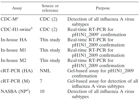
Specimens that gave a positive result for influenza A virus by the DFA, CDC-M, or RVP assay but were not subtyped as seasonal H1 or H3 viruses by the RVP assay were subjected to real-time RT-PCR assays, as described previ-ously (2), for the typing of seasonal H1 and H3 subtypes. The protocol for these typing assays was distributed in December 2008 through U.S. Public Health Service laboratories and the WHO Global Influenza Surveillance Network; it has been approved for use by the Food and Drug Administration. All influenza A virus-positive samples for which a valid subtype by the RVP assay or the real-time typing methods was not obtained were subjected to conventional RT-PCR (cRT-PCR) assays targeting the HA and M genes for amplification and sequenc-ing, as described below. Table 1 provides a list of the assays used for this validation study and clarifies the source and purpose of each test.
cRT-PCR amplification and confirmation of pandemic (H1N1) 2009 virus by sequencing.cRT-PCR was performed by using a One-Step RT-PCR kit (Qiagen, Mississauga, Ontario, Canada) in a total volume of 50l. This included 10l of 5⫻RT-PCR buffer, 10l of Q solution, 2l of 10 mM deoxynucleoside triphos-phates, 0.125l of 40 U/l RNaseOUT (Invitrogen), 2l of One-Step RT-PCR enzyme mix, 3.0l each of 10M primers, and 15l of RNase-free water. Primers H1_F_swine and H1_R_swine were provided by the National Microbi-ology Laboratory (NML; Public Health Agency of Canada, Winnipeg, Manitoba, Canada; unpublished data) for specific amplification of a 517-bp fragment of the HA gene of pandemic (H1N1) 2009 virus. Primers published previously (7) were used for amplification of a 244-bp fragment of the M gene of all influenza A virus subtypes, including pandemic (H1N1) 2009 virus. The thermal cycling conditions comprised a 30-min RT step at 50°C; a 15-min initial PCR activation step at 95°C; and 40 cycles of 94°C for 30 s, 50°C for 30 s, and 72°C for 60 s each,
followed by a 10-min extension at 72°C. All thermal cycling was performed with a model 2720 thermal cycler (Applied Biosystems [ABI], Foster City, CA). The PCR products were subjected to gel electrophoresis for visualization of the products. For confirmation, the products were purified with QIAquick columns (Qiagen) and sequenced in both directions by using an ABI Prism BigDye Terminator (version 3.1) cycle sequencing kit in an ABI Prism 3130-Avant genetic analyzer on a 50-cm array. The sequencing reaction was performed according to the manufacturer’s protocol in a final volume of 10l with approx-imately 2 ng of amplified product, BigDye Terminator (version 3.1) premix, and 0.64 pmol of the primer used for RT-PCR. Unincorporated nucleotides were purified by using a BigDye XTerminator purification kit (ABI). The data were collected by using ABI software (version 2.0). The sequences were analyzed by using Sequencing Analysis software (version 5.3; ABI), and alignments were performed by using the ClustalW program (version 1.4) included in the BioEdit package (version 7.0.0; http://www.mbio.ncsu.edu/BioEdit/bioedit.html). The se-quences were compared to the previously published sese-quences of pandemic (H1N1) 2009 virus and seasonal influenza virus subtypes available from the GenBank database and the Global Initiative on Sharing Avian Influenza Data (GISAID; http://platform.gisaid.org). Sequences with⬎99% nucleotide identity to database submissions were confirmed to be positive for pandemic (H1N1) 2009 virus.
Comparison of different assays for confirmation of pandemic (H1N1) 2009 virus.A subset of the samples confirmed to be positive for pandemic (H1N1) 2009 virus by sequencing (n⫽39 of 97 samples identified during this time period) and 6 samples that gave a positive result for influenza A virus but could not be subtyped as seasonal H1 or H3 virus by the real-time typing assays or as pandemic (H1N1) 2009 virus by cRT-PCR and sequencing were included in the evaluation and validation of the three in-house assays described here. The results were compared with those of the swH1 assay published previously (2); that assay is referred to as the CDC-H1-swine assay in this report. Assay details are described below. Table 1 provides the list of assays used in this comparison study. Of the 45 specimens included in this assay validation, 30 were nasopharyngeal swab specimens collected in universal transport medium (UTM; Copan Diag-nostics Inc., Murrieta, CA), 10 were throat swab specimens in UTM, 4 were formalin-fixed tissue samples, and 1 was a respiratory swab specimen of unknown source that was received in UTM.
[image:2.585.300.542.87.263.2](i) Sample preparation.Respiratory samples were pretreated with 25l of Qiagen protease (0.01 milli-Anson units/l) in a thermomixer (Eppendorf, West-bury, NY) at 56°C and 1,000 rpm for 15 min or until the specimen cleared. Nucleic acid was extracted from the treated samples by using an easyMAG automated extractor (bioMe´rieux, Durham, NC), according to the manufactur-er’s instructions. The extracted nucleic acid was eluted in 110l from a sample input volume of 200l. Tissue samples were extracted by using the total nucleic
TABLE 1. Real-time and conventional RT-PCR assays used
for the study
Assay Source or
reference Purpose
CDC-M
aCDC (2)
Detection of all influenza A virus
subtypes
CDC-H1-swine
bCDC (2)
Real-time RT-PCR for
pH1N1_2009
cconfirmation
In-house HA
This study
Real-time RT-PCR for
pH1N1_2009 confirmation
In-house M1
This study
Real-time RT-PCR for
pH1N1_2009 confirmation
In-house M2
This study
Real-time RT-PCR for
pH1N1_2009 confirmation
cRT-PCR (HA)
NML
Gel-based assay for pH1N1_2009
confirmation
cRT-PCR (M)
7
Gel-based assay for detection of all
influenza A virus subtypes
NASBA (NP
d)
10
Detection of all influenza A virus
subtypes
aThe CDC-M assay was referred to as the InfA assay in the original publica-tion (2).
b
The CDC-H1-swine assay was referred to as the swH1 assay in the original publication (2).
c
pH1N1_2009, abbreviation for pandemic (H1N1) 2009 virus. d
NP, nucleoprotein gene.
on May 16, 2020 by guest
http://jcm.asm.org/
acid extraction protocol for tissue specimens by using the QIAamp kit (Qiagen), according to the manufacturer’s instructions.
(ii) Design of primers and probes for in-house real-time RT-PCR assays.
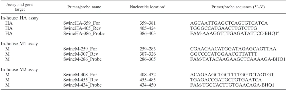
Three sets of primers and probes were designed for this study. All available HA and M gene sequences from GenBank and GISAID (as of 25 April 2009) were aligned, and the three sets of primers and probes were designed to amplify pandemic (H1N1) 2009 virus specifically by using the Primer Express (version 3.0) program (ABI). These primers and probes are listed in Table 2. A review of the sequences submitted to the online databases up to 20 May 2009 confirmed that these in-house-designed primers and probes would detect pandemic (H1N1) 2009 virus sequences available up to that date.
The in-house HA assay targets the 5⬘end of the HA gene, and the in-house M1 and M2 assays target the 5⬘end of the M gene. These assays are henceforth referred to as the in-house HA, in-house M1, and in-house M2 assays. All assays utilized minor groove binding hydrolysis probes and were labeled with 6-car-boxyfluorescein as the reporter dye at the 5⬘end and a black hole quencher at the 3⬘end. The primers were synthesized at the University Core DNA Services (University of Calgary, Calgary, Alberta, Canada), and the probes were synthe-sized by ABI.
(iii) Real-time RT-PCR for detection of pandemic (H1N1) 2009 virus. A one-step RT-PCR method was used for the detection of pandemic (H1N1) 2009 virus by the in-house-developed assays targeting the HA and M genes. The master mixture contained the TaqMan one-step RT-PCR master mix, the Multi-Scribe enzyme mixture, and the primers and probes at final concentrations of 0.8
M and 0.2M, respectively. RT-PCR was performed in an SDS 7500 system in optical tubes from ABI by using 5l of extracted nucleic acid and 20l of the master mixture. The cycling conditions followed the TaqMan universal amplifi-cation protocol, according to the manufacturer’s instructions. The reaction com-ponents for the CDC-M assay were the same as those described above. The cycling conditions included a RT reaction at 48°C for 30 min, followed by enzyme inactivation at 95°C for 10 min. The template was denatured at 95°C for 15 s, annealing and data acquisition were performed at 55°C, and extension was performed at 72°C for 45 cycles.
In addition to the three in-house assays, a published real-time RT-PCR assay designed for the subtyping of pandemic (H1N1) 2009 virus (the CDC-H1-swine assay) (2) was also performed with the available positive samples used in this validation study. This assay was performed on the SDS 7500 system by using the one-step RT-PCR master mixture (ABI) and the protocol provided by the CDC. This protocol was optimized by using an Invitrogen SuperscriptIII Platinum one-step quantitative kit that has been shown to have comparable results on thermocycler systems such as ABI real-time PCR systems (systems 7000, 7300, 7500, etc.), Bio-Rad real-time PCR detection systems (the iQ or iQ5 system), or Stratagene quantitative PCR instruments (the MX4000, MX3000, or MX3005 instrument). Our experiments comparing the use of the one-step RT-PCR master mixture from ABI with the Invitrogen SuperscriptIII Platinum one-step quantitative kit on the 7500 SDS system showed that the performance of the assays were similar (data not shown). Table 1 provides a list of the assays used for this validation study and clarifies the source and purpose of each test.
Limit of detection, sensitivity, specificity, reproducibility, and efficiency of the real-time RT-PCR assays.The pandemic (H1N1) 2009 virus was propagated to 4⫻106
50% tissue culture infectious doses (TCID50)/ml at the NML, and nucleic acid extracted from this harvest was kindly provided to the public health laboratories involved in pandemic (H1N1) 2009 virus testing in Canada for use as control material. This extract was used as the template for all sensitivity studies. Tenfold serial dilutions from 10⫺4
to 10⫺8
of the extract were made in PCR-grade water to which carrier RNA (Qiagen) was added to a final concen-tration of 1g/l. All extracts were tested in triplicate using three independent runs by the new in-house RT-PCR assays (the HA, M1, and M2 assays) and the CDC RT-PCR assays (the CDC-M and CDC-H1-swine assays) described above with 5l of template per reaction mixture (5l of template at a dilution of 10⫺4 contains 2⫻100
TCID50). The sensitivity is reported in terms of the TCID50, as a cloned plasmid is not yet available; the ratio between the TCID50values and the genomic copy numbers can vary between strains and harvests; thus, these numbers cannot be considered absolute and provide only a relative comparison between the different assays used. These dilutions were also tested by the RVP assay and an influenza A virus nucleic acid sequence-based amplification (NASBA) assay targeting the nucleoprotein gene (10) to determine the limit of detection for pandemic (H1N1) 2009 virus. Six specimens that were influenza A virus positive by the screening assays but that could not be subtyped as human H1 or H3 virus by the real-time typing assays or confirmed to be the pandemic (H1N1) 2009 virus by cRT-PCR were tested by the new in-house and CDC-H1-swine assays to investigate if these assays could identify additional pandemic (H1N1) 2009 cases.
The specificities of the newly developed assays were determined by testing extracts of cultures from a variety of influenza A virus strains, including A/Bei-jing/95-like, A/Shangdong/11742/95-like, A/Taiwan/95-like, A/Texas/95-like, A/Johannesburg/96-like, A/Sydney/98-like, A/New Caledonia/00-like, A/Panama/ 03-like, and A/Brisbane/59/07-like strains. In addition, 22 extracts from harvests of patient specimens that were positive for influenza A virus, including subtype H1N1, H1N2, and H3N2 viruses recovered from 1995 to 2009, were used to determine that the pandemic (H1N1) 2009 virus-specific assays did not detect seasonal influenza A viruses. These specimens included four influenza A viruses of the H3N2 subtype and two of the H1N1 subtype that were cocirculating in the community during the same time period as the pandemic (H1N1) 2009 virus after 24 April 2009. The extract from an influenza A virus (H3N2) of probable swine origin designated A/Canada/1158/2006 isolated from a child living on a farm in Canada (14) was tested to determine the specificities of the assays. Samples with high copy numbers of common respiratory pathogens, including influenza B virus; parainfluenza virus types 1, 2, 3, and 4B; respiratory syncytial viruses A and B; human coronavirus NL63, 229E, and OC43; rhinovirus type 1B; coxsackievirus type A16; human metapneumovirus; adenovirus type 2; bocavirus; Chlamy-dophila pneumoniae;Legionella pneumophila; andMycoplasma pneumoniae, were also tested by the in-house HA, M1, and M2 assays and the CDC-H1-swine assay. The reproducibilities of all the real-time RT-PCR assays described above on the SDS 7500 system were evaluated in two independent runs by using three positive clinical specimens with a range of viral loads tested in five replicates.
[image:3.585.42.541.80.237.2]Tenfold serial dilutions of the pandemic (H1N1) 2009 virus control nucleic
TABLE 2. Primers and probes designed in-house for detection of pandemic (H1N1) 2009 virus
Assay and gene
target Primer/probe name Nucleotide location
a Primer/probe sequence (5⬘–3⬘)
In-house HA assay
HA
SwineHA-359_For
359–381
AGCAATTGAGCTCAGTGTCATCA
HA
SwineHA-405_Rev
405–424
TGGGCCATGAACTTGTCTTG
HA
SwineHA-386_Probe
386–403
FAM-AAAGGTTTGAGATATTCC-BHQ1
bIn-house M1 assay
M
SwineM-259_For
259–283
CGAACAACATGGATAGAGCAGTTAA
M
SwineM-307_Rev
307–326
GGCCCCATGGAACGTTATTT
M
SwineM-286_Probe
286–305
FAM-TATACAAGAAGCTCAAAAGA-BHQ1
In-house M2 assay
M
SwineM-408_For
408–432
ACAGAAGCTGCTTTTGGTCTAGTGT
M
SwineM-455_Rev
455–485
TGAGACCGATGCTGTGAATCA
M
SwineM-434_Probe
434–450
FAM-TGCCACTTGTGAACAGA-BHQ1
aThe nucleotide positions for the HA and M genes are based on sequences obtained from GISAID corresponding to EPI176470 HA A/California/04/ 2009 EPI_ISL_29573 2009712049_seg4 H1N1 and EPI176471 MP A/California/04/2009 EPI_ISL_29573 2009712049_seg7 H1N1, respectively.
bThe minor groove binding hydrolysis probes were labeled with 6-carboxyfluorescein (FAM) at the 5⬘end and a black hole quencher (BHQ1) at the 3⬘end.
on May 16, 2020 by guest
http://jcm.asm.org/
acid diluted in carrier RNA were used to determine the dynamic range of the in-house assays and the CDC-M and CDC-H1-swine assays. The efficiency of the PCR was calculated on the basis of the crossing cycle threshold (CT) values obtained.
Data analysis.Samples which gave equivocal results by the RVP assay were excluded from the sensitivity and specificity analyses; equivocal results were defined by the manufacturer to be those with a median fluorescent intensity (MFI) of between 150 and 300. The McNemar test was used to test for a significant difference between the detection of influenza A virus by the RVP assay and the CDC-M assay by using any nucleic acid amplification test (NAT) result positive for influenza A virus as a true-positive result. The differences in CDC-MCTvalues for samples that gave negative, equivocal, or positive results by the RVP assay were compared by the Kruskal-Wallis test. TheCTvalues of the various real-time assays for the 39 samples confirmed to be positive for pandemic (H1N1) 2009 virus were compared by the Friedman test.
RESULTS
Detection of influenza A virus by RVP and CDC-M assays.
During the 25-day study period, 5,436 respiratory specimens
from Alberta, Northwest Territories, Yukon, and Nunavut
were tested for respiratory viruses at ProvLab by the RVP
and/or CDC-M assay. A total of 217 (4.0%) samples were
positive for influenza A virus. Of the 1,465 specimens tested by
this method, 3 samples were initially identified to be positive
for virus by DFA, 156 were positive by both the RVP and the
CDC-M assays, 29 were positive by the CDC-M assay (of which
17 were negative and 12 gave an equivocal result by the RVP
assay), 1 was positive by the RVP assay but negative by the
CDC-M assay, and 28 were positive by the RVP assay but no
CDC-M assay was performed with these samples. The
H-typ-ing results for the 217 influenza A virus-positive samples were
as follows: 97 were confirmed to be pandemic (H1N1) 2009
virus; 80 were confirmed to be seasonal H3 virus; 28 were
confirmed to be seasonal H1 virus; and 9 were unresolved,
likely because of low viral loads. HA typing of the virus in the
remaining three specimens was not completed because they
were duplicate samples from patients who had tested positive
for pandemic (H1N1) 2009 virus.
For the 4,128 samples tested by both the CDC-M and the
RVP assays, the sensitivity of the RVP assay was 90.2% (157/
174 samples; 95% confidence interval [CI], 84.8 to 94.2%). The
sensitivity of the CDC-M assay was 99.4% (173/174 samples;
95% CI, 96.8 to 100%). The specificity was 100% (3,954/3,954
samples) for both assays (
n
⫽
4,128;
P
⬍
0.001, McNemar
test).
Of the 17 specimens that gave a negative result for influenza
A virus by the RVP assay but a positive result by the CDC-M
assay, 2 were typed as pandemic (H1N1) 2009 virus, 5 as
seasonal H3 virus, and 1 as seasonal H1 virus; the types in the
remaining 9 specimens were unresolved because of low viral
loads. Of the 12 specimens with an equivocal result by the RVP
assay but a positive result by the CDC-M assay, 7 were typed as
pandemic (H1N1) 2009 virus and 5 as seasonal H3 virus. The
median
C
Tvalues obtained by the CDC-M assay for samples
that were negative by the RVP assay and equivocal by the RVP
assay or that had concordant influenza A virus-positive results
by both the RVP and the CDC-M assays were 36.0 (range, 31.6
to 38.0), 34.7 (range, 29.7 to 36.6), and 26.4 (range, 13.5 to
36.2), respectively (
P
⬍
0.001, Kruskal-Wallis test). The one
sample that was influenza A virus positive by the RVP assay
but negative by the CDC-M assay was confirmed to be
pan-demic (H1N1) 2009 virus.
Assessment of pandemic (H1N1) 2009 virus RT-PCR assay
performance.
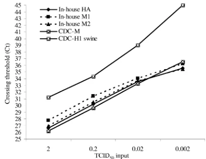
The results for the 10-fold serial dilutions tested
in nine replicates are shown in Table 3 and Fig. 1. Table 3
shows the number of replicates that were positive at each
template concentration. The NASBA signal and MFI for the
RVP assay are also shown in Table 3. The
C
Tvalues obtained
for the real-time RT-PCR assays performed on the SDS 7500
system at each template concentration are given in Fig. 1. All
assays except the NASBA assay had comparable end-point
sensitivities and were able to detect the control pandemic
(H1N1) 2009 virus-positive nucleic acid reproducibly with an
input of 2
⫻
10
⫺2TCID
50
; the NASBA assay detected seven of
the nine replicates.
[image:4.585.42.551.83.152.2]The newly designed assays did not amplify representative
specimens of seasonal influenza A viruses, including subtypes
TABLE 3. Limit of detection analysis for the different assays
Virus concn (no. of TCID50s/5l
a )
No. of positive replicates of nine tested for the following amplification assay:
In-house HA In-house M1 In-house M2 CDC-M CDC-H1-swine RVP (MFI) NASBA (signalb )
2
⫻
10
09
9
9
9
9
9 (7,302)
9 (2.89)
2
⫻
10
⫺19
9
9
9
9
9 (2,680)
9 (2.73)
2
⫻
10
⫺29
9
9
9
9
9 (433)
7 (2.46)
2
⫻
10
⫺34
4
5
6
2
0
3 (2.28)
a
Five microliters of template was used per reaction mixture. b
The fluorescence signal above the background.
FIG. 1. Comparison of
CT
values for detection of serial dilutions of
pandemic (H1N1) 2009 virus. The
C
Tvalues are shown for the
hydro-lysis probe-based assays performed on the SDS 7500 system. The mean
C
Tvalue from the replicates is indicated. The number of replicates
with a positive result for each dilution is indicated in Table 3.
on May 16, 2020 by guest
http://jcm.asm.org/
[image:4.585.314.524.515.677.2]H1N1, H1N2, and H3N2 recovered from 1995 to 2008;
anti-genic drift variants; an H3N2 virus of swine origin isolated in
2006; and the other viral and bacterial respiratory pathogens
tested. This indicates that the newly designed assays show
100% specificity for the detection of pandemic (H1N1) 2009
virus. The assays did not detect the human influenza A virus
H1N1 and H3N2 subtypes, which were cocirculating with
pan-demic (H1N1) 2009 virus between 24 April and 18 May 2009 in
Alberta.
Three clinical specimens were tested by using the SDS 7500
system in five replicates in two independent runs. The
coeffi-cient of variation for the
C
Tvalues ranged from 0.36% to
3.13% for all five assays tested, showing that the assays have
good reproducibilities.
All the assays were able to detect 10-fold serial dilutions of
the control material over 7 log units of template dilution from
4
⫻
10
6TCID
50
/ml to 4
⫻
10
0
TCID
50
/ml when 5
l of
template was used. On the basis of these
C
Tvalues, the PCR
amplification efficiency of the different assays ranged from
82.17% to 91.04%. A representative example of an
amplifica-tion curve for the in-house HA assay and the standard curve
generated by using these dilutions is given in Fig. 2.
Comparison of RT-PCR results for detection of pandemic
(H1N1) 2009 virus-positive specimens.
A subset of all positive
samples detected was used for the validation study. Extracts
from 39 specimens positive for influenza A virus by the
CDC-M screening assay and confirmed to be positive for
pan-demic (H1N1) 2009 virus by cRT-PCR were retrospectively
tested by the CDC-H1-swine assay and the three in-house PCR
assays. The
C
Tvalues obtained by the CDC-M assay ranged
from 17.1 to 36.6 (median, 27.1). Of these 39 specimens, 35
were influenza A virus positive, 3 were equivocal, and 1 was
negative by the RVP assay. As expected, none of the pandemic
(H1N1) 2009 virus-positive samples were typed by the RVP
assay (subtyping is based on human influenza virus sequences).
The MFI values for the positive samples ranged from 313 to
9,835 (median, 7,151). One specimen containing the pandemic
(H1N1) 2009 virus sequence was negative for influenza A virus
by the RVP assay but gave a
C
Tof 33.53 by the CDC-M assay,
suggesting the presence of a low viral load. These results are
summarized in Table 4.
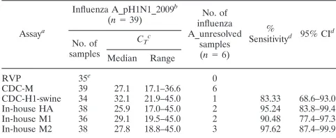
The
C
Tvalues obtained by the different assays (the CDC-M,
CDC-H1-swine, and three in-house assays) for the detection of
virus in these 39 samples are shown in Table 4 (
P
⬍
0.001,
FIG. 2. Representative amplification curves for 10-fold dilutions of template RNA obtained by the in-house HA assay and the standard curve
generated by using these dilutions. Tenfold serial dilutions of template starting at 4
⫻
10
6TCID
50
/ml were tested by using 5
l of template per
reaction mixture. The features of the standard curve were as follows: slope,
⫺
3.56;
R
2, 0.99.
on May 16, 2020 by guest
http://jcm.asm.org/
Friedman test). The virus could not be detected in one
speci-men by any of the real-time assays for pandemic (H1N1) 2009
virus. This specimen had given a weak positive result for
in-fluenza A virus by the CDC-M assay (
C
Tvalue, 36.57) and an
equivocal MFI of 153 by the RVP assay. The reason that the
virus in this sample was missed by the pandemic (H1N1) 2009
virus-specific assays was likely due to the presence of a low viral
load combined with additional freeze-thawing, leading to
nu-cleic acid degradation before the sample was tested by the
pandemic (H1N1) 2009 virus-specific real-time assays. The
in-house HA and M2 assays gave positive results for 38
speci-mens, and the
C
Tvalues were comparable to those from the
CDC-M assay (Table 4). The in-house M1 assay gave positive
results for 36 of the 39 samples containing pandemic (H1N1)
2009 virus sequences (
C
Tvalues for the samples in which virus
was not detected by the CDC-M assay, 34.80, 34.96, and 36.57).
The
C
Tvalues obtained by the in-house M1 assay were higher
than those obtained by the CDC-M and in-house HA and M2
assays. The CDC-H1-swine assay gave positive results for 34
samples containing pandemic (H1N1) 2009 virus sequences
(
C
Tvalues, 34.53, 33.73, 34.65, 34.42, and 36.57 by the CDC-M
assay for the 5 samples in which virus was not detected by the
CDC-H1-swine assay). These results suggest that the in-house
HA and M2 assays have sensitivities comparable to the
sensi-tivity of the CDC-M assay and provide slightly better
sensitiv-ities than the in-house M1 and CDC-H1-swine assays for the
detection of pandemic (H1N1) 2009 virus in clinical samples,
as summarized in Table 4.
Six specimens that were influenza A virus positive by the
CDC-M assay but that could not be subtyped as human H1,
H3, or pandemic (H1N1) 2009 virus were tested by the new
real-time assays to investigate if they provided enhanced
sen-sitivity. These specimens gave positive results for influenza A
virus by the CDC-M assay and had
C
Tvalues that ranged from
31.64 to 37.64 (median, 36.02), but they were all influenza A
virus negative by the RVP assay. Of these six nontypeable
specimens, one was positive for pandemic (H1N1) 2009 virus
by all four real-time PCR assays, one gave a positive result for
pandemic (H1N1) 2009 virus by the three in-house assays, and
the in-house M2 assay detected an additional pandemic
(H1N1) 2009 virus-positive specimen. The
C
Tvalues (by the
CDC-M assay) of three samples which could not be typed by
the new pandemic (H1N1) 2009 virus RT-PCR assays ranged
from 36.00 to 37.64, suggesting that they had very low viral
loads. Thus, additional testing by our newly developed assays
identified three new cases of pandemic (H1N1) 2009 virus
infection.
When the 42 positive specimens (39 confirmed to be positive
by cRT-PCR and 3 that were positive by one or more
pan-demic [H1N1] 2009 virus real-time typing assays) are included,
the sensitivities for the cRT-PCR, CDC-H1-swine, in-house
HA, in-house M1, and in-house M2 assays were 92.86% (95%
CI, 80.5 to 98.5%), 83.33% (95% CI, 68.6 to 93.0%), 95.24%
(95% CI, 83.8 to 99.4%), 90.48% (95% CI, 77.4 to 97.3%), and
97.62% (95% CI, 87.4 to 99.9%), respectively. This
informa-tion is also included in Table 4.
DISCUSSION
Influenza A virus pandemics can arise when genetic
reas-sortment of the influenza A virus in bird and swine populations
leads to the emergence of a novel virus to which majority of the
population is susceptible. Two distinct lineages of swine
influ-enza A viruses of the H1N1 subtype currently circulate within
the North American and European pig populations (6).
Avail-able sequence data indicate that six segments of the currently
circulating pandemic (H1N1) 2009 virus are related to swine
viruses from North America and that the NA and M genes are
related to swine viruses from Europe/Asia (15). Early reports
concerning the pandemic potential of the circulating pandemic
(H1N1) 2009 virus have suggested that the transmissibility of
this virus is substantially higher than that of seasonal influenza
virus (8).
NATs are increasingly being used for the identification of
respiratory virus infections, including those caused by influenza
viruses. NATs provide a rapid and sensitive means of detection
of the etiological agent and are not easily compromised by
sample quality or the timing of collection. Compared with
traditional methods for respiratory virus detection (culture,
antigen detection), NATs have enhanced sensitivities,
specific-ities, and turnaround times. For novel viruses such as
pan-demic (H1N1) 2009 virus, it is important that a rapid diagnosis
be provided without the need for culture to ensure that these
tests can be performed even in laboratories without biosafety
level 2 facilities.
In our diagnostic algorithm, the CDC-M and RVP assays
were used to screen patient specimens for influenza A virus.
Positive specimens were subtyped to identify seasonal H1 and
H3 viruses and pandemic (H1N1) 2009 virus. The higher
sen-sitivity of the CDC-M assay for the detection of influenza A
virus compared with that of the RVP assay did not seem to be
related to the H types but seemed to be more directly linked to
the amount of virus in the sample, as estimated by the
C
Tvalues. Sensitive real-time assays for the subtyping of seasonal
H1 and H3 viruses but not pandemic (H1N1) 2009 virus were
already available at the time of this study. Initial identification
and confirmation of pandemic (H1N1) 2009 virus were
per-TABLE 4. Analysis of influenza A virus-positive specimens tested
for pandemic (H1N1) 2009 virus
Assaya
Influenza A_pH1N1_2009b
(n⫽39) influenzaNo. of A_unresolved
samples (n⫽6)
%
Sensitivityd 95% CId No. of
samples
CT c
Median Range
RVP 35e 0
CDC-M 39 27.1 17.1–36.6 6
CDC-H1-swine 34 32.1 21.9–45.0 1 83.33 68.6–93.0 In-house HA 38 25.9 17.0–45.0 2 95.24 83.8–99.4 In-house M1 36 29.1 19.5–45.0 2 90.48 77.4–97.3 In-house M2 38 27.8 18.8–45.0 3 97.62 87.4–99.9
aThe CDC-M and RVP assays were performed for the detection of influenza A virus, and gel/sequencing methods (cRT-PCR) were used for the prospective confirmation of pandemic (H1N1) 2009 virus. Detection of the pandemic (H1N1) 2009 virus by the real-time assays was performed retrospectively.
bpH1N1_2009, abbreviation for pandemic (H1N1) 2009 virus. cThe median and range ofC
Tvalues is indicated for the different assays (P⬍ 0.001, Friedman test). ACTvalue of 45 was used to represent a negative result. dThe sensitivity and 95% CI calculations were performed by using 42 samples with positive results (39 samples confirmed to be positive by cRT-PCR and 3 samples positive by one or more pandemic关H1N1兴2009 real-time typing assays). eOf the 39 samples tested by the RVP assay, 35 were positive, 3 had a MFI defined as equivocal, and 1 was negative by the RVP assay.
on May 16, 2020 by guest
http://jcm.asm.org/
[image:6.585.42.283.90.189.2]formed by sequencing, which is labor-intensive, involves
mul-tiple steps, and is not readily available in many diagnostic
laboratories. In order to facilitate rapid subtyping, we designed
three real-time RT-PCR assays based on the available
se-quences for pandemic (H1N1) 2009 virus recovered at the
outset of the outbreak. Comparison of the performance
char-acteristics of those assays to the performance charchar-acteristics of
assays provided by the CDC for the detection of all influenza
virus subtypes (the CDC-M assay) and the specific detection of
pandemic (H1N1) 2009 virus (CDC-H1-swine assay) showed
that these in-house assays have comparable (and perhaps
en-hanced) sensitivity for the identification of individuals infected
with pandemic (H1N1) 2009 virus. Due to the limited
avail-ability of positive specimens, a relatively small number was
used to validate the new assays, but these tests will be
moni-tored prospectively for additional validation data. Although we
have not compared our newly developed assay with those
re-ported previously, the limit-of-detection studies suggest that
the assay has a sensitivity comparable to that of other reported
methods for the detection of nucleic acid for pandemic (H1N1)
2009 virus (13). These new assays will be used in conjunction
with the influenza A virus screening and seasonal H1/H3
sub-typing assays for the diagnosis and surveillance of pandemic
(H1N1) 2009 virus. The assays were specific and did not show
any cross-reaction with common respiratory viruses or a variety
of influenza A viruses circulating from 1995 to 2009, including
human H1 and H3 virus subtypes cocirculating with the
pan-demic (H1N1) 2009 virus.
As the influenza season begins in the Southern Hemisphere,
there may be further opportunities for the pandemic (H1N1)
2009 virus to reassort and mutate. A range of good diagnostic
tools for tracking cases will be important in our future planning
for the pandemic and the management of this (and other)
novel influenza viruses. Assays targeting conserved genes (e.g.,
the M, nucleoprotein, and polymerase genes) are useful for the
screening of patient specimens for influenza A virus but will
not differentiate between seasonal influenza A viruses and
viruses such as the pandemic (H1N1) 2009 virus. However,
they are less likely to be prone to problems relating to
se-quence variations and point mutations. Assays targeting
sur-face genes, such as the HA gene, are important for subtyping
and antigenic characterization but may be subject to sequence
variation, especially in the region where short hydrolysis
probes bind to the target sequence. Single point mutations may
disrupt primer or probe binding. Thus, a combination of
screening and typing assays provides an optimal diagnostic
algorithm for the detection of influenza A viruses and
moni-toring of pandemic (H1N1) 2009 virus infections.
The future evolution of pandemic (H1N1) 2009 virus and,
thus, its transmissibility, antigenicity, virulence, and antiviral
resistance are impossible to predict (8, 9). These factors can
vary depending on whether the antigenic changes in the virus
are incremental or if the mutations in the viral genome go from
point to point in a confined space (1, 17). The tracking of
sequence changes will be vital to anticipating the pandemic
potential of this novel subtype of influenza A virus.
ACKNOWLEDGMENTS
We thank all staff at ProvLab for their efforts with accessioning, data
entry, testing, and reporting of results for all the samples during the
time of enhanced laboratory testing and surveillance. We thank the
technologists in the Molecular Diagnostics Division for excellent
tech-nical support. Special thanks go to Kara Tokaryk and Sandy Shokoples
for their technical support.
REFERENCES
1.Allen, J. E., S. N. Gardner, E. A. Vitalis, and T. R. Slezak.2009. Conserved amino acid markers from past influenza pandemic strains. BMC Microbiol.
9:77.
2.Anonymous.2009. Emergence of a novel swine-origin influenza A (H1N1) virus in humans. N. Engl. J. Med.360:2605–2615.
3.Centers for Disease Control and Prevention.2009. Outbreak of swine-origin influenza A (H1N1) virus infection—Mexico, March–April 2009. MMWR Morb. Mortal. Wkly. Rep.58:467–470.
4.Centers for Disease Control and Prevention.2009. Update: novel influenza A (H1N1) virus infections—worldwide, May 6, 2009. MMWR Morb. Mortal. Wkly. Rep.58:453–458.
5.de Wit, E., and R. A. M. Fouchier.2008. Emerging influenza. J. Clin. Virol.
41:1–6.
6.Dunham, E. J., V. G. Dugan, E. K. Kaser, S. E. Perkins, I. H. Brown, E. C. Holmes, and J. K. Taubenberger.2009. Different evolutionary trajectories of European avian-like and classical swine H1N1 influenza A viruses. J. Virol.
83:5485–5494.
7.Fouchier, R. A., T. M. Bestebroer, S. Herfst, K. L. Van Der, G. F. Rimmel-zwaan, and A. D. Osterhaus.2000. Detection of influenza A viruses from different species by PCR amplification of conserved sequences in the matrix gene. J. Clin. Microbiol.38:4096–4101.
8.Fraser, C., C. A. Donnelly, S. Cauchemez, W. P. Hanage, K. Van, T. D. Hollingsworth, J. Griffin, R. F. Baggaley, H. E. Jenkins, E. J. Lyons, T. Jombart, W. R. Hinsley, N. C. Grassly, F. Balloux, A. C. Ghani, N. M. Ferguson, A. Rambaut, O. G. Pybus, H. Lopez-Gatell, C. M. Pluche-Aranda, I. B. Chapela, E. P. Zavala, D. M. Guevara, F. Checchi, E. Garcia, S. Hugonnet, and C. Roth.2009. Pandemic potential of a strain of influenza A (H1N1): early findings. Science324:1557–1561.
9.Gallaher, W. R.2009. Towards a sane and rational approach to management of influenza H1N1 2009. Virol. J.6:51.
10.Moore, C., S. Hibbitts, N. Owen, S. A. Corden, G. Harrison, J. Fox, C. Gelder, and D. Westmoreland.2004. Development and evaluation of a real-time nucleic acid sequence based amplification assay for rapid detection of influenza A. J. Med. Virol.74:619–628.
11.Myers, K. P., C. W. Olsen, and G. C. Gray.2007. Cases of swine influenza in humans: a review of the literature. Clin. Infect. Dis.44:1084–1088. 12.Pabbaraju, K., K. L. Tokaryk, S. Wong, and J. D. Fox.2008. Comparison of
the Luminex xTAG respiratory viral panel with in-house nucleic acid ampli-fication tests for diagnosis of respiratory virus infections. J. Clin. Microbiol.
46:3056–3062.
13.Poon, L. L., K. H. Chan, G. J. Smith, C. S. Leung, Y. Guan, K. Y. Yuen, and J. S. Peiris.2009. Molecular detection of a novel human influenza (H1N1) of pandemic potential by conventional and real-time quantitative RT-PCR assays. Clin. Chem.55:1555–1558.
14.Robinson, J. L., B. E. Lee, J. Patel, N. Bastien, K. Grimsrud, R. F. Seal, R. King, F. Marshall, and Y. Li.2007. Swine influenza (H3N2) infection in a child and possible community transmission, Canada. Emerg. Infect. Dis.
12:1865–1870.
15.Trifonov, V., H. Khiabanian, B. Greenbaum, and R. Rabadan.2009. The origin of the recent swine influenza A(H1N1) virus infecting humans. Euro. Surveill.14:19193.
16.Tumpey, T. M., and J. A. Belser.2009. Resurrected pandemic influenza viruses. Annu. Rev. Microbiol.63:79–98.
17.Wikramaratna, P. S., and S. Gupta.2009. Influenza outbreaks. Cell. Micro-biol.11:1016–1024.