Screening Methods for Vancomycin Resistance in Enterococci
Thamara M. Wijesuriya,aPeta Perry,aTodd Pryce,aJohn Boehm,aIan Kay,aJames Flexman,aGeoffrey W. Coombs,a,b Paul R. Ingrama,c
Department of Microbiology & Infectious Diseases, Royal Perth Hospital, Perth, Australiaa
; Australian Collaborating Centre for Enterococcus and Staphylococcus (ACCESS) Typing and Research, Curtin University, Perth, Australiab
; School of Pathology and Laboratory Medicine, University of Western Australia, Perth, Australiac
Active surveillance is part of a multifaceted approach used to prevent the spread of vancomycin-resistant enterococci (VRE). The
impact of fecal density, the vancomycin MIC of the isolate, and the vancomycin concentration in liquid medium on test
perfor-mance are uncertain. Using fecal specimens spiked with a collection of 18 VRE (predominantly
vanB
) with a wide vancomycin
MIC range, we compared the performances of commercial chromogenic agars (CHROMagar VRE, chromID VRE, Brilliance
VRE, and VRE Select) and 1 liquid medium (Enterococcosel enrichment broth) for VRE detection. The specificity of solid media
was excellent; however, the sensitivity at 48 h varied from 78 to 94%. Screening using liquid medium was less sensitive than
screening with solid media, particularly as the vancomycin content increased. Sensitivity declined (i) as the fecal VRE density
decreased, (ii) when the media were assessed at 24 h (versus 48 h), and (iii) for isolates with a low vancomycin MIC (sensitivity,
25 to 75% versus 100% for isolates with vancomycin MIC of
<
16 mg/liter versus
>
32 mg/liter on solid medium using 10
6CFU/ml of feces). Depending on local epidemiology and in particular VRE vancomycin MICs, the sensitivity of culture-based
methods for VRE screening of stool or rectal specimens may be suboptimal, potentially facilitating secondary transmission.
A
ctive surveillance is part of a multifaceted infection control
approach used to prevent the spread of vancomycin-resistant
enterococci (VRE) (
1
,
2
). The ability to detect VRE-colonized
pa-tients allows prompt implementation of infection control
mea-sures to interrupt the transmission cycle, whereas exclusion of
VRE colonization reduces the impact of such activity on patient
care and hospital workflow.
A variety of in-house and commercial chromogenic solid and
liquid media are available for VRE screening in stool or rectal swab
specimens. Test performance depends upon a number of variables
that may relate to the patient, the specimen, the assay, or the
iso-late. Previous studies have suggested that the vancomycin MIC of
VRE is a determinant of test sensitivity and that the optimal
screening method is hence likely to be dependent upon local VRE
epidemiology (
3
,
4
). Historically, Australian VRE epidemiology
has differed from that in either North America or Europe (
5–9
), as
it is dominated by
vanB Enterococcus faecium
, of which certain
clones have low vancomycin MICs, creating unique challenges for
detection during active surveillance (
4
,
10
,
11
). In the 2011
Aus-tralian Group on Antimicrobial Resistance
Enterococcus
Sepsis
Surveillance program, which examined enterococci obtained
from blood cultures from 29 institutions across Australia (
12
), 20
of 124 (16.1%)
vanB E. faecium
isolates had a vancomycin MIC at
or below the Clinical and Laboratory Standards Institute (CLSI)
susceptibility breakpoint of
ⱕ
4 mg/liter (
13
). A further 33 isolates
(26.6%) had a vancomycin MIC within the CLSI intermediate
category of 8 to 16 mg/liter. However, recent studies have
demon-strated a significant presence of
vanB E. faecium
in both North
America and Europe (
3
,
14
,
15
).
vanB
VRE are now more
preva-lent than
vanA
VRE in several European centers, including
Swe-den (
16
), Spain (
17
), and Germany (
18
), while recent Canadian
national surveillance demonstrates that
vanB
strains constitute
10% of all their VRE (
19
).
The purpose of this study was to compare the performances of
four commercial chromogenic VRE agars and a liquid medium for
VRE screening of fecal specimens. CHROMagar VRE (CHROMagar,
Paris, France), chromID VRE (bioMérieux, Marcy l’Étoile, France),
Brilliance VRE (Oxoid, Basingstoke, United Kingdom), VRE Select
(Bio-Rad Laboratories, Hercules, CA, USA), and Enterococcosel
en-richment broth (EVB; Becton, Dickinson, Cockeysville, MD, USA)
with various vancomycin concentrations were evaluated using fecal
specimens spiked with various concentrations of a panel of 18
well-characterized
vanA
- or
vanB
-positive enterococcal isolates with a
broad vancomycin MIC range.
MATERIALS AND METHODS
A VRE was defined as an enterococcal isolate that possessed either the
vanAorvanBgene regardless of the vancomycin MIC and its relationship to susceptibility breakpoints. This definition has practical validity, as from an infection control perspective, the ability for the resistance mechanism to disseminate is dependent upon the presence of the gene, not the resis-tance phenotype. Additionally, thevanAandvanBgenes are inducible, and hence, MIC expression may be variable (4).
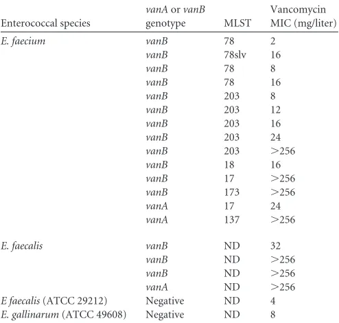
Eighteen well-characterized enterococcal strains (14E. faeciumand 4
Enterococcus faecalisstrains) that reflected contemporary Australian VRE epidemiology were selected from the Australian Collaborating Centre for
EnterococcusandStaphylococcusSpecies (ACCESS) Typing and Research collection (Table 1). An enterococcal isolate possessing thevanCgene with a vancomycin MIC in the CLSI intermediate category was included in the study (Enterococcus gallinarumATCC 49608), as was a fully suscep-tible enterococcal strain (E. faecalisATCC 29212). The vancomycin MIC of each isolate was determined by Etest (bioMérieux, France).vanAand
Received8 January 2014 Returned for modification2 February 2014 Accepted30 April 2014
Published ahead of print28 May 2014
Editor:K. C. Carroll
Address correspondence to Thamara M. Wijesuriya, [email protected].
Copyright © 2014, American Society for Microbiology. All Rights Reserved.
doi:10.1128/JCM.00021-14
on May 16, 2020 by guest
http://jcm.asm.org/
vanBgene PCR and multilocus sequence typing (MLST) was performed using previously described methods (20,21).
Randomly selected inpatient fecal samples were tested for the presence ofvanAandvanBgenes (22). Three negative samples were pooled to make a fecal suspension to which each test isolate was added to create working concentrations of 104CFU/ml and 106CFU/ml of feces, the latter being
the usual VRE fecal density in patients colonized with VRE (23). Ten microliters of each spiked fecal suspension was directly inoculated onto four commercial chromogenic VRE agars (CHROMagar VRE, chromID VRE, Brilliance VRE, and VRE Select). All plates were incubated accord-ing to the manufacturers’ recommendations. Growth was assessed at 24 and 48 h. Recovery of the original test isolate was established by identifi-cation of suspect isolates (based on their colony morphology as per the manufacturer’s instructions) using matrix-assisted laser desorption ion-ization–time of flight mass spectrometry (MALDI-TOF MS) and PCR for
vanAandvanBgenes.
The performance of each direct plating method was assessed accord-ing to its sensitivity for recovery of the test VRE isolate, heterogeneity of recovered VRE colonies, ability to suppress fecal flora, and ability to sup-press growth of the isolate possessingvanCwith an elevated vancomycin MIC (E. gallinarumATCC 49608). In order to facilitate the analysis of the impact of vancomycin MIC on the sensitivity of each method, isolates were arbitrarily categorized as having vancomycin MICs in the low (⬍16 mg/liter), medium (16 to 32 mg/liter), or high (⬎32 mg/liter) range.
Ten microliters of spiked fecal suspensions was also inoculated into EVB containing esculin, bile, sodium azide, and either 4, 6, or 8 mg/liter of vancomycin. Following 24 and 48 h of incubation at 35°C, broths that displayed evidence of esculin hydrolysis (i.e., turned black) were subcul-tured onto solid agar. The presence of the test VRE isolate was confirmed as previously. The vancomycin concentration of EVBs was assessed by high-performance liquid chromatography on a weekly basis without evi-dence of significant change in concentrations (data not provided).
RESULTS
Table 2
summarizes the sensitivity for recovery of the VRE isolates
from the solid media using a fecal density of 10
4CFU/ml. At 48 h,
the sensitivity of CHROMagar VRE (94%) and that of chromID
VRE (94%) were superior to that of either Brilliance VRE (83%)
or VRE Select (78%). When the same analysis was done using a
higher fecal density of 10
6CFU/ml (
Table 3
), the only difference at
48 h was an improvement in the sensitivity of VRE Select to 94%.
Tables 4
and
5
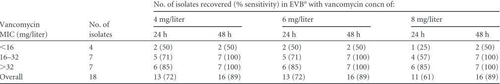
demonstrate that regardless of the fecal density
used, at 48 h the recovery of VRE from liquid medium was
con-sistently lower than that for commercial chromogenic agars and
was particularly poor when the EVB vancomycin concentration
was highest (8 mg/liter).
The impact of the test isolate’s vancomycin MIC category was
remarkably consistent, with low-MIC isolates being recovered less
frequently. The reduced sensitivity was evident across both fecal
densities, regardless of the commercial chromogenic agar used or
the vancomycin concentration of the EVBs (
Tables 2
to
5
).
All four commercial solid media suppressed fecal flora,
result-ing in no background growth on the plates.
E. faecalis
ATCC 29212
was suppressed on all four media.
E. gallinarum
ATCC 49608
growth was present after 24 h of incubation on CHROMagar VRE
and VRE Select but was easily distinguished from either
E. faecalis
or
E. faecium
on the basis of colony color as per the manufacturer’s
instructions. Brilliance VRE and chromID VRE suppressed
E.
gal-linarum
at 48 h.
CHROMagar VRE gave minimal colony heterogeneity (size
and color), whereas VRE Select gave marked colony variation
(three types) that persisted even at 48 h. Brilliance VRE and
chromID VRE gave a slight variation initially, consisting of one
or two colonies of a lighter-than-expected color, which
re-solved after 48 h.
DISCUSSION
[image:2.585.40.287.78.313.2]Numerous variables impact the performance of phenotypic
screening methods, making comparisons difficult. By using a
ho-mogenous fecal suspension spiked with a large collection of
well-characterized VRE isolates, our study allowed us to closely
exam-ine the impact of several of these variables on test performance,
TABLE 2Recovery from solid media using an inoculum of 104CFU/ml of test strain, categorized according to the vancomycin MIC of the test
isolates
Vancomycin MIC (mg/liter)
No. of isolates
No. of isolates recovered (% sensitivity)
CHROMagar VRE chromID VRE Brilliance VRE VRE Select
24 h 48 h 24 h 48 h 24 h 48 h 24 h 48 h
⬍16 4 2 (50) 3 (75) 2 (50) 3 (75) 1 (25) 1 (25) 0 1 (25)
16–32 7 7 (100) 7 (100) 6 (85) 7 (100) 7 (100) 7 (100) 2 (28) 6 (85)
⬎32 7 7 (100) 7 (100) 7 (100) 7 (100) 7 (100) 7 (100) 7 (100) 7 (100)
Overall 18 16 (89) 17 (94) 15 (83) 17 (94) 15 (83) 15 (83) 9 (50) 14 (78)
TABLE 1Characteristics of the study isolatesa
Enterococcal species
vanAorvanB
genotype MLST
Vancomycin MIC (mg/liter)
E. faecium vanB 78 2
vanB 78slv 16
vanB 78 8
vanB 78 16
vanB 203 8
vanB 203 12
vanB 203 16
vanB 203 24
vanB 203 ⬎256
vanB 18 16
vanB 17 ⬎256
vanB 173 ⬎256
vanA 17 24
vanA 137 ⬎256
E. faecalis vanB ND 32
vanB ND ⬎256
vanB ND ⬎256
vanA ND ⬎256
E faecalis(ATCC 29212) Negative ND 4
E. gallinarum(ATCC 49608) Negative ND 8
a
MLST, multilocus sequence type; slv, single-locus variant; ND, not done.
on May 16, 2020 by guest
http://jcm.asm.org/
[image:2.585.41.545.636.724.2]including the vancomycin MIC of the isolate, the fecal density of
the test isolate, and the vancomycin concentration of the liquid
medium.
When using a fecal VRE density of 10
6CFU/ml, and when
results were read at 48 h, the sensitivities of CHROMagar VRE,
chromID VRE, and VRE Select were excellent (94%). However,
the sensitivity at 24 h ranged from 67 to 94%, which has important
implications for hospitals that depend upon short turnaround
times for results of VRE screening. According to the
manufactur-er’s instructions, only VRE Select is recommended to be incubated
for less than 48 h. Our findings suggest that chromogenic media
may need to be incubated for 48 h to ensure adequate sensitivity.
In keeping with other studies, we found that the sensitivity of
the liquid medium and some solid media was reduced as the fecal
density of VRE was lowered, which may be influenced by
concur-rent antibiotic use by the patient (
24
). The advantage of
identify-ing patients with low fecal VRE densities is uncertain, as such
patients may be less likely to contribute to secondary VRE
trans-mission.
In this study, we showed that all solid media were suboptimal
for detection of enterococci with vancomycin MICs of
⬍
16 mg/
liter. Low sensitivity for recovery of
vanB
VRE with low
vancomy-cin MICs from commercial chromogenic agars has been
demon-strated previously (
3
,
25
). In both of these studies, the sensitivity
ranged from 94 to 98%; however, these studies are not necessarily
comparable to our study for several reasons. First, they inoculated
plates with pure cultures (versus spiked fecal specimens); next,
they did not attempt to use a standardized inoculum; and finally,
they assessed a more limited number of chromogenic agars. The
likely explanation for our study findings is that the vancomycin
concentration used in the screening media was above the
vanco-mycin MIC for a proportion of our test isolates. VRE screening
media usually contain vancomycin concentrations ranging from 4
to 64 mg/liter (
26
). ChromID VRE contains 8 mg/liter
vancomy-cin (
27
); however, we were unable to establish the glycopeptide
content of the remaining three commercial solid media.
Interest-ingly, supplementation of solid media with oxgall has been
dem-onstrated to improve detection of
vanB
VRE and so may offer a
means of improving test sensitivity (
28
). The reduced ability of
screening media to detect isolates with low MICs is also an issue
for detection of carbapenemase-producing
Enterobacteriaceae
(
29
) and methicillin-resistant
Staphylococcus aureus
(
30
).
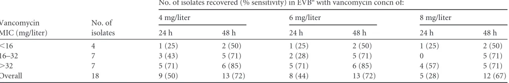
Compared to screening with solid media, screening using EVB
was less sensitive regardless of the vancomycin concentration in
the broth. This is in contrast to results of Drews and colleagues
(
31
), who reported 100% sensitivity for EVB when they tested 52
stool samples and rectal swabs. In keeping with North American
VRE epidemiology, all their strains were
vanA
VRE, whereas we
focused on
vanB
VRE, which usually have lower vancomycin
MICs than
vanA
VRE, potentially explaining our contradictory
findings. Not surprisingly, the combination of low VRE fecal
den-sity (10
4CFU/ml) and high vancomycin concentration in broth (8
mg/liter) was associated with the lowest sensitivity (28% at 24 h
and 67% at 48 h). A low vancomycin MIC of the test isolate
ap-peared to adversely influence EVB test performance, with a
sensi-tivity of
ⱕ
50% for detection of isolates with MIC of
⬍
16 mg/liter.
The sensitivity of EVB improved with increased incubation time;
thus, in the setting of VRE with low vancomycin MICs, EVB
should be read at 48 h.
The specificity of the solid media was excellent, with complete
suppression of all fecal microbiota members. Breakthrough
growth of
E. gallinarum
ATCC 49608 on two of the media may
have occurred, as this isolate has a vancomycin MIC that likely
exceeded the vancomycin concentration on the screening media.
However, the differing appearances of
E. gallinarum
,
E. faecium
,
and
E. faecalis
on the chromogenic agars would prevent this from
translating into increased laboratory workload. Previous studies
of the commercial chromogenic agars used in our study had found
significant breakthrough growth of yeasts and Gram-negative
ba-cilli (
27
,
32
,
33
). However, these isolates were easily
distinguish-TABLE 3Recovery from solid media using an inoculum of 106CFU/ml of test strain, categorized according to the vancomycin MIC of the test
isolates
Vancomycin MIC (mg/liter)
No. of isolates
No. of isolates recovered (% sensitivity)
CHROMagar VRE chromID VRE Brilliance VRE VRE Select
24 h 48 h 24 h 48 h 24 h 48 h 24 h 48 h
⬍16 4 2 (50) 3 (75) 3 (75) 3 (75) 1 (25) 1 (25) 0 3 (75)
16–32 7 7 (100) 7 (100) 7 (100) 7 (100) 7 (100) 7 (100) 5 (71) 7 (100)
⬎32 7 7 (100) 7 (100) 7 (100) 7 (100) 7 (100) 7 (100) 7 (100) 7 (100)
[image:3.585.41.545.87.175.2]Overall 18 16 (89) 17 (94) 17 (89) 17 (94) 15 (83) 15 (83) 12 (67) 17 (94)
TABLE 4Recovery from Enterococcosel enrichment broth using an inoculum of 104CFU/ml of test strain, categorized according to the
vancomycin concentration in the broth and the vancomycin MIC of the test isolates
Vancomycin MIC (mg/liter)
No. of isolates
No. of isolates recovered (% sensitivity) in EVBawith vancomycin concn of:
4 mg/liter 6 mg/liter 8 mg/liter
24 h 48 h 24 h 48 h 24 h 48 h
⬍16 4 1 (25) 2 (50) 1 (25) 2 (50) 1 (25) 2 (50)
16–32 7 3 (43) 5 (71) 2 (28) 5 (71) 0 5 (71)
⬎32 7 5 (71) 6 (85) 5 (71) 6 (85) 4 (57) 5 (71)
Overall 18 9 (50) 13 (72) 8 (44) 13 (72) 5 (28) 12 (67)
aEVB, Enterococcosel enrichment broth.
on May 16, 2020 by guest
http://jcm.asm.org/
[image:3.585.42.546.632.715.2]able from VRE, and thus no additional workup was required. This
discrepancy could have arisen because of the organisms present in
the fecal suspension used in our study.
The explanation for the colony variations observed with some
solid media warrants further study, as it potentially creates an
increased workload for laboratory staff when selecting colonies
from the agar for additional workup. It may reflect the influence of
proprietary ingredients within different chromogenic agars or
heterogeneity that may be due to the presence of subpopulations
with various vancomycin MICs.
In conclusion, while all four chromogenic media
demon-strated excellent specificity, their ability to detect VRE with low
vancomycin MICs is suboptimal. Screening using EVB was less
sensitive than with any of the solid media, particularly when the
broth contained high vancomycin concentrations. Our study
highlights the limitations of phenotypic methods that rely upon
their glycopeptide content to select for growth of VRE whose
van-comycin MIC is variable. Genotypic methods that directly detect
the
vanA
and/or
vanB
gene may circumvent this problem,
al-though detection of the
vanB
gene should still be accompanied by
confirmatory culture to ensure specificity, as other members of
the bowel microbiota may also possess the
vanB
gene (
34
).
ACKNOWLEDGMENTS
We thank Bio-Rad, bioMérieux, and Oxoid for providing the media for the study and Julie Pearson from ACCESS Typing and Research for pro-viding the test isolates.
REFERENCES
1.Christiansen KJ, Tibbett PA, Beresford W, Pearman JW, Lee RC, Coombs GW, Kay ID, O’Brien FG, Palladino S, Douglas CR, Mont-gomery PD, Orrell T, Peterson AM, Kosaras FP, Flexman JP, Heath CH, McCullough CA.2004. Eradication of a large outbreak of a single strain ofvanBvancomycin-resistantEnterococcus faeciumat a major Aus-tralian teaching hospital. Infect. Control Hosp. Epidemiol.25:384 –390. http://dx.doi.org/10.1086/502410.
2.Siegel JD, Rhinehart E, Jackson M, Chiarello L, Healthcare Infection Control Practices Advisory Committee.2007. Management of multi-drug-resistant organisms in health care settings, 2006. Am. J. Infect. Con-trol35:S165–S193.http://dx.doi.org/10.1016/j.ajic.2007.10.006. 3.Klare I, Fleige C, Geringer U, Witte W, Werner G.2012. Performance
of three chromogenic VRE screening agars, two Etest®vancomycin
pro-tocols, and different microdilution methods in detecting vanB genotype
Enterococcus faeciumwith varying vancomycin MICs. Diagn. Microbiol. Infect. Dis. 74:171–176. http://dx.doi.org/10.1016/j.diagmicrobio.2012 .06.020.
4.Pendle S, Jelfs P, Olma T, Su Y, Gilroy N, Gilbert GL.2008. Difficulties in detection and identification ofEnterococcus faeciumwith low-level in-ducible resistance to vancomycin, during a hospital outbreak. Clin. Mi-crobiol. Infect.14:853– 857.http://dx.doi.org/10.1111/j.1469-0691.2008 .02052.x.
5.Leclercq R, Derlot E, Duval J, Courvalin P. 1988. Plasmid-mediated resistance to vancomycin and teicoplanin inEnterococcus faecium. N. Engl. J. Med.319:157–161.http://dx.doi.org/10.1056/NEJM198807213190307. 6.Uttley AH, Collins CH, Naidoo J, George RC. 1988.
Vancomycin-resistant enterococci. Lanceti:57–58.
7.Frieden TR, Munsiff SS, Low DE, Willey BM, Williams G, Faur Y, Eisner W, Warren S, Kreiswirth B.1993. Emergence of vancomycin-resistant enterococci in New York City. Lancet342:76 –79.http://dx.doi .org/10.1016/0140-6736(93)91285-T.
8.Hidron AI, Edwards JR, Patel J, Horan TC, Sievert DM, Pollock DA, Fridkin SKNational Healthcare Safety Network Team, Participating National Healthcare Safety Network Facilities. 2008. NHSN annual update: antimicrobial-resistant pathogens associated with healthcare-associated infections: annual summary of data reported to the National Healthcare Safety Network at the Centers for Disease Control and Preven-tion, 2006-2007. Infect. Control Hosp. Epidemiol.29:996 –1011.http://dx .doi.org/10.1086/591861.
9.Johnson PD, Ballard SA, Grabsch EA, Stinear TP, Seemann T, Young HL, Grayson ML, Howden BP.2010. A sustained hospital outbreak of vancomycin-resistantEnterococcus faeciumbacteremia due to emergence ofvanB E. faeciumsequence type 203. J. Infect. Dis.202:1278 –1286.http: //dx.doi.org/10.1086/656319.
10. Christiansen KJ, Turnidge JD, Bell JM, George NM, Pearson JC, Aus-tralian Group on Antimicrobial Resistance.2007. Prevalence of antimi-crobial resistance inEnterococcusisolates in Australia, 2005: report from the Australian Group on Antimicrobial Resistance. Commun. Dis. Intell. Q. Rep.31:392–397.http://www.health.gov.au/internet/main/publishing .nsf/Content/cda-cdi3104-pdf-cnt.htm/$FILE/cdi3104f.pdf.
11. Bell J, Turnidge J, Coombs G, O’Brien F.1998. Emergence and epide-miology of vancomycin-resistant enterococci in Australia. Commun. Dis. Intell.22:249 –252.
12. Coombs GW, Pearson JC, Daley DA, Le T, Robinson OJ, Gottlieb T, Howden BP, Johnson PD, Bennett CM, Stinear TP, Turnidge JD, Australian Group on Antimicrobial Resistance.2014. Molecular epide-miology of enterococcal bacteremia in Australia. J. Clin. Microbiol.52: 897–905.http://dx.doi.org/10.1128/JCM.03286-13.
13. CLSI. 2012. Performance standards for antimicrobial susceptibility testing. Twenty-second informational supplement. M100-S22. CLSI, Wayne, PA.
14. Mendes RE, Woosley LN, Farrell DJ, Sader HS, Jones RN. 2012. Oritavancin activity against susceptible and vancomycin-resistant Enterococci with molecularly characterized glycopeptide re-sistance genes recovered from bacteremic patients, 2009-2010. Anti-microb. Agents Chemother.56:1639 –1642.http://dx.doi.org/10.1128 /AAC.06067-11.
15. Jones RN, Sader HS, Flamm RK.2013. Update of dalbavancin spectrum and potency in the USA: report from the SENTRY Antimicrobial Surveil-lance Program (2011). Diagn. Microbiol. Infect. Dis.75:304 –307.http: //dx.doi.org/10.1016/j.diagmicrobio.2012.11.024.
16. Soderblom T, Aspevall O, Erntell M, Hedin G, Heimer D, Hokeberg I, Kidd-Ljunggren K, Melhus A, Olsson-Liljequist B, Sjogren I, Smedje-gard J, Struwe J, Sylvan S, Tegmark-Wisell K, Thore M.2010. Alarming spread of vancomycin resistant enterococci in Sweden since 2007. Euro Surveill.15:pii:19620.http://www.eurosurveillance.org/ViewArticle.aspx ?ArticleId⫽19620.
[image:4.585.41.545.90.175.2]17. Lopez M, Cercenado E, Tenorio C, Ruiz-Larrea F, Torres C. 2012. Diversity of clones and genotypes among vancomycin-resistant clinical TABLE 5Recovery from Enterococcosel enrichment broth using an inoculum of 106CFU/ml of test strain, categorized according to the
vancomycin concentration in the broth and the vancomycin MIC of the test isolates
Vancomycin MIC (mg/liter)
No. of isolates
No. of isolates recovered (% sensitivity) in EVBawith vancomycin concn of:
4 mg/liter 6 mg/liter 8 mg/liter
24 h 48 h 24 h 48 h 24 h 48 h
⬍16 4 2 (50) 2 (50) 2 (50) 2 (50) 1 (25) 2 (50)
16–32 7 5 (71) 7 (100) 5 (71) 7 (100) 4 (57) 7 (100)
⬎32 7 6 (85) 7 (100) 6 (85) 7 (100) 6 (85) 7 (100)
Overall 18 13 (72) 16 (89) 13 (72) 16 (89) 11 (61) 16 (89)
aEVB, Enterococcosel enrichment broth.
on May 16, 2020 by guest
http://jcm.asm.org/
Enterococcusisolates recovered in a Spanish hospital. Microb. Drug Resist. 18:484 – 491.http://dx.doi.org/10.1089/mdr.2011.0203.
18. Werner G, Klare I, Fleige C, Geringer U, Witte W, Just HM, Ziegler R. 2012. Vancomycin-resistant vanB-type Enterococcus faecium isolates ex-pressing varying levels of vancomycin resistance and being highly preva-lent among neonatal patients in a single ICU. Antimicrob. Resist. Infect. Control1:21.http://dx.doi.org/10.1186/2047-2994-1-21.
19. McCracken M, Wong A, Mitchell R, Gravel D, Conly J, Embil J, Johnston L, Matlow A, Ormiston D, Simor AE, Smith S, Du T, Hizon R, Mulvey MR, Canadian Nosocomial Infection Surveillance Program. 2013. Molecular epidemiology of vancomycin-resistant enterococcal bac-teraemia: results from the Canadian Nosocomial Infection Surveillance Program, 1999-2009. J. Antimicrob. Chemother.68:1505–1509.http://dx .doi.org/10.1093/jac/dkt054.
20. Palladino S, Kay ID, Costa AM, Lambert EJ, Flexman JP.2003. Real-time PCR for the rapid detection ofvanAandvanBgenes. Diagn. Microbiol. Infect. Dis.45:81– 84.http://dx.doi.org/10.1016/S0732-8893(02)00505-9. 21. Homan WL, Tribe D, Poznanski S, Li M, Hogg G, Spalburg E, Van
Embden JD, Willems RJ.2002. Multilocus sequence typing scheme for
Enterococcus faecium. J. Clin. Microbiol.40:1963–1971.http://dx.doi.org /10.1128/JCM.40.6.1963-1971.2002.
22. Palladino S, Kay ID, Flexman JP, Boehm I, Costa AM, Lambert EJ, Christiansen KJ.2003. Rapid detection ofvanAandvanBgenes directly from clinical specimens and enrichment broths by real-time multiplex PCR assay. J. Clin. Microbiol.41:2483–2486.http://dx.doi.org/10.1128 /JCM.41.6.2483-2486.2003.
23. Kuch A, Stefaniuk E, Ozorowski T, Hryniewicz W.2009. New selective and differential chromogenic agar medium, chromID VRE, for screening vancomycin-resistant Enterococcus species. J. Microbiol. Methods77: 124 –126.http://dx.doi.org/10.1016/j.mimet.2009.01.004.
24. D’Agata EM, Gautam S, Green WK, Tang YW. 2002. High rate of false-negative results of the rectal swab culture method in detection of gastrointestinal colonization with vancomycin-resistant enterococci. Clin. Infect. Dis.34:167–172.http://dx.doi.org/10.1086/338234. 25. Hegstad K, Giske CG, Haldorsen B, Matuschek E, Schonning K,
Leegaard TM, Kahlmeter G, Sundsfjord A, on behalf of the NordicAST VRE Detection Study Group.2014. Performance of the EUCAST disk diffusion method, CLSI agar screen and VITEK 2 automated antimicro-bial susceptibility testing system in detection of clinical isolates of
entero-cocci with low and medium level VanB-type vancomycin resistance: a multicenter study. J. Clin. Microbiol.52:1582–1589.http://dx.doi.org/10 .1128/JCM.03544-13.
26. Nelson RR.1998. Selective isolation of vancomycin-resistant enterococci. J. Hosp. Infect.39:13–18.http://dx.doi.org/10.1016/S0195-6701(98)90238-9. 27. Grabsch EA, Ghaly-Derias S, Gao W, Howden BP.2008. Comparative
study of selective chromogenic (chromID VRE) and bile esculin agars for isolation and identification ofvanB-containing vancomycin-resistant en-terococci from feces and rectal swabs. J. Clin. Microbiol.46:4034 – 4036. http://dx.doi.org/10.1128/JCM.00944-08.
28. Grabsch EA, Chua K, Xie S, Byrne J, Ballard SA, Ward PB, Grayson ML.2008. Improved detection of vanB2-containingEnterococcus faecium
with vancomycin susceptibility by Etest using oxgall supplementation. J. Clin. Microbiol.46:1961–1964.http://dx.doi.org/10.1128/JCM.01778-07. 29. Nordmann P, Poirel L.2013. Strategies for identification of carbapen-emase-producing Enterobacteriaceae. J. Antimicrob. Chemother.68:487– 489.http://dx.doi.org/10.1093/jac/dks426.
30. Yamada K, Inuzuka K, Tatsumi N, Sanzen I, Ohkura T, Okamoto A, Hasegawa T, Ohta M.2010. Evaluation of selection media for the detec-tion of borderline MRSA. J. Infect. Chemother.16:19 –24.http://dx.doi .org/10.1007/s10156-009-0009-0.
31. Drews SJ, Johnson G, Gharabaghi F, Roscoe M, Matlow A, Tellier R, Richardson SE.2006. A 24-hour screening protocol for identification of vancomycin-resistantEnterococcus faecium. J. Clin. Microbiol.44:1578 – 1580.http://dx.doi.org/10.1128/JCM.44.4.1578-1580.2006.
32. Peltroche-Llacsahuanga H, Top Weber-Heynemann JJ, Lutticken R, Haase G.2009. Comparison of two chromogenic media for selective iso-lation of vancomycin-resistant enterococci from stool specimens. J. Clin. Microbiol.47:4113– 4116.http://dx.doi.org/10.1128/JCM.00882-09. 33. Ledeboer NA, Das K, Eveland M, Roger-Dalbert C, Mailler S, Chatellier
S, Dunne WM.2007. Evaluation of a novel chromogenic agar medium for isolation and differentiation of vancomycin-resistantEnterococcus fae-ciumandEnterococcus faecalisisolates. J. Clin. Microbiol.45:1556 –1560. http://dx.doi.org/10.1128/JCM.02116-06.
34. Graham M, Ballard SA, Grabsch EA, Johnson PD, Grayson ML.2008. High rates of fecal carriage of nonenterococcalvanBin both children and adults. Antimicrob. Agents Chemother.52:1195–1197.http://dx.doi.org /10.1128/AAC.00531-07.