CROSSTALK BETWEEN SOLUBLE FACTORS AND
CELL-CELL INTERACTIONS: IMPLICATIONS FOR
CELL CYCLE CONTROL AND TUMOR DEVELOPMENT
Thesis by
Nicholas Graham
In partial fulfillment of the requirements for the degree of
Doctor of Philosophy
CALIFORNIA INSTITUTE OF TECHNOLOGY Pasadena, California
2007
ACKNOWLEDGMENTS
I would first and foremost like to thank my research advisor, Anand Asthagiri. His boundless passion for science, combined with a deep sense of precision, keen insight, and technical expertise have continually amazed me. It has been an honor to work with Anand, and his guidance, scientifically and professionally, has been invaluable during my time at Caltech.
Additionally, I thank my thesis committee, Mark Davis, Christina Smolke and David Tirrell, for their valuable comments and advice. The Caltech Department of Chemical Engineering is a wonderful environment that fosters both academic and social collaboration, and that is due in large part to the example that these and other faculty have set.
My colleagues in the Asthagiri group have been an invaluable technical resource, as well as great friends. I have been privileged to watch the group to grow from infancy into a fully-fledged research lab, and I could not have succeeded at Caltech without their enthusiasm, curiosity, helpfulness, and company over poco machos from Ernie’s.
No thesis would be possible without the help of the countless professionals who keep Caltech running smoothly. In particular, I would like to thank Kathy Bubash and Martha Hepworth for their help navigating every aspect of graduate studies, Efrain Hernandez for always bringing a sunny, optimistic attitude to the sub-basement, and Joe, Terry, and Moses in the chemistry stock room for keeping me in reagents (and fantasy football advice).
Mangion, and Erin Guidry for being unabashed food and beer snobs, as well as great friends who helped me to keep life at Caltech in perspective. Also deserving of specific mention are the men of FBOD, the crew from Imperial Palace (the casino and the softball team), and the Friday afternoon Wash U alumni swim team. Finally, I should also thank Sunday Night Baseball, Mannyz House, Apartment 402, beach ball costumes, Jeff Murdock, Coachella, Tuesday night karaoke in Burbank, Scrubs, Dr. Kang-Yell Choi, Kim Sung-Eun and the Laboratory of Molecular Complex Control at Yonsei University, TiVo, and Pete’s cat kitty.
Of course, none of this would have been possible without the support of my parents and sister. You have set a great example of hard work, humility, patience and love that I’m still trying to emulate. Although I still don’t have a real job, I couldn’t have gotten this far without you and your encouragement!
ABSTRACT
Crosstalk between Soluble Factors and Cell-Cell Interactions: Implications for Cell Cycle Control and Tumor Development
April 2007
Nicholas Graham, B.S., Washington University in St. Louis, M.S., Chemical Engineering, California Institute of Technology Ph.D., Chemical Engineering, California Institute of Technology
Precise and dynamic control of cell behaviors, including proliferation, adhesion, and migration, is required for proper tissue organization and homeostasis. A key element to understanding how cellular functions are controlled lies in uncovering the topology of the molecular signaling networks that couple environmental signals to cellular responses. In this study, we have parsed the signaling networks involved in cell cycle regulation and tumor development and uncovered novel mechanisms of crosstalk between soluble factors and cell-cell interactions.
transcription using different sub-cellular pools of β-catenin. Because hyperactive β -catenin signaling drives proliferation in cancer, this suggests that attenuation of β-catenin signaling may require different therapeutic strategies for EGF- and Wnt-driven tumors.
Since β-catenin signaling can be antagonized by sequestration with the cell-cell contact protein E-cadherin at the plasma membrane, proliferative signals mediated by β-catenin may regulate growth suppression at high cell density, a property of normal cells that is often lost during tumorigenesis. Indeed, in non-tumorigenic epithelial cells, we demonstrate that E-cadherin is upregulated in contexts where β-catenin signaling and DNA synthesis are suppressed. Additionally, exogenous E-cadherin suppresses proliferation with a strict requirement for β-catenin binding. Future studies to test the hypothesis that E-cadherin regulates the growth of normal cells will benefit from a quantitative assay developed to measure E-cadherin:β-catenin complexes. Such quantitative measurements are likely to be important because contact-mediated growth suppression by E-cadherin is coupled with a density-dependent, ligand-depletion mechanism that concomitantly regulates proliferation.
Finally, we demonstrate that EGF and other soluble factors synergistically control cell-cell interactions governing organization of normal epithelial cells into multicellular structures. Notably, this behavior resembles the program initiated during metastatic cancer, thus illustrating the flexibility of the epithelial phenotype even in non-cancerous cells. Together, these studies illustrate how the topology of molecular signaling networks can couple environmental cues including soluble extracellular factors and cell-cell
TABLE OF CONTENTS
Acknowledgments... iii
Abstract... v
Table of Contents... vii
List of Tables... xiii
List of Figures... xiv
Abbreviations... xvii
Chapter
I. Introduction... I-1 1. Introduction... I-1 2. Mechanisms of cell-cell adhesion... I-2 3. The canonical Wnt pathway: soluble ligands promote signaling through the
cell contact protein β-catenin... I-3 4. E-cadherin and β-catenin in normal and pathological contexts... I-4 4.1. De-regulation of β-catenin signaling drives proliferation... I-5 4.2. Cadherins suppress tumorigenesis... I-6 5. Current unresolved questions involving crosstalk between
soluble factors and cell-cell interactions... I-7 6. Current results... I-10 7. References... I-12
II. EGF-mediated Tcf/Lef transcriptional activity is essential but not sufficient for cell cycle progression in non-transformed mammary epithelial cells... II-1 1. Introduction... II-2 2. Materials and Methods... II-6
2.4. Retroviral Infection... II-7 2.5. GSK3β Serine 9 Phosphorylation Assay... II-7 2.6. ERK Signaling Assay... II-8 2.7. Cell Lysis... II-8 2.8. Reporter Assays... II-8 2.9. Integrated Reporter Response... II-9 2.10. Western Blotting... II-9 2.11. DNA Synthesis... II-9 2.12. Immunofluorescence... II-10 3. Results... II-11
3.1. Re-entry into the Cell Cycle Correlates with Tcf/Lef Reporter
Activity... II-11 3.2. EGF Independently Induces Tcf/Lef Transcriptional Activity
and DNA Synthesis... II-13 3.3. Tcf/Lef Transcriptional Activity Is Required for EGF-mediated
DNA Synthesis... II-17 3.4. EGF-mediated Activation of Tcf/Lef Transcriptional Activity Is
Upstream of Cyclin D1 Promoter Activity... II-21 4. Discussion... II-22 5. Acknowledgments... II-27 6. References... II-28
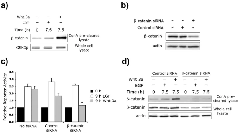
III. EGF and Wnt 3a differentially regulate Tcf/Lef transcription with
implications for tumor development... III-1 1. Introduction... III-2 2. Results... III-4 2.1. EGF activates Tcf/Lef transcriptional activity in 293T-EGFR cells... III-4 2.2. EGF and Wnt 3a additively activate Tcf/Lef transcription... III-6 2.3. Wnt 3a and EGF activate Tcf/Lef transcription via different
2.5. PKC, but not PKA, is required for EGF- and Wnt 3a-mediated
Tcf/Lef transcription... III-13 2.6. Src family kinase activity is required for EGF-, but not Wnt 3a-,
mediated Tcf/Lef transcription... III-15 3. Discussion... III-17
3.1. Physiological implications of EGF and Wnt co-regulation of
Tcf/Lef transcription... III-19 3.2. Mechanisms underlying EGF/Wnt crosstalk in regulating Tcf/Lef
transcriptional activity... III-21 4. Experimental Procedures... III-26 4.1. Antibodies and Reagents... III-26 4.2. Cell Culture... III-26 4.3. Plasmid Constructs... III-27 4.4. Cell Lysis... III-27 4.5. ConA Fractionation... III-28 4.6. Reporter Assays... III-28 4.7. Immunoblotting... III-29 4.8. siRNA knockdown of β-catenin... III-29 5. Acknowledgments... III-29 6. Supplemental Data... III-30 7. References... III-32
2.6. Retroviral Infection... IV-7 2.7. Data Analysis and Statistical Calculations... IV-7 3. Results and Discussion... IV-8
3.1. Development and validation of a quantitative microtiter ELISA for E-cadherin:β-catenin protein complexes... IV-8 3.2. Compatibility of the protein complex ELISA with the standard
sandwich ELISAs... IV-14 3.3. Quantitative comparision of E-cadherin:β-catenin interactions in
transformed versus non-transformed cells... IV-16 3.4. Quantitative analysis of the effect of constitutively-active Src on
E-cadherin:β-catenin interactions... IV-19 4. Conclusions... IV-23 5. Acknowledgments... IV-24 6. Supplemental Data... IV-25 7. References... IV-26
V. Mechanisms underlying growth saturation of epithelial cells... V-1 1. Introduction... V-2 2. Results... V-6 2.1. Non-tumorigenic epithelial cells growth arrest at high cell density... V-6 2.2. Receptor-mediated signaling is qualitatively similar in low- and
high-density cells... V-9 2.3. Increased expression of endogenous E-cadherin at high cell density
inversely correlates with low levels of DNA synthesis and β-catenin:Tcf/Lef signaling... V-11 2.4. Exogenous E-cadherin inhibits DNA synthesis... V-13 2.5. Exogenous E-cadherin constructs interfere with EGFR signaling... V-16 2.6. Growth factor availability contributes to growth arrest... V-17 2.7. Diminished Akt, but not ERK, activity correlates with growth factor
3.1. Cadherins as anti-proliferative signals: Modulation of Tcf/Lef
transcription via cell-cell contact... V-25 3.2. Cadherins as anti-proliferative signals: Cell-cell contact mechanisms
independent of Tcf/Lef transcription... V-28 3.3. Density-dependent models of growth suppression: Phosphatase-
mediated inhibition of mitogenic signaling pathways... V-30 3.4. Mitogenic ligand depletion as a mediator of growth suppression... V-31 4. Future Work... V-32 4.1. The role of ERK and Akt in growth suppression... V-32 4.2. Mechanisms underlying upregulation of endogenous E-cadherin... V-34 4.3. Functional significance of E-cadherin upregulation for growth
suppression... V-35 5. Conclusions and Acknowledgments... V-37 6. Experimental Procedures... V-37 6.1. Antibodies... V-37 6.2. Cell Culture... V-37 6.3. Plasmid Constructs... V-37 6.4. Retroviral Infection... V-38 6.5. Cell Lysis... V-38 6.6. Immunoblotting... V-39 6.7. Reporter Assays... V-40 6.8. DNA Synthesis Measurements... V-40 6.9. Immunofluorescence... V-41 6.10. Immunoprecipitation... V-41 7. References... V-42
2.3. Pharmacological Inhibition... VI-4 3. Results and Discussion... VI-5 3.1. Growth Medium controls the reversible formation of cell colonies... VI-5 3.2. EGF, but not serum, can prevent cell aggregation... VI-6 3.3. EGF and ChT induce mild dissociation of cell colonies... VI-7 3.4. EGF and ChT cooperate to induce synergistic cell scattering... VI-8 3.5. MAPK and PI3K control various aspects of cell scattering... VI-10 3.6. Relevance of cell scattering to EMT... VI-11 4. Future Work... VI-13
LIST OF TABLES
Number Page
IV-1. Quantitative performance of protein complex and sandwich ELISAs... IV-12 VI-1. Potential Scatter Metric: Average and percent standard deviation of
LIST OF FIGURES
Number Page
I-1 Generalized structure of Adherens Junctions... I-2 I-2 The canonical Wnt signaling pathway... I-4 II-1. TOPFLASH and FOPFLASH reporter activity in SW480 and
MCF-10A cells... II-12 II-2. Dominant-negative Tcf4 effect on TOPFLASH reporter... II-13 II-3. Growth medium constituents vary in the ability to induce Tcf/Lef
transcriptional activity, GSK3β phosphorylation, and DNA synthesis... II-16 II-4. Dominant-negative Tcf4 blocks DNA synthesis... II-18 II-5. Dominant-negative Tcf4 does not affect EGF-mediated EGF receptor
and ERK phosphorylation... II-21 II-6. Tcf/Lef involvement in cyclin D1 promoter activity... II-22 II-7. Proposed model for the strict requirement of Tcf/Lef signaling for
EGF-mediated cell cycle progression... II-26 III-1. EGF induces Tcf/Lef transcriptional activity in 293T-EGFR cells... III-5 III-2. Wnt 3a and EGF cooperate to activate Tcf/Lef transcriptional activity... III-7 III-3. Wnt 3a and EGF activate Tcf/Lef transcription via different
mechanisms... III-9 III-4. ERK is required for both EGF- and Wnt-3a-mediated
Tcf/Lef signaling... III-12 III-5. PKC, but not PKA, is required for EGF- and Wnt-3a-mediated Tcf/Lef
transcriptional activity... III-14 III-6. ERK, but not Wnt 3a, requires Src family kinase activity to activate Tcf/Lef transcriptional activity... III-16 III-7. EGF and Wnt 3a activate Tcf/Lef signaling via a distinct but partially
overlapping network... III-18 III-S1. Wnt 5a does not activate Tcf/Lef signaling... III-30 III-S2. Neither Wnt 3a nor EGF induces phosphorylation of GSK3β on
III-S3. ConA pre-clearing of whole cell lysate depletes E-cadherin... III-30 III-S4. ERK signaling is required for EGF-mediated Tcf/Lef signaling... III-31 III-S5. The PKC inhibitor Calphostin C inhibits EGF-mediated Tcf/Lef
signaling only at high concentrations... III-31 III-S6. PKC does not lie upstream of EGF-mediated ERK activation,
but Src possibly does... III-32 III-S7. PKC and Src are not required for Wnt 3a-mediated stabilization of
β-catenin... III-32 IV-1. Antigen capture and protein:protein co-capture. ... IV-10 IV-2. Detection of E-cadherin:β-catenin protein complexes by protein
complex ELISA... IV-11 IV-3. Specificity test for E-cadherin:β-catenin ELISA... IV-14 IV-4. Detection of E-cadherin and β-catenin total protein levels by
sandwich ELISA... IV-15 IV-5. Quantitative comparison of the levels of E-cadherin:β-catenin
complexes, E-cadherin and β-catenin expression in normal and
tumorigenic cell lines... IV-18 IV-6. Quantifying the effect of constitutively-active Src on cellular levels of
E-cadherin:β-catenin complexes and the expression of E-cadherin and β-catenin... IV-21 IV-S1. Validation of standard sandwich and protein complex ELISAs in
normal and tumorigenic cell lines... IV-25 IV-S2. Validation of standard sandwich and protein complex ELISAs in cells
expressing constitutively-active Src ... IV-26 V-1. Quantification of intercellular contact by measuring cell density in
MCF-10A... V-7 V-2. DNA synthesis is inversely correlated with cell density ... V-8 V-3. EGFR, ERK, and Akt signaling at various cell densities... V-10 V-4. β-catenin-mediated transcription is inversely correlated with
V-5. Expression of endogenous E-cadherin, but not β-catenin, is cell density-dependent... V-13 V-6. Full-length E-cadherin, but not the cytodomain-truncated mutant,
reduces DNA synthesis... V-15 V-7. Exogenous E-cadherin constructs do not affect ERK despite affecting EGFR and Akt phosphorylation... V-17 V-8. Growth factor concentration-dependent saturation of cell growth... V-18 V-9. Cell colony formation does not affect EGF-mediated proliferation... V-19 V-10. Growth factor availability determines the cell density at growth
saturation... V-21 V-11. Akt signaling, but not ERK signaling, may control cell density at
saturation... V-22 V-12. Growth inhibition at high cell density in MCF-10A... V-24 VI-1. Reversible formation of epithelial cell colonies by growth medium
starvation or stimulation... VI-5 VI-2. EGF, but not serum, prevents cell island aggregation... VI-6 VI-3. EGF and ChT are the only components of growth medium that induce
cell colony dissociation... VI-7 VI-4. EGF and ChT can synergize to induce cell island dissociation... VI-8 VI-5. PI3K and MAPK are required for different aspects of GM-induced
cell scattering... VI-11 VI-6. Average neighbor centroid distance as a metric for quantification of
ABBREVIATIONS
Ab Antibody
APC Adenomatous polyposis coli gene product
BrdU Bromodeoxyuridine
cAMP Cyclic adenosine monophosphate
ChT Cholera toxin
CREB cAMP-responsive element-binding protein DAPI 4’,6-diamidino-2’-phenylindole-dihydrochloride
E- Epithelial
EGF Epidermal growth factor
EGFR EGF receptor
EGTA Ethylene glycol tetraacetic acid ELISA Enzyme-linked immunosorbent assay EMT Epithelial-mesenchymal transition ERK Extracellular signal-regulated kinase
Frz Frizzled
GM Growth medium
GSK3β Glycogen synthase kinase 3β
HEPES 4-(2-hydroxyethyl)-1-piperazineethanesulfonic acid HGF Hepatocyte growth factor
IgG Immunoglobulin G
IP Immunoprecipitation
Lef Lymphoid enhancer factor LRP Low-density lipoprotein receptor MOI Multiplicity of infection
PAGE Poly-acrylamide gel electrophoresis pAkt Phospho-serine 473 Akt
PBS Phosphate-buffered saline
PCR Polymerase chain reaction PKA cAMP-dependent protein kinase PKC Protein kinase C
pRb Retinoblastoma protein RTK Receptor tyrosine kinase RT-PCR Reverse transcription PCR shRNA Short hairpin RNA
siRNA Short interfering RNA SDS Sodium dodecyl sulfate S.E. Standard error
TBS Tris-buffered saline
TBST TBS plus 0.5% (v/v) Tween-20 Tcf T-cell factor
TGFβ Transforming growth factor β VE Vascular endothelial
VEGF VE growth factor VEGFR VEGF receptor
Chapter I. Introduction
1. Introduction
Precise and dynamic control of fundamental cell processes, including
proliferation, adhesion and migration, is required for proper organization and homeostasis
of mammalian organisms. De-regulation of the mechanisms regulating these behaviors
underlies many pathologies, including cancer. For example, proliferation of
non-cancerous mammalian cells requires properly-timed mitogenic signals, as well as
avoidance of anti-proliferative stimuli, from the cellular microenvironment. However,
cancer cells circumvent these requirements, becoming both self-sufficient in mitogenic
signals and insensitive to anti-proliferative signals, permitting unchecked cell growth
(Hanahan and Weinberg, 2000). With a fundamental understanding of the mechanisms
underlying cell behaviors, it may be possible to manipulate these same processes, either
for therapeutic benefit or technological applications. A key component of this strategy
involves elucidating the topology of the molecular networks that regulate cellular
functions.
In this study, we sought to identify mechanisms of crosstalk between soluble
factors and cell-cell interactions that regulate the ability of cells to proliferate, migrate,
and mediate intercellular adhesion. Probing these mechanisms, we elucidate the
architecture of sophisticated molecular circuits that enable biological systems to control
cell behaviors and guide multicellular organization. Notably, these studies highlight
achieve aberrant cellular behaviors. To motivate these studies, it is useful to have some
background information on the key molecular players.
2. Mechanisms of cell-cell adhesion: Adherens junctions
One of the hallmarks of epithelial tissues is tight intercellular adhesion. Cell-cell
contact not only permits epithelial tissues to serve as a physical barrier, but also encodes
biochemical signals that regulate cell behaviors such as proliferation. Although there are
several adhesive structures present in epithelial cells, including tight junctions,
desmosomes, and gap junctions, the structure that is primarily responsible for
intercellular adhesion is the adherens junction.
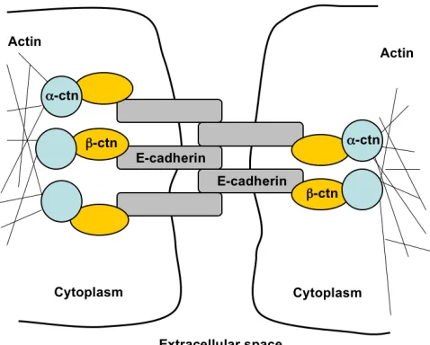
Adherens junctions are composed of cadherin and catenin proteins (Figure I-1).
Cadherins are single-pass transmembrane glycoproteins that bind homotypically to
[image:20.612.110.349.393.585.2]cadherins on neighboring cells in a calcium-dependent manner (Angst et al., 2001). The
Figure I-1. Generalized structure of adherens junctions
The molecular constituents of adherens junctions are the cadherin and catenin proteins. Cadherins span the plasma membrane and bind to cadherins on neighboring cells. The cadherin intracellular domain binds β
-catenin (β-ctn), which links to the
actin cytoskeleton through α-catenin
intracellular tail of cadherins binds β-catenin, which then recruits α-catenin and links to
the actin cytoskeleton. As such, cadherin-mediated contacts link the cytoskeletons of
neighboring cells and impart a structural rigidity to cell-cell contacts.
E-(epithelial)cadherin is the predominant cadherin family member expressed in epithelial
cells.
3. The canonical Wnt pathway: soluble ligands promote signaling through the cell
contact protein β-catenin.
In addition to its adhesive role at the plasma membrane, β-catenin can function as
a transcriptional activator when localized to the nucleus (Figure I-2). A key constraint on
β-catenin-mediated transcription is the stability of β-catenin in the cytoplasm. In the
absence of soluble Wnt factors, cytosolic β-catenin is phosphorylated on N-terminal
serine and threonine residues by a multiprotein complex consisting of axin, APC, and
glycogen synthase kinase 3β (GSK3β). Phosphorylated β-catenin is then ubiquitinated
and degraded by the proteasome. Notably, this active degradation mechanism keeps
cytosolic concentrations of β-catenin very low.
Signaling events that inhibit this degradation machinery, such as those initiated by
a subset of Wnt family ligands, stabilize β-catenin. This allows β-catenin to accumulate
and translocate to the nucleus, where it binds to the Tcf/Lef family of transcription
factors. Together, this bipartite transcription factor induces expression of genes including
1998). The activation of gene transcription by β-catenin:Tcf/Lef complexes is generally
referred to as β-catenin signaling; when Wnt ligands are the agonist of β-catenin
[image:22.612.175.473.186.401.2]signaling, this process is known as Wnt signaling.
Figure I-2. The canonical Wnt signaling pathway
In the absence of Wnt ligands, cytosolic β-catenin (β-ctn) is phosphorylated by a multiprotein complex
consisting of APC, axin, and GSK3β. Phosphorylated β-catenin is then degraded by the proteasome,
keeping cytosolic concentrations of β-catenin low. When Wnt ligands bind to the co-receptor complex of
Frizzled (Frz) and LRP 5/6, the cytosolic degradation machinery is inhibited, allowing β-catenin to accumulate in the cytoplasm and translocate to the nucleus. In the nucleus, β-catenin binds to the Tcf/Lef
family of transcription factors and mediates expression of target genes including cyclin D1 and c-myc.
4. E-cadherin and β-catenin in normal and pathological contexts
E-cadherin and β-catenin play prominent roles in both embryonic development
and carcinogenesis (Clevers, 2006; Halbleib and Nelson, 2006; Wijnhoven et al., 2000).
Morphogenetic and oncogenic signals are transmitted by both the adhesive function of
cadherins and the nuclear signaling activity of β-catenin. Because of the functional
mutations that mimic these cues - regulate cell behaviors by regulating cadherin-mediated
cell-cell interactions and β-catenin signaling.
4.1. De-regulation of β-catenin signaling drives proliferation.
Mutations that abnormally stabilize β-catenin and hard-wire Tcf/Lef transcription
into a constitutively activate state occur in a diverse range of cancer types, implying a
functional link between β-catenin signaling and tumor development. One such
mechanism commonly found in breast cancers is autocrine secretion of β-catenin
signaling agonists, including Wnt ligands (Bafico et al., 2004). Confirming that
overexpression of β-catenin agonists can induce transformation, mammary-tissue-specific
overexpression of Wnt-1 induces adenocarcinomas in mouse models (Tsukamoto et al.,
1988). Consistent with these findings, studies using stabilized mutants of β-catenin or
Tcf/Lef-VP16 fusion constructs have affirmed the capacity of β-catenin signaling to
transform established cell lines and primary cells (Aoki et al., 1999; Kolligs et al., 1999;
Orford et al., 1999).
In fact, antagonizing β-catenin signaling appears to be an effective method to curb
the growth of cancer cell lines afflicted by elevated levels of nuclear β-catenin.
Inhibitors of soluble Wnt factors decrease cell growth of human breast cancers that
exhibit autocrine Wnt signaling (Bafico et al., 2004). Furthermore, overexpression of
proteins – such as full-length E-cadherin or a truncated mutant that retains β-catenin
binding – sequester stabilized β-catenin at the plasma membrane, precluding its
colorectal cancer cell lines (Gottardi et al., 2001; Orsulic et al., 1999; Sadot et al., 1998).
Although the role of β-catenin in hyperproliferation of cancer cells is well
established, the role of β-catenin and Tcf/Lef transcription factors in cell cycle
progression of normal mammalian cells is only recently becoming apparent.
Immunohistochemical data have shown that epithelial precursor cells in the intervillus
regions of the small intestine may require β-catenin signaling for self-renewal (van de
Wetering et al., 2002). In addition, Tcf4 knock-out mice lack proliferating stem cells and
possess only differentiated villus cells, suggesting a causal role for Tcf/Lef in governing
stem cell lineage commitment (Korinek et al., 1998). In addition to intestinal epithelia,
Tcf/Lef signaling is involved in lineage commitment of human epidermal stem cells
(Chenn, 2002; Chenn and Walsh, 2002; Hari et al., 2002; Zhu and Watt, 1996; Zhu and
Watt, 1999), hematopoietic stem cells (Reya et al., 2003) and embryonic stem cells
(Kielman et al., 2002). In all these cases, the upstream ligands that regulate β-catenin
signaling are either Wnt or unidentified.
4.2. Cadherins suppress tumorigenesis.
In general, the attenuation of cell-cell adhesion plays a critical role in both early
and late stages of oncogenesis (Wijnhoven et al., 2000). At early steps, reduced
intercellular adhesion may attenuate contact-inhibition of proliferation, permitting
unregulated cell division and tumor formation; at later stages, reduced cell-cell adhesion
is often associated with invasion, metastasis, and poor patient prognosis (Christofori and
transcriptional inactivation (Giroldi et al., 1997; Hennig et al., 1996; Ji et al., 1997), but it
is not clear whether loss of E-cadherin is a prerequisite for cancer progression or merely a
consequence of the dedifferentiation that occurs during cancer progression (Wijnhoven et
al., 2000). Since re-expression of E-cadherin inhibits invasion (Vleminckx et al., 1991)
and tumorigenicity (Navarro et al., 1991) of some cancers, loss of E-cadherin may have a
dual effect, permitting motility and invasion, as well as relaxing the constraints on
proliferation (Sasaki et al., 2000).
The growth-suppressive effects of cadherins has been attributed to both
sequestration of β-catenin outside of the nucleus (Sasaki et al., 2000; Stockinger et al.,
2001) and the attenuation of receptor tyrosine kinase (RTK) (Grazia Lampugnani et al.,
2003; Lampugnani et al., 2006; Qian et al., 2004; Takahashi and Suzuki, 1996). By
inhibiting proliferation, cadherins may also play a role in contact inhibition of
proliferation, whereby cells growth arrest even in the presence of mitogenic ligands
(Motti et al., 2005; St Croix et al., 1998; Stockinger et al., 2001). Thus,
cadherin-mediated cell-cell contacts may antagonize intracellular signaling pathways and
subsequent cell responses that are initiated by soluble factors.
5. Current unresolved questions involving crosstalk between soluble factors and
cell-cell interactions
The classical agonists of β-catenin transcriptional activity are the Wnt ligands
including the epidermal growth factor (EGF), also provoke β-catenin signaling (Lu et al.,
2003; Muller et al., 2002). If indeed non-Wnt ligands such as EGF can induce β-catenin
transcriptional activity, it is unclear whether they utilize the canonical Wnt mechanism
that stabilizes cytoplasmic β-catenin. In the case of EGF, this question is particularly
interesting because EGF is known to inactivate GSK3β (Eldar-Finkelman et al., 1995),
the kinase which primes cytosolic β-catenin for degradation (Aberle et al., 1997).
Investigating this mechanism may shed insight on whether β-catenin is a primed or
non-primed substrate of GSK3β (Ding et al., 2000; Liu et al., 2002). If EGF-mediated
transactivation of β-catenin does not involve Wnt-like mechanisms such as the
stabilization of β-catenin, what mechanisms are important? One possibility is that EGF
transactivates β-catenin by modulating the adhesive and transcriptional properties of β
-catenin through tyrosine phosphorylation (Harris and Peifer, 2005).
Whatever the mechanism of EGF-mediated β-catenin transactivation, it would
also be interesting to test whether Wnt ligands and RTK ligands can co-regulate β
-catenin:Tcf/Lef transcription. Some reports have suggested that specific signals
downstream of RTKs, including constitutively-active Ras, can cooperate with
constitutive inhibition of GSK3β to induce synergistic β-catenin signaling (Chen et al.,
2000; Desbois-Mouthon et al., 2001). However, constitutive activation or inhibition of
signaling pathways is clearly different from intracellular signals mediated by soluble
ligands, precluding assessment of whether RTK and Wnt ligands co-regulate Tcf/Lef
In cancer, hyperactive β-catenin signaling drives unchecked proliferation
(Clevers, 2006). Because the targets of β-catenin transcription include proteins that are
ubiquitously required for cell cycle progression (e.g., cyclin D1, c-myc), the untested
hypothesis remains that β-catenin signaling is important for proliferation of normal cells.
Correlations between serum-mediated proliferation and Tcf/Lef transcriptional activity
have been demonstrated in an engineered mammary cell system (Stockinger et al., 2001);
however, the expression of a c-Fos:estradiol receptor fusion protein in these cells
precludes an assessment of whether β-catenin nuclear activity is involved in proliferation,
since c-Fos itself is itself critically involved in cell cycle control (Cook et al., 1999).
Thus, it remains to be tested whether β-catenin signaling is involved in proliferation of
normal cells, and if so, whether non-Wnt ligands utilize β-catenin:Tcf/Lef transcription to
regulate passage through the cell cycle.
Because β-catenin signaling can be attenuated by binding to E-cadherin at the
plasma membrane, the interplay between proliferative signals mediated by β-catenin and
contact-induced, anti-proliferative signals may regulate growth. One cellular process that
may be regulated by this mechanism is contact inhibition of proliferation, a property of
normal cells that is often lost during tumorigenesis. As such, in the context of confluent,
growth-arrested, epithelial cell monolayers, do cadherins antagonize β-catenin signaling
and thereby inhibit proliferation? To test the hypothesis that E-cadherin regulates the
growth of normal cells, it may be necessary to develop quantitative assays to measure the
distinguish between contact-mediated growth suppression and alternative mechanisms
that concomitantly block proliferation of normal cells at high density.
Finally, although cell-cell interactions may regulate signaling initiated by soluble
factors, the converse is also true. In particular, EGF signaling in carcinoma cells
promotes the dissociation of cell-cell junctions, as seen in epithelial-mesenchymal
transition (EMT) (Boyer et al., 1997; Edme et al., 2002; Lu et al., 2003), a process
whereby tumor cells lose their epithelial characteristics and acquire invasive
mesenchymal phenotypes. Although hyperactive EGF signaling induces cell scatter in
epithelial cells (Khoury et al., 2001), it is not clear whether the soluble factor EGF
initiates similar phenomena in normal epithelia, or whether EGF cooperates with other
signaling pathways to induce synergistic responses.
6. Current results
In this report, we have investigated the molecular networks that control
fundamental cellular processes including proliferation, adhesion, and multicellular
organization. Chapter II elucidates how the soluble factor EGF promotes proliferation
through the cell-cell contact protein β-catenin. In fact, transactivation of β
-catenin:Tcf/Lef target genes is an essential signal for EGF-mediated proliferation of
normal cells. Because Wnt ligands are the classical activators of β-catenin:Tcf/Lef
transcription, Chapter III compares and contrasts the mechanisms by which Wnt 3a and
illustrate the sophisticated molecular circuitry that regulates activation of β
-catenin:Tcf/Lef transcription and highlight the importance of this process for proliferation
of normal cells.
One of the key mechanisms regulating β-catenin signaling may be tuning the
ability of E-cadherin to bind β-catenin. Thus, in Chapter IV, a quantitative method for
measuring the association of endogenous E-cadherin and β-catenin is developed. In two
case studies closely related to cancer cell biology, we use this quantitative method to
observe the regulation of adherens junctions in vivo. Because E-cadherin can attenuate β
-catenin signaling, this suggests that E-cadherin:β-catenin interactions may mediate
growth suppression of normal cells at high density, a property of normal cells that is often
lost during tumorigenesis. In Chapter V, evidence for both contact-dependent and
density-dependent mechanisms of growth inhibition in normal cells is presented.
Finally, in Chapter VI, we probe the role of soluble ligands in promoting
aggregation of individual epithelial cells into multicellular structures with extensive
intercellular adhesions. We demonstrate that EGF and other soluble factors
synergistically govern the cell-cell interactions that guide multicellular organization.
Notably, this behavior resembles the program initiated during metastatic cancer, thus
illustrating the flexibility of the epithelial phenotype even in non-cancerous cells.
Together, these studies illustrate how the topology of molecular signaling
7. References
Aberle, H., A. Bauer, J. Stappert, A. Kispert, and R. Kemler. 1997. β-catenin is a target
for the ubiquitin-proteasome pathway. Embo J. 16:3797-804.
Angst, B.D., C. Marcozzi, and A.I. Magee. 2001. The cadherin superfamily. J Cell Sci.
114:625-6.
Aoki, M., A. Hecht, U. Kruse, R. Kemler, and P.K. Vogt. 1999. Nuclear endpoint of Wnt
signaling: neoplastic transformation induced by transactivating
lymphoid-enhancing factor 1. Proc Natl Acad Sci U S A. 96:139-44.
Bafico, A., G. Liu, L. Goldin, V. Harris, and S.A. Aaronson. 2004. An autocrine
mechanism for constitutive Wnt pathway activation in human cancer cells.
Cancer Cell. 6:497-506.
Boyer, B., S. Roche, M. Denoyelle, and J.P. Thiery. 1997. Src and Ras are involved in
separate pathways in epithelial cell scattering. Embo J. 16:5904-13.
Chen, R.H., W.V. Ding, and F. McCormick. 2000. Wnt signaling to β-catenin involves
two interactive components. Glycogen synthase kinase-3β inhibition and
activation of protein kinase C. J Biol Chem. 275:17894-9.
Chenn, A. 2002. Making a bigger brain by regulating cell cycle exit. Science. 298:766-7.
Chenn, A., and C.A. Walsh. 2002. Regulation of cerebral cortical size by control of cell
Christofori, G., and H. Semb. 1999. The role of cell-adhesion molecule E-cadherin as a
tumour suppressor gene. Trends Biochem Sci. 24:73-6.
Clevers, H. 2006. Wnt/β-catenin signaling in development and disease. Cell. 127:469-80.
Cook, S.J., N. Aziz, and M. McMahon. 1999. The repertoire of fos and jun proteins
expressed during the G1 phase of the cell cycle is determined by the duration of
mitogen-activated protein kinase activation. Mol Cell Biol. 19:330-41.
Desbois-Mouthon, C., A. Cadoret, M.J. Blivet-Van Eggelpoel, F. Bertrand, G. Cherqui,
C. Perret, and J. Capeau. 2001. Insulin and IGF-1 stimulate the β-catenin pathway
through two signalling cascades involving GSK-3β inhibition and Ras activation.
Oncogene. 20:252-9.
Ding, V.W., R.H. Chen, and F. McCormick. 2000. Differential regulation of glycogen
synthase kinase 3β by insulin and Wnt signaling. J Biol Chem. 275:32475-81.
Edme, N., J. Downward, J.P. Thiery, and B. Boyer. 2002. Ras induces NBT-II epithelial
cell scattering through the coordinate activities of Rac and MAPK pathways. J
Cell Sci. 115:2591-601.
Eldar-Finkelman, H., R. Seger, J.R. Vandenheede, and E.G. Krebs. 1995. Inactivation of
glycogen synthase kinase-3 by epidermal growth factor is mediated by
mitogen-activated protein kinase/p90 ribosomal protein S6 kinase signaling pathway in
Giroldi, L.A., P.P. Bringuier, M. de Weijert, C. Jansen, A. van Bokhoven, and J.A.
Schalken. 1997. Role of E boxes in the repression of E-cadherin expression.
Biochem Biophys Res Commun. 241:453-8.
Gottardi, C.J., E. Wong, and B.M. Gumbiner. 2001. E-cadherin suppresses cellular
transformation by inhibiting β-catenin signaling in an adhesion-independent
manner. J Cell Biol. 153:1049-60.
Grazia Lampugnani, M., A. Zanetti, M. Corada, T. Takahashi, G. Balconi, F. Breviario,
F. Orsenigo, A. Cattelino, R. Kemler, T.O. Daniel, and E. Dejana. 2003. Contact
inhibition of VEGF-induced proliferation requires vascular endothelial cadherin,
β-catenin, and the phosphatase DEP-1/CD148. J Cell Biol. 161:793-804.
Halbleib, J.M., and W.J. Nelson. 2006. Cadherins in development: cell adhesion, sorting,
and tissue morphogenesis. Genes Dev. 20:3199-214.
Hanahan, D., and R.A. Weinberg. 2000. The hallmarks of cancer. Cell. 100:57-70.
Hari, L., V. Brault, M. Kleber, H.Y. Lee, F. Ille, R. Leimeroth, C. Paratore, U. Suter, R.
Kemler, and L. Sommer. 2002. Lineage-specific requirements of β-catenin in
neural crest development. J Cell Biol. 159:867-80.
Harris, T.J., and M. Peifer. 2005. Decisions, decisions: β-catenin chooses between
He, T.C., A.B. Sparks, C. Rago, H. Hermeking, L. Zawel, L.T. da Costa, P.J. Morin, B.
Vogelstein, and K.W. Kinzler. 1998. Identification of c-MYC as a Target of the
APC Pathway. Science. 281:1509-1512.
Hennig, G., O. Lowrick, W. Birchmeier, and J. Behrens. 1996. Mechanisms identified in
the transcriptional control of epithelial gene expression. J Biol Chem.
271:595-602.
Ji, X., A.S. Woodard, D.L. Rimm, and E.R. Fearon. 1997. Transcriptional defects
underlie loss of E-cadherin expression in breast cancer. Cell Growth Differ.
8:773-8.
Khoury, H., D.L. Dankort, S. Sadekova, M.A. Naujokas, W.J. Muller, and M. Park. 2001.
Distinct tyrosine autophosphorylation sites mediate induction of epithelial
mesenchymal like transition by an activated ErbB-2/Neu receptor. Oncogene.
20:788-99.
Kielman, M.F., M. Ridanpaa, C. Gaspar, N. van Poppel, C. Breukel, S. van Leeuwen,
M.M. Taketo, S. Roberts, R. Smits, and R. Fodde. 2002. Apc modulates
embryonic stem-cell differentiation by controlling the dosage of β-catenin
signaling. Nat Genet. 32:594-605.
Kolligs, F.T., G. Hu, C.V. Dang, and E.R. Fearon. 1999. Neoplastic transformation of
RK3E by mutant β-catenin requires deregulation of Tcf/Lef transcription but not
Korinek, V., N. Barker, P. Moerer, E. van Donselaar, G. Huls, P.J. Peters, and H.
Clevers. 1998. Depletion of epithelial stem-cell compartments in the small
intestine of mice lacking Tcf-4. Nat Genet. 19:379-83.
Lampugnani, M.G., F. Orsenigo, M.C. Gagliani, C. Tacchetti, and E. Dejana. 2006.
Vascular endothelial cadherin controls VEGFR-2 internalization and signaling
from intracellular compartments. J Cell Biol. 174:593-604.
Liu, C., Y. Li, M. Semenov, C. Han, G.H. Baeg, Y. Tan, Z. Zhang, X. Lin, and X. He.
2002. Control of β-Catenin Phosphorylation/Degradation by a Dual-Kinase
Mechanism. Cell. 108:837-847.
Lu, Z., S. Ghosh, Z. Wang, and T. Hunter. 2003. Downregulation of caveolin-1 function
by EGF leads to the loss of E-cadherin, increased transcriptional activity of
β-catenin, and enhanced tumor cell invasion. Cancer Cell. 4:499-515.
Motti, M.L., D. Califano, G. Baldassarre, A. Celetti, F. Merolla, F. Forzati, M.
Napolitano, B. Tavernise, A. Fusco, and G. Viglietto. 2005. Reduced E-cadherin
expression contributes to the loss of p27kip1-mediated mechanism of contact inhibition in thyroid anaplastic carcinomas. Carcinogenesis. 26:1021-34.
Muller, T., G. Bain, X. Wang, and J. Papkoff. 2002. Regulation of epithelial cell
migration and tumor formation by β-catenin signaling. Exp Cell Res. 280:119-33.
Navarro, P., M. Gomez, A. Pizarro, C. Gamallo, M. Quintanilla, and A. Cano. 1991. A
role for the E-cadherin cell-cell adhesion molecule during tumor progression of
Orford, K., C.C. Orford, and S.W. Byers. 1999. Exogenous Expression of β-Catenin
Regulates Contact Inhibition, Anchorage-independent Growth, Anoikis, and
Radiation-induced Cell Cycle Arrest. J Cell Biol. 146:855-867.
Orsulic, S., O. Huber, H. Aberle, S. Arnold, and R. Kemler. 1999. E-cadherin binding
prevents β-catenin nuclear localization and β-catenin/LEF-1-mediated
transactivation. J Cell Sci. 112 ( Pt 8):1237-45.
Qian, X., T. Karpova, A.M. Sheppard, J. McNally, and D.R. Lowy. 2004.
E-cadherin-mediated adhesion inhibits ligand-dependent activation of diverse receptor
tyrosine kinases. Embo J. 23:1739-84.
Reya, T., A.W. Duncan, L. Ailles, J. Domen, D.C. Scherer, K. Willert, L. Hintz, R.
Nusse, and I.L. Weissman. 2003. A role for Wnt signalling in self-renewal of
haematopoietic stem cells. Nature. 423:409-14.
Sadot, E., I. Simcha, M. Shtutman, A. Ben-Ze'ev, and B. Geiger. 1998. Inhibition of
β-catenin-mediated transactivation by cadherin derivatives. Proc Natl Acad Sci U S
A. 95:15339-44.
Sasaki, C.Y., H. Lin, P.J. Morin, and D.L. Longo. 2000. Truncation of the extracellular
region abrogrates cell contact but retains the growth-suppressive activity of
E-cadherin. Cancer Res. 60:7057-65.
Shtutman, M., J. Zhurinsky, I. Simcha, C. Albanese, M. D'Amico, R. Pestell, and A.
Ben-Ze'ev. 1999. The cyclin D1 gene is a target of the β-Catenin / LEF-1 pathway.
St Croix, B., C. Sheehan, J.W. Rak, V.A. Florenes, J.M. Slingerland, and R.S. Kerbel.
1998. E-Cadherin-dependent growth suppression is mediated by the
cyclin-dependent kinase inhibitor p27KIP1. J Cell Biol. 142:557-71.
Stockinger, A., A. Eger, J. Wolf, H. Beug, and R. Foisner. 2001. E-cadherin regulates cell
growth by modulating proliferation-dependent β-catenin transcriptional activity. J
Cell Biol. 154:1185-96.
Takahashi, K., and K. Suzuki. 1996. Density-dependent inhibition of growth involves
prevention of EGF receptor activation by E-cadherin-mediated cell-cell adhesion.
Exp Cell Res. 226:214-22.
Tetsu, O., and F. McCormick. 1999. β-Catenin regulates expression of cyclin D1 in colon
carcinoma cells. Nature. 398:422-426.
Tsukamoto, A.S., R. Grosschedl, R.C. Guzman, T. Parslow, and H.E. Varmus. 1988.
Expression of the int-1 gene in transgenic mice is associated with mammary gland
hyperplasia and adenocarcinomas in male and female mice. Cell. 55:619-25.
van de Wetering, M., E. Sancho, C. Verweij, W. de Lau, I. Oving, A. Hurlstone, K. van
der Horn, E. Batlle, D. Coudreuse, A.P. Haramis, M. Tjon-Pon-Fong, P. Moerer,
M. van den Born, G. Soete, S. Pals, M. Eilers, R. Medema, and H. Clevers. 2002.
The β-catenin/TCF-4 complex imposes a crypt progenitor phenotype on colorectal
Vleminckx, K., L. Vakaet, Jr., M. Mareel, W. Fiers, and F. van Roy. 1991. Genetic
manipulation of E-cadherin expression by epithelial tumor cells reveals an
invasion suppressor role. Cell. 66:107-19.
Wijnhoven, B.P., W.N. Dinjens, and M. Pignatelli. 2000. E-cadherin-catenin cell-cell
adhesion complex and human cancer. Br J Surg. 87:992-1005.
Zhu, A.J., and F.M. Watt. 1996. Expression of a dominant negative cadherin mutant
inhibits proliferation and stimulates terminal differentiation of human epidermal
keratinocytes. J Cell Sci. 109 ( Pt 13):3013-23.
Zhu, A.J., and F.M. Watt. 1999. β-catenin signalling modulates proliferative potential of
human epidermal keratinocytes independently of intercellular adhesion.
Chapter II. EGF-mediated Tcf/Lef transcriptional activity is essential but not sufficient for cell cycle progression in non-transformed mammary epithelial cells
Abstract
Because β-catenin target genes such as cyclin D1 are involved in cell cycle
progression, we examined whether β-catenin has a more pervasive role in normal cell
proliferation, even upon stimulation by non-Wnt ligands. Here, we demonstrate that
epidermal growth factor (EGF) stimulates T-cell factor/lymphoid enhancer factor
(Tcf/Lef) transcriptional activity in nontransformed mammary epithelial cells
(MCF-10A), and that its transcriptional activity is essential for EGF-mediated progression
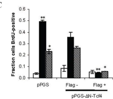
through G1/S phase. Thus, expression of dominant-negative Tcf4 blocks EGF-mediated
Tcf/Lef transcriptional activity and bromodeoxyuridine uptake. In fact, the importance of
EGF-mediated Tcf/Lef transcriptional activity for cell cycle progression may lie further
upstream at the G1/S phase transition. We demonstrate that dominant-negative Tcf4
inhibits a reporter of cyclin D1 promoter activity in a dose-dependent manner.
Importantly, dominant-negative Tcf4 suppresses EGF-mediated cell cycle activity
specifically by thwarting EGF-mediated Tcf/Lef transcriptional activity, not by broader
effects on EGF signaling. Thus, although expression of dominant-negative Tcf4 blocks
EGF-mediated TOPFLASH activation, it has no effect on either EGF receptor or ERK
phosphorylation, further underscoring the fact that Tcf/Lef-mediated transcription is
essential for cell cycle progression, even when other pro-mitogenic signals are at normal
sufficient for cell cycle progression. Serum also stimulates Tcf/Lef transcriptional
activation in MCF-10A cells but is unable to promote DNA synthesis. Taken together,
our data support a model wherein EGF promotes Tcf/Lef transcriptional activity, and this
signal is essential but not sufficient for cell cycle activity.
Reprinted with permission from N.A. Graham and A.R. Asthagiri from The Journal of
Biological Chemistry (2004).
1. Introduction
β-catenin is a 90-kDa intracellular protein whose functions range from stabilization of
cell-cell adhesion to control over gene expression. These functions are tightly regulated
through its association with various proteins such as the transmembrane protein
E-cadherin and Tcf/Lef transcription factors (1,2). E-E-cadherin is a major constituent of
adherens junctions where it promotes epithelial cell-cell contact through homotypic
interactions mediated by its extracellular domain (3). Meanwhile, its cytoplasmic domain
binds to β-catenin, whose association with α-catenin and other structural proteins bridges
E-cadherin-mediated contacts to the actin cytoskeleton (4). In addition to regulation by
sequestration to the plasma membrane, β-catenin is tightly regulated by cytosolic
degradation via a multiprotein complex consisting of Axin, APC, and glycogen synthase
kinase 3β (GSK3β) (5). Signaling events that inhibit this cytosolic degradation
machinery, such as those initiated by a subset of Wnt family ligands, help to stabilize
Tcf/Lef family of transcription factors.
Mutations that abnormally stabilize β-catenin occur in a diverse range of cancer
types. In colorectal carcinomas and melanomas, these mutations include the loss and/or
truncation of APC and mutations among critical N-terminal serine residues of β-catenin
whose phosphorylation flags it for ubiquitin-mediated degradation (5-7). Evidence of β
-catenin stabilization has also been shown in hepatomas and prostate cancers wherein loss
of axin and PTEN, respectively, leads to accumulation of nuclear β-catenin and increased
Tcf/Lef-mediated transcription (8,9). In the mammary gland, transgenic expression of
Wnt family ligands induces mammary adenocarcinomas in mice (10). Consistent with
this finding, mammary-tissue-specific overexpression of a constitutively stable β-catenin
mutant induces hyperplasia and adenocarcinoma in the mammary gland (11). Finally,
studies using stabilized mutants of β-catenin or Tcf/Lef-VP16 fusion constructs have
affirmed the capacity of this signaling pathway to transform established cell lines and
primary cells (12-14).
In fact, antagonizing β-catenin signaling appears to be an effective method to curb the
growth of cancer cell lines afflicted by elevated levels of nuclear β-catenin. Exogenous
expression of APC, axin, or PTEN reinstates β-catenin turnover and suppresses growth of
hepatocellular and prostate carcinoma cells (8,9). Inhibition of integrin-linked kinase, a
serine/threonine kinase that inhibits GSK3β and thereby stabilizes β-catenin, reduces
growth in prostate cancer lines (15). Finally, overexpression of proteins, such as
β/γ-catenin-binding domain, sequesters stabilized β-catenin, precludes its association with Tcf/Lef
transcription factors, and effectively inhibits proliferation of colorectal cancer cell lines
(16-18). Although the transformation potential of β-catenin has been closely examined,
the role of β-catenin and Tcf/Lef transcription factors in cell cycle progression among
normal mammalian cells is just now beginning to emerge. Immunohistochemical data
have shown that self-propagating precursor cells in the intervillus regions of the small
intestine epithelium – but not the well-differentiated cells at the villi tip – exhibit nuclear
β-catenin and express several Tcf/Lef target genes, including c-myc and CD44 (19). In
addition, Tcf4 knock-out mice lack proliferating stem cells and possess only
differentiated villus cells, suggesting a causal role for Tcf/Lef in governing stem cell
lineage commitment (20). In addition to intestinal epithelia, Tcf/Lef signaling is involved
in lineage commitment of human epidermal stem cells (21-25), hematopoietic stem cells
(26), and embryonic stem cells (27). However, the ligand(s) implicated in stimulating
Tcf/Lef signaling and dictating stem cell fate are largely unknown, although Wnt is
clearly involved in some instances (26,28).
It is unclear whether non-Wnt ligands also utilize the Tcf/Lef pathway to regulate
proliferation. Recently, a correlation between serum-mediated proliferation and Tcf/Lef
transcriptional activity has been suggested in a study using an engineered mammary cell
system (29). These cells express a c-Fos-estradiol receptor fusion protein that permits
switching from epithelial to fibroblastoid phenotype upon estradiol-mediated activation
of c-Fos (30). In both phenotypes, conditions that inhibited proliferation, such as serum
for β-catenin in serum-induced cell cycle progression was not clearly established in the
epithelial cell phenotype. Taken together, inducible activation of c-Fos, which is a
component of the AP-1 transcriptional machinery and itself critically involved in cell
cycle control (31), and the inability of β-catenin suppression to consistently inhibit
proliferation preclude an assessment of whether β-catenin nuclear activity is
mechanistically involved in proliferation.
Interestingly, several reports have indicated that specific growth factors such as
insulin and insulin-like growth factor I induce β-catenin transcriptional activity (32).
Although these studies were conducted with cancer cell lines lacking normal β-catenin
degradation machinery, HGF and certain members of the Wnt family of ligands induce
β-catenin transcriptional activity in normal cells (33,34). Although the importance of
HGF-mediated β-catenin signaling for normal cell cycle progression has not been examined,
certain members of the Wnt family of ligands regulate proliferation in a
β-catenin-dependent manner (35). Nevertheless, because β-catenin target genes include c-myc and
cyclin D1, whose protein products are ubiquitously crucial for cell cycle progression
(36-38), the untested hypothesis remains that β-catenin has a more pervasive role in normal
epithelial cell proliferation, even in response to growth-stimulating cues from non-Wnt
ligands.
We examined this hypothesis pertaining to the role of β-catenin in cell cycle
progression in the normal mammary epithelial cell line MCF-10A. We demonstrate that
necessary but not sufficient for cell cycle progression of normal epithelial cells. Thus,
inhibition of Tcf/Lef transcriptional activity using dominant-negative Tcf4 prevents
EGF-mediated cell cycle progression. Since dominant-negative Tcf4 inhibits cyclin D1
promoter activity and BrdU uptake without affecting other EGF-mediated signals such as
ERK that also regulate proliferation, we conclude that Tcf/Lef-mediated transcription is
required for cell cycle progression.
2. Experimental Procedures
2.1. Antibodies
The following antibodies were used in this study: actin (Santa Cruz),
anti-BrdU (Roche Applied Science), anti-ERK2 (Santa Cruz), anti-GSK3β (BD Transduction
Laboratories), anti-phospho-Ser9-GSK3β (BIOSOURCE), monoclonal and polyclonal
anti-FLAG (Sigma), anti-phospho-ERK1/2 (Thr202/Tyr204) (Cell Signaling
Technology), anti-phosphotyrosine (Santa Cruz), and anti-Tcf4 (Upstate Biotechnology,
Inc.).
2.2. Cell Culture
SW480 cells were cultured in Dulbecco’s modified Eagle’s medium
supplemented with 4 mM L-glutamine (Invitrogen), 10% (v/v) fetal bovine serum
(Invitrogen), and 1% (v/v) penicillin/streptomycin (Invitrogen). MCF-10A cells were
cultured in Dulbecco’s modified Eagle’s medium/Ham’s F-12 containing HEPES and
L-glutamine (Invitrogen) supplemented with 5% (v/v) horse serum (Invitrogen), 20 ng/ml
µg/ml insulin (Sigma), and 1% (v/v) penicillin/streptomycin. For serum starvation, the
cells were washed twice in PBS and then cultured in Dulbecco’s modified Eagle’s
medium/Ham’s F-12 supplemented with 1% (v/v) penicillin/streptomycin and 0.1%
bovine serum albumin (Sigma) for 24 h.
2.3. Plasmid Constructs
pcDNA-myc-ΔN-Tcf4 was generously provided by K. W. Kinzler (Johns Hopkins
University) (7). pPGS and pPGS-ΔNTcf4 were kindly donated by E. Fearon (University
of Michigan, Ann Arbor) (14). VSV-G and gag-pol vectors were gifts from D. Schaffer
(University of California, Berkeley). Luciferase-based reporters pTOPFLASH and
pFOPFLASH were purchased from Upstate Biotechnology, Inc., whereas 1745CD1 was
a gift from R. Pestell (Georgetown University, Washington, D.C.) (39).
2.4. Retroviral Infection
Retrovirus was produced by either by single transfection of the packaging cell line
293GPG with 15 µg of retroviral plasmid (40) or by triple transfection of 293T cells with
5 µg each of VSV-G, gag-pol and a retroviral vector using LipofectAMINE (Invitrogen).
For infection, MCF-10A cells were incubated with retrovirus-containing medium and 8
µg/ml polybrene for 24 h.
2.5. GSK3β Serine 9 Phosphorylation Assay
MCF-10A cells were plated at a subconfluent density (105 cells/35-mm dish) and allowed to adhere for 48 h, followed by serum starvation for 24 h. The cells were
stimulated with either full growth medium or serum-free medium supplemented with
either 10 µg/ml insulin or 20 ng/ml EGF and then lysed in modified RIPA buffer at
2.6. ERK Signaling Assay
MCF-10A cells were plated at a subconfluent density (105 cells/35-mm dish),
allowed to adhere for 24 h, and then infected with retrovirus encoding pPGS or
pPGS-FLAG-ΔN-Tcf4 at multiplicity of infection equal to 1. Twenty-four hours after infection,
the cells were starved in serum-free medium for 24 h, stimulated with 20 ng/ml EGF in
serum-free medium, and then lysed in modified RIPA buffer at desired times.
2.7. Cell Lysis
The stimulated cells were washed twice in ice-cold PBS and scraped into cold
lysis buffer. After incubating on ice for 15 min, the cell lysates were clarified by
centrifugation, and the supernatant was collected as whole cell lysate. The protein
concentrations were determined using BCA reagents (Sigma). The samples prepared to
assay EGF-mediated activation of the ERK pathway were lysed in modified RIPA buffer
(50 mM Tris-Cl (pH 7.5), 150 mM NaCl, 1% Triton X-100, 0.5% Nonidet P-40, 0.25%
sodium deoxycholate, 50 mM β-glycerophosphate (pH 7.3), 10 mM NaPP, 30 mM NaF,
1 mM benzamidine, 2 mM EGTA, 1 mM sodium orthovanadate, 1 mM dithiothreitol, 5
µg/ml aprotinin, 5 µg/ml leupeptin, 1 µg/ml pepstatin, and 1 mM phenylmethylsulfonyl
fluoride). Finally, cell lysis for all reporter measurements was performed in 1X passive
lysis buffer provided by the manufacturer (Promega).
2.8. Reporter Assays
SW480 or MCF-10A cells were plated at a subconfluent density (105 cells/35-mm
dish) and co-transfected with 1 µg of the appropriate reporter and 0.1 µg of pRL-TK
using FuGENE 6 (Roche Applied Science). SW480 cells were always maintained in
serum-starved for 24 h, stimulated with appropriate medium, and lysed at desired times.
In both cases, reporter activity was measured using the dual luciferase assay according to
the manufacturer instructions (Promega). To normalize for potential variations in
transfection or lysis efficiency, luciferase signals were normalized to control Renilla
luciferase signal.
2.9. Integrated Reporter Response
The reporter signal response above its initial value was integrated numerically
over time as follows,
!
R t
( )
"R t( )
0 dt= 12k=1R t
( )
k +R t( )
k"1 n#
$ % & ' ()"
(
n"1)
R t( )
0$ % & & ' ( ) )*t
0 12
+
(Eq. II-1)where R(t) is the reporter signal, R(t0) is its basal, initial value, tk is the time ranging from
0 to 12 h in discrete intervals of Δt (3 h), n is the number of time points (n = 5), and k is
the index of summation.
2.10. Western Blotting
Whole cell lysates were resolved by SDS-PAGE on 7.5-10% gels and blotted onto
polyvinylidene difluoride membrane (Bio-Rad). The membranes were blocked overnight
and then incubated sequentially with primary and corresponding horseradish
peroxidase-conjugated secondary antibody. The blots were treated with SuperSignal West Femto
Substrate (Pierce) and imaged on VersaDoc 3000 (Bio-Rad) using Quantity One software
(Bio-Rad).
2.11. DNA Synthesis
DNA synthesis was assayed by either [3H]thymidine or BrdU incorporation. In both cases, MCF-10A cells were seeded at the indicated cell densities. After 24 h, the
day, the cells were serum-starved. Notably, the 48 h of duration between cell seeding and
serum starvation was chosen to match the time required for plating and transfecting cells
in reporter assays, allowing direct comparison between DNA synthesis and reporter
experiments. Following 24 h of serum starvation, the cells were stimulated with
appropriate medium. Sixteen hours after stimulation, the medium was replaced with
identical medium supplemented with either 10 µCi/ml [3H]thymidine (ICN Biomedicals)
or 10 µmol/liter BrdU (Roche Applied Science) and further incubated for 6 h. In the case
of [3H]thymidine incorporation, the cells were washed twice in ice-cold PBS, incubated in 5% trichloroacetic acid for 20 min at 4 °C, washed twice with cold 70% ethanol, and
incubated with 0.1 M NaOH, 2% Na2CO3, and 1% SDS for 30 min at 37 °C. The
solution was collected and mixed with CytoScint (ICN Biomedicals) for scintillation
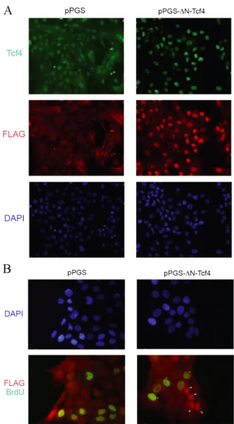
counting. For BrdU detection, the cells were fixed and co-stained with DAPI, anti-BrdU
antibody, and polyclonal anti-FLAG antibody. The number of nuclei stained positive for
BrdU and FLAG were quantified in 3-10 different fields on 2-5 independent trials using
the Zeiss Axiovert 200M microscope.
2.12. Immunofluorescence
For Tcf/FLAG co-staining, the cells grown on glass coverslips were washed three
times in ice-cold PBS, fixed in 4% formalin in PBS, and permeabilized in 0.2% Triton
X-100. After blocking overnight in BB (10% goat serum, 0.1% bovine serum albumin in
PBS), the coverslips were sequentially incubated with primary and corresponding Alexa
dye-labeled secondary antibodies (Molecular Probes). Following antibody incubations,
the coverslips were stained with DAPI (Sigma) and mounted using Prolong Anti-Fade
in 4% formalin and 0.2% Triton X-100, respectively, and then incubated with polyclonal
FLAG antibody and Alexa 594-conjugated secondary antibody. The antibodies were
then fixed in 4% formalin, followed by a second fixation in 15 mM glycine in 70%
ethanol (pH 2). The coverslips were incubated with monoclonal BrdU antibody and then
Alexa 488-conjugated secondary antibody, stained with DAPI, and mounted as described
above.
3. Results
3.1. Re-entry into the Cell Cycle Correlates with Tcf/Lef Reporter Activity
Target genes for Tcf/Lef include cyclin D1 and c-myc, suggesting a role for this
family of transcription factors in cell cycle progression not only among cancer cells with
stabilized nuclear β-catenin, but also among normal epithelial cells. Therefore, we
determined whether a correlation exists between Tcf/Lef transcriptional activity and cell
cycle progression in nontransformed mammary epithelial (MCF-10A) cells. Tcf/Lef
transcriptional activity was monitored with TOPFLASH reporter (7), a plasmid
containing consensus Tcf-binding sites upstream of the luciferase gene. In contrast, the
negative control FOPFLASH reporter carries mutations at these Tcf/Lef-binding sites.
Performance of TOPFLASH and FOPFLASH reporters was confirmed in SW480 colon
carcinoma cells in which TOPFLASH, but not FOPFLASH, is constitutively active
because of a truncation of the APC gene and consequent stabilization of β-catenin (7)
(Fig. II-1A). Subconfluent MCF-10A cells transfected with TOPFLASH or FOPFLASH
cell cycle by treatment with growth medium. As shown in Fig. II-1B, growth medium
stimulation activated TOPFLASH reporter, which gradually increased to a
near-maximum level within the first 9 h. Meanwhile, FOPFLASH negative control reporter
did not respond to growth medium stimulation. Taken together, this establishes a
correlation between re-entry into the cell cycle and Tcf/Lef-mediated transcription.
FIG. II-1. TOPFLASH and FOPFLASH reporter activity in SW480 and MCF-10A cells
A, TOPFLASH, but not FOPFLASH, reporter is triggered in SW480 colon carcinoma cells. SW480 cells were co-transfected with 0.1 µg of pRL-TK and 1 µg of either TOPFLASH or FOPFLASH. Forty-eight
hours after transfection, the cells were lysed, and the ratio of luciferase to Renilla luciferase signal was quantified. B, TOPFLASH, but not FOPFLASH, reporter is activated upon growth medium stimulation of normal mammary epithelial cells. MCF-10A cells were co-transfected with 0.1 µg of pRL-TK and 1 µg of
TOPFLASH () or FOPFLASH (). After serum starvation, the cells were stimulated with growth
medium and luciferase:Renilla luciferase signal ratio was quantified at desired time points. Reporter activity relative to the TOPFLASH response at 6 h is shown. The error bars represent ± S.E. from two to
five independent experiments. The asterisk denotes p < 0.05 (Student’s t-test) in comparing TOPFLASH signal to the zero time response.
To confirm further that the observed TOPFLASH signal was specifically monitoring
Tcf/Lef transcription factor activity, a dominant-negative Tcf4 construct (myc-ΔN-Tcf4)
was employed. This construct possesses the DNA-binding domain of Tcf4, but lacks the
N-terminal 31 amino acids that mediate its association with its transactivating catenin
SW480 cells (Fig. II-2A). Co-transfection of ΔN-Tcf4 into MCF-10A cells decreased
growth medium-induced TOPFLASH response in a dose-dependent fashion (Fig. II-2B),
indicating that the TOPFLASH signal was mediated specifically by Tcf/Lef transcription
factors.
FIG. II-2. Dominant-negative Tcf4 effect on TOPFLASH reporter
A, dominant-negative Tcf4 inhibits TOPFLASH signal in SW480 colon carcinoma cells. SW480 cells were co-transfected with 1 µg of TOPFLASH, 0.1 µg of pRL-TK, and either 0.5 µg of empty vector
(pcDNA) or dominant-negative Tcf4 (ΔN-Tcf4). Forty-eight hours after transfection, the luciferase:Renilla luciferase signal ratio was quantified. B, dominant-negative Tcf4 inhibits growth medium-mediated TOPFLASH signal in MCF-10A cells. MCF-10A cells were co-transfected with 1 µg of TOPFLASH, 0.1 µg of pRL-TK, and different amounts (0, 0.05, 0.1, and 0.5 µg) of dominant-negative Tcf4 (ΔN-Tcf4), always with a balancing amount (0.5, 0.45, 0.4, and 0 µg, respectively) of empty vector (pcDNA).
Serum-starved cells were stimulated with growth medium for 9 h, after which the luciferase:Renilla luciferase signal ratio was quantified. Co-transfection with increasing amount of ΔN-Tcf4 correspondingly attenuated TOPFLASH induction by growth medium. The error bars indicate ± S.E. (n = 3). The asterisk indicates p
< 0.01 (Student’s t-test).
3.2. EGF Independently Induces Tcf/Lef Transcriptional Activity and DNA Synthesis
Because MCF-10A growth medium contains a complex mixture of stimuli, including
serum factors, insulin, and EGF, it is unclear whether a single constituent is capable of
inducing Tcf/Lef transcriptional activity and, moreover, whether the same constituent
[image:50.612.140.512.198.399.2]constituent of growth medium separately, and Tcf/Lef transcriptional activity and DNA
synthesis were assessed by measuring TOPFLASH reporter signal and [3H]thymidine
uptake, respectively. EGF independently induced TOPFLASH signal to a level distinctly
above the corresponding FOPFLASH control (Fig. II-3A). At early times, EGF-mediated
TOPFLASH signal mirrors growth-medium-induced TOPFLASH activity. However,
whereas full growth medium sustains TOPFLASH signal to 24 h (Fig. II-1A), EGF
promotes a transient signal that reaches its peak intensity of nearly 3-fold above basal
level at 3 h. Meanwhile, in contrast to EGF, insulin-mediated TOPFLASH activation
more closely matches the FOPFLASH negative control, except at 3 h, where a transient
signal that is 50% of the EGF-mediated TOPFLASH signal is observed. Taken together,
growth medium constituents quantitatively vary in their ability to promote Tcf/Lef
transcriptional activity, with EGF, more so than insulin, resembling the response to full
growth medium.
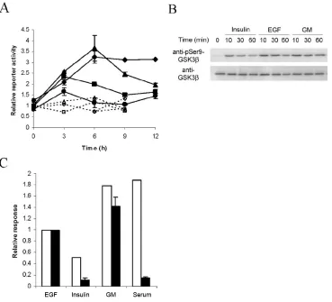
The current paradigm for Wnt-mediated Tcf/Lef transcriptional activity involves
inhibition of GSK3β-mediated phosphorylation of β-catenin, which in turn stabilizes β
-catenin and ultimately enables its translocation into the nucleus (41-43). Interestingly,
both EGF and insulin have been reported to inhibit GSK3β kinase activity toward primed
substrates by inducing phosphorylation of GSK3β at serine 9 (44-48). Because there is
conflicting evidence as to whether β-catenin qualifies as a primed or nonprimed substrate
of GSK3β (49-52), we investigated whether EGF, insulin, and growth medium affected
GSK3β phosphorylation at serine 9 in a manner that is quantitatively consistent with their