ISSN Print: 2160-5866
DOI: 10.4236/jbbs.2018.812040 Dec. 10, 2018 641 Journal of Behavioral and Brain Science
Mammalian Auditory Cortex Structure as the
Basis of Cortical Sound Processing
Gleb Khorunzhii, Marina Egorova
*I.M. Sechenov Institute of Evolutionary Physiology and Biochemistry, RAS, Saint-Petersburg, Russia
Abstract
The basic morphological aspects of auditory cortex organization in different orders of eutherian mammals are considered in the present review. The modern data describing a partitioning of mammalian auditory cortex into subfields are presented. A detailed observation of the structural organization of primary auditory cortex is given, as well as a review of recent morphologi-cal data about secondary auditory areas. Another section describes the system of auditory cortical projections. The data are considered from the perspective of possible homologies existing between the auditory cortices in different mammalian species.
Keywords
Auditory Cortex, Primary and Secondary Auditory Cortical Fields, Auditory Cortical Projections
1. Preface
At present, there are extensive data about the morphological structure of the mammalian auditory cortex obtained in numerous studies with different ex-perimental approaches and methods [1]-[20]. The set of experimental objects used in this research is large. Despite this, information about partitioning of the auditory cortex into separate sub-regions and about its connectivity among dif-ferent mammalian orders has not been summarized in detail. Our review aims to systematically present the available data about the structural organization and connectivity of the auditory cortex in eutherian mammals.
The first part of this review contains a detailed description of the auditory fields’ disposition in the temporal lobe of the neocortex among a significant number of mammalian species. The common scheme of the auditory cortex spa-tial organization inispa-tially proposed by Woolsey and Walzl (1942) for cats [21]
How to cite this paper: Khorunzhii, G. and Egorova, M. (2018) Mammalian Audi-tory Cortex Structure as the Basis of Corti-cal Sound Processing. Journal of Behavioral and Brain Science, 8, 641-673.
https://doi.org/10.4236/jbbs.2018.812040
Received: November 5, 2018 Accepted: December 7, 2018 Published: December 10, 2018 Copyright © 2018 by authors and Scientific Research Publishing Inc. This work is licensed under the Creative Commons Attribution International License (CC BY 4.0).
http://creativecommons.org/licenses/by/4.0/
DOI: 10.4236/jbbs.2018.812040 642 Journal of Behavioral and Brain Science and later applied to other animals [2]-[20][22] [23][24] was used as a frame-work for the present review. For each mammalian order, we separately describe primary and secondary auditory fields as well as the auditory parabelt, their dis-position within the auditory cortex, tonotopic organization and the relative size of these areas.
The second chapter presents data on the connectivity patterns of the auditory cortex. First, we focused on the projection system of the cat auditory cortex, which is the most studied among all eutherians. Second, we observed connec-tions in the auditory cortex regarding their source contribuconnec-tions to the total volume of the auditory cortical projections.
In both chapters, we focused on basic aspects of auditory cortical structure in the mammalian phylogenetic tree rather than on their functional organization. The data systematised here provide a necessary base for an in-depth analysis of acoustic information processing in the auditory cortex and for an integrative scheme of sound encoding by the brain’s auditory centres.
2. Introduction
The neocortical temporal lobe, which receives large inputs from the thalamic medial geniculate body (MGB) and contains neurons responding to acoustic stimulation, is generally classified as the auditory cortex [25]. A number of ana-tomical, physiological and behavioural studies, some of which were performed over 100 years ago, repeatedly showed that the auditory cortex is a complex structure consisting of several fields [26][27][28][29]. These fields can be dis-tinguished on the basis of their cytoarchitectonics, connectivity, functional map-ping and neuronal processing.
Systematic studies of the auditory cortex’s functional organization began in 1942 with a study by Woolsey and Walzl in which the authors mapped a distri-bution of the slow-wave evoked potentials in the ectosylvian region of the cat cerebral cortex under local electrical stimulation of the auditory nerve fibres in a cochlear labyrinth [6]. The topography of frequency representation (i.e., its high-ordered spatial distribution within the auditory cortex) was shown by Woolsey and Walzl for the first time. Based on the results of evoked potential recordings in the cat auditory cortex [21][22][23], its cytoarchitectonic analysis [30] and MGB retrograde degenerations evoked by local lesions of auditory cor-tical areas [31][32], Woolsey suggested a scheme of the cat auditory cortex’s or-ganization (Figure 1).
In accordance with this scheme, the auditory cortical area was divided into a primary auditory field (A1) and several other fields surrounding A1. Woolsey adapted the model of cat auditory cortex to other mammals, especially to pri-mates [33][34][35][36].
DOI: 10.4236/jbbs.2018.812040 643 Journal of Behavioral and Brain Science Figure 1. Representation of the cat auditory cortical area, suggested by Woolsey [22]. AI—primary auditory field (black filling). Dashed and dotted lines indicate approximate borders of areas. Ep—posterior ectosylvian gyrus, AII—secondary auditory field, SF—suprasylvian peripheral area, Ins—insular area, AIII—temporal auditory field, Assoc—associative cortex. MI—precentral motor cortical area, Late VII—visual cortex. In tonotopic auditory fields the direction of frequency increase is indicated (from A to B). d—dorsal direction, v—ventral direction, r—rostral direction, c—caudal direction. Modi-fied from: [49].
developed cortical layers II and IV receiving a large pool of both specific and non-specific thalamocortical projections. Primary auditory fields have a conio-cortical cellular structure, receive direct inputs from MGB and differ from other auditory fields by a number of biochemical properties. Coniocortical cells in the neocortex usually demonstrate a small percentage of large pyramidal neurons and are predominated by densely distributed small stellate cells. The basic source of the primary auditory cortex afferents is a main lemniscal auditory path-way—the system of ascending projections rising from the auditory brainstem and reaching MGB as part of the lateral lemniscus fibres. The secondary audi-tory belt is usually located on the periphery of the primary audiaudi-tory core. Due to its cytoarchitectonics, the auditory belt is defined as agranular neocortex with the thick cortical layer III (pyramidal) where there is a high spatial density of cells, including large pyramidal neurons. The extensive development of cortical layer III is provided by many associative and commissural fibres, located within this layer. Secondary auditory fields receive a large set of afferents from different parts of MGB and cortical primary auditory fields.
DOI: 10.4236/jbbs.2018.812040 644 Journal of Behavioral and Brain Science modalities but demonstrates a strong response to acoustic stimuli [25]. It seems that the diffuse auditory system doesn’t have direct connections with primary auditory areas but receives rich afferentation from the secondary auditory cor-tex, medial pulvinar, thalamic suprageniculate nucleus and nucleus limitans. Weak afferent inputs to the auditory parabelt rising from dorsal and medial parts of MGB have also been described [40]. Currently, the non-lemniscal audi-tory pathway is known as a part of a higher order stage of processing, constitut-ing a secondary system capable of processconstitut-ing more complex aspects of auditory scene analysis [1]. On a cortical level, this integrative system of sound processing is closely associated with the auditory belt and parabelt [1][37][38].
Thus, as the main integrative centre of the mammalian auditory system, audi-tory cortex provides the basement for hierarchical (serial) cortical sound proc-essing [15][16], being involved in the different aspects of acoustic information analysis, which become more complex and more integrative from the primary auditory cortex up to auditory parabelt.
3. Subdividing the Auditory Cortex into Fields in Different
Mammalian Orders
It was shown in numerous studies of the auditory cortex in eutherian mammals over the last 50 years that all of the studied animals had auditory cortices con-sisting of several separate auditory fields [2]-[20][22][23][24]. The number of fields increases with the evolutionary development from 2 in insectivores to 4 - 7 in rodents and from 6 to more than 8 in carnivores and primates, respectively [41]. This is related to the increase in the relative size of cerebral cortex in dif-ferent mammalian orders. The only exception is marsupial mammals, for which only the primary auditory field has been shown [42][43].
3.1. Auditory Cortical Fields in Insectivores
Among different insectivore species, the auditory cortex has been studied in hedgehogs and shrews. The auditory cortex of the long-eared hedgehog (He-miechinus auritus) is located in a lateral part of the medial temporal lobe, medial to sulcus olfactorius [44]. The primary auditory cortex boundaries in this animal were defined by electrophysiological recordings of single neurons and by label-ling with horse-radish peroxidase of MGB projections to the auditory cortex. Recordings of the neuronal responses evoked by tonal stimuli revealed the spa-tial distribution of frequency representations within the primary auditory cortex, with high frequencies located caudally and low frequencies located rostrally. Another auditory area was found caudal to the primary auditory field. Neurons in this area responded to sounds, but ordered distribution of frequencies was not shown [44].
parie-DOI: 10.4236/jbbs.2018.812040 645 Journal of Behavioral and Brain Science tal-ventral (PV) cortical areas bordering the auditory cortex of this animal [45] [46].
In the northern short-tailed shrew (Blarina brevicauda), the auditory cortex was found in an extremely caudolateral part of the neocortex. The properties of single cells in the shrew auditory cortex remain poorly studied [46].
3.2. The Auditory Cortical Fields in Carnivores
Woolsey defined four tonotopic (or cochleotopic) fields in the cat auditory cor-tex containing a representation of the whole frequency range of a cat’s hearing: the primary auditory field (A1), secondary auditory field (A2), posterior ectosyl-vian field (EP) and suprasylectosyl-vian peripheral field (SF) (Figure 1) [21] [22][23]. Woolsey also included in the cat auditory cortex a temporal auditory field (T) similar to the field A3 in the dog’s auditory cortex [7], insular region (Ins) [47] and associative cortex, which were studied in detail by Thompson and Sindberg (1960) [48] (Figure 1). Auditory evoked potentials were also recorded in the precentral motor cortex (Figure 1) and visual cortex [25]. Further data from electrophysiological mapping of the cat auditory cortex [9][49][50][51] slightly modified the original Woolsey’s scheme, bringing it to the present state (Figure 2). Thus, the A1 location defined by Woolsey and Walzl did not change dra-matically. Another full frequency representation was revealed rostrally to the A1 in the region of the anterior ectosylvian gyrus, which was first mapped by Woolsey as part of the suprasylvian peripheral region. This area was mapped in more detail by Knight (1977) [52] as well as by Reale and Imig (1980) [9], who named it the anterior auditory field (AAF) (Figure 2). Aside from these two primary fields, two other tonotopically organized auditory areas were related with the core regions of the cat auditory cortex, the posterior (P) and ventropos-terior (VP) fields [9][49]. The auditory belt surrounding the primary auditory cortex was subdivided into secondary (A2), ventral (Ve), temporal (Te) and in-sular (Ins) fields, and the posterior ectosylvian region was additionally divided into dorsal (EPD), medium (EPM) and ventral (EPV) parts of posterior ectosyl-vian gyrus [50][51]. It is noteworthy that, in a study performed by Reale and Imig in 1980 [9], the posterior ectosylvian area was identified as a dorsal audi-tory field. The dorsal region of this field corresponding to EPD was defined as a dorsal auditory zone (DZ) based on the analysis of functional properties of its neurons [17][53].
DOI: 10.4236/jbbs.2018.812040 646 Journal of Behavioral and Brain Science Figure 2. Auditory cortical fields in the cat [50]. Tonotopically organized fields, receiving inputs from the ventral MGB, are marked with grey filling. Non-tonotopically organized fields, receiving inputs from other MGB subregions, are marked by light grey. AI—primary auditory field; AAF—anterior auditory field; AII—secondary auditory field; EPD—posterior ectosylvian gyrus, dorsal part; EPI—posterior ectosylvian gyrus, intermediate part; EPV—posterior ectosylvian gyrus, ventral part; Ins—insular cortex; Te—temporal cortex; P—posterior auditory field; Ve—ventral auditory field; VP—ventroposterior auditory field; SF—suprasylvian fringe area; D—dorsal direction, C—caudal direction.
in dogs occupies the same cortical area as EP in the cat auditory cortex, and the A3 field in the dog cortex corresponds to the temporal auditory field (Te) in cats. In the ferret auditory cortex studied by autoradiography [54] and neuronal activity recordings under sound stimulation [18][55] [56][57] [58], three dis-tinct areas were revealed near the medial ectosylvian gyrus. The first of them is the medial ectosylvian gyrus (MEG), which is identified as a primary auditory cortex. Within this area, two tonotopically organized auditory fields were de-scribed, A1 and AAF [18][58]. The second area is located in the anterior ecto-sylvian gyrus and includes anterior ventral (AVF) and anterior dorsal (ADF) auditory fields [18][58]. The last large auditory area was found in the posterior ectosylvian gyrus, which includes posterior pseudosylvian field (PPF) and poste-rior suprasylvian field (PSF) [18][57] [58]. PPF and PSF are tonotopically or-ganized. In AVF and ADF, the tonotopy was not shown [18][58].
3.3. Auditory Cortical Fields in Rodents
DOI: 10.4236/jbbs.2018.812040 647 Journal of Behavioral and Brain Science [77][78][79][80][81], squirrels [82][83][84] and chinchillas [85][86][87]. In all studied rodent species, primary and secondary auditory cortical fields were found. The number of auditory cortical fields varies among different rodent spe-cies.
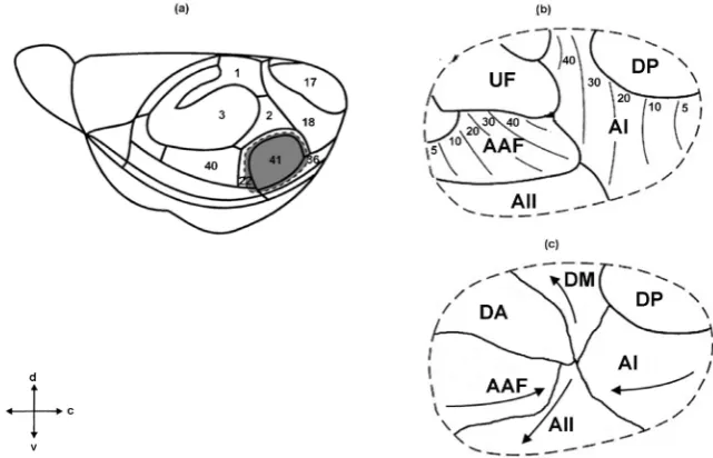
In the house mouse auditory cortex (Figure 3(a) and Figure 3(b)), three pri-mary auditory fields (pripri-mary auditory field (A1), anterior auditory field (AAF), ultrasound field (UF)) and two secondary auditory fields (dorsoposterior field (DP) and secondary auditory field (A2)) were defined [12][70]. Two core fields are tonotopically organized, A1 and AAF (located rostrally to A1). UF is located dorsorostrally to A1. The dorsocaudal border of A1 is adjacent to the DP field. The ventral border of A1 flanks A2 (Figure 3(b)). Recently, this scheme was modified by Tsukano and colleagues (Figure 3(c)) [88][89][90]. Using the op-tical imagination method based on flavoprotein fluorescence imaging (FFI), the authors identified six distinct fields in the mouse auditory cortex. Four of them are tonotopically organized: A1, A2, AAF and dorsomedial field (DM), while two fields (DP and dorsoanterior field (DA)) have no tonotopy (Figure 3(c)). A1, AAF and A2 include distinct ultrasonic frequency bands with CFs over 40 kHz. DM and DA also include neurons responding to the ultrasound stimuli. It was assumed that the region first annotated as the UF was a mixture of the DA and high frequency bands of the DM [90] (Figure 3(b) and Figure 3(c)).
The rat auditory cortex contains at least four primary auditory fields: A1 and AAF, which are typical for most rodents, and also suprarhinal auditory field (SRAF) and posterior auditory field (PAF) [63][91][92][93]. In addition, Pro-fant and colleagues referred to an area in the rat primary auditory cortex as “unspecific auditory region” (UR) [93]. The primary auditory cortex is sur-rounded by a number of secondary fields localized in the temporal cortical areas Te2 and Te3 [62][93][94].
In the auditory cortex of the Mongolian gerbil, seven fields were identified [12][95][96] [97]. Two of them demonstrated strong tonotopy and, similar to the mouse auditory cortex, were named the primary auditory field (A1) and an-terior auditory field (AAF), with AAF located more rostrally. Primary auditory fields were surrounded by several secondary areas named the ventral (V), ven-troposterior (VP), dorsoposterior (DP), anterior-ventral (AV) and dorsal (D) auditory fields.
DOI: 10.4236/jbbs.2018.812040 648 Journal of Behavioral and Brain Science Figure 3. Auditory cortical fields in the mouse. (a): lateral view of the left-side cortex with areas shown that are delimitated on the basis of cytoarchitectonic criteria [13]. Auditory cortex (field 41) is marked with grey filling. (b): original map of spatial organization of primary and secondary auditory cortical fields in the house mouse, obtained by Stiebler et al.[13]. (c): the scheme of the mouse auditory cortex, suggested by Tsukano et al. Modi-fied from: [90]. AI—primary auditory field, AAF—anterior auditory field, UF—ultrasound field, AII—secondary auditory field, DP—dorsoposterior auditory field, DM—dorsomedial auditory field, DA—dorsoanterior auditory field. On (b) the regular tonotopy in AI and AAF is shown. On (c) arrows indicate the direction of frequencies increase in AI, AAF, DM and AII. d—dorsal direction, v—ventral direction, r—rostral direction, c—caudal direction.
colleagues proposed to subdivide the secondary auditory cortex of guinea pigs into eight separate fields [80].
In the grey squirrel, a region of the temporal cortex responding to acoustic stimuli was separated in a similar manner to other rodents into subregions on the basis of their physiological and cytoarchitectonic properties [82] [83] [84]. One of these subregions, containing the full map of frequency representations, was considered by Merzenich and colleagues to be homologous to the A1 field of the cat and primates [85]. In the rostral field (R) located rostrally to A1, tonotopy was also shown [86]. Among secondary auditory fields in the grey squirrel, up to seven separate belt fields are identified [87].
In the auditory cortical area of the chinchilla, three tonotopically organized fields are described: A1, AAF (located rostrally to A1) and the secondary field (A2) located ventrally to the core areas [85][86][87]. Organization of the audi-tory belt areas in the chinchilla cortex remains unclear.
3.4. Auditory Cortical Fields in Chiropterans
orien-DOI: 10.4236/jbbs.2018.812040 649 Journal of Behavioral and Brain Science tation behaviour when searching for prey (echolocation), which makes them es-pecially interesting as a model for hearing physiology. Among all mammalian orders, different aspects of the morphofunctional organization of the auditory cortex in chiropterans were studied in the greatest detail, which is reflected in numerous studies of bats (Microchiroptera) [98]-[110].
The auditory cortex of different bat species contains from four to six auditory fields. The largest number of fields (six) was shown in the auditory cortex of the
Phyllostomidae family [106][107][111]. The German neurobiologists Esser and
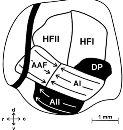
Eiermann mapped the auditory cortex of the leaf-nosed bat (Carollia perspe-cilata) and defined six separate areas: four primary and two secondary auditory fields (Figure 4) [106][107]. The authors referred to the primary auditory areas A1, AAF and the high-frequency field (HF), which contained neurons respond-ing to tone frequencies in the range of 65 - 97 kHz. HF in turn was subdivided into two separate auditory fields (HFI and HFII) based on the differences in the distributions of the minimal neuronal response thresholds across these fields. As for the secondary auditory cortex in this animal, the secondary auditory field (A2) and dorsoposterior field (DP) were identified [106][107].
The insectivore bats of the Microchiroptera suborder, such as the mustached bat, great horseshoe bat and little brown bat attracted the attention of neurosci-entists due to their hunting behaviour based on the echolocation mechanism [98] [99] [101] [102] [104][112]-[119]. Specific cortical organization in these animals is conditioned by its specialization to the processing of orientation calls, emitted by the bat itself during hunting, and orientation call reflections from multiple obstacles (i.e., echo signal). The temporal lobes of chiropterans are dis-tinguished by their size among all neocortical formations. Their relative size in bats is much larger than in all other mammalian orders, which is directly related to the overrepresentation of the auditory system at the cortical level in these animals [102][114][120].
In the auditory cortex of insectivore bats, the primary auditory field (A1) contains several tonotopic areas. These areas have disproportionately large fre-quency representations of the orientation call, echo signal and the frefre-quency ra-tios between them [101] [102][109][110][121]-[126]. Anterior and posterior regions of A1 in the mustached bat and the central area DSCF located between them (famous for its concentric tonotopic structure) could be referred to as A1 “subfields” [109][122]. The A1 is surrounded by several other auditory cortical areas involved in target movement processing. Tonotopy has not been shown in these fields [109][122][123][124]. It is notable that the identification of differ-ent auditory cortical areas in bats was based rather on their functional speciali-zation in echolocation than on the separation into primary and secondary areas.
3.5. Auditory Cortical Fields in Other Mammalian Species
DOI: 10.4236/jbbs.2018.812040 650 Journal of Behavioral and Brain Science Figure 4. Auditory cortical fields of leaf-nosed bat Carollia
perspe-cillata auditory cortex [107]. The primary auditory fields are: AI, AAF, HFI and HFII. The secondary auditory fields (AII and DP) are filled by black. The direction of frequencies increase in each tonotopically organized field is indicated by arrows. The dark line in the left part of figure is the blood vessel. d—dorsal direction, v—ventral direction, r—rostral direction, c—caudal direction.
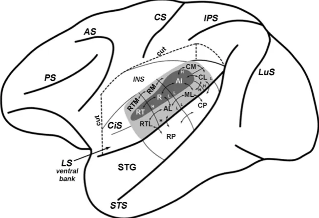
The organization of the auditory cortex in primates was clarified by the results of studies in New World monkeys [35] [129]-[134], Old World monkeys [36] [135]-[145], chimpanzee [35] [140] [146] [147] and galago [148] [149] [150] [151]. The main part of primate cortex where sound-evoked responses were re-corded was the superior temporal gyrus, located deep within a lateral fissure (Figure 5). The primary auditory cortex is located along the inferior margin of the lateral sulcus. It contains three fields: primary auditory field (A1), rostral field (R) and rostrotemporal field (RT), all of which are elongated in the ros-tro-caudal direction in the sulcus plane. As in most of other mammals, A1 is lo-cated the most caudally. Field R is placed rostrally to A1, and field RT is lolo-cated rostrally to field R [25][40] [136][138][139][148][152][153]. Primary audi-tory cortex in primates, due to its cytoarchitectonic properties and connectivity patterns, is regarded as a homological region of Brodmann’s Area 41 in the hu-man neocortex [25] [146] [147]. Within the primate secondary auditory belt surrounding primary auditory cortex, up to eight different fields are defined on the basis of their tonotopy, connections and architectonical features [25] [40] [138][147].
DOI: 10.4236/jbbs.2018.812040 651 Journal of Behavioral and Brain Science Figure 5. The scheme of macaque brain left hemisphere, demonstrating the location and intrinsic connections of auditory cortical fields [140]. The dorsal bank of lateral sulcus is removed to make available the superior temporal cortical zone (i.e. the ventral fringe of lateral sulcus). The floor and the outer bank of circular sulcus are smoothed to show the auditory fields located medially. The auditory core is filled by dark grey. AI—primary auditory field, R—rostral field, RT—rostrotemporal field. The auditory belt is shown by light grey filling. CM—caudomedial belt, CL—caudolateral belt, ML—medium lateral belt, AL—anterolateral belt, RTL—rostrotemporolateral belt, RTM—rostrotemporomedial belt, RM—rostromedial secondary auditory area. The auditory parabelt is not colored. The parabelt area occupies the surface of superior temporal gyrus (STG), exposed on this scheme, and consists of rostral parabelt (RP) and caudal parabelt (CP). The core fields are projecting to the surrounding auditory belt (the direction of their efferents is indicated by arrows). The inputs, received by parabelt from lateral and medial auditory belt, are also shown by arrows. Connections, occurring between auditory parabelt and secondary belt areas, located medially, are not illustrated to maintain the clarity of the scheme. The fre-quency gradients are indicated by characters H (high frequencies) and L (low frequen-cies). Abbreviations: PS—principal sulcus; AS—arcuate sulcus; CS—central sulcus; IPS—interparietal sulcus; LuS—lunate sulcus; STS—superior temporal sulcus; LS—lateral sulcus; CiS—circular sulcus; INS—insular cortex.
in these areas. Hackett and colleagues subdivided the auditory parabelt into ros-tral (RP) and caudal (CP) subregions due to differences in connectivity of these areas [154]. The main part of the rostral parabelt projects to the rostral secon-dary auditory fields, whereas the connections of caudal parabelt were concen-trated in the caudal region of secondary auditory cortex. However, there were not clear differences found in the connectivity of these two parabelt areas [40], [147].
4. Connectivity of the Auditory Cortex
path-DOI: 10.4236/jbbs.2018.812040 652 Journal of Behavioral and Brain Science ways, and also with other fields of the auditory cortex by corticocortical fibres. The methods of retrograde degeneration and tracer injections (i.e., the injection of fluorescent and radioactive labels or labelling by horse-radish peroxidase) were used for identifying pathways. Early studies of cat and primate auditory cortices included only tracer injections and weren’t combined with electro-physiological mapping of the auditory cortex. Thus, the axonal terminals distri-bution in cortical fields and their functional maps couldn’t be obtained in these studies [155]-[163]. Later, simultaneous auditory cortex electrophysiological mapping and anatomical connectivity studies in a single animal were performed. This approach revealed that both the set and the spatial distribution of afferent and efferent projections in the auditory cortex strictly correlated with the func-tional organization of the auditory cortex and with the properties of synaptic terminals of cortical neurons. A combination of auditory cortex microelectrode mapping and morphological studies of the auditory cortical field projections to thalamus [17] [53] [164] [165] [166][167] [168], midbrain [164][169] [170] [171][172][173] basal ganglia [174] and other auditory, sensory and associative neocortical areas [17] [53] [175] [176] [177] [178] [179] revealed two main properties of this connected system. First, auditory cortical connections are a highly-ordered cochleotopic system. Neurons tuned to similar characteristic frequencies (CFs) are strictly (often reciprocally) connected to each other form-ing a common network, whereas neurons respondform-ing to different CFs remain relatively isolated from each other. Second, the auditory cortical projections are both highly convergent pathways and highly divergent ones [25].
4.1. Primary Auditory Cortex Connections
The system of primary auditory cortex afferents and efferents is well-studied [17] [173][179]. The scheme of the cat auditory cortex afferent projections is presented in Figure 6.
4.1.1. Thalamic Projections to the Primary Auditory Cortex
DOI: 10.4236/jbbs.2018.812040 653 Journal of Behavioral and Brain Science Figure 6. The main afferent projections received by the cat AI and AAF, rising from tha-lamic nuclei and cortical areas. The difference between AI and AAF in the set of their af-ferents is shown. Cortical sources of primary AC afaf-ferents are: ventroposterior auditory field (VP), posterior auditory field (P), secondary auditory field (AII), perirhinal cortex (PRh), cingular cortex (Cg) and auditory dorsal zone (DZ). Thalamic nuclei, projecting to the primary AC, are: ventral MGB (V), medial MGB (M), dorsal MGB (D) and MGB ros-tral pole (RP, the lateral part of thalamic nuclei posterior group). The area of each con-tour is proportional to the relative strength of the projections from each of thalamic or cortical source. Dashed lines divide projection sources (thalamus, ipsilateral cortex, con-tralateral cortex). AI is shown by grey filling. Modified from: [179].
afferents (Figure 6). About 75% of the AAF projections come from tonotopically organized ventral and rostral MGB regions, which are also called the lateral part of caudal thalamic nuclei [180]. A quarter of thalamic inputs to AAF are formed by projections from dorsal and medial MGB regions, where ordered tonotopy wasn’t found [17]. It seems that the differences in thalamic afferentation found between A1 and AAF partially explain the different neuronal properties, which was revealed in these fields by Imaizumi and colleagues [179][181].
The organization of thalamic inputs to the primary auditory cortex in mice was studied by using different types of retrograde tracers such as biotinylated dextran amine (BDA) and Alexa fluor-conjugated cholera toxin B subunit (CTB) [90][182][183]. The data obtained in these studies showed that A1 receives tha-lamic inputs mainly from the middle part of the ventral MGB region, whereas DM receives projections from the rostral compartment of the ventral MGB re-gion [90] [183]. Tsukano and colleagues also reported that the ventral MGB neurons projecting to A1 were distributed along the dorsoventral axis, while the ventral MGB neurons projecting to the DM were topographically distributed along the ventrolateral-dorsomedial axis [90]. Horie and colleagues injected BDA into different parts of the mouse AAF in regard to previously identified tonotopic organization of this field and showed that the majority of labelled MGB neurons were located in the medial compartment of ventral MGB [183]. They also reported that neurons of the ventral MGB region projecting to the AAF were located along the mediolateral axis differing from the units projecting to the A1, which were distributed along the dorsoventral axis [183].
4.1.2. Corticocortical Projections in Primary Auditory Fields
re-DOI: 10.4236/jbbs.2018.812040 654 Journal of Behavioral and Brain Science ceive various excitatory and inhibitory afferent inputs from other cortical re-gions including secondary auditory fields, sensory cortices of other modalities, associative areas and limbic cortex [17][53][179][184]. In cats, strong projec-tions to A1 and AAF from both ipsilateral and contralateral P, VP and A2 areas were shown [17] (Figure 6). It was found that the ipsilateral DZ in the cat audi-tory cortex is reciprocally connected with A1 [53][179] (Figure 6). Shmigidina revealed the direct projections connecting rat primary auditory cortical area with both rostral and caudal regions of cingular cortex [175] (Figure 6). In addition, the vast afferent inputs to the rat AAF from perirhinal cortex, which is probably involved in learning and memory, were described by Lindquist and colleagues [185] (Figure 6).
The neurons in A1 and AAF receive strong afferents from the corresponding areas of the contralateral hemisphere, with the A1 units mainly projecting to the contralateral A1, and AAF neurons projecting their afferents to the contralateral AAF [17] (Figure 6). The evidence of high-ordered spatial distribution of con-nections within the auditory cortex was first obtained in a study of the cat audi-tory cortex, which showed that the electrical stimulation of certain cortical re-gions in one field evoked the response in a restricted area of another field [186]. Later, the organization of the main corticocortical pathways was studied in detail by electrophysiological recordings of sound-evoked single neurons’ responses followed by the labelling of studied projections [176][177]. Imig and Brugge electrophysiologically mapped large regions of cat A1, AAF, P and A2 simulta-neously by injecting small amounts of the radioactive labelled proline in A1 along the 7.5 kHz isofrequency contour. After injection, the radioactive label was found in AAF and P regions with neurons tuned to 7.5 kHz [176]. These results revealed two properties of corticocortical auditory pathways. First, corticocorti-cal connections are topographic—A1 neurons address their projections to the AAF and P units tuned to similar CFs. Second, corticocortical projections are divergent. A single injection of a small amount of radioactive proline in A1 was followed by the axons’ labelling along the whole length of the isofrequency con-tour.
In another study, small doses of labelled proline were injected into the AAF and P regions in which the preliminary electrophysiological mapping of neu-ronal frequency tuning was performed. In both cases, the non-homogeneous distribution of the AAF and P projections occurred only in certain zones of the A1 field where the representations of corresponding frequencies were located. The clearly non-homogeneous distribution of corticocortical connections was shown after the injection of significant doses of labelled proline into the contra-lateral A1 [25].
DOI: 10.4236/jbbs.2018.812040 655 Journal of Behavioral and Brain Science AAF. In addition, Bizley and colleagues reported that A1 neurons are mainly projected to the belt areas in the posterior ectosylvian gyrus, whereas the AAF units project to the secondary auditory areas located close to both caudal and rostral ectosylvian gyrus [58]. Based on the differences obtained for the A1 and AAF projections to other auditory fields of ferret cortex, it was suggested that there are two independent pathways of acoustic information processing. A1 is involved with one of them, and AAF is involved with another [17][58].
4.1.3. Internal Connections within the Primary Auditory Fields
Primary auditory cortex neurons are connected with each other by a set of in-ternal pathways, and distribution within the primary fields corresponds to their tonotopic organization. Labelling of internal connections in the cat A1 with horse-radish peroxidase showed that the majority of labelled fibres were ob-served along the isofrequency contours [187][188]. In addition, neurons of cat A1 located in the medial ectosylvian gyrus had branched dendrites oriented along the dorsoventral axis [187].
Anatomical studies of the primary auditory cortex cellular structure and cor-tical external and internal connectivity showed that, in addition to the horizontal ordering of the auditory cortex structure (i.e., cortical layers), there is a high-ordered vertical system of cortical neurons from all layers which are com-bined into vertically oriented groups of cells [161]. The vertically-oriented cellu-lar group with weak horizontal connectivity consisting of cells from different cortical layers interconnected by vast projections along the vertical axis was named a “vertical column of neocortex”. The term “column” at the first time was used by von Economo and Koskinas for the definition of the vertical rows of neocortical cells [189]. Lorento de No first suggested the vertical model of neo-cortical organization based on the results of his own study of interneuronal connectivity within the neocortex made by the Golgi method of tissue staining [190]. Vertical columns are shown for a wide variety of neocortical areas and therefore are the universal principle of cerebral cortex structural organization. The results of many electrophysiological experiments have shown that the verti-cal cellular column is a main functional unit in the auditory cortex underlying the importance of columnar organization studies for understanding the mecha-nisms of acoustic information cortical processing [99] [135] [191] [192]. It seems more reasonable to consider in greater detail the morpho-functional or-ganization of cortical columns as functional units of primary and secondary auditory fields when describing a functional organization of these areas.
4.1.4. Corticotectal Projections
DOI: 10.4236/jbbs.2018.812040 656 Journal of Behavioral and Brain Science the VI layer of neocortex [173]. It was shown in a cat [196][197] and several species of rodents (guinea pig [169] [173], rat [194] [198], gerbil [199] and mouse [200]) that the A1 efferents project to the dorsomedial region of the ipsi-lateral inferior colliculus in the same way as in primates [8]. The contralateral inferior colliculus central nucleus (ICC) also receives inputs from A1, but they are weaker than ipsilateral ones [8]. The important property of the corticotectal connectivity system is the highly-ordered spatial distribution of projections cor-responding to tonotopic organization observed in A1 and ICC.
4.1.5. Ultrasound Field Connections
The primary auditory cortex area containing neurons sensible to ultrasound was first observed in the auditory cortex of the echolocating mustached bat. This area was named the DSCF region. Neurons in this region were tuned to the sec-ond harmonic of the echo-signal (i.e., to frequencies of about 61 kHz) [101] [104][114].
The areas of the mustached bat auditory cortex containing neurons tuned to high sound frequencies, especially regions specialized to the processing of the location call harmonic component (so-called CF-CF area), have an enormous set of afferents originating from different structures [201]. Namely, the CF-CF area receives afferent inputs from several subdivisions of MGB, the thalamic part of the reticular formation, striatum, pontine nuclei and claustrum as well as weaker inputs from both the inferior and superior colliculi, periaqueductal grey, pretec-tal region, intralaminar thalamic nuclei and the fronpretec-tal lobe of the neocortex. In addition, connections of this area with other auditory cortical fields were shown—for example, with the FM-FM area specialized to analysis of fre-quency-modulated components of location call and with contralateral the CF-CF area [201]. In other chiropterans, connectivity in the auditory cortex ultrasound fields is poorly studied.
The second experimental subject using ultrasounds channel for communica-tion with natural environments is the house mouse. UF of the mouse auditory cortex receives a large set of afferent projections. Most of them are inputs from the ipsilateral and contralateral MGB, ipsilateral primary and secondary auditory cortex and afferents from the contralateral UF field [69]. Furthermore, the weak efferent projections from sensory cortical areas of other modalities (e.g., soma-tosensory cortex) and from dorsal associative cortex are also addressed to the mouse UF [69].
4.2. Secondary Auditory Cortex Connections
As mentioned above, the set of afferent inputs to secondary auditory fields along with their cytoarchitectonics are the main criteria by which the auditory belt ar-eas are distinguished from the core and parabelt arar-eas. Connections of the sec-ondary auditory fields differ in diffuse spatial distribution and in a high diversity of their sources.
DOI: 10.4236/jbbs.2018.812040 657 Journal of Behavioral and Brain Science receive thalamic inputs from the thalamus ventral nucleus and medial MGB [177][202] [203]. Projections of ventral MGB to P and VP are much weaker than ones addressed to the primary auditory fields. At the same time, pathways originating from the nuclei surrounding the ventral MGB region along its dorsal, caudal and ventral borders are relatively powerful. A2 receives thalamic inputs from several groups of MGB nuclei, including the ventrolateral nucleus and the caudal part of the dorsal nucleus. Projections of the medial MGB nucleus to A2 have also been shown [17][25].
Furthermore, it was shown that the auditory belt areas are connected with the primary auditory fields, associative cortex, limbic cortical area and with each other by vast corticocortical projections [184]. Thus, the cat A2 field receives large afferent inputs from all tonotopically organized primary auditory areas, weak projections from P and inputs from temporal and insulate areas of limbic cortex. The P field receives inputs from A1, AAF and VP as well as weak and diffuse projections from A2, DZ, temporal and insulate cortex and from the posterior ectosylvian gyrus [184]. It is noteworthy that in cats, the strongest connections between the primary and secondary auditory cortex were described for tonotopically organized primary and secondary areas, which are likely inter-connected by the powerful system of reciprocal projections [17].
4.3. Corticostriatal Connections
Reale and Imig in 1983 determined the set and distribution of projections ad-dressed by different areas of cat auditory cortex (A1, AAF, P and VP) to basal ganglia [188]. They showed that A1 and AAF neurons representing all frequency ranges of cats’ hearing project to dorsal striatum regions (i.e., to caudate nucleus and putamen). Efferents of the P field were observed in the caudate nucleus, pu-tamen and amygdala lateral nucleus. Neurons tuned to low and middle frequen-cies sent their axons to the caudate nucleus. Neurons tuned to high and middle frequencies addressed their projections to the amygdala lateral nucleus. The ax-ons of neurax-ons responding to all sound frequencies presented in the P field were addressed to the putamen. Projections of the VP field represented the whole frequency range of VP. They terminated on neurons of the ventral putamen and the amygdala lateral nucleus. There were not any axons of VP neurons found in the caudate nucleus [188].
5. Conclusion
DOI: 10.4236/jbbs.2018.812040 658 Journal of Behavioral and Brain Science
Acknowledgements
This work was supported by RFBR (projects No. 12-04-00969 and 18-15-00188) and state budget for 2018-2020 (reg. No. АААА-А18-118013090245-6).
Conflicts of Interest
The authors declare no conflicts of interest regarding the publication of this pa-per.
References
[1] Carbajal, G.V. and Malmierca, M.S. (2018) The Unique Role of the Non-Lemniscal Pathway on Stimulus-Specific Adaptation (SSA) in the Auditory System. Proceed-ings of International Symposium on Auditory and Audiological Research, 6, 95-106. [2] Serkov, F.N. (1977) Electrophysiology of Higher Levels of the Auditory System.
Naukova Dumka, Kiev. (In Russian)
[3] Serkov, F.N. (1986) Cortical Inhibition. Naukova Dumka, Kiev. (In Russian) [4] Vartanian, I.A. (1978) Slukhovoi analiz slozhnykh zvukov (Auditory Analysis of
Complex Sounds). Nauka, Leningrad. (In Russian)
[5] Vartanian, I.A. and Shmigidina, G.N. (1991) The Structural-Functional Organiza-tion of the Auditory Cortex in Rats. Zhurnal evoliutsionnoi biokhimii i fiziologii, 27, 344-350. (In Russian)
[6] Egorova, M.A. (2005) Frequency Selectivity of Neurons of the Primary Auditory Field (A1) and Anterior Auditory Field (AAF) in the Auditory Cortex of the House Mouse (Mus musculus). Journal of Evolutionary Biochemistry and Physiology, 41, 379-382. https://doi.org/10.1007/s10893-005-0085-4
[7] Tunturi, A.R. (1960) Anatomy and Physiology of the Auditory Cortex. In: Thomas, C.C., Eds., Neural Mechanisms of the Auditory and Vestibular Systems, Springfield, Illinois, 181-200.
[8] Rockel, A.J. and Jones, E.G. (1973) The Neuronal Organization of the Inferior Col-liculus of the Adult Cat. I. The Central Nucleus. Journal of Comparative Neurology, 147, 11-60. https://doi.org/10.1002/cne.901470103
[9] Reale, R.A. and Imig, T.J. (1980) Tonotopic Organization in Auditory Cortex of the Cat. Journal of Comparative Neurology, 192, 265-291.
https://doi.org/10.1002/cne.901920207
[10] Redies, H., Sieben, U. and Creutzfeldt, O.D. (1989) Functional Subdivisions in the Auditory Cortex of the Guinea Pig. Journal of Comparative Neurology, 282, 473-488. https://doi.org/10.1002/cne.902820402
[11] Haase, H. and Ehret, G. (1990) Lateralization of Sound Perception in the Brain of the Mouse (Mus musculus) In: Perception-Cognition: Proceedings of the 18th Goettingen Neurobiological Conference, Thieme Verlag, Stuttgart, 150-151. [12] Thomas, H., Tillein, J., Heil, P. and Scheich, H. (1993) Functional Organization of
Auditory Cortex in the Mongolian Gerbil (Meriones unguiculatus). I. Electro-physiological Mapping of Frequency Representation and Distinction of Fields.
European Journal of Neuroscience, 5, 882-897.
https://doi.org/10.1111/j.1460-9568.1993.tb00940.x
DOI: 10.4236/jbbs.2018.812040 659 Journal of Behavioral and Brain Science
Analysis of Frequency Representation. Journal of Comparative Physiology A, 181, 559-571. https://doi.org/10.1007/s003590050140
[14] Wallace, M.N., Rutkowski, R.G. and Palmer, A.R. (2000) Identification and Local-ization of Auditory Areas in Guinea Pig Cortex. Experimental Brain Research, 132, 445-456. https://doi.org/10.1007/s002210000362
[15] Loftus, W.C. and Sutter, M.L. (2001) Spectrotemporal Organization of Excitatory and Inhibitory Receptive Fields of Cat Posterior Auditory Field Neurons. Journal of Neurophysiology, 86, 475-491. https://doi.org/10.1152/jn.2001.86.1.475
[16] Linden, J.F., Liu, R.C., Sahani, M. and Schreiner, C.E. (2003) Spectrotemporal Structure of Receptive Fields in Areas AI and AAF of Mouse Auditory Cortex.
Journal of Neurophysiology, 90, 2660-2675. https://doi.org/10.1152/jn.00751.2002
[17] Lee, C.C., Imaizumi, K., Schreiner, C.E. and Winer, J.A. (2004) Concurrent Tonotopic Processing Streams in Auditory Cortex. Cerebral Cortex, 14, 441-451.
https://doi.org/10.1093/cercor/bhh006
[18] Bizley, J.K., Nodal, F.R., Nelken, I. and King, A.J. (2005) Functional Organization of Ferret Auditory Cortex. Cerebral Cortex, 15, 1637-1653.
https://doi.org/10.1093/cercor/bhi042
[19] Taniguchi, I., Sugimoto, S., Hess, A., Horikawa, J., Hosokawa, Y. and Scheich, H. (2005) Spatio-Temporal Pattern in the Guinea Pig Auditory Cortex. In: Auditory Cortex: A Synthesis of Human and Animal Research, Lawrence Erlbaum Associates Inc., Mahwah-London, 315-330.
[20] Christianson, G.B., Sahani, M. and Linden, J.F. (2011) Depth-Dependent Temporal Response Properties in Core Auditory Cortex. Journal of Neuroscience, 31, 12837-12848. https://doi.org/10.1523/JNEUROSCI.2863-11.2011
[21] Woolsey, C.N. and Walzl, E.M. (1942) Topical Projection of Nerve Fibres from Lo-cal Regions of the Cochlea to the Cerebral Cortex of the Cat. Bulletin of the Johns Hopkins Hospital, 71, 315-344.
[22] Woolsey, C.N. (1960) Organization of Cortical Auditory System: A Review and a Synthesis. In: Rasmussen, G.L. and Windle, W.F., Eds., Neural Mechanisms of the Auditory and Vestibular Systems, Charles C Thomas, Springfield, IL, 165-180. [23] Woolsey, C.N. (1961) Organization of Cortical Auditory System. In: Rosenblith,
W.A., Ed., Sensory Communication, MIT Press, Cambridge, MA, 235-257.
[24] O’Connel, M., Falchier, A., McGinnis, T., Schroeder, C.E. and Lakatos, P. (2011) Dual Mechanism of Neuronal Ensemble Inhibition in Primary Auditory Cortex.
Neuron, 69, 805-817. https://doi.org/10.1016/j.neuron.2011.01.012
[25] Brugge, J.F. and Reale, R.A (1985) Auditory Cortex. In: Peters, A. and Jones, E.G., Eds., Cerebral Cortex, Plenum Press, New York, 229-271.
https://doi.org/10.1007/978-1-4757-9619-3_6
[26] Campbell, A.W. (1905) Histological Studies on the Localization of Cerebral Func-tion. Cambridge University Press, Cambridge.
[27] Kornmüller, A.E. (1937) Die bioelektrischen Erscheinungen der Hirnrindenfelder: mit allgemeineren Ergebnissen zur Physiologie und Pathophysiologie des zentral-nervösen Griseum. Georg Thieme Verlag. (In German)
[28] Bremer, F. and Dow, R.S. (1939) The Cerebral Acoustic Area of the Cat. A Com-bined Oscillographic and Cytoarchitectonic Study. Journal of Neurophysiology, 2, 308-318. https://doi.org/10.1152/jn.1939.2.4.308
DOI: 10.4236/jbbs.2018.812040 660 Journal of Behavioral and Brain Science
[30] Rose, J.E. (1949) The Cellular Structure of the Auditory Region of the Cat. Journal of Comparative Neurology, 91, 409-439. https://doi.org/10.1002/cne.900910305
[31] Rose, J.E. and Woolsey, C.N. (1949) Organization of the Mammalian Thalamus and Its Relationships to the Cerebral Cortex. Electroencephalography and Clinical Neu-rophysiology, 1, 391-404. https://doi.org/10.1016/0013-4694(49)90212-6
[32] Rose, J.E. and Woolsey, C.N. (1958) Cortical Connections and Functional Organi-zation of Thalamic Auditory System in the Cat. In: Harlow, H.F. and Woolsey, C.N., Eds., Biological and Biochemical Bases of Behavior, University of Wisconsin Press, Madison, 127-150.
[33] Hind, J.E., Benjamin, R.M. and Woolsey, C.N. (1958) Auditory Cortex of the Squir-rel Monkey (Saimiri sciureus). Federation Proceedings, 17, 71.
[34] Woolsey, C.N., Adrian, H. and Lifschitz, W. (1964) Activity of Neuronal Units in the Auditory area of the Cerebellum of Decerebrate Cats. Science, 146, 435-436. [35] Woolsey, C.N. (1971) Tonotopic Organization of the Auditory Cortex. In: Sachs,
M.B., Ed., Physiology of the Auditory System: A Workshop, National Educational Consultants, Baltimore, 271-282.
[36] Woolsey, C.N. and Walzl, E.M. (1982) Cortical Auditory Area of Macaca mulatta
and Its Relation to the Second Somatic Sensory Area (Sm II). In: Woolsey, C.N., Ed., Cortical Sensory Organization, Humana Press, Totowa, NJ, 231-256.
https://doi.org/10.1007/978-1-4612-5817-9_8
[37] Graybiel, A.M. (1973) The Thalamo-Cortical Projection of the So-Called Posterior Nuclear Group: A Study with Anterograde Degeneration Methods in the Cat. Brain Research, 49, 229-244. https://doi.org/10.1016/0006-8993(73)90420-4
[38] Graybiel, A.M. (1974) Studies on the Anatomical Organization of Posterior Assotia-tion Cortex. In: The Neurosciences Third Study Program, The MIT Press, Cam-bridge, 205-214.
[39] Phillips, D.P. and Irvine, D.R.F. (1982) Properties of Single Neurons in the Anterior Auditory Field (AAF) of Cat Cerebral Cortex. Brain Research, 248, 237-244.
https://doi.org/10.1016/0006-8993(82)90581-9
[40] Kaas, J.H. and Hackett, T.A. (2005) Subdivisions and Connections of Auditory Cortex in Primates: A Working Model. In: Auditory Cortex. A Synthesis of Human and Animal Research, Lawrence Erlbaum Associates Inc., Mahwah-London, 7-26. [41] Kanwal, J.S. and Ehret, G. (2010) Communication Sounds and Their Cortical
Rep-resentation. In: The Auditory Cortex, Springer, New York, 343-368.
[42] Gates, G.R. and Aitkin, L.M. (1982) Auditory Cortex in the Marsupial Possum,
Trichosurus vulpecula. Hearing Research, 7, 1-11.
https://doi.org/10.1016/0378-5955(82)90078-8
[43] Aitkin, L.M., Irvine, D.R.F., Nelson, J.E., Merzenich, M.M. and Clarey, J.C. (1986) Frequency Representation in the Auditory Midbrain and Forebrain of a Marsupial, the Northern Native Cat (Dasyurus hallucatus). Brain, Behavior and Evolution, 29, 17-28. https://doi.org/10.1159/000118669
[44] Batzri-Izraeli, R., Kelly, J.B., Glendenning, K.K., Masterton, R.B. and Wollberg, Z. (1990) Auditory Cortex of the Long-Eared Hedgehog (Hemiechinus auritus): I. Boundaries and Frequency Representation. Brain, Behavior and Evolution, 36, 237-248. https://doi.org/10.1159/000115310
DOI: 10.4236/jbbs.2018.812040 661 Journal of Behavioral and Brain Science https://doi.org/10.1002/(SICI)1096-9861(20000529)421:2<256::AID-CNE10>3.0.CO ;2-Y
[46] Catania, K.C. (2005) Evolution of Sensory Specializations in Insectivores. The Ana-tomical Record. Part A, Discoveries in Molecular, Cellular, and Evolutionary Biol-ogy, 287A, 1038-1050. https://doi.org/10.1002/ar.a.20265
[47] Goldberg, J., Diamond, I. and Neff, W. (1957) Auditory Discrimination after Abla-tion of Temporal and Insular Cortex in Cat. Federation Proceedings, 16, 204-205. [48] Thompson, R.F. and Sindberg, R.M. (1960) Auditory Response Fields in
Associa-tion and Motor Cortex of Cat. Journal of Neurophysiology, 23, 87-105.
https://doi.org/10.1152/jn.1960.23.1.87
[49] Imig, T.J. and Morel, A. (1983) Organization of the Thalamocortical Auditory Sys-tem in the Cat. Annual Review of Neuroscience, 6, 95-120.
https://doi.org/10.1146/annurev.ne.06.030183.000523
[50] Read, H.L., Winer, J.A. and Schreiner, C.E. (2002) Functional Architecture of Auditory Cortex. Current Opinion in Neurobiology, 12, 433-440.
https://doi.org/10.1016/S0959-4388(02)00342-2
[51] Hall, A.J. (2015) Hierarchical Organization in Auditory Cortex of the Cat Using High-Field Functional Magnetic Resonance Imaging. PhD Thesis,Western Univer-sity, London, Ontario, Canada.
[52] Knight, P.L. (1977) Representation of the Cochlea within the Anterior Auditory Field (AAF) of the Cat. Brain Research, 130, 447-467.
https://doi.org/10.1016/0006-8993(77)90108-1
[53] Middlebrooks, J.C. and Zook, J.M. (1983) Intrinsic Organization of the Cat’s Medial Geniculate Body Defined by Projections to Binaural Response-Specific Bands in the Primary Auditory Cortex. Journal of Neuroscience, 3, 203-224.
https://doi.org/10.1523/JNEUROSCI.03-01-00203.1983
[54] Wallace, M.N., Roeda, D. and Harper, M.S. (1997) Deoxyglucose Uptake in the Ferret Auditory Cortex. Experimental Brain Research, 117, 488-500.
https://doi.org/10.1007/s002210050245
[55] Kelly, J.B., Judge, P.W. and Phillips, D.P. (1986) Representation of the Cochlea in Primary Auditory Cortex of the Ferret (Mustela putorius). Hearing Research, 24, 111-115. https://doi.org/10.1016/0378-5955(86)90054-7
[56] Kelly, J.B. and Judge, P.W. (1994) Binaural Organization of Primary Auditory Cor-tex in the Ferret (Mustela putorius). Journal of Neurophysiology, 71, 904-913.
https://doi.org/10.1152/jn.1994.71.3.904
[57] Nelken, I., Bizley, J.K., Nodal, F.R., Ahmed, B., Schnupp, J.W.H. and King, A.J. (2004) Large-Scale Organization of Ferret Auditory Cortex Revealed Using Con-tinuous Acquisition of Intrinsic Optical Signals. Journal of Neurophysiology, 92, 2574-2588. https://doi.org/10.1152/jn.00276.2004
[58] Bizley, J.K., Bajo, V.M., Nodal, F.R. and King, A.J. (2015) Cortico-Cortical Connec-tivity within Ferret Auditory Cortex. Journal of Comparative Neurology, 523, 2187-2210. https://doi.org/10.1002/cne.23784
[59] Krieg, W.J.S. (1946) Connections of Cerebral Cortex. I. The Albino Rat. A. Topog-raphy of the Cortical Areas. Journal of Comparative Neurology, 84, 221-275.
https://doi.org/10.1002/cne.900840205
DOI: 10.4236/jbbs.2018.812040 662 Journal of Behavioral and Brain Science
[61] Vartanyan, I.A. and Shmigidina, G.N. (1995) Neurobiological Basis for Acoustical Communication. Comparative Physiological Review of Structures and Mechanisms.
Journal of Evolutionary Biochemistry and Physiology, 31, 677-684.
[62] Zilles, K., Zilles, B. and Schleicher, A. (1980) A Quantitative Approach to Cytoar-chitectonics. VI. The Areal Pattern of the Cortex of the Albino-Rat. Anatomy and Embryology, 159, 335-360. https://doi.org/10.1007/BF00317655
[63] Sally, S.L. and Kelly, J.B. (1988) Organization of Auditory Cortex in the Albino Rat: Sound Frequency. Journal of Neurophysiology, 59, 1627-1638.
https://doi.org/10.1152/jn.1988.59.5.1627
[64] Villa, A.E.P. (2005) Spatio-Temporal Patterns of Spike Occurrences in Freely-Moving Rats Associated to Perception of Human Vowels. In: König,R., Heil, P., Budinger, E. and Scheich, H., Eds., Auditory Cortex: Towards a Synthesis of Human and Animal Research, Lawrence Erlbaum Associates Inc., Mahwah-London, 275-293. [65] Polley, D.B., Read, H.L., Storace, D.A. and Merzenich, M.M. (2007)
Multiparamet-ric Auditory Receptive Field Organization across Five Cortical Fields in the Albino Rat. Journal of Neurophysiology, 97, 3621-3638.
https://doi.org/10.1152/jn.01298.2006
[66] Caviness, V.S. (1975) Architectonic Map of Neocortex of the Normal Mouse. Jour-nal of Comparative Neurology, 164, 247-263.
https://doi.org/10.1002/cne.901640207
[67] Ehret, G. (1987) Left Hemisphere Advantage in the Mouse Brain for Recognizing Ultrasonic Communication Calls. Nature, 325, 249-251.
https://doi.org/10.1038/325249a0
[68] Stiebler, I. (1987) A Distinct Ultrasound-Processing Area in the Auditory Cortex of the Mouse. The Science of Nature, 74, 96-97. https://doi.org/10.1007/BF00366088
[69] Hofstetter, K.M. and Ehret, G. (1992) The Auditory Cortex of the Mouse: Connec-tions of the Ultrasonic Field. Journal of Comparative Neurology, 323, 370-386.
https://doi.org/10.1002/cne.903230306
[70] Willott, J.F., Aitkin, L.M. and McFadden, S.L. (1993) Plasticity of Auditory Cortex Associated with Sensorineural Hearing Loss in Adult C57BL/6J Mice. Journal of Comparative Neurology, 329, 402-411. https://doi.org/10.1002/cne.903290310
[71] Shen, J., Xu, Z. and Yao, Y. (2000) Topography of Acoustic Response Characteris-tics in the Auditory Cortex of the Kunming Mouse. Chinese Science Bulletin, 45, 443-448. https://doi.org/10.1007/BF02884948
[72] Malinina, E.S. (2005) Responses of Mouse Auditory Cortex Neurons to Signals with Regular Varying Spectrum. Sensory Systems, 19, 240-244. (In Russian)
[73] Malinina, E.S. (2006) Processing of Spectral Localization-Informative Changes in Sound Signals by Neurons of Inferior Colliculus and Auditory Cortex of the House Mouse Mus musculus. Journal of Evolutionary Biochemistry and Physiology, 42, 604-618. https://doi.org/10.1134/S0022093006050103
[74] Rothschild, G., Nelken, I. and Mizrahi1, A. (2010) Functional Organization and Population Dynamics in the Mouse Primary Auditory Cortex. Nature Neuroscience, 13, 353-362. https://doi.org/10.1038/nn.2484
[75] Schulze, H., Ohl, F.W., Heil, P. and Scheich, H. (1997) Field-Specific Responses in the Auditory Cortex of the Unanesthestized Mongolian Gerbil to Tones and Slow Frequency Modulations. Journal of Comparative Physiology A, 181, 573-589.
https://doi.org/10.1007/s003590050141
DOI: 10.4236/jbbs.2018.812040 663 Journal of Behavioral and Brain Science
Effects of Local Manipulation of Inhibition in Gerbil Auditory Cortex. Brain Re-search, 831, 184-199. https://doi.org/10.1016/S0006-8993(99)01440-7
[77] Hellweg, F.C., Koch, R. and Vollrath, M. (1977) Representation of the Cochlea in the Neocortex of Guinea Pigs. Experimental Brain Research, 29, 467-474.
https://doi.org/10.1007/BF00236184
[78] Wallace, M.N., Rutkowski, R.G. and Palmer, A.R. (1999) A Ventrodorsal Belt Is Adjacent to the Guinea Pig Primary Auditory Cortex. NeuroReport, 10, 2095-2099.
https://doi.org/10.1097/00001756-199907130-00019
[79] Horikawa, J., Hosokawa, Y., Nasu, M. and Taniguchi, I. (1997) Optical Study of Spatiotemporal Inhibition Evoked by Two-Tone Stimuli in the Guinea Pig Auditory Cortex.Journal of Comparative Physiology A, 181, 677-684.
https://doi.org/10.1007/s003590050149
[80] Horikawa, J., Hess, A., Nasu, M., Hosokawa, Y., Scheich, H. and Taniguchi, I. (2001) Optical Imaging of Neuronal Activity in Multiple Auditory Cortical Fields of Guinea Pigs. NeuroReport, 12, 3335-3339.
https://doi.org/10.1097/00001756-200110290-00038
[81] Hosokawa, Y., Sugimoto, S., Kubota, M., Taniguchi, I. and Horikawa, J. (2004) Op-tical Imaging of Binaural Interaction in Multiple Fields of the Guinea Pig Auditory Cortex. NeuroReport, 15, 1093-1097.
https://doi.org/10.1097/00001756-200405190-00002
[82] Kaas, J.H., Hall, W.C. and Diamond, I.T. (1972) Visual Cortex of the Grey Squirrel (Sciureus carolinensis): Architectonic Subdivisions and Connections from Visual Thalamus. Journal of Comparative Neurology, 145, 273-306.
https://doi.org/10.1002/cne.901450303
[83] Merzenich, M.M., Kaas, J.H. and Roth, G.L. (1976) Auditory Cortex in the Grey Squirrel: Tonotopic Organization and Architectonic Fields. Journal of Comparative Neurology, 166, 387-401. https://doi.org/10.1002/cne.901660402
[84] Luethke, L.E., Krubitzer, L.A. and Kaas, J.H. (1988) Cortical Connections of Elec-trophysiologically and Architectonically Defined Subdivisions of Auditory Cortex in Squirrels. Journal of Comparative Neurology, 268, 181-203.
https://doi.org/10.1002/cne.902680205
[85] Harrison, R.V., Kakigi, A., Hirakawa, H., Harel, N. and Mount, R.J. (1996) Tonotopic Mapping in Auditory Cortex of the Chinchilla. Hearing Research, 100, 157-163. https://doi.org/10.1016/0378-5955(96)00120-7
[86] Harel, N., Mori, N., Sawada, S., Mount, R.J. and Harrison, R.V. (2000) Three Dis-tinct Auditory Areas of Cortex (AI, AII, and AAF) Defined by Optical Imaging of Intrinsic Signals. NeuroImage, 11, 302-312. https://doi.org/10.1006/nimg.1999.0537
[87] Pienkowski, M. and Harrison, R.V. (2005) Tone Frequency Maps and Receptive Fields in the Developing Chinchilla Auditory Cortex. Journal of Neurophysiology, 93, 454-466. https://doi.org/10.1152/jn.00569.2004
[88] Tsukano, H., Horie, M., Bo, T., Uchimura, A., Hishida, R., Kudoh, M., et al. (2015) Delineation of a Frequency-Organized Region Isolated from the Mouse Primary Auditory Cortex. Journal of Neurophysiology, 113, 2900-2920.
https://doi.org/10.1152/jn.00932.2014
[89] Tsukano, H., Horie, M., Hishida, R., Takahashi, K., Takebayashi, H. and Shibuki, K. (2016) Quantitative Map of Multiple Auditory Cortical Regions with a Stereotaxic Fine-Scale Atlas of the Mouse Brain. Scientific Reports, 6, Article No. 22315.
DOI: 10.4236/jbbs.2018.812040 664 Journal of Behavioral and Brain Science
[90] Tsukano, H., Horie, M., Ohga, S., Takahashi, K., Kubota, Y., Hishida, R., et al.
(2017) Reconsidering Tonotopic Maps in the Auditory Cortex and Lemniscal Auditory Thalamus in Mice. Frontiers in Neural Circuits, 11, 14.
[91] Rutkowski, R.G., Miasnikov, A.A. and Weinberger, N.M. (2003) Characterisation of Multiple Physiological Fields within the Anatomical Core of Rat Auditory Cortex.
Hearing Research, 181, 116-130. https://doi.org/10.1016/S0378-5955(03)00182-5
[92] Pandya, P.K., Rathbun, D.L., Moucha, R., Engineer, N.D. and Kilgard, M.P. (2008) Spectral and Temporal Processing in Rat Posterior Auditory Cortex. Cerebral Cor-tex, 18, 301-314. https://doi.org/10.1093/cercor/bhm055
[93] Profant, O., Burianova, J. and Syka, J. (2013) The Response Properties of Neurons in Different Fields of the Auditory Cortex in the Rat. Hearing Research, 296, 51-59.
https://doi.org/10.1016/j.heares.2012.11.021
[94] Zilles, K. and Wree, A. (1985) Cortex: Areal and Laminar Structure. In: The Rat Nervous System. Forebrain and Midbrain, Academic Press, New York, 375-392. [95] Budinger, E., Heil, P. and Scheich, H. (2000) Functional Organization of Auditory
Cortex in the Mongolian Gerbil (Meriones unguiculatus). IV. Connections with Anatomically Characterized Subcortical Structures. European Journal of Neurosci-ence, 12, 2452-2474. https://doi.org/10.1046/j.1460-9568.2000.00143.x
[96] Sakai, M. and Suga, N. (2002) Centripetal and Centrifugal Reorganizations of Fre-quency Map of Auditory Cortex in Gerbils. Proceedings of the National Academy of Sciences of the United States of America, 99, 7108-7112.
https://doi.org/10.1073/pnas.102165399
[97] Schulze, H., Hess, A., Ohl, F.W. and Scheich, H. (2002) Superposition of Horse-shoe-Like Periodicity and Linear Tonotopic Maps in Auditory Cortex of the Mon-golian Gerbil. European Journal of Neuroscience, 15, 1077-1084.
https://doi.org/10.1046/j.1460-9568.2002.01935.x
[98] Suga, N. (1965) Functional Properties of Auditory Neurons in the Cortex of Echo-Locating Bats. The Journal of Physiology, 181, 671-700.
https://doi.org/10.1113/jphysiol.1965.sp007791
[99] Suga, N. (1977) Amplitude Spectrum Representation in the Doppler-Shifted-CF Processing Area of the Auditory Cortex of the Mustache Bat. Science, 196, 64-67.
https://doi.org/10.1126/science.190681
[100]Burikova, N.V. (1974) Cytoarchitectonics and Afferent Projections of Several Cen-ters of the Auditory System in Bats. PhD Thesis, Leningrad State University, Lenin-grad. (In Russian)
[101]Suga, N. and Jen, P.H.S. (1976) Disproportionate Tonotopic Representation for Processing Species-Specific CF-FM Sonar Signals in the Mustache Bat Auditory Cortex. Science, 194, 542-544. https://doi.org/10.1126/science.973140
[102]Ostwald, J. (1980) The Functional Organization of the Auditory Cortex in the CF-FM Bat Rhinolophus ferrumequinum. In: Busnel, R.G. and Fish, J.F., Eds.,
Animal Sonar Systems, Plenum Press, New York, 953-955.
https://doi.org/10.1007/978-1-4684-7254-7_66
[103]Ostwald, J. (1984) Tonotopical Organization and Pure Tone Response Characteris-tics of Single Units in the Auditory Cortex of the Greater Horseshoe Bat. Journal of Comparative Physiology A, 155, 821-834. https://doi.org/10.1007/BF00611599
[104]Suga, N., Kuzirai, K. and O’Neill, W.E. (1981) How Biosonar Information Is Repre-sented in the Bat Cerebral Cortex. In: Syka, J. and Aitkin, L., Eds., Neuronal Mecha-nisms of Hearing, Plenum Press, New York, 197-219.
DOI: 10.4236/jbbs.2018.812040 665 Journal of Behavioral and Brain Science
[105]Esser, K.-H. and Kiefer, R. (1996) Detection of Frequency Modulation in the FM-Bat Phyllostomus discolor. Journal of Comparative Physiology A, 178, 787-796.
https://doi.org/10.1007/BF00225827
[106]Eiermann, A. and Esser, K.-H. (1996) Tonotopic Organization and Parcellation of Auditory Cortex in the FM-Bat Carollia perspicillata.In: Elsner, N. and Schnitztler, H.-U. (Eds.), Göttingen Neurobiology Report. Proceedings of the 24th Göttingen Neurobiological Conference, Thieme Verlag, Stuttgart, 237.
[107]Esser, K.-H. and Eiermann, A. (1999) Tonotopic Organization and Parcellation of Auditory Cortex in the FM-Bat Carollia perspicillata. European Journal of Neuro-science, 11, 3669-3682. https://doi.org/10.1046/j.1460-9568.1999.00789.x
[108]Esser, K.H., Condon, C.J., Suga, N. and Kanwal, J. (1997) Syntax Processing by Auditory Cortical Neurons in the FM-FM Area of the Mustached Bat Pteronotus parnellii. Proceedings of the National Academy of Sciences of the United States of America, 94, 14019-14024. https://doi.org/10.1073/pnas.94.25.14019
[109]Kanwal, J.S., Fitzpatrick, D.C. and Suga, N. (1999) Facilitatory and Inhibitory Fre-quency Tuning of Combination-Sensitive Neurons in the Primary Auditory Cortex of Mustached Bats. Journal of Neurophysiology, 82, 2327-2345.
https://doi.org/10.1152/jn.1999.82.5.2327
[110]Ma, X. and Suga, N.J. (2004) Lateral Inhibition for Center-Surround Reorganization of the Frequency Map of Bat Auditory Cortex. Journal of Neurophysiology, 92, 3192-3199. https://doi.org/10.1152/jn.00301.2004
[111]Hoffmann, S., Baier, L., Borina, F., Schuller, G., Wiegrebe, L. and Firzlaff, U. (2008) Psychophysical and Neurophysiological Hearing Thresholds in the Bat Phyllosto-mus discolor. Journal of Comparative Physiology A, 194, 39-47.
https://doi.org/10.1007/s00359-007-0288-9
[112]Galambos, R. and Griffin, D.R. (1940) The Supersonic Cries of Bats. The Anatomi-cal Record, 78, 95-96.
[113]Galambos, R. and Griffin, D.R. (1942) Obstacle Avoidance by Flying Bats: The Cries of Bats. Journal of Experimental Zoology, 89, 475-490.
https://doi.org/10.1002/jez.1400890308
[114]Suga, N. (1988) Auditory Neuroethology and Speech Processing: Complex Sound Processing by Combination-Sensitive Neurons. In: Edelman, G.M., Gall, W.E. and Cowan, W.M., Eds., Auditory Function. Neurobiological Bases of Hearing, John Wiley & Sons, New York, 679-720.
[115]Airapetianz, E.S. and Konstantinov, A.I. (1970) Echolocation in Nature. Nauka, Leningrad. (In Russian)
[116]Airapetianz, E.S. and Konstantinov, A.I. (1974) Echolocation in Nature. An English Translation of the National Technical Information Service, Springfield, VA, Joint Publications Research Service, 63328-1.
[117]Suga, N. and O’Neill, W.E. (1980) Auditory Processing of Echoes: Representation of Acoustic Information from the Environment in the Bat Cerebral Cortex. In: Busnel, R.G. and Fish, J.F., Eds., Animal Sonar Systems, Springer, Boston, MA, 589-611.
https://doi.org/10.1007/978-1-4684-7254-7_26
[118]Konstantinov, A.I., Makarov, A.K. and Movchan, E.V. (1988) Sensory System for Echolocation in Horseshoe Bats. Russian Academy of Sciences, Moscow. (In Rus-sian)
[119]Suga, N. and Ma, X. (2003) Multiparametric Corticofugal Modulation and Plasticity in the Auditory System. Nature Reviews Neuroscience, 4, 783-794.
DOI: 10.4236/jbbs.2018.812040 666 Journal of Behavioral and Brain Science
[120]Nikitenko, M.F. (1969) The Evolution and the Brain. Minsk. (In Russian)
[121]Suga, N. and Manabe, T. (1982) Neural Basis of Amplitude-Spectrum Representa-tion in Auditory Cortex of the Mustached Bat. Journal of Neurophysiology, 47, 225-255. https://doi.org/10.1152/jn.1982.47.2.225
[122]Suga, N. (1984) The Extent to Which Biosonar Information Is Represented in the Bat Auditory Cortex. In: Edelman, G.M., Gall, W.E. and Cowan, W.M., Eds., Dy-namic Aspects of Neocortical Function, John Wiley & Sons, New York, 315-373. [123]Fitzpatrick, D.C., Kanwal, J.S., Butman, J.A. and Suga, N. (1993)
Combina-tion-Sensitive Neurons in the Primary Auditory Cortex of the Mustached Bat.
Journal of Neuroscience, 13, 931-940.
https://doi.org/10.1523/JNEUROSCI.13-03-00931.1993
[124]Fitzpatrick, D.C., Olsen, J.F. and Suga, N. (1998) Connections among Functional Areas in the Mustached Bat Auditory Cortex. Journal of Comparative Neurology, 391, 366-396.
[125]Ma, X. and Suga, N. (2007) Multiparametric Corticofugal Modulation of Collicular Duration-Tuned Neurons: Modulation in the Amplitude Domain. Journal of Neu-rophysiology, 97, 3722-3730. https://doi.org/10.1152/jn.01268.2006
[126]Ma, X. and Suga, N. (2009) Specific and Nonspecific Plasticity of the Primary Auditory Cortex Elicited by Thalamic Auditory Neurons. Journal of Neuroscience, 29, 4888-4896. https://doi.org/10.1523/JNEUROSCI.0167-09.2009
[127]Kraus, N. and Disterhoft, J.F. (1982) Response Plasticity of Single Neurons in Rabbit Auditory Association Cortex during Tone-Signalled Learning. Brain Research, 246, 205-215. https://doi.org/10.1016/0006-8993(82)91168-4
[128]McMullen, N.T. and Glaser, E.M. (1982) Tonotopic Organization of Rabbit Audi-tory Cortex. Experimental Neurology, 75, 208-220.
https://doi.org/10.1016/0014-4886(82)90019-X
[129]Imig, T.J., Ruggero, M.A., Kitzes, L.M., Javel, E. and Brugge, J.F. (1977) Organiza-tion of Auditory Cortex in the Owl Monkey (Aotus trivirgatus). Journal of Com-parative Neurology, 171, 111-128. https://doi.org/10.1002/cne.901710108
[130]Morel, A. and Kaas, J.H. (1992) Subdivisions and Connections of Auditory Cortex in Owl Monkeys.Journal of Comparative Neurology, 318, 27-63.
https://doi.org/10.1002/cne.903180104
[131]De La Mothe, L.A., Blumell, S., Kajikawa, Y. and Hackett, T.A. (2006) Thalamic Connections of the Auditory Cortex in Marmoset Monkeys: Core and Medial Belt Regions. Journal of Comparative Neurology, 496, 72-96.
https://doi.org/10.1002/cne.20924
[132]Bendor, D. and Wang, X. (2008) Neural Response Properties of Primary, Rostral, and Rostrotemporal Core Fields in the Auditory Cortex of Marmoset Monkeys.
Journal of Neurophysiology, 100, 888-906. https://doi.org/10.1152/jn.00884.2007
[133]De la Mothe, L.A., Blumell, S., Kajikawa, Y. and Hackett, T.A. (2012) Thalamic Connections of Auditory Cortex in Marmoset Monkeys: Lateral Belt and Parabelt Regions. The Anatomical Record, 295, 822-836. https://doi.org/10.1002/ar.22454
[134]Rajan, R., Dubaj, V., Reser, D.H. and Rosa, M.G. (2013) Auditory Cortex of the Marmoset Monkey-Complex Responses to Tones and Vocalizations under Opiate Anaesthesia in Core and Belt Areas. European Journal of Neuroscience, 37, 924-941. https://doi.org/10.1111/ejn.12092
![Figure 1. Representation of the cat auditory cortical area, suggested by Woolsey fied from: [22]](https://thumb-us.123doks.com/thumbv2/123dok_us/9244887.412106/3.595.207.538.71.308/figure-representation-auditory-cortical-area-suggested-woolsey-fied.webp)
![Figure 2. Auditory cortical fields in the cat [50]. Tonotopically organized fields, receiving inputs from the ventral MGB, are marked with grey filling](https://thumb-us.123doks.com/thumbv2/123dok_us/9244887.412106/6.595.209.537.76.304/figure-auditory-cortical-tonotopically-organized-receiving-ventral-filling.webp)