Sex differences in learned fear expression and extinction involve altered gamma oscillations in medial prefrontal cortex
†Georgina E. Fenton1, David M. Halliday2, Rob Mason3, Timothy W. Bredy4, 5, *Carl W. Stevenson1
1School of Biosciences, University of Nottingham, Sutton Bonington Campus, Loughborough, LE12
5RD, UK
2Department of Electronics, University of York, Heslington, York, YO10 5DD, UK
3School of Life Sciences, University of Nottingham, Queen’s Medical Centre, Nottingham, NG7
2UH, UK
4Queensland Brain Institute, University of Queensland, Brisbane, Queensland 4072, Australia
5Department of Neurobiology and Behavior, University of California, Irvine, Irvine CA, 92697
Article type: Short Communication
Running title: Sex differences in learned fear & mPFC activity
Keywords: sex differences, fear conditioning, extinction, prelimbic, infralimbic, gamma oscillations
†Current Address:
Department of Neuroscience, Psychology and Behaviour University of Leicester
University Road Leicester LE1 9HN UK
Email: [email protected]
*Corresponding Author:
Carl W. Stevenson School of Biosciences University of Nottingham Sutton Bonington Campus Loughborough
LE12 5RD UK
Abstract
Sex differences in learned fear expression and extinction involve the medial prefrontal cortex
(mPFC). We recently demonstrated that enhanced learned fear expression during auditory fear
extinction and its recall is linked to persistent theta activation in the prelimbic (PL) but not infralimbic
(IL) cortex of female rats. Emerging evidence indicates that gamma oscillations in mPFC are also
implicated in the expression and extinction of learned fear. Therefore we re-examined our in vivo
electrophysiology data and found that females showed persistent PL gamma activation during
extinction and a failure of IL gamma activation during extinction recall. Altered prefrontal gamma
oscillations thus accompany sex differences in learned fear expression and its extinction. These
findings are relevant for understanding the neural basis of post-traumatic stress disorder, which is
1. Introduction
Women show a greater prevalence of post-traumatic stress disorder (PTSD) than men [Glover
et al. 2015; Maeng and Milad 2015], yet the neural mechanisms underlying this sex difference remain
unclear. PTSD is characterized by impaired fear extinction [Milad et al. 2009; Jovanovic and
Norrholm 2011]. This is the reduction in learned fear that results from repeated non-reinforced
presentations of the conditioned stimulus (CS) [Herry et al. 2010]. PTSD is also associated with
dysfunction of the medial prefrontal cortex (mPFC), a heterogeneous brain area important for
mediating the expression and extinction of learned fear. Whereas the anterior cingulate cortex (ACC)
is involved in learned fear expression, its extinction requires the ventromedial prefrontal cortex
(vmPFC) [Linmann et al. 2012; Mueller et al. 2014]. Importantly, ACC and vmPFC dysfunction are
observed in PTSD [Shin et al. 2009; Milad et al. 2009]. Accumulating evidence indicates sex
differences in fear extinction that involve mPFC [Baran et al. 2010; Zeidan et al. 2011; Merz et al.
2012; Baker-Andresen et al. 2013; Rey et al. 2014]. However, the functional roles of different mPFC
subregions in mediating sex differences in learned fear expression and extinction remain unclear.
Using in vivo electrophysiology, we have recently shown that sex differences in learned fear
expression are linked to altered mPFC theta oscillations (4-12 Hz) in rats [Fenton et al. 2014].
Compared to males, females exhibited more fear during auditory fear extinction and its recall which
was accompanied by persistent theta activation in prelimbic (PL) cortex, the rodent homolog of ACC.
In contrast, we found no sex differences in theta activity in infralimbic (IL) cortex, the homologous
area to vmPFC [Vidal-Gonzalez et al. 2006; Sierra-Mercado et al. 2011]. Compared to males, females
also showed more contextual fear before extinction and extinction recall associated with persistent PL
theta activation, whereas there was no accompanying sex difference in IL theta activation. Given the
lack of sex differences in IL theta activity observed before and during extinction and its recall, we
speculated that the enhanced fear shown by females was due to impaired contextual regulation of
extinction rather than an extinction deficit per se. However, another possibility is that other types of
oscillatory activity in mPFC are linked to sex differences in fear extinction.
Gamma oscillations (30-120 Hz) play a key role in prefrontal-dependent cognitive functions
and gamma synchrony between mPFC and other inter-connected areas are involved in various
memory processes [Harris & Gordon 2015], including learned fear inhibition [Lesting et al. 2011;
Courtin et al. 2014b; Stujenske et al. 2014; Wang et al. 2015]. Interestingly, emerging evidence
indicates that oscillatory activity at lower gamma frequencies (~30-50 Hz) in mPFC plays a role in the
extinction of learned fear. In mice, impaired fear extinction is associated with enhanced gamma
activation in PL [Fitzgerald et al. 2014]. In humans, gamma activation in vmPFC accompanies the
recall of fear extinction, while unsuccessful fear extinction recall is associated with a failure of
vmPFC gamma activation [Mueller et al. 2014]. Therefore gamma oscillations in PL and IL might
also be implicated in sex differences in learned fear expression and its extinction. Here we
re-examined the local field potential (LFP) activity data from our recent study and analyzed PL and IL
gamma oscillations before and during auditory fear extinction and its recall in males and females.
2. Material and methods
2.1. Animals and surgery
Young adult male and age-matched naturally cycling female Lister hooded rats (Harlan, UK)
were used in these experiments, which were conducted with internal ethical approval and in
accordance with the Animals (Scientific Procedures) Act 1986, UK. Electrodes were implanted into
PL and IL under anesthesia and with analgesia as previously described [Fenton et al. 2014]. Rats were
singly housed during recovery and behavioural testing, which started 10-14 days after surgery.
2.2. Behavioural testing
Auditory fear conditioning, extinction and extinction recall testing were conducted using two
chambers described in detail elsewhere [Stevenson et al. 2009]. On Day 0 rats were habituated to
contexts A and B (15 min each). On Day 1 rats underwent tone habituation (five tones alone; 30 s, 80
dB, 4 kHz, 2 min inter-trial interval (ITI)) followed by auditory fear conditioning (five tones
co-terminating with footshock; 1 s, 0.5 mA, 2 min ITI) in context A. On Days 2 and 15 rats underwent
extinction training and recall testing, respectively (30 tones alone; 30 sec ITI), in context B (Fig 1A).
prior to the first tone onset) and during tone presentations on Days 2 and 15, was scored manually and
served as an index of learned fear.
2.3. In vivo electrophysiology
During behavioural testing LFP activity was recorded by connecting the electrodes to a
preamplifier linked to a Plexon Recorder system (Plexon Inc, TX), via a headstage and a commutator.
LFPs were band-pass filtered at 0.7-170 Hz and digitized at 1 kHz. All electrode placements in PL
and IL were verified histologically as described previously [Fenton et al. 2014]. Only data from rats
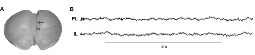
with confirmed electrode placements in PL and IL (Fig 2A) was included in the analysis.
2.4. Behavioural data analysis
The mean of freezing during two consecutive tones on Days 2 and 15 was used in the
statistical analysis. Spontaneous fear recovery was calculated by dividing mean freezing during the
first two tones on Day 15 by mean freezing during the last two tone-shock pairings on Day 1 and
expressed as a percentage. Contextual fear before extinction and extinction recall was determined by
assessing freezing before the first tone onset on Days 2 and 15. Freezing and spontaneous fear
recovery were expressed as the mean + SEM. Sex differences in tone-induced freezing during
conditioning, extinction and extinction recall were analyzed separately using two-way analysis of
variance (ANOVA). Sex differences in spontaneous fear recovery were analyzed using an unpaired
t-test. Differences in tone-induced freezing between extinction and extinction recall were analyzed
separately in males and females using two-way ANOVA. Sex differences in contextual fear before
extinction and extinction recall were analyzed using two-way ANOVA. Post-hoc comparisons were
conducted using Bonferroni’s testing. The significance level for all comparisons was set at P < 0.05.
2.5. In vivo electrophysiology data analysis
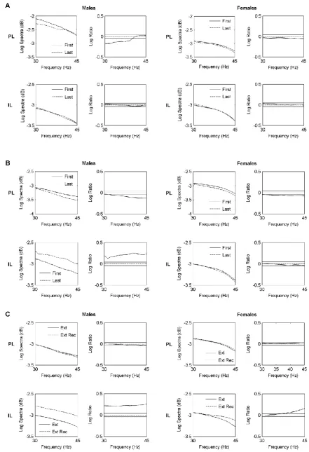
Using multi-taper spectral analysis [Fenton et al. 2013], spectral estimates of LFP activity in
PL and IL (Fig 2B) were generated during early and late extinction and extinction recall by taking the
mean of the first two and last two tones on Days 2 and 15 and pooling across males or females.
Similarly, spectral estimates of LFP activity in the 2 min periods before tone onset on Days 2 and 15
were generated and pooled across males or females. Differences in LFP power during early vs late
determined separately in males or females using the log ratio difference of spectra test [Diggle, 1990]
and quantified statistically using 95% confidence intervals [Stevenson et al. 2007, 2008; Fenton et al.
2013, 2014]. The LFP analysis was restricted to the 30-45 Hz band, which coincides with the lower
gamma frequencies recently implicated in fear extinction [Fitzgerald et al. 2014; Mueller et al. 2014].
3. Results
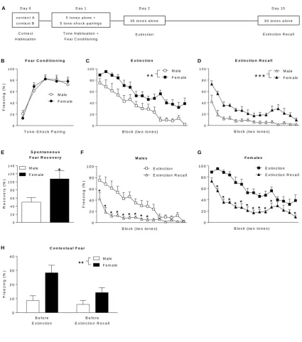
3.1. Females exhibit enhanced learned fear expression during extinction and extinction recall
Before presenting our mPFC gamma activity findings we first summarize the behavioural
results from our recent study [Fenton et al. 2014]. We found no differences in freezing between males
(n=9) and females (n=10) during the presentation of tones alone (data not shown) or tone-shock
pairings during fear conditioning (Fig 1B). Females did show significantly greater tone-induced
freezing during extinction (main effect of sex: F(1,17)=13.63, P<0.01; Fig 1C) and extinction recall
(main effect of sex: F(1,17)=27.41, P<0.001; Fig 1D), indicating that females showed enhanced learned
fear expression during extinction and its later recall. Females also showed enhanced spontaneous
recovery of fear over time after extinction, as indicated by a significantly increased percentage of fear
recovered during early extinction recall relative to late fear conditioning (t17=2.39, P<0.05; Fig 1E).
Despite this sex difference in spontaneous fear recovery both males (Fig 1F) and females (Fig 1G)
showed savings of extinction, as indicated by a significant decrease in tone-induced freezing during
extinction recall compared to extinction (males: day x block interaction: F(14,112)=5.89, P<0.0001;
Bonferroni’s post-hoc test, Blocks 1-9: P<0.05) (females: day x block interaction: F(14,126)=2.48,
P<0.001; Bonferroni’s post-hoc test: Blocks 2-11 and 15, P<0.05). Females also showed enhanced
contextual fear before extinction and its recall, as indicated by a significant increase in freezing during
the 2 min periods before tone presentations (main effect of sex: F(1,17)=8.70, P<0.01; Fig 1H).
3.2. Females show persistent PL gamma activation during extinction and a failure of IL gamma
activation during extinction retrieval
Changes in mPFC gamma activity during extinction are shown in Fig 3A. In males, gamma
activity in PL was significantly decreased during late compared to early extinction (P<0.05), whereas
activity in PL and IL, along with the low levels of PL-IL gamma synchrony that were observed
throughout behavioural testing (coherence<0.1; data not shown), suggest that volume conduction of
gamma oscillations between PL and IL was negligible. Females also showed no difference in gamma
activity in IL between late and early extinction. However, in contrast to males, PL gamma activity did
not change between late and early extinction in females. Sex differences were also observed in mPFC
gamma activity during extinction recall (Fig 3B). In males, gamma activity in PL was significantly
decreased during late compared to early extinction recall (P<0.05); conversely, IL gamma activity was
significantly increased during late compared to early extinction recall (P<0.05). Females also showed
a significant decrease in gamma activity in PL during late compared to early extinction recall
(P<0.05). However, there was no difference in IL gamma activity between late and early extinction
recall in females. Changes in mPFC gamma activity before extinction and extinction recall are shown
in Fig 3C. In males, there was no difference in gamma activity in PL before extinction recall
compared to before extinction, while IL gamma activity was significantly increased before extinction
recall compared to before extinction (P<0.05). Females also showed no difference in PL gamma
activity before extinction recall compared to before extinction, and a significant increase in gamma
activity in IL before extinction recall compared to before extinction (P<0.05).
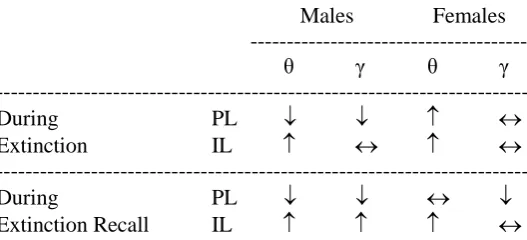
4. Discussion
We found similarities but also some notable differences between mPFC gamma and theta
activation before and during extinction and its recall in males. As with theta activity, we found that PL
gamma activity decreased during extinction and extinction recall and that gamma activity in IL
increased during extinction recall, in keeping with the known roles of PL and IL in learned fear
expression and extinction, respectively [Vidal-Gonzalez et al. 2006; Sierra-Mercado et al. 2011;
Fenton et al. 2014]. However, we found no change in gamma activity in IL during extinction, which is
different to the IL theta activation that we observed previously. This suggests that IL gamma
oscillations might be preferentially involved in extinction memory rather than learning. We also found
greater IL gamma activation before extinction recall compared to before extinction, which is similar
before extinction recall compared to before extinction, whereas previously we found less PL theta
activation before extinction recall compared to before extinction. This suggests that, in contrast to
theta activity, PL gamma activity may not be involved in the contextual regulation of learned fear.
We also found sex differences in mPFC gamma activation during extinction and extinction
recall. There was no change in PL gamma activation during extinction in females, which resembles
our theta activation findings. In contrast to theta activity, females showed a small but significant
decrease in PL gamma activity during extinction recall. As we observed in males, females showed no
change in IL gamma activation during extinction, which again contrasts with the increased theta
activation that we found previously. The most striking sex difference observed was related to IL
gamma activity during extinction recall. In contrast to the gamma activation in males, there was no
change in gamma activity in IL during extinction recall in females. This differs from the increase in
IL theta activity that we observed previously during extinction recall in females. We also found no sex
differences in PL or IL gamma activity before extinction recall compared to before extinction. Taken
together, these results indicate that, compared to males, females showed sustained PL gamma
activation during extinction and a failure of IL gamma activation during extinction recall.
Based on these new results and the emerging evidence implicating mPFC gamma oscillations
in learned fear expression and extinction [Fitzgerald et al. 2014; Mueller et al. 2014], it is worth
revisiting how we interpreted our behavioural results in light of our previous theta activity findings
[Fenton et al. 2014]. We hypothesized that females showed enhanced learned fear expression during
extinction and its recall as a result of impaired contextual regulation of extinction instead of resistance
to extinction. This idea was based largely on our observation that sex differences in learned fear
before and during extinction and extinction recall were associated with changes in PL, but not IL,
theta activation. It also fits with evidence indicating a role for PL in the contextual renewal of fear
after extinction [Orsini et al. 2011; Sharpe and Killcross 2015], together with known sex differences
in contextual fear processing [Maren et al. 1994; Gupta et al. 2001]. Our finding of enhanced
conditioned fear accompanied by sustained PL gamma activation during extinction in females adds to
evidence indicating an important role for PL activity in learned fear expression [Vidal-Gonzalez et al.
differences in fear extinction showed that enhanced gamma activation in PL is also associated with
impaired extinction [Fitzgerald et al. 2014]. Although initial inspection of our behavioural results
seems to agree with previous studies showing impaired fear extinction in females [Baran et al. 2009,
2010; Baker-Andresen et al. 2013], closer scrutiny of our data suggests that females exhibited intact
extinction encoding. Both males and females showed extinction savings and there were no sex
differences in IL activation during extinction. Females did exhibit enhanced spontaneous recovery of
fear after extinction and a failure of IL gamma activation during extinction recall. Interestingly, the
recall of fear extinction is linked to vmPFC gamma activation and unsuccessful extinction recall is
associated with failed gamma activation in this region in humans [Mueller et al. 2014]. This suggests
that female rats may have a specific impairment in the retrieval of extinction memory.
5. Conclusions
The present results confirm and extend our previous findings demonstrating sex differences in
learned fear expression and extinction involving altered theta and gamma oscillations in mPFC
(summarized in Table 1). Females showed enhanced conditioned fear and impaired extinction recall
associated with persistent PL activation and a failure of IL activation, respectively, although the
possibility remains that these sex differences may also involve changes in the contextual regulation of
fear extinction. These findings have potential translational relevance given that PTSD is more
prevalent in women, characterized by impaired inhibition of learned fear, and associated with mPFC
dysfunction [Milad et al. 2009; Shin et al. 2009; Glover et al. 2015; Jovanovic and Norrholm 2011;
Maeng and Milad 2015]. Further research is needed to determine the mechanisms underlying sex
differences in learned fear inhibition and mPFC activity. Local GABA interneurons in mPFC, which
regulate learned fear and its extinction, also modulate theta and gamma oscillations locally [Sangha et
al. 2012; Courtin et al. 2014a; Glykos et al. 2015]. Moreover, functional connectivity within the
neural circuitry mediating learned fear inhibition, which includes reciprocal projections between
mPFC, amygdala and hippocampus [Herry et al. 2010], involves synchronized activity at theta and
gamma frequencies [Lesting et al. 2011; Courtin et al. 2014b; Stujenske et al. 2014; Wang et al.
as hippocampus and amygdala function, have been reported [Maren et al. 1994; Gupta 2001; Blume et
al. 2013; Cholanian et al. 2014; Marron Fernandez de Velasco et al. 2015]. Determining the role of
gonadal hormones in mediating sex differences in the neural circuitry underpinning learned fear
inhibition is another key issue to investigate in the future, given the evidence indicating their
involvement in GABA signalling and neural circuit function during fear extinction [Zeidan et al.
2011; Merz et al. 2012; Cholanian et al. 2014; Rey et al. 2014; Mackenzie and Maguire, 2014].
Although we found sex differences in learned fear inhibition and mPFC gamma oscillations without
accounting for variations in the phase of the females’ estrous cycle, growing evidence indicates an
important role for estrogen in modulating fear extinction [Glover et al. 2015; Maeng and Milad 2015].
Acknowledgements
This research was supported by the Australian Research Council (DP1096148; TWB) and a
References
Baker-Andresen D, Flavell CR, Li X, Bredy TW. 2013. Activation of BDNF signaling prevents the
return of fear in female mice. Learn Mem 20:237-240.
Baran SE, Armstrong CE, Niren DC, Conrad CD. 2010. Prefrontal cortex lesions and sex differences
in fear extinction and perseveration. Learn Mem 17:267-278.
Baran SE, Armstrong CE, Niren DC, Hanna JJ, Conrad CD. 2009. Chronic stress and sex differences
on the recall of fear conditioning and extinction. Neurobiol Learn Mem 91:323-332.
Benchenane K, Tiesinga PH, Battaglia FP. 2011. Oscillations in the prefrontal cortex: a gateway to
memory and attention. Curr Opin Neurobiol 21:475-485.
Blume SR, Hetzel A, Rosenkranz JA. 2013. Sex differences in the basolateral amygdala. SFN
Abstract 673.01.
Cholanian M, Lobzova A, Das B, Yelleswarapu C, Donaldson ST. 2014. Digital holographic
microscopy discriminates sex differences in medial prefrontal cortex GABA neurons following
amphetamine sensitization. Pharmacol Biochem Behav 124:326-332.
Courtin J, Chaudun F, Rozeske RR, Karalis N, Gonzalez-Campo C, Wurtz H, Abdi A, Baufreton J,
Bienvenu TC, Herry C. 2014a. Prefrontal parvalbumin interneurons shape neuronal activity to drive
fear expression. Nature 505:92-96.
Courtin J, Karalis N, Gonzalez-Campo C, Wurtz H, Herry C. 2014b. Persistence of amygdala gamma
oscillations during extinction learning predicts spontaneous fear recovery. Neurobiol Learn Mem
113:82-89.
Diggle PJ. 1990. Spectral analysis. In: Diggle PJ, editor. Time series. A biostatistical introduction.
New York: Oxford University Press. p. 94–133.
Fenton GE, Pollard AK, Halliday DM, Mason R, Bredy TW, Stevenson CW. 2014. Persistent
prelimbic cortex activity contributes to enhanced learned fear expression in females. Learn Mem
21:55-60.
Fenton GE, Spicer CH, Halliday DM, Mason R, Stevenson CW. 2013. Basolateral amygdala activity
Fitzgerald PJ, Whittle N, Flynn SM, Graybeal C, Pinard CR, Gunduz-Cinar O, Kravitz AV,
Singewald N, Holmes A. 2014. Prefrontal single-unit firing associated with deficient extinction in
mice. Neurobiol Learn Mem 113:69-81.
Glover EM, Jovanovic T, Norrholm SD. 2015. Estrogen and extinction of fear memories: implications
for posttraumatic stress disorder treatment. Biol Psychiatry 78:178-185.
Glykos V, Whittington MA, LeBeau FE (2015) Subregional differences in the generation of fast
network oscillations in the rat medial prefrontal cortex (mPFC) in vitro. J Physiol 593:3597-3615.
Gupta RR, Sen S, Diepenhorst LL, Rudick CN, Maren S. 2001. Estrogen modulates sexually
dimorphic contextual fear conditioning and hippocampal long-term potentiation (LTP) in rats. Brain
Res 888:356-365.
Harris AZ, Gordon JA. 2015. Long-range neural synchrony in behavior. Annu Rev Neurosci
38:171-94.
Herry C, Ferraguti F, Singewald N, Letzkus JJ, Ehrlich I, Lüthi A. 2010. Neuronal circuits of fear
extinction. Eur J Neurosci 31:599-612.
Jovanovic T, Norrholm SD. 2011. Neural mechanisms of impaired fear inhibition in posttraumatic
stress disorder. Front Behav Neurosci 5:44.
Lesting J, Narayanan RT, Kluge C, Sangha S, Seidenbecher T, Pape HC. 2011. Patterns of coupled
theta activity in amygdala-hippocampal-prefrontal cortical circuits during fear extinction. PLoS One
6:e21714.
Linnman C, Zeidan MA, Furtak SC, Pitman RK, Quirk GJ, Milad MR. 2012. Resting amygdala and
medial prefrontal metabolism predicts functional activation of the fear extinction circuit. Am J
Psychiatry 169:415-423.
MacKenzie G, Maguire J. 2014. The role of ovarian hormone-derived neurosteroids on the regulation
of GABAA receptors in affective disorders. Psychopharmacology 231:3333-3342.
Maeng LY, Milad MR. 2015. Sex differences in anxiety disorders: Interactions between fear, stress,
Maren S, De Oca B, Fanselow MS. 1994. Sex differences in hippocampal long-term potentiation
(LTP) and Pavlovian fear conditioning in rats: positive correlation between LTP and contextual
learning. Brain Res 661:25-34.
Marron Fernandez de Velasco E, Hearing M, Xia Z, Victoria NC, Luján R, Wickman K. 2015. Sex
differences in GABABR-GIRK signaling in layer 5/6 pyramidal neurons of the mouse prelimbic
cortex. Neuropharmacology 95:353-360.
Merz CJ, Tabbert K, Schweckendiek J, Klucken T, Vaitl D, Stark R, Wolf OT. 2012. Neuronal
correlates of extinction learning are modulated by sex hormones. Soc Cogn Affect Neurosci
7:819-830.
Milad MR, Pitman RK, Ellis CB, Gold AL, Shin LM, Lasko NB, Zeidan MA, Handwerger K, Orr SP,
Rauch SL. 2009. Neurobiological basis of failure to recall extinction memory in posttraumatic stress
disorder. Biol Psychiatry 66:1075-1082.
Mueller EM, Panitz C, Hermann C, Pizzagalli DA. 2014. Prefrontal oscillations during recall of
conditioned and extinguished fear in humans. J Neurosci 34:7059-7066.
Orsini CA, Kim JH, Knapska E, Maren S. 2011. Hippocampal and prefrontal projections to the basal
amygdala mediate contextual regulation of fear after extinction. J Neurosci 31:17269-17277.
Rey CD, Lipps J, Shansky RM. 2014. Dopamine D1 receptor activation rescues extinction
impairments in low-estrogen female rats and induces cortical layer-specific activation changes in
prefrontal-amygdala circuits. Neuropsychopharmacology 39:1282-1289.
Sangha S, Ilenseer J, Sosulina L, Lesting J, Pape HC. 2012. Differential regulation of glutamic acid
decarboxylase gene expression after extinction of a recent memory vs. intermediate memory. Learn
Mem 19:194-200.
Sharpe M, Killcross S. 2015. The prelimbic cortex uses contextual cues to modulate responding
towards predictive stimuli during fear renewal. Neurobiol Learn Mem 118:20-29.
Shin LM, Lasko NB, Macklin ML, Karpf RD, Milad MR, Orr SP, Goetz JM, Fischman AJ,
Rauch SL, Pitman RK. 2009. Resting metabolic activity in the cingulate cortex and vulnerability to
Sierra-Mercado D, Padilla-Coreano N, Quirk GJ. 2011. Dissociable roles of prelimbic and infralimbic
cortices, ventral hippocampus, and basolateral amygdala in the expression and extinction of
conditioned fear. Neuropsychopharmacology 36:529-538.
Stevenson CW, Spicer CH, Mason R, Marsden CA. 2009. Early life programming of fear
conditioning and extinction in adult male rats. Behav Brain Res 205:505-510.
Stevenson CW, Halliday DM, Marsden CA, Mason R. 2008. Early life programming of hemispheric
lateralization and synchronization in the adult medial prefrontal cortex. Neuroscience 155:852-863.
Stevenson CW, Halliday DM, Marsden CA, Mason R. 2007. Systemic administration of the
benzodiazepine receptor partial inverse agonist FG-7142 disrupts corticolimbic network interactions.
Synapse 61:646-663.
Stujenske JM, Likhtik E, Topiwala MA, Gordon JA. 2014. Fear and safety engage competing patterns
of theta-gamma coupling in the basolateral amygdala. Neuron 83:919-933.
Vidal-Gonzalez I, Vidal-Gonzalez B, Rauch SL, Quirk GJ. 2006. Microstimulation reveals opposing
influences of prelimbic and infralimbic cortex on the expression of conditioned fear. Learn Mem
13:728-733.
Wang ME, Yuan RK, Keinath AT, Ramos Álvarez MM, Muzzio IA. 2015. Extinction of learned fear
induces hippocampal place cell remapping. J Neurosci 35:9122-9136.
Zeidan MA, Igoe SA, Linnman C, Vitalo A, Levine JB, Klibanski A, Goldstein JM, Milad MR. 2011.
Estradiol modulates medial prefrontal cortex and amygdala activity during fear extinction in women
Figure and Table Legends
Fig 1. Sex differences in learned fear before and during extinction and its later recall. (A) Schematic representation of the behavioural paradigm used. (B) There were no sex differences in freezing in
response to tone-shock pairings during auditory fear conditioning. Females showed greater freezing in
response to tone presentations during (C) extinction (**P<0.01) and (D) extinction recall
(***P<0.001). E) Females showed greater spontaneous fear recovery (i.e. freezing during early
extinction recall relative to late fear conditioning; *P<0.05). Compared to extinction, freezing during
tone presentations was decreased during extinction recall in (F) males (*P<0.05) and (G) females
(*P<0.05). (H) Females showed greater freezing before tone presentations during extinction and
extinction recall (**P<0.01) (figure adapted from [Fenton et al. 2014]).
Fig 2. (A) Representative example of electrode placements in PL and IL (indicated by the arrows). (B) Sample LFP traces recorded from PL and IL (figure adapted from [Fenton et al. 2014]).
Fig 3. Sex differences in mPFC gamma oscillations during extinction and its later recall. (A) Pooled gamma power spectra (left) and log ratio plots for pairwise comparisons of spectra (right) in PL (top)
and IL (bottom) during the first and last tone blocks during extinction in males and females. Solid
horizontal lines in the log ratio plots represent the upper and lower 95% confidence limits; positive
log ratio values indicate increased power during the last compared to the first tone block, whereas
negative values indicate decreased power during the last compared to the first tone block. In males,
PL gamma power decreased during extinction, whereas gamma power in PL showed no change
during extinction in females. There was no change in IL gamma power during extinction in males or
females. (B) Pooled gamma power spectra (left) and log ratio plots (right) in PL (top) and IL (bottom)
during the first and last tone blocks during extinction recall. Gamma power in PL decreased during
extinction recall in males and, to a lesser extent, in females. In contrast, whereas IL gamma power
increased during extinction recall in males, there was no change in gamma power in IL during
extinction recall in females. (C) Pooled gamma power spectra (left) and log ratio plots (right) in PL
(top) and IL (bottom) before tone presentations during extinction (Ext) and extinction recall (Ext
Rec). Positive log ratio values indicate increased power before extinction recall compared to before
before extinction. There was no change in PL gamma power before extinction recall compared to
before extinction in males or females. Gamma power in IL increased before extinction recall
compared to before extinction in males and, to a lesser extent, in females.
Figure 1.
F e a r C o n d it i o n i n g
0 2 0 4 0 6 0 8 0 1 0 0
F re e z in g ( % )
T o n e - S h o c k P a ir in g M a le F e m a le
E x t in c t io n
0 2 0 4 0 6 0 8 0
1 0 0 M a le
F e m a le * *
B lo c k ( tw o to n e s )
E x t in c t io n R e c a ll
0 2 0 4 0 6 0 8 0 1 0 0
M a le F e m a le * * *
B lo c k ( tw o to n e s )
S p o n t a n e o u s F e a r R e c o v e r y
0 2 0 4 0 6 0 8 0 1 0 0 1 2 0 1 4 0
R e c o v e ry ( % )
M a le F e m a le *
M a le s
0 2 0 4 0 6 0 8 0
1 0 0 E x tin c tio n E x tin c tio n R e c a ll
F re e z in g ( % ) * * * * * * * * *
B lo c k ( tw o to n e s )
F e m a le s
0 2 0 4 0 6 0 8 0 1 0 0
E x tin c tio n E x tin c tio n R e c a ll
* * * * * * * * * * *
B lo c k ( tw o to n e s )
B C D
E F G
A D a y 0
C o n te x t H a b itu a tio n
c o n t e x t A c o n te x t B
D a y 1 5 t o n e s a lo n e + 5 to n e -s h o c k p a ir in g s
T o n e H a b itu a tio n + F e a r C o n d itio n in g
D a y 2
3 0 to n e s a lo n e
E x tin c tio n
D a y 1 5
3 0 to n e s a lo n e
E x tin c tio n R e c a ll
H C o n t e x t u a l F e a r
B e fo r e E x tin c tio n
B e fo r e E x tin c tio n R e c a ll 0
1 0 2 0 3 0
4 0 M a le
F e m a le
Table 1.
Males Females
---
θ γ θ γ
---
During PL
Extinction IL
---
During PL
Extinction Recall IL