ISSN Print: 2161-7325
DOI: 10.4236/ojpsych.2018.83023 Jul. 24, 2018 263 Open Journal of Psychiatry
Autism: A Different Vision
Golder N. Wilson
*, Vijay S. Tonk
Department of Pediatrics, Texas Tech University Health Science Centers, Lubbock, USA
Abstract
Genomic analysis has emphasized the enormous genetic contribution to aut-ism spectrum disorders, with over 80% of patients having changes demonstr-able by high resolution chromosome (microarray) analysis or whole exome sequencing. An overview of these genetic changes demonstrates the expected role of synaptic transmission in autism and, together with clinical observa-tions, emphasizes the importance of visual input on developing sensory sys-tems and social responses. Neonatal recognition of autism predisposition through genetic analysis could allow sensory stimulation therapies during pe-riods of neuroplasticity, an approach analogous to strabismus correction be-fore the cortical dissociation of the deviant eye.
Keywords
Autism, Autism Spectrum Disorders, Genomics, Array-Comparative Genomic Hybridization, Microarray Analysis, Whole Exome Sequencing, Synaptic Transmission
1. Introduction
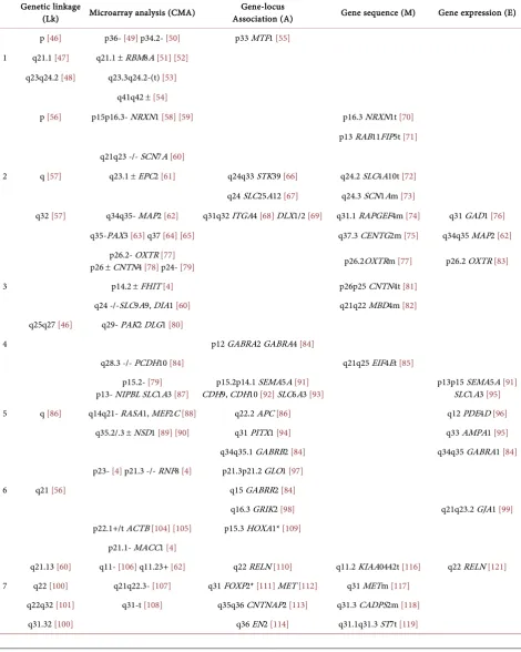
Recent advances in genetics and neuroscience, when focused by the appropriate clinical prism, reveal a glimpse of gold beneath the multicolored autism spec-trum [1]. A profusion of low-frequency genetic changes [2] [3] [4] ends the dark of vaccine myths [5] and heralds a sunrise of early screening for autism suscep-tibility. The new challenge is to integrate highlighted genes with neurodevelop-mental pathways, achieving early diagnosis and remodeling of neural architec-ture for the 1% of children who will become autistic [6]. This review updates autism genomics with an organizing hypothesis: Autism is an emergent disorder that reflects inborn errors of the sensory nervous system. We will catalogue ge-netic changes and their roles in neural patterning or synapse transmission (Table 1 and Table 2), examine environmental influences that could interact
How to cite this paper: Wilson, G.N. Tonk, V.S. (2018) Autism: A Different Vision. Open Journal of Psychiatry, 8, 263-296.
https://doi.org/10.4236/ojpsych.2018.83023
Received: February 24, 2018 Accepted: July 21, 2018 Published: July 24, 2018
Copyright © 2018 by authors and Scientific Research Publishing Inc. This work is licensed under the Creative Commons Attribution International License (CC BY 4.0).
DOI: 10.4236/ojpsych.2018.83023 264 Open Journal of Psychiatry with these genomic changes (Table 3), emphasize that timely stimulation thera-pies could remodel plastic neural pathways, and finish with an old medical dic-tum: Study the patient.
Table 1. Genetic alterations in patients with autism and without recognized genetic disorders.
Genetic linkage
(Lk) Microarray analysis (CMA) Association (A) Gene-locus Gene sequence (M) Gene expression (E) p [46] p36- [49] p34.2- [50] p33 MTF1 [55]
1 q21.1 [47] q21.1 ± RBM8A [51] [52]
q23q24.2 [48] q23.3q24.2-(t) [53]
q41q42 ±[54]
p [56] p15p16.3- NRXN1 [58] [59] p16.3 NRXN1t [70]
p13 RAB11FIP5t [71]
q21q23 -/- SCN7A [60]
2 q [57] q23.1 ±EPC2 [61] q24q33 STK39 [66] q24.2 SLC4A10t [72]
q24 SLC25A12 [67] q24.3 SCN1Am [73]
q32 [57] q34q35- MAP2 [62] q31q32 ITGA4 [68] DLX1/2 [69] q31.1 RAPGEF4m [74] q31 GAD1 [76]
q35-PAX3 [63] q37 [64] [65] q37.3 CENTG2m [75] q34q35 MAP2 [62]
p26.2- OXTR [77]
p26 ± CNTN4 [78] p24- [79] p26.2OXTRm [77] p26.2 OXTR [83]
3 p14.2 ± FHIT [4] p26p25 CNTN4t [81]
q24 -/-SLC9A9, DIA1 [60] q21q22 MBD4m [82]
q25q27 [46] q29- PAK2 DLG1 [80]
4 p12 GABRA2 GABRA4 [84]
q28.3 -/- PCDH10 [84] q21q25 EIF4Et [85]
p15.2- [79]
p13- NIPBL SLC1A3 [87] CDH9, CDH10 p15.2p14.1 SEMA5A [92] SLC6A3 [91][93] p13p15 SEMA5A SLC1A3 [95][91]
5 q [86] q14q21- RASA1, MEF2C [88] q22.2 APC [86] q12 PDE4D [96]
q35.2/.3 ± NSD1 [89] [90] q31 PITX1 [94] q33 AMPA1 [95]
q34q35.1 GABRB2 [84] q34q35 GABRA1 [84]
p23- [4] p21.3 -/- RNF8 [4] p21.3p21.2 GLO1 [97]
6 q21 [56] q15 GABRR2 [84]
q16.3 GRIK2 [98] q21q23.2 GJA1 [99]
p22.1+/t ACTB [104] [105] p15.3 HOXA1* [109]
p21.1- MACC1 [4]
q21.13 [60] q11- [106] q11.23+ [62] q22 RELN [110] q11.2 KIAA0442t [116] q22 RELN [121]
7 q22 [100] q21q22.3- [107] q31 FOXP2* [111] MET [112] q31 METm [117]
q22q32 [101] q31-t [108] q35q36 CNTNAP2 [113] q31.3 CADPS2m [118]
DOI: 10.4236/ojpsych.2018.83023 265 Open Journal of Psychiatry Continued
q31q34 [102] q31/q31.2 WNT2* [115] q32 SSBP1 T2R3t [120]
q35q36 [103]
8 p23.1- MCPH1 [122] [123] q21.13 FABP5 (FABP7) [125] q21MMP16t [117]
q22.1q23- [124] q23 CSMD3t [124]
9 p13.1 [60]
q34.3 [60] q34.3 GRIN [95]
p12p11- WAC [126] p11.23 GAD2 [131]
10 q11.2- CHAT SLC18A3 [127] q11.1TRIP8 REEP3t [129]
q22q23- [128] q23.31 PTENm [130]
11 p12p13 [60] p12p13- [4] p13 BDNF [132] p15.5 SCTm [133]
12 q14.2 [134] p13.33/.32- CACNA1C [135] q14q15 AVPR1A [136] p13.3 CACNA1Cm [137]
13 q12.2 [60] q13.2 NBEAt [139]
q14.2q14.1 [138] q14.2q14.1- [4]
14 q11.2- CHD8 [4] [140]
q11q13 [141] q11.2 ± CYFIP1, NIPA1 [142] q11.2q12 GABRB3* GABRG3 [149]
15 q11q13+ UBE3A [60] [143]
q13.2 ± CHRNA7 [4] [144] q11q13 UBE3A [150] q13.1 APBA2m [151]
q22- PTPN9 [145] q24- [146]
q25.2 [147] q26qter- [148]
p13 [152] p11.2 ± SH2B [3] [4] [153] p11.2 PRKCB1 [158] p11.2 PRKCB1 [158]
16 p13.1 ± NDE1 [4] [154] p13 GRIN2A ABAT [159] p13.3 A2BP1t [160]
q23.2- CMIP [155]
q24.2/.3- [156] [157]
p13.1- [162] p13.3+ [163]
17 q11 [60] p11.2 ± NF1 [164] [165] q11.1q12 SLC6A4 [167] q11.1q12 SLC6A4m [169]
q21 [161] q12- [166] q21.32 ITGB3 [168]
18 q12-t [170] q11.2q12 AQP4 [171]
19 p [172] p13.13/.12- AKAP8 [173]
20 p13- [4]
21 p13q11 [174] q21 NCAM2 q21.1q21.3- [176][175]
22 q11.2 ±[177] q13.1 ADSLm [179]
q13.3- SHANK3 [4] [178] q13.33 SHANK3m [180]
p22.33- NLGN4 [181] p22.33 NLGN4m [188]
p22.2p22.3 ± STS, NLGN4,
VCX cluster [182] p21.3 IL1RAPL1mt [189]
X p22.12+ RPS6KA3 [183] p11.23 MAOA [187] p21.3 ARXm [190]
DOI: 10.4236/ojpsych.2018.83023 266 Open Journal of Psychiatry Continued
q12q13.3+ [185] q24q26 UPF3Bm [192]
q13q21+ [186] q28 FMR2m [193]
q28+ [194] q28 MECP2m [194] [195]
Y q11.2 NLGN4Ym [196]
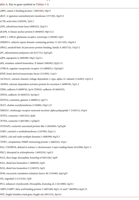
[image:4.595.60.543.249.745.2]Genetic loci are listed as p for chromosome short arm, q for long arm, numbers for bands, gene symbols as in Online Mendelian Inheritance in Man (http://www.omim.org/) referenced with OMIM numbers in the appended Table A; linkage includes traditional linkage studies focused on one locus and whole genome association linkage studies that analyze as many as 200 loci at once; microarray analysis loci defined by + for microduplication, - for micro-deletion, -/- for homozygous micromicro-deletion, -t for translocation and microdeletion; gene locus-association, allele association study with specific genes indi-cated (*when negative association studies also reported); gene sequence studies show m for mutation, t for a gene disrupted by translocation; gene expres-sion involves measurement of RNA or protein species, often in brain.
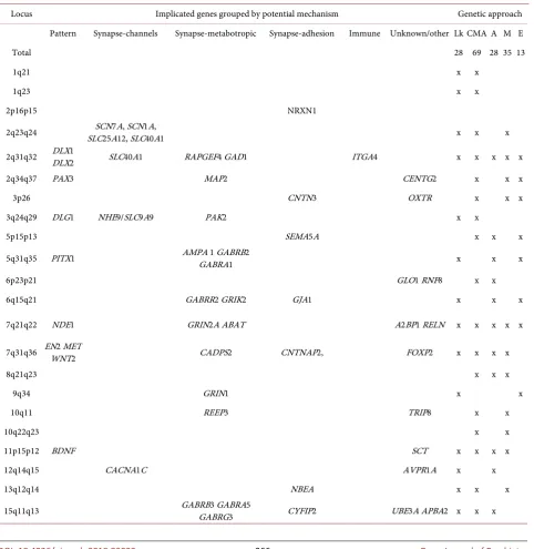
Table 2. Summary of loci implicated in autism.
Locus Implicated genes grouped by potential mechanism Genetic approach
Pattern Synapse-channels Synapse-metabotropic Synapse-adhesion Immune Unknown/other Lk CMA A M E
Total 28 69 28 35 13
1q21 x x
1q23 x x
2p16p15 NRXN1
2q23q24 SLC25A12, SLC40A1 SCN7A, SCN1A, x x x
2q31q32 DLX1 DLX2 SLC40A1 RAPGEF4 GAD1 ITGA4 x x x x x
2q34q37 PAX3 MAP2 CENTG2 x x x
3p26 CNTN3 OXTR x x x
3q24q29 DLG1 NHE9/SLC9A9 PAK2 x x
5p15p13 SEMA5A x x x
5q31q35 PITX1 AMPA 1 GABRB2 GABRA1 x x x
6p23p21 GLO1 RNF8 x x
6q15q21 GABRR2 GRIK2 GJA1 x x x
7q21q22 NDE1 GRIN2A ABAT A2BP1 RELN x x x x x
7q31q36 EN2 MET WNT2 CADPS2 CNTNAP2, FOXP2 x x x x
8q21q23 x x x
9q34 GRIN1 x x
10q11 REEP3 TRIP8 x x
10q22q23 x x
11p15p12 BDNF SCT x x x x
12q14q15 CACNA1C AVPR1A x x
13q12q14 NBEA x x x
DOI: 10.4236/ojpsych.2018.83023 267 Open Journal of Psychiatry Continued
16p13p11 PRKCB1 x x x x x
17q11q12 RAI1 SLC6A4 x x x
17q21 ITGB3 x x
18q12 AQP4 x x
22q11q13 SHANK3 ADSL x x
Xp22 NLGN4 IL1IRAPL1 VCX x x
Xp11 WNK3 MAOA PHF8 x x
Xq13 NLGN3 x x
Loci implicated by 2 genetic approaches are taken from Table 1, loci and gene symbols explained in the appended Table A with numbers assigned in Online
Mendelian Inheritance in Man (http://www.omim.org/). Loci implicated by all 5 genetic approaches are highlighted in dark grey, those with 3 - 4 in light
[image:5.595.59.534.312.566.2]gray. Genetic approaches include Lk, whole genome linkage/association; CMA, aCGH-microarray analysis; A, association; M, mutation defined by DNA sequencing; E, expression studies (usually in brain).
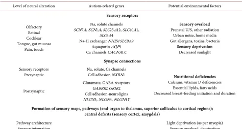
Table 3. Potential environmental factors highlighted by autism-related genes.
Level of neural alteration Autism-related genes Potential environmental factors Sensory receptors
Olfactory Retinal Cochlear Tongue, gut mucosa
Pain, touch
Na, solute channels SCN7A, SCN1A, SLC25A12, SLC40A1,
SLC6A4
Na-H exchanger NHE9/SLC9A9 Aquaporin AQP4 Ca channels CACNA1C
Sensory overload Prenatal U/S, other radiation
Urban noise, home media Gut allergens, toxins, bacteria
Sensory deprivation Decreased sunlight Synapse connections
Sensory receptors
Presynaptic Na, solute, Ca channels Cell adhesion NXRN1 Nutritional deficiencies Calcium, vitamin D deficiencies
Essential lipids, fatty acids
Decreased breast-feeding initiation and duration Postsynaptic
Glutamate, GABA receptors GABRR2 GRIK2 Cell adhesion-neuroligins NLGN3, NLGN4, NLGN4Y
Formation of sensory maps, pathways (end-organ to thalamus, superior colliculus to cortical regions); central deficits (sensory cortex, amygdala)
Pathway architecture
Sensory integration Light deprivation (as per myopia) Sensory overload, deprivation
Gut-brain connections Interleukin receptor IL1IRAPL1 Integrins ITGA4 ITGB3 Immune/inflammatory factors Rare vaccine reactions
Neuroendocrine Thyroid receptor TRIP, Secretin SCT Maternal hypothyroidism, diabetes Parental/social interaction (infant
touch, gaze stimulation) Oxytocin receptor OXTR Community, family deficiencies Maternal depression/drug use Gene symbols are defined in appended Table A.
spi-DOI: 10.4236/ojpsych.2018.83023 268 Open Journal of Psychiatry rit versus that of substance: “[The advent of pathology] … meant that the rela-tion between visible and invisible—which is necessary to all concrete know-ledge—changed its structure, revealing by gaze and language what had pre-viously been below and beyond their domain.”
There is a parallel between the flawed gaze and interaction of autistic children and the searching gaze of their students, striving to merge mute with savant in a language of mind and molecules.
2. The Rationale for Sensory Deficits
Frequent encounters with patients having autism and early visual deficits, sup-ported by research demonstrating face perception changes [12] emphasize vision as a key problem in autism [13] [14] [15]. Certainly vision is pivotal for child development, perhaps foreshadowed by its catalysis of novelty during the Cam-brian explosion [16], and recapitulated when a child’s poor eye contact forecasts altered communication and social interaction. The optic nerve does contain 38% of cranial nerve fibers [17] but broader neurosensory disruption is suggested by the hypersensitivities of hearing or touch that often precede an autism diagnosis
[18].
The many disorders with neurosensory deficits and autism include mixed hearing loss, strabismus, cataracts, or nystagmus in Down, Prader-Willi, Wil-liams, and other chromosomal syndromes [7] [8] [19]. Sensory compensation may be evidenced by hyperacusis in Williams syndrome and by photophobia in Smith-Lemli-Opitz syndrome [20]. Vision impairment and autism are notable in aniridia, Leber amaurosis, Mobius syndrome, or thalidomide embryopathy [15]
and, with some controversy about autism diagnosis, in the congenitally blind (10% - 15% autism prevalence) [21] [22] or deaf (7% autism prevalence with re-ciprocal frequencies of 7.9% - 18.5% mild/moderate and 3.5% profound hearing loss in autistic children) [23]. Congenitally blind children also have delayed ma-ternal attachment, articulation problems, idiosyncratic language (“verbalisms”), stereotypy (repetitive movements or “blindisms”), and exploration of space with their hands (haptic perception) that mimic symptoms of autism [21].
DOI: 10.4236/ojpsych.2018.83023 269 Open Journal of Psychiatry
3. Neurosensory Measures Can Refine Autism Classification
and Provide Earlier Diagnosis of Susceptibility
The classic autism triad described by Kanner in 1943 consisted of abnormal ver-bal/nonverbal communication, abnormal social interaction, and repetitive movements or routines, expanded a year later by Asperger to include social im-maturity, eccentric behaviors, and restricted interests in children with normal cognitive function [31]. The DSM IV grouped several autism conditions with Rett syndrome in a pervasive developmental disorder (PDD) category, a mis-match improved by DSM V that also replaced Asperger disorder with the de-scription of high-functioning autism [32] [33]. Insightful clinical delineation should separate patients with autism and global intellectual disability (ID) like those with Rett or fragile X syndromes [34] [35] from those with selective defi-cits (perhaps denoted as pure or primary autism). Defining essential autism pathways would allow explanation of its significant frequency in almost any dis-order with ID, just as the focus on young people with heart attacks defined ge-netic hypercholesterolemia [36] and explained its occurrence in diabetes melli-tus.
Autism diagnosis is currently based on subjective observation and parental questionnaires, with intentions to bring diagnosis from the standard age of 3 - 4 years to 18 months [31]. Novel neurophysiologic and neuro-imaging techniques
[27] [37] could provide objective diagnosis much earlier, defining a new catego-ry of selective developmental disorders (SDD) based on subtle neurosensocatego-ry deficits. Eye-tracking technologies reveal different gaze behaviors at age six months [38] illustrating a potential for early functional diagnosis heralded by genomic screening. This would be followed by remodeling of plastic neural pathways [37] [39] by stimulation therapies analogous to the eye patch for stra-bismus [40] or the cochlear implant for deafness [41]. A more definitive classifi-cation would include the timing of autistic symptoms, extent of ID, types of neurosensory deficits, and underlying disorders as presently known or newly de-fined.
4. The New Genomics of Autism
DOI: 10.4236/ojpsych.2018.83023 270 Open Journal of Psychiatry Table 1 presents a detailed list of chromosome regions and/or gene sequences highlighted in patients diagnosed with autism who do not have recognized con-ditions like Rett or fragile X syndromes [35]; gene symbols in Table 1 and Table 2 are explained in the appended Table A. Placing the autism diagnosis first, then the genetic finding avoids controversy about whether disorders like fragile X cause behaviors that merit a typical autism diagnosis [19]. Chromosome loci and genes are ordered by chromosome position (p for short arm, q for long arm, numbers reflecting bands) and partitioned in five columns based on the tech-nology employed—first by genetic linkage that is now accelerated by whole ge-nome association studies [43], second by microarray analysis/CMA [2] [3] [4], third by association with particular genes in the manner of the HLA B27 marker with ankylosing spondylitis [43], fourth and most convincing by showing muta-tions or translocamuta-tions that disrupt particular genes [44], and fifth by showing altered gene expression [44]. As reviewed previously [2] and summarized in Ta-ble 2, Table 1 lists 69 genomic regions solidly implicated in autism by recurring microduplications or microdeletions detected by microarray analysis and another 35 documented by gene sequence mutation or disruption (transloca-tion), supporting the polygenic inheritance implied by population studies. Less definitive are the loci implicated in autism by linkage (28 loci), association (28 loci), or expression studies (13 loci), the latter encompassing the sparsely ex-plored domain of epigenetic influence [34].
The left column of Table 2 lists loci implicated by two or more genetic ap-proaches, drawn from the detailed genetic changes listed in Table 1. Genes within the implicated regions listed in columns related to their potential influ-ence on brain development and function, their symbols defined in appended
Table A. Table 2 lists 30 loci implicated by at least two linkage, genomic, or ex-pression techniques with 3 regions (2q31q32, 7q21q22, and 16p11p13) hig-hlighted by all 5 genetic approaches and others (2q, 3p, 5q, 7q, 15q, 16p, 17q, and X) implicated by several. Genes within these susceptibility domains can be grouped by their potential regulation of early pattern, synaptogenesis, or other functions, discussed below from the perspective of neurosensory development.
Early
pattern
genes.
Correlating with sensory importance [16] is theout-side-in development of the nervous system, beginning with dorsal ectoderm that becomes midline neural plate with flanking neural folds and neural crest. Poten-tial neural patterning genes include
PAX
3 (paired-box gene 3),PITX
1 (paired-like homeodomain transcription factor-1),EN
2 (engrailed-2),WNT
2 (wingless-type MMTV integration site family, member 2), and theMET
pro-to-oncogene among others in Table 2.PAX
3 murine [197] andWNT
3 avianDOI: 10.4236/ojpsych.2018.83023 271 Open Journal of Psychiatry before the anterior neuropore is closed at 25 days post-conception. Segmenta-tion into fore- and hind-brain segments involves the sonic hedgehog
SHH
gene that is deleted in some cases of holoprosencephaly malformation [201] and sig-nal molecules in theHOXA
2, bone morphogenetic protein (BMP
), and hedge-hog families guide dorsoventral differentiation of the spinal cord, telencephalon, and hypothalamus [202]. TheSHH
,BMP
, andWNT
genes all have roles in pat-terning the cerebral cortex or pallium [201] [202].Cephalic proliferation of the dorsal neural tube and the embryonic head fold bring dorsolateral optic and otic vesicles to their respective adult anterior and lateral positions. As the otic vesicles migrate ventrally, the branchial arches/pha- ryngeal grooves ascend anteriorly and reach toward olfactory and cochlear or-gans with mouth and ear canals. While the optic cups induce surface ectoderm to form the surface structures of vision (cornea, iris, lens), the olfactory nerves extend to cluster at the nasal cribiform plate near palate and tongue. The sensory organs of sight, smell, hearing, and taste all derive from neurectoderm and pro-duce analogous neuron patterns, each with unique metabotropic receptors [37]
that extend sensory maps from receptor to tract to processing center to sensory cortex [203] [204] [205] [206], each with unique connections that will be custo-mized by experience-directed neural activity [37] [39] [203] [204] [205] [206].
Concordant molding of cerebral and optic pattern is illustrated by the sin-gle-eyed cyclops malformation that reflects underlying holoprosencephaly, an anomaly caused by mutations in the hedgehog pathway or by defective choles-terol synthesis in Smith-Lemli-Opitz syndrome with its frequent autism [20] [201]. The cholesterol moieties required for
SHH
action [201], like folic acid prevention of neural tube defects and the fetal brain anomalies with maternal diabetes, establish links between nutrients and brain development analogous to the enhancement of visual acuity and cognitive outcomes by essential fatty acids[207]. Cephalic enlargement relates to other genes listed in Table 1—
NDE
1 in-teracts withDISC
1 (deleted in schizophrenia) to increased cortex gyral differen-tiation and size [208] while mice withPTEN
mutations have macrocephaly[209]; humans with the latter mutations can manifest macrocephaly and autism
[210].
5. Synapse and Connectivity Genes
The human brain contains over 100 trillion synapse units that are organized by morphogen, guidance, and cell adhesion molecules to produce highly specific neural connections and pathways [37] [204]. Neurons employ successive depo-larization of Na/K chloride voltage-gated channels to jump synapses electrically or release neurotransmitters that trigger responses on the post-synaptic mem-brane [37]. Genes from Table 1 and Table 2 that could regulate sensory receptor activation and synapse transmission include those encoding channel proteins
SLC
40A
1,SCN7A
,SCN
1A
,SLC
25A
12,SLC
40A
1NHE
9/SLC
9A
9 (SCN
forDOI: 10.4236/ojpsych.2018.83023 272 Open Journal of Psychiatry 4). Others encode the metabotropic receptors/regulators
GRIN
1,GRIN
2A
(glu-tamate receptors), theGABRA
1,GABRR
2,GABRB
3,GABRA
5, andGABRG
3 gamma-aminobutyric acid receptors, and the calcium-dependent activator pro-teinCADPS
2. Several encode cell adhesion molecules includingNRXN
1 (neu-rexin 1),CNTN
3 andCNTNAP
2 (contactin or contactin-associated proteins),GJA
1 (gap junction alpha-1/connexin 43),SEMA
5A
(semaphorin 5A),SHANK
3 (SH3 and multiple ankyrin repeat domains 3), and the neuroliginsNLGN
4 andNLGN
3.Autism often involves altered connectivity [26] [35] but the challenge is to as-sociate specific synaptic pathways with specific disorders and molecular deficits, e. g., the autism-associated neuroligin mutation that depletes its protein at neu-ronal surfaces [211]. Certain neurotransmitters like GABA are not only message but medium, playing structural roles in synapse maturation and stabilization. Down-regulation of the GABA-A alpha3 subunit decreases the number of GA-BAergic inhibitory synapses with mismatched synapse formation [212] while abnormal clustering of neuroligin-2 was observed when postsynaptic mem-branes were deprived of GABA-A receptors [213].
In
vivo
techniques [27] [37]should allow localization of altered receptors and synapse transmission in autis-tic patients.
6. Exploring Stimulation Therapies
As correlation of sensory pathways with particular synaptic molecules progresses, knowledge of experience-directed sculpting of these pathways is also expanding. Modulation of neuron clusters called barrels in the sensory cortex of rats can be achieved by ablation or stimulation of particular snout whiskers, and the plasticity of these barrel maps documented by
in
vivo
photon or fluorescent imaging rather than postmortem histology [204]. The sculpting of sensory cor-tex maps in response to passive or training inputs is achieved by rapid long-term potentiation or depression of excitatory (NMDA/glutamate) and inhibitory (GABA) synaptic circuits as well as by slower rearrangement of synaptic connec-tions [204] [205] [206]—processes involving genes that are well-represented inTable 1. A critical junction of change seems to be the post-synaptic dendritic spines, elements of excitatory sensory synapses that enlarge or contact in con-junction with synapse electrical activity [204]. The dependence of oriented cor-tex networks on sensory stimuli can be demonstrated by exposing kittens to one visual stimulus orientation or by connecting developing ferret retina to their au-ditory cortex; the kittens demonstrate a customized visual cortex and the ferrets an auditory cortex patterned by visual stimuli [204].
simpli-DOI: 10.4236/ojpsych.2018.83023 273 Open Journal of Psychiatry fies storage in memory [215]. Moreover, the ability of visual stimuli to elicit re-producible, topographically unique fMRI patterns in the human visual cortex required conscious recognition of the stimulus as a face or house; control images that exploited binocular fusion to obliterate image recognition elicited non- specific fMRI activity in face-sensitive visual areas [216]. Completing the loop is a modification of visual perception by prior, experience-sculpted activity pat-terns—“predictive codes” that anticipate and facilitate recognition of familiar stimuli [217]. Such experiments preview neurodevelopmental cycles—input to perception to memory/experience to tailored perception—that would progres-sively mold the sensory processing maps used for language response and social interaction.
Developmental, experience-directed sculpting of sensory pathways is also demonstrated by perception of facial emotional expressions [218] and individu-als with autism do show alterations in face and face emotion perception [12] [219] [220] with the expected changes in cortical connectivity [221] [222]. Changes in auditory processing and pitch discrimination [24] [25] have been demonstrated, along with impaired perception of linguistic and social auditory stimuli that may relate to song practice and, in birds, the FOXP2 gene [223]. Subtle auditory processing deficits could relate to noise sensitivities and musical savant tendencies in Williams syndrome and other disorders with frequent aut-ism symptoms [223] [224] [225] [226].
Mirror neuron alterations have been claimed [224] or denied [225] in autism, but such changes, guided by the sensorimotor cortex [226], could certainly lead to altered voice inflection, language idiosyncracies, fine motor differences, and production of repetitive movements by sensorimotor disconnection. The smaller cerebellum [227] that stands out among cortex and amygdale volume differences in autism [27] may seem to contradict the hypothesis of sensory deficits unless coordinated development of sensory and motor pathways is recognized [228].
The preceding information shows how congenital deficits in detecting or processing sensory inputs would have cumulative effects, sometimes exacerbated by environmental factors, and emerge as the cardinal communication and social impairments of autism. A clear research pathway would combine genetic screening for autism susceptibility with neurosensory measures to document early sensory/perception deficits. Novel stimulation therapies to promote genesis of face or voice recognition pathways, not to be confused with discredited ocular therapies [229], would be employed before critical periods of neuroplasticity have expired. The occasional successes of sensory stimulation [18] or Applied Behavior Analysis/ABA [31] therapies may foreshadow more targeted strategies that make primary autism as treatable as strabismus [40] or deafness [41]; these approaches could substantially improve function in autistic children with global ID.
7. Genomic Guides to Environmental Factors
influ-DOI: 10.4236/ojpsych.2018.83023 274 Open Journal of Psychiatry ence, approached through the framework of gene-environmental interactions and pharmacogenomics (Table 3). Sensory injury by overwhelming inputs could implicate increasing use of fetal ultrasound [230], higher radiation exposure
[231] or urban noise. Reduced sensory inputs could implicate reduced exposure to outdoor light as suggested for myopia [232] and proven for vitamin D defi-ciency [233]. These factors highlight several calcium-dependent genes from Ta-ble 1 (the
CACNA
1C
calcium channel, theCADPS
2 calcium-dependent activa-tor protein for secretion, and several neurotransmitter or adhesion-related genes). Deficits in gut sensation from smell/taste in the upper tract to mucosal receptors in the lower tract could explain early feeding and gastrointestinal problems that stoked fears of vaccine injuries in autism [5] [234] a “gut-brain” connection or immune pathogenesis [34] [235] involving nutrient deficiencies, food allergens, and toxins could be examined using candidate genes likeITGB
3 (integrin beta-3) orIL
1IRAPL
1 (interleukin1 receptor accessory protein-like, calcium-related) from Table 1. However, recognition of the low frequency of any single causal factor should restrict trial of alternative therapies/elimination diets to those with the relevant genetic changes.A complex area of environmental interaction concerns abnormal socialization as a core symptom of autism. Social deficits may relate to a lack of joint atten-tion—the parallel processing of information about one’s own and other people’s visual attention—that begins developing at 4 - 6 months of infancy [236] [237]. Joint attention is the beginning of self-other perception, and it may be depen-dent on early social visual pursuit that can be measured by eye-tracking [27] [237] Although most would reject Bettleheim’s emphasis on “refrigerator moth-ers” [234], coincidence of loci for schizophrenia and autism including the cad-herin pathway [9] could underlie altered maternal-infant bonding and decreased infant stimulation. Altered face processing in parents of autistic children [238]
and trends toward isolated family units and single parents could be additional factors that combine with genetic predisposition to increase autism prevalence.
8. Study the Patient
Now that whole genome techniques have defined chromosome regions and genes associated with autism as a broad phenotype, correlation of genetic changes with scrupulously defined patients is needed [239]. Trends toward pro-prietary genomic databases should be reversed so that benign CNVs and muta-tions can be distinguished from those related to autism, with or without global ID. The contribution of individual genes within aneuploid segments, easily iden-tified through appropriate genome browsers [44], will likely require CSF RNA/protein expression studies [235]. These gaps in genomic knowledge em-phasize that prenatal genetic screening should target autism as susceptibility ra-ther than disease, coupled with novel ra-therapies modeled by early enzyme sup-plementation in cystic fibrosis [240].
DOI: 10.4236/ojpsych.2018.83023 275 Open Journal of Psychiatry perhaps the greatest asset for future research. Autism registries should be viewed as dynamic resources where calibrated natural histories, morphologic changes, and behavioral symptoms are regrouped using the latest neurogenetic tech-niques. Particularly important is the single case, for patients are the elements of medicine and case presentations its essence, case reports [88] pure cultures compared to the mixed flora of group studies. Among the large registries may be some striking child who reveals a key scotoma of autism like the amnesiac H. M.
[37] did for memory and the lobectomized Phineas Gage did for behavior [241]. As the geneticist Richard Goldschmidt stated [242]:
“Progress in biology is derived from cooperation of observation, experiment, and constructive thinking and none of these can
claim
primary
. A good observa-tion may lead to results which a meaningless experiment cannot achieve, and a good idea or analysis may accomplish with one strike what a thousand experi-ments cannot do. This truism, obvious as it is in the history of all sciences, is frequently forgotten in this era of overestimation of new techniques, which are tools of progress only when in the hands of constructive thinkers. We must therefore take whatever material is available in any field and try to use it to its full extent, subject to critical evaluation.”References
[1] Sacrey, L.A., Bennett, J.A. and Zwaigenbaum, L. (2015) Early Infant Development
and Intervention for Autism Spectrum Disorder. JournalofChildNeurology, 30,
1921-1929.https://doi.org/10.1177/0883073815601500
[2] Wyandt, H.E., Wilson, G.N. and Tonk, V.S. (2017) Human Chromosome Variation:
Heteromorphism, Polymorphism, and Pathogenesis. Springer Nature, Singapore. https://doi.org/10.1007/978-981-10-3035-2
[3] Weiss, L.A., Shen, Y., Korn, J.M., etal. (2008) Association between Microdeletion
and Microduplication at 16p11.2 and Autism. TheNewEnglandJournalof
Medi-cine, 358, 667-675.https://doi.org/10.1056/NEJMoa075974
[4] Sebat, J., Lakshmi, B. and Malhotra, D. (2007) Strong Association of DeNovo Copy Number Mutations with Autism. Science, 316, 445-449.
https://doi.org/10.1126/science.1138659
[5] Offit, P.A. (2008) Autism’s False Prophets: Bad Science, Risky Medicine, and the Search for a Cure. Columbia University Press, New York.
https://doi.org/10.7312/offi14636
[6] Autism and Developmental Disabilities Monitoring Network Surveillance Year 2006
Principal Investigators; Centers for Disease Control and Prevention CDC (2009) Prevalence of Autism Spectrum Disorders—Autism and Developmental Disabilities
Monitoring Network, United States, 2006. MMWR Surveillance Summaries, 58,
1-24.
[7] Kumar, R.A. and Christian, S.L. (2009) Genetics of Autism Spectrum Disorders. CurrentNeurologyandNeuroscienceReports, 9, 188-197.
https://doi.org/10.1007/s11910-009-0029-2
[8] Muhle, R., Trentacoste, S.V. and Rapin, I. (2004) The Genetics of Autism.
Pedia-trics, 113, e472-e486.https://doi.org/10.1542/peds.113.5.e472
Schizophre-DOI: 10.4236/ojpsych.2018.83023 276 Open Journal of Psychiatry nia and Bipolar Disorder. GenomeMedicine, 30, 102.
http://genomemedicine.com/content/1/10/102
[10] Guilmatre, A., Dubourg, C., Mosca, A.L., etal. (2009) Recurrent Rearrangements in Synaptic and Neurodevelopmental Genes and Shared Biologic Pathways in Schi-zophrenia, Autism, and Mental Retardation. Archives ofGeneral Psychiatry, 66, 947-956.https://doi.org/10.1001/archgenpsychiatry.2009.80
[11] Foucault, M. (1994) The Birth of the Clinic. An Archaeology of Medical Perception. Random House, New York, xii.
[12] Wallace, S., Coleman, M. and Bailey, A. (2008) Face and Object Processing in Aut-ism Spectrum Disorders. AutismResearch, 1, 43-51.https://doi.org/10.1002/aur.7 [13] Simmons, D.R., Robertson, A.E., McKay, L.S., etal. (2009) Vision in Autism
Spec-trum Disorders. VisionResearch, 49, 2705-2739. https://doi.org/10.1016/j.visres.2009.08.005
[14] Hobson, R.P. and Bishop, M. (2003) The Pathogenesis of Autism: Insights from Congenital Blindness. PhilosophicalTransactionsoftheRoyalSocietyB: Biological Sciences, 358, 335-344.https://doi.org/10.1098/rstb.2002.1201
[15] Miller, M.T., Strömland, K., Gillberg, C., etal. (1998) The Puzzle of Autism: An
Ophthalmologic Contribution. TransactionsoftheAmericanOphthalmological
So-ciety, 96, 369-385.
[16] Parker, A. (2003) In the Blink of an Eye: How Vision Sparked the Big Bang of Evo-lution. Basic Books, New York.
[17] Bruesch, S.R. and Arey, L.B. (1942) The Number of Myelinated and Unmyelinated
Fibers in the Optic Nerve of Vertebrates. Journalof ComparativeNeurology, 77, 169-191.https://doi.org/10.1002/cne.900770310
[18] Ben-Sasson, A., Hen, L., Fluss, R., etal. (2009) A Meta-Analysis of Sensory
Modula-tion Symptoms in Individuals with Autism Spectrum Disorders. JournalofAutism
andDevelopmentalDisorders, 39, 1-11. https://doi.org/10.1007/s10803-008-0593-3
[19] Skuse, D.H. (2007) Rethinking the Nature of Genetic Variability to Autism
Spec-trum Disorders. TrendsGenetics, 23, 387-395. https://doi.org/10.1016/j.tig.2007.06.003
[20] Anstey, A.V. and Taylor, C.R. (1999) Photosensitivity in the Smith-Lemli-Opitz Syndrome: The US Experience of a New Congenital Photosensitivity Syndrome. JournaloftheAmericanAcademyofDermatology, 41, 121-123.
https://doi.org/10.1016/S0190-9622(99)70420-2
[21] Davidson, P.W. (1999) Visual Impairment and Blindness. In: Levine, M.D., Carey,
W.B., Crocker, A.C. and Gross, R.T., Eds., Developmental-Behavioral Pediatrics, WB Saunders Co, Philadelphia, 778-788.
[22] Mukaddes, N.M., Kilincaslan, A., Kucukyazici, G., etal. (2007) Autism in Visually Impaired Individuals. PsychiatryandClinicalNeurosciences, 61, 39-44.
https://doi.org/10.1111/j.1440-1819.2007.01608.x
[23] Rosenhall, U., Nordin, V., Sandstrom, M., etal. (1999) Autism and Hearing Loss. JournalofAutismandDevelopmentalDisorders, 29, 349-357.
https://doi.org/10.1023/A:1023022709710
[24] Ceponiene, R., Lepistö, T., Shestakova, A., etal. (2003) Speech-Sound-Selective Au-ditory Impairment in Children with Autism: They Can Perceive But Do Not Attend. ProceedingsoftheNationalAcademyofSciencesoftheUnitedStatesofAmerica, 100, 5567-5572.https://doi.org/10.1073/pnas.0835631100
DOI: 10.4236/ojpsych.2018.83023 277 Open Journal of Psychiatry Pitch in Children with Autism Spectrum Disorders. ClinicalNeurophysiology, 119, 1720-1731.https://doi.org/10.1016/j.clinph.2008.01.108
[26] Minshew, N.J. and Williams, D.L. (2007) The New Neurobiology of Autism: Cortex,
Connectivity, and Neuronal Organization. JAMANeurology, 64, 945-950. https://doi.org/10.1001/archneur.64.7.945
[27] Williams, D. (2007) Understanding Autism and Related Disorders: What Has
Im-aging Taught Us? NeuroimagingClinicsofNorthAmerica, 17, 495-ix. https://doi.org/10.1016/j.nic.2007.07.007
[28] Flagg, E.J., Cardy, J.E., Roberts, W. and Roberts, T.P. (2005) Language Lateraliza-tion Development in Children with Autism: Insights from the Late Field Magne-toencephalogram. NeuroscienceLetters, 386, 82-87.
https://doi.org/10.1016/j.neulet.2005.05.037
[29] Neville, H. and Bavelier, D. (2002) Human Brain Plasticity: Evidence from Sensory Deprivation and Altered Language Experience. Progress in BrainResearch, 138, 177-188.https://doi.org/10.1016/S0079-6123(02)38078-6
[30] Kuban, K.C., O’Shea, T.M., Allred, E.N., et al. (2009) Positive Screening on the Modified Checklist for Autism in Toddlers M-CHAT in Extremely Low Gestational Age Newborns. JournalofPediatrics, 154, 535-540.
https://doi.org/10.1016/j.jpeds.2008.10.011
[31] Greenspan, S.I., Brazelton, T.B., Cordero, J., etal. (2008) Guidelines for Early Iden-tification, Screening, and Clinical Management of Children with Autism Spectrum Disorders. Pediatrics, 121, 828-829.https://doi.org/10.1542/peds.2007-3833
[32] American Psychiatric Association (2000) Diagnostic and Statistical Manual of
Mental Disorders, 4th Edition, Text Revision. American Psychiatric Association, Washington DC.
[33] American Psychiatric Association, DSM V Development.
http://www.dsm5.org/Pages/Default.aspx
[34] Derecki, N.C., Privman, E. and Kipnis, J. (2010) Rett Syndrome and Other Autism
Spectrum Disorders—Brain Diseases of Immune Malfunction? Molecular
Psychia-try, 15, 355-363.https://doi.org/10.1038/mp.2010.21
[35] Pfeiffer, B.E. and Huber, K.M. (2009) The State of Synapses in Fragile X Syndrome. Neuroscientist, 15, 549-567.https://doi.org/10.1177/1073858409333075
[36] Goldstein, J.L. and Brown, M.S. (2009) The LDL Receptor. Arteriosclerosis, Thrombosis, andVascularBiology, 29, 431-438.
https://doi.org/10.1161/ATVBAHA.108.179564
[37] Kandel, E.R. and Squire, L.R. (2000) Neuroscience: Breaking down Scientific
Bar-riers to the Study of Brain and Mind. Science, 290, 1113-1120. https://doi.org/10.1126/science.290.5494.1113
[38] Young, G.S., Merin, N., Rogers, S.J. and Ozonoff, S. (2009) Gaze Behavior and Af-fect at 6 Months: Predicting Clinical Outcomes and Language Development in Typ-ically Developing Infants and Infants at Risk for Autism. Developmental Science, 12, 798-814.https://doi.org/10.1111/j.1467-7687.2009.00833.x
[39] Wiesel, T.N. and Hubel, D.H. (1965) Comparison of the Effects of Unilateral and
Bilateral Eye Closure on Cortical Unit Responses in Kittens. Journalof Neurophysi-ology, 28, 1029-1040.https://doi.org/10.1152/jn.1965.28.6.1029
[40] Altemeier, W.A. and Altemeier, L. (2009) How Can Early Intensive Training Help a
DOI: 10.4236/ojpsych.2018.83023 278 Open Journal of Psychiatry [41] Ryugo, D.K., Kretzmer, E.A. and Niparko, J.K. (2005) Restoration of Auditory
Nerve Synapses in Cats by Cochlear Implants. Science, 310, 1490-1491. https://doi.org/10.1126/science.1119419
[42] Sánchez-Sánchez, S.M., Magdalon, J., Griesi-Oliveira, K., etal. (2018) Rare RELN Variants Affect Reelin-DAB1 Signal Transduction in Autism Spectrum Disorder. HumanMutation.https://doi.org/10.1002/humu.23584
[43] Hardy, J. and Singleton, A. (2009) Genome-Wide Association Studies and Human
Disease. TheNewEnglandJournalofMedicine, 360, 1759-1768. https://doi.org/10.1056/NEJMra0808700
[44] Genome browser at U of California Santa Clara. http://www.genome.ucsc.edu/
[45] Goldenberg, P. (2018) An Update on Common Chromosome Microdeletion and
Microduplication Syndromes. PediatricAnnals, 47, e198-e203. https://doi.org/10.3928/19382359-20180419-01
[46] Auranen, M., Vanhala, R., Varilo, T., etal. (2002) A Genomewide Screen for Aut-ism-Spectrum Disorders: Evidence for a Major Susceptibility Locus on Chromo-some 3q25-27. TheAmericanJournalofHumanGenetics, 71, 777-790.
https://doi.org/10.1086/342720
[47] Szatmari, P., Paterson, A.D., Zwaigenbaum, L., etal.; Autism Genome Project Con-sortium (2007) Mapping Autism Risk Loci Using Genetic Linkage and Chromo-somal Rearrangements. NatureGenetics, 39, 319-328.
https://doi.org/10.1038/ng1985
[48] Buxbaum, J.D., Silverman, J., Keddache, M., etal. (2004) Linkage Analysis for Aut-ism in a Subset Families with Obsessive-Compulsive Behaviors: Evidence for an Autism Susceptibility Gene on Chromosome 1 and Further Support for Susceptibil-ity Genes on Chromosomes 6 and 19. MolecularPsychiatry, 9, 144-150.
https://doi.org/10.1038/sj.mp.4001465
[49] Gajecka, M., Mackay, K.L. and Shaffer, L.G. (2007) Monosomy 1p36 Deletion
Syn-drome. AmericanJournalofMedicalGeneticsPartC: SeminarsinMedical Genet-ics, 145C, 346-356.https://doi.org/10.1002/ajmg.c.30154
[50] Lee, M.S., Kim, Y.J., Kim, E.J. and Lee, M.J. (2015) Overlap of Autism Spectrum Disorder and Glucose Transporter 1 Deficiency Syndrome Associated with a Hete-rozygous Deletion at the 1p34.2 Region. JournaloftheNeurologicalSciences, 356, 212-214.https://doi.org/10.1016/j.jns.2015.06.041
[51] Mefford, H.C., Sharp, A.J., Baker, C., etal. (2008) Recurrent Rearrangements of
Chromosome 1q21.1 and Variable Pediatric Phenotypes. TheNewEnglandJournal
ofMedicine, 359, 1685-1699.https://doi.org/10.1056/NEJMoa0805384
[52] Papoulidis, I., Oikonomidou, E., Orru, S., etal (2013) Prenatal Detection of TAR Syndrome in a Fetus with Compound Inheritance of an RBM8A SNP and a 334-kb Deletion: A Case Report. MolecularMedicineReports, 9, 163-165.
https://doi.org/10.3892/mmr.2013.1788
[53] Della Monica, M., Lonardo, F., Faravelli, F., etal. (2007) A Case of Autism with an Interstitial 1q Deletion (1q23.3-24.2) and a De Novo Translocation of Chromo-somes 1q and 5q. AmericanJournalofMedicalGeneticsPartA, 143A, 2733-2737. https://doi.org/10.1002/ajmg.a.32006
[54] Slavotinek, A.M. (2008) Novel Microdeletion Syndromes Detected by Chromosome
Microarrays. HumanGenetics, 124, 1-17. https://doi.org/10.1007/s00439-008-0513-9
DOI: 10.4236/ojpsych.2018.83023 279 Open Journal of Psychiatry Metabolism Genes and Autism. JournalofChildNeurology, 19, 413-417.
https://doi.org/10.1177/088307380401900603
[56] Philippe, A., Martinez, M., Guilloud-Bataille, M., et al. (1999) Paris Autism Re-search International Sibpair Study: Genome-Wide Scan for Autism Susceptibility Genes. HumanMolecularGenetics, 8, 805-812.https://doi.org/10.1093/hmg/8.5.805 [57] Shao, Y., Wolpert, C.M., Raiford, K.L., etal. (2002) Genomic Screen and Follow-Up
Analysis for Autistic Disorder. AmericanJournalofMedicalGenetics, 114, 99-105. https://doi.org/10.1002/ajmg.10153
[58] Zahir, F.R., Baross, A., Delaney, A.D., etal. (2008) A Patient with Vertebral,
Cogni-tive and Behavioural Abnormalities and a De Novo Deletion of NRXN1-Alpha.
JournalofMedicalGenetics, 45, 239-243. https://doi.org/10.1136/jmg.2007.054437 [59] Rajcan-Separovic, E., Harvard, C., Liu, X., etal. (2007) Clinical and Molecular
Cy-togenetic Characterisation of a Newly Recognised Microdeletion Syndrome Involv-ing 2p15-16.1. JournalofMedicalGenetics, 44, 269-276.
https://doi.org/10.1136/jmg.2006.045013
[60] Morrow, E.M., Yoo, S.-Y., Flavell, S.W., etal. (2008) Identifying Autism Loci and Genes by Tracing Recent Shared Ancestry. Science, 321, 218-223.
https://doi.org/10.1126/science.1157657
[61] Newbury, D.F., etal.; International Molecular Genetic Study of Autism Consortium (2009) Mapping of Partially Overlapping DeNovo Deletions across an Autism Sus-ceptibility Region (AUTS5) in Two Unrelated Individuals Affected by
Develop-mental Delays with Communication Impairment. American Journal of Medical
Genetics, 149A, 588-597.https://doi.org/10.1002/ajmg.a.32704
[62] Mukaetova-Ladinska, E.B., Arnold, H., Jaros, E., Perry, R. and Perry, E. (2004) Depletion of MAP2 Expression and Laminar Cytoarchitectonic Changes in Dorso-lateral Prefrontal Cortex in Adult Autistic Individuals. NeuropathologyandApplied Neurobiology, 30, 615-623.https://doi.org/10.1111/j.1365-2990.2004.00574.x [63] Borg, I., Squire, M., Menzel, C., etal. (2002) A Cryptic Deletion of 2q35 Including
Part of the PAX3 Gene Detected by Breakpoint Mapping in a Child with Autism and a DeNovo 2;8 Translocation. JournalofMedicalGenetics, 39, 391-399. https://doi.org/10.1136/jmg.39.6.391
[64] Moog, U., Arens, Y.H.J.M., van Lent-Albrechts, J.C.M., etal. (2005) Subtelomeric Chromosome Aberrations: Still a Lot to Learn. ClinicalGenetics, 68, 397-407. https://doi.org/10.1111/j.1399-0004.2005.00506.x
[65] Lukusa, T., Vermeesch, J.R., Holvoet, M., Fryns, J.P. and Devriendt, K. (2004) Dele-tion 2q37.3 and Autism: Molecular Cytogenetic Mapping of the Candidate Region for Autistic Disorder. GeneticCounseling, 15, 293-301.
[66] Ramoz, N., Cai, G., Reichert, J.G., Silverman, J.M. and Buxbaum, J.D. (2008) An Analysis of Candidate Autism Loci on Chromosome 2q24-q33: Evidence for Asso-ciation to the STK39 Gene. AmericanJournalofMedicalGeneticsPartB: Neurop-sychiatricGenetics, 147B, 1152-1158.https://doi.org/10.1002/ajmg.b.30739
[67] Ramoz, N., Cai, G., Reichert, J.G., etal. (2004) Linkage and Association of the
Mi-tochondrial Aspartate/Glutamate Carrier SLC25A12 Gene with Autism. American
Journal of Psychiatry, 161, 662-669.https://doi.org/10.1176/appi.ajp.161.4.662 [68] Correia, C., Coutinho, A.M., Almeida, J., etal. (2009) Association of the Alpha4
In-tegrin Subunit Gene (ITGA4) with Autism. AmericanJournalofMedicalGenetics
DOI: 10.4236/ojpsych.2018.83023 280 Open Journal of Psychiatry [69] Liu, X., Novosedlik, N., Wang, A., etal. (2009) The DLX1 and DLX2 Genes and Susceptibility to Autism Spectrum Disorders. EuropeanJournalofHumanGenetics, 17, 228-235. https://doi.org/10.1038/ejhg.2008.148
[70] Kim, H.-G., Kishikawa, S., Higgins, A.W., etal. (2008) Disruption of Neurexin 1
Associated with Autism Spectrum Disorder. TheAmericanJournalofHuman
Ge-netics, 82, 199-207. https://doi.org/10.1016/j.ajhg.2007.09.011
[71] Roohi, J., Tegay, D.H., Pomeroy, J.C., etal. (2008) A DeNovo Apparently Balanced Translocation [46,XY,t(2;9)(p13;p24)] Interrupting RAB11FIP5 Identifies a
Poten-tial Candidate Gene for Autism Spectrum Disorder. AmericanJournalofMedical
GeneticsPartB: NeuropsychiatricGenetics, 147B, 411-417. https://doi.org/10.1002/ajmg.b.30755
[72] Gurnett, C.A., Veile, R., Zempel, J., etal. (2008) Disruption of Sodium Bicarbonate Transporter SLC4A10 in a Patient with Complex Partial Epilepsy and Mental Re-tardation. JAMANeurology, 65, 550-553. https://doi.org/10.1001/archneur.65.4.550 [73] Weiss, L.A., Escayg, A., Kearney, J.A., et al. (2003) Sodium Channels SCN1A,
SCN2A and SCN3A in Familial Autism. MolecularPsychiatry, 8, 186-194. https://doi.org/10.1038/sj.mp.4001241
[74] Bacchelli, E., Blasi, F., Biondolillo, M., etal.; International Molecular Genetic Study of Autism Consortium (IMGSAC) (2003) Screening of Nine Candidate Genes for autism on Chromosome 2q Reveals Rare Nonsynonymous Variants in the cAMP-GEFII Gene. MolecularPsychiatry, 8, 916-924.
https://doi.org/10.1038/sj.mp.4001340
[75] Wassink, T.H., Piven, J., Vieland, V.J. etal. (2005) Evaluation of the Chromosome
2q37.3 Gene CENTG2 as an Autism Susceptibility Gene. AmericanJournalof
Med-icalGeneticsPartB: NeuropsychiatricGenetics, 136B, 36-44. https://doi.org/10.1002/ajmg.b.30180
[76] Zhubi, A., Chen, Y., Guidotti, A. and Grayson, D.R. (2017) Epigenetic Regulation of RELN and GAD1 in the Frontal Cortex (FC) of Autism Spectrum Disorder (ASD) Subjects. International Journal of Developmental Neuroscience, 62, 63-72. https://doi.org/10.1016/j.ijdevneu.2017.02.003
[77] Gregory, S.G., Connelly, J.J., Towers, A.J., etal. (2009) Genomic and Epigenetic Evidence for Oxytocin Receptor Deficiency in Autism. BMC Medicine, 7, 62. [78] Fernandez, T., etal. (2008) Disruption of Contactin 4 (CNTN4) Results in
Deve-lopmental Delay and Other Features of 3p Deletion Syndrome. The American
JournalofHumanGenetics, 74, 1286-1293.https://doi.org/10.1086/421474
[79] Qiao, Y., Riendeau, N., Koochek, M., etal. (2009) Phenomic Determinants of
Ge-nomic Variation in Autism Spectrum Disorders. JournalofMedical Genetics, 46,
680-688. https://doi.org/10.1136/jmg.2009.066795
[80] Willatt, L., Cox, J., Barber, J., etal. (2005) 3q29 Microdeletion Syndrome: Clinical
and Molecular Characterization of a New Syndrome. The American Journal of
HumanGenetics, 77, 154-60.https://doi.org/10.1086/431653
[81] Roohi, J., Montagna, C., Tegay, D.H., etal. (2009) Disruption of Contactin 4 in 3 Subjects with Autism Spectrum Disorder. JournalofMedicalGenetics, 46, 176-182. https://doi.org/10.1136/jmg.2008.057505
[82] Cukier, H.N., Rabionet, R., Konidari, I., etal. (2009) Novel Variants Identified in Methyl-CpG-Binding Domain Genes in Autistic Individuals. Neurogenetics Online.
http://www.springerlink.com/content/k089753tr5150g4q/fulltext.pdf
Re-DOI: 10.4236/ojpsych.2018.83023 281 Open Journal of Psychiatry
ceptor Gene (OXTR) in Caucasian Children and Adolescents with Autism.
Neuros-cienceLetters, 417, 6-9.https://doi.org/10.1016/j.neulet.2007.02.001
[84] Ma, D.Q., Whitehead, P.L., Menold, M.M., etal. (2005) Identification of Significant Association and Gene-Gene Interaction of GABA Receptor Subunit Genes in Aut-ism. TheAmericanJournalofHumanGenetics, 77, 377-388.
https://doi.org/10.1086/433195
[85] Neves-Pereira, M., Müller, B., Massie, D., et al. (2009) Deregulation of EIF4E: A Novel Mechanism for Autism. JournalofMedicalGenetics, 46, 759-765.
https://doi.org/10.1136/jmg.2009.066852
[86] Buxbaum, J.D., Silverman, J.M., Smith, C.J., etal. (2001) Evidence for a
Susceptibil-ity Gene for Autism on Chromosome 2 and for Genetic HeterogeneSusceptibil-ity. The
Ameri-canJournalofHumanGenetics, 68, 1514-1520.https://doi.org/10.1086/320588 [87] Sobreira, N., Walsh, M.F., Batista, D., Wang, T., etal. (2009) Interstitial Deletion
5q14.3-q21 Associated with Iris Coloboma, Hearing Loss, Dental Anomaly, Mod-erate Intellectual Disability, and Attention Deficit and Hyperactivity Disorder. AmericanJournalofMedicalGenetics, 149A, 2581-2583.
https://doi.org/10.1002/ajmg.a.33079
[88] Tonk, V., Kyhm, J.H., Gibson, C.E. and Wilson, G.N. (2011) Interstitial Deletion 5q14.3q21.3 with MEF2C Haploinsufficiency and Mild Phenotype: When More Is Less. AmericanJournalofMedicalGenetics, 155A, 1437-1441.
https://doi.org/10.1002/ajmg.a.34012
[89] Kurotaki, N., Harada, N., Shimokawa, O., etal. (2003) Fifty Microdeletions among 112 Cases of Sotos Syndrome: Low Copy Repeats Possibly Mediate the Common Deletion. HumanMutation, 22, 378-387.https://doi.org/10.1002/humu.10270
[90] Dikow, N. (2013) The Phenotypic Spectrum of Duplication 5q35.2-q35.3
Encom-passing NSD1: Is It Really a Reversed Sotos Syndrome? AmericanJournalof
Medi-calGeneticsPartA, 161A, 2158-2166.https://doi.org/10.1002/ajmg.a.36046 [91] Weiss, L.A. and Arking, D.E.; Gene Discovery Project of Johns Hopkins & the
Aut-ism Consortium (2009) A Genome-Wide Linkage and Association Scan Reveals Novel Loci for Autism. Nature, 461, 802-808.
https://doi.org/10.1038/nature08490
[92] Wang, K., Zhang, H., Ma, D., et al. (2009) Common Genetic Variants on 5p14.1 Associate with Autism Spectrum Disorders. Nature, 459, 528-533.
https://doi.org/10.1038/nature07999
[93] Gadow, K.D., Roohi, J., DeVincent, C.J. and Hatchwell, E. (2008) Association of ADHD, Tics, and Anxiety with Dopamine Transporter (DAT1) Genotype in Aut-ism Spectrum Disorder. JournalofChildPsychologyandPsychiatry, 49, 1331-1338. https://doi.org/10.1111/j.1469-7610.2008.01952.x
[94] Philippi, A., Tores, F., Carayol, J., etal. (2007) Association of Autism with Poly-morphisms in the Paired-Like Homeodomain Transcription Factor 1 (PITX1) on
Chromosome 5q31: A Candidate Gene Analysis. BMCMedicalGenetics, 8, 74.
[95] Purcell, A.E., Jeon, O.H., Zimmerman, A.W., etal. (2001) Postmortem Brain
Ab-normalities of the Glutamate Neurotransmitter System in Autism. Neurology, 57,
1618-1628.https://doi.org/10.1212/WNL.57.9.1618
[96] Braun, N.N., Reutiman, T.J., Lee, S., etal. (2007) Expression of Phosphodiesterase 4 Is Altered in the Brains of Subjects with Autism. NeuroReport, 18, 1841-1844. https://doi.org/10.1097/WNR.0b013e3282f16dca
Sin-DOI: 10.4236/ojpsych.2018.83023 282 Open Journal of Psychiatry gle Nucleotide Polymorphism in Glyoxalase I as Autism Susceptibility Factor. AmericanJournalofMedicalGenetics, 131A, 11-17.
https://doi.org/10.1002/ajmg.a.30349
[98] Jamain, S., Betancur, C., Quach, H., etal. (2002) Paris Autism Research Interna-tional Sibpair (PARIS) Study. Linkage and Association of the Glutamate Receptor 6 Gene with Autism. MolecularPsychiatry, 7, 302-310.
https://doi.org/10.1038/sj.mp.4000979
[99] Fatemi, S.H., Folsom, T.D., Reutiman, T.J. and Lee, S. (2008) Expression of Astro-cytic Markers Aquaporin 4 and Connexin 43 Is Altered in Brains of Subjects with Autism. Synapse, 62, 501-507.https://doi.org/10.1002/syn.20519
[100]International Molecular Genetic Study of Autism Consortium (2001) Further
Cha-racterization of the Autism Susceptibility Locus AUTS1 on Chromosome 7q.
Hu-manMolecularGenetics, 10, 973-982. https://doi.org/10.1093/hmg/10.9.973 [101]Trikalinos, T.A., Karvouni, A., Zintzaras, E., et al. (2006) A Heterogeneity-Based
Genome Search Meta-Analysis for Autism-Spectrum Disorders. Molecular
Psychia-try, 11, 29-36.https://doi.org/10.1038/sj.mp.4001750
[102]Lamb, J.A., Barnby, G., Bonora, E., et al. (2005) International Molecular Genetic Study of Autism Consortium: Analysis of IMGSAC Autism Susceptibility Loci: Evi-dence for Sex Limited and Parent of Origin Specific Effects. JournalofMedical Ge-netics, 42, 132-137.https://doi.org/10.1136/jmg.2004.025668
[103]Molloy, C.A., Keddache, M. and Martin, L.J. (2005) Evidence for Linkage on 21q
and 7q in a Subset of Autism Characterized by Developmental Regression.
Molecu-larPsychiatry, 10, 741-746.https://doi.org/10.1038/sj.mp.4001691
[104]Goitia, V., Oquendo, M. and Stratton, R. (2015) Case of 7p22.1 Microduplication
Detected by Whole Genome Microarray (REVEAL) in Workup of Child Diagnosed with Autism. CaseReportsinGenetics, 212436.
https://doi.org/10.1155/2015/212436
[105]Bayou, N., M’rad, R., Belhaj, A., etal. (2008) DeNovo Balanced Translocation t (7;16) (p22.1; p11.2) Associated with Autistic Disorder. JournalofBiomedicineand Biotechnology, 2008, 231904.https://doi.org/10.1155/2008/231904
[106]Edelmann, L., Prosnitz, A., Pardo, S., etal. (2007) An Atypical Deletion of the Wil-liams-Beuren Syndrome Interval Implicates Genes Associated with Defective Vi-suospatial Processing and Autism. JournalofMedicalGenetics, 44, 136-143. https://doi.org/10.1136/jmg.2006.044537
[107]Yu, C.-E., Dawson, G., Munson, J., etal. (2002) Presence of Large Deletions in Kin-dreds with Autism. TheAmericanJournalofHumanGenetics, 71, 100-115. https://doi.org/10.1086/341291
[108]Cukier, H.N., Skaar, D.A., Rayner-Evans, M.Y., et al. (2009) Identification of Chromosome 7 Inversion Breakpoints in an Autistic Family Narrows Candidate Region for Autism Susceptibility. AutismResearch, 2, 258-266.
https://doi.org/10.1002/aur.96
[109]Ingram, J.L., Stodgell, C.J., Hyman, S.L., etal. (2000) Discovery of Allelic Variants
of HOXA1 and HOXB1, Genetic Susceptibility to Autism Spectrum Disorders.
Te-ratology, 62, 393-405.
https://doi.org/10.1002/1096-9926(200012)62:6<393::AID-TERA6>3.0.CO;2-V [110]Persico, A.M., D’Agruma, L., Maiorano, N., et al. (2001) Collaborative Linkage
Study of Autism. Reelin Gene Alleles and Haplotypes as a Factor Predisposing to Autistic Disorder. MolecularPsychiatry, 6, 150-159.
DOI: 10.4236/ojpsych.2018.83023 283 Open Journal of Psychiatry [111]Gong, X., Jia, M., Ruan, Y., etal. (2004) Association between the FOXP2 Gene and
Autistic Disorder in Chinese Population. AmericanJournal of MedicalGenetics
PartB: NeuropsychiatricGenetics, 127B, 113-116. https://doi.org/10.1002/ajmg.b.20162
[112]Jackson, P.B., Boccuto, L., Skinner, C., et al. (2009) Further Evidence that the rs1858830 C Variant in the Promoter Region of the MET Gene Is Associated with Autistic Disorder. AutismResearch, 2, 232-236.https://doi.org/10.1002/aur.87 [113]Arking, D.E., Cutler, D.J., Brune, C.W., etal. (2008) A Common Genetic Variant in
the Neurexin Superfamily Member CNTNAP2 Increases Familial Risk of Autism. TheAmericanJournalofHumanGenetics, 82, 160-164.
https://doi.org/10.1016/j.ajhg.2007.09.015
[114]Benayed, R., Gharani, N., Rossman, I., et al. (2005) Support for the Homeobox Transcription Factor Gene ENGRAILED 2 as an Autism Spectrum Disorder Sus-ceptibility Locus. TheAmericanJournalofHumanGenetics, 77, 851-868. https://doi.org/10.1086/497705
[115]Wassink, T.H., Piven, J., Vieland, V.J., etal. (2001) Evidence Supporting WNT2 as
an Autism Susceptibility Gene. American Journal of Medical Genetics, 105,
406-413. https://doi.org/10.1002/ajmg.1401
[116]Kalscheuer, V.M., FitzPatrick, D., Tommerup, N., etal. (2007) Mutations in Autism
Susceptibility Candidate 2 (AUTS2) in Patients with Mental Retardation. Hum
Ge-net, 121, 501-509.https://doi.org/10.1007/s00439-006-0284-0
[117]Campbell, D.B., Sutcliffe, J.S., Ebert, P.J., etal. (2006) A Genetic Variant that
Dis-rupts MET Transcription Is Associated with Autism. Proceedingsofthe National
AcademyofSciencesoftheUnitedStatesofAmerica, 103, 16834-16839. https://doi.org/10.1073/pnas.0605296103
[118]Sadakata, T., Washida, M., Iwayama, Y., etal. (2007) Autistic-Like Phenotypes in
Cadps2-Knockout Mice and Aberrant CADPS2 Splicing in Autistic Patients.
Jour-nalofClinicalInvestigation, 117, 931-943. https://doi.org/10.1172/JCI29031 [119]Vincent, J.B., Herbrick, J.A., Gurling, H.M.D., etal. (2000) Identification of a Novel
Gene on Chromosome 7q31 that Is Interrupted by a Translocation Breakpoint in an Autistic Individual. AmericanJournalofHumanGenetics, 67, 510-514.
https://doi.org/10.1086/303005
[120]Tentler, D., Brandberg, G., Betancur, C., etal. (2001) A Balanced Reciprocal Trans-location t(5;7)(q14;q32) Associated with Autistic Disorder: Molecular Analysis of the Chromosome 7 Breakpoint. AmericanJournalofMedicalGenetics, 105, 729-736. https://doi.org/10.1002/ajmg.1607
[121]Fatemi, S.H., Snow, A.V., Stary, J.M., etal. (2005) Reelin Signaling Is Impaired in Autism. BiologicalPsychiatry, 57, 777-787.
https://doi.org/10.1016/j.biopsych.2004.12.018
[122]Ozgen, H.M., van Daalen, E., Bolton, P.F., etal. (2009) Copy Number Changes of the Microcephalin 1 Gene (MCPH1) in Patients with Autism Spectrum Disorders. ClinicalGenetics, 76, 348-356.https://doi.org/10.1111/j.1399-0004.2009.01254.x [123]Molck, M.C., Monteiro, F.P., Simioni, M. and Gil-da-Silva-Lopes, V.L. (2015)
8p23.1 Interstitial Deletion in a Patient with Congenital Cardiopathy, Neurobeha-vioral Disorders, and Minor Signs Suggesting 22q11.2 Deletion Syndrome. Journal ofDevelopmental&BehavioralPediatrics, 36, 544-548.
[124]Floris, C., Rassu, S., Boccone, L., Gasperini, D., Cao, A. and Crisponi, L. (2008) Two Patients with Balanced Translocations and Autistic Disorder: CSMD3 as a
DOI: 10.4236/ojpsych.2018.83023 284 Open Journal of Psychiatry JournalofHumanGenetics, 16, 696-704.https://doi.org/10.1038/ejhg.2008.7 [125]Maekawa, M., Iwayama, Y., Arai, R., et al. (2010) Polymorphism Screening of
Brain-Expressed FABP7, 5 and 3 Genes and Association Studies in Autism and Schizophrenia in Japanese Subjects. JournalofHumanGenetics, 55, 127-130. https://doi.org/10.1038/jhg.2009.133
[126]Wentzel, C., Rajcan-Separovic, E., Ruivenkamp, C.A.L., etal. (2011) Genomic and Clinical Characteristics of Six Patients with Partially Overlapping Interstitial Dele-tions at 10p12p11. EuropeanJournalofHumanGenetics, 19, 959-964.
https://doi.org/10.1038/ejhg.2011.71
[127]Stankiewicz, P., Kulkarni, S., Dharmadhikari, A.V., etal. (2012) Recurrent Deletions and Reciprocal Duplications of 10q11.21q11.23 Including CHAT and SLC18A3 Are Likely Mediated by Complex Low-Copy Repeats. HumanMutation, 33, 165-179. https://doi.org/10.1002/humu.21614
[128]Balciuniene, J., Feng, N., Iyadurai, K., etal. (2007) Recurrent 10q22-q23 Deletions: A Genomic Disorder on 10q Associated with Cognitive and Behavioral Abnormali-ties. TheAmericanJournalofHumanGenetics, 80, 938-947.
https://doi.org/10.1086/513607
[129]Castermans, D., Vermeesch, J.R. and Fryns, J.P. (2007) Identification and Characte-rization of the TRIP8 and REEP3 Genes on Chromosome 10q21.3 as Novel Candi-date Genes for Autism. EuropeanJournalofHumanGenetics, 15, 422-431. https://doi.org/10.1038/sj.ejhg.5201785
[130]Varga, E.A., Pastore, M., Prior, T., etal. (2009) The Prevalence of PTEN Mutations in a Clinical Pediatric Cohort with Autism Spectrum Disorders, Developmental Delay, and Macrocephaly. GeneticsinMedicine, 11, 111-117.
https://doi.org/10.1097/GIM.0b013e31818fd762
[131]Fatemi, S.H., Halt, A.R., Stary, J.M., etal. (2002) Glutamic Acid Decarboxylase 65 and 67 kDa Proteins Are Reduced in Autistic Parietal and Cerebellar Cortices. Bio-logicalPsychiatry, 52, 805-810.https://doi.org/10.1016/S0006-3223(02)01430-0 [132]Gadow, K.D., Roohi, J., DeVincent, C.J., et al. (2009) Association of COMT
(Val158Met) and BDNF (Val66Met) Gene Polymorphisms with Anxiety, ADHD
and Tics in Children with Autism Spectrum Disorder. JournalofAutismand
Deve-lopmentalDisorders, 39, 1542-1551.https://doi.org/10.1007/s10803-009-0794-4 [133]Yamagata, T., Aradhya, S., Mori, M., etal. (2002) The Human Secretin Gene: Fine
Structure in 11p15.5 and Sequence Variation in Patients with Autism. Genomics,
80, 185-194.https://doi.org/10.1006/geno.2002.6814
[134]Ma, D.Q., Cuccaro, M.L., Jaworski, J.M., etal. (2007) Dissecting the Locus
Hetero-geneity of Autism: Significant Linkage to Chromosome 12q14. Molecular
Psychia-try, 12, 376-384.https://doi.org/10.1038/sj.mp.4001927
[135]Fanizza, I., Bertuzzo, S., Beri, S., etal. (2014) Genotype-Phenotype Relationship in a Child with 2.3 Mb DeNovo Interstitial 12p13.33-p13.32 Deletion. EuropeanJournal ofMedicalGenetics, 57, 334-338.https://doi.org/10.1016/j.ejmg.2014.04.009 [136]Israel, S., Lerer, E., Shalev, I., etal. (2008) Molecular Genetic Studies of the Arginine
Vasopressin 1a Receptor (AVPR1a) and the Oxytocin Receptor (OXTR) in Human
Behaviour: From Autism to Altruism with Some Notes in between. Progress in
BrainResearch, 170, 435-449.https://doi.org/10.1016/S0079-6123(08)00434-2 [137]Splawski, I., Timothy, K.W., Sharpe, L.M., etal. (2004) Ca(V)1.2 Calcium Channel
DOI: 10.4236/ojpsych.2018.83023 285 Open Journal of Psychiatry [138]Ritvo, E.R., Mason-Brothers, A., Menkes, J.H. and Sparkes, R.S. (1988) Association
of Autism, Retinoblastoma, and Reduced Esterase D activity. ArchivesofGeneral
Psychiatry, 45, 600.
[139]Castermans, D., Wilquet, V., Parthoens, E., etal. (2003) The Neurobeachin Gene Is Disrupted by a Translocation in a Patient with Idiopathic Autism. Journalof Medi-calGenetics, 40, 352-356.https://doi.org/10.1136/jmg.40.5.352
[140]Zahir, F., Firth, H.V., Baross, A., etal. (2007) Novel Deletions of 14q11.2 Associated with Developmental Delay, Cognitive Impairment and Similar Minor Defects in Three Children. JournalofMedicalGenetics, 44, 556-561.
https://doi.org/10.1136/jmg.2007.050823
[141]Shao, Y., Cuccaro, M.L., Hauser, E.R., etal. (2003) Fine Mapping of Autistic
Dis-order to Chromosome 15q11-q13 by Use of Phenotypic Subtypes. TheAmerican
JournalofHumanGenetics, 72, 539-548. https://doi.org/10.1086/367846
[142]Doornbos, M., Sikkema-Raddatz, B., Ruijvenkamp, C.A., etal. (2009) Nine Patients with a Microdeletion 15q11.2 between Breakpoints 1 and 2 of the Prader-Willi Crit-ical Region, Possibly Associated with Behavioural Disturbances. EuropeanJournal ofMedicalGenetics, 52, 108-115.https://doi.org/10.1016/j.ejmg.2009.03.010 [143]Kalsner, L. and Chamberlain, S.J. (2015) Prader-Willi, Angelman, and 15q11-q13
Duplication Syndromes. PediatricClinicsofNorthAmerica, 62, 587-606. https://doi.org/10.1016/j.pcl.2015.03.004
[144]Ben-Shachar, S., Lanpher, B., German, J.R., etal. (2009) Microdeletion 15q13.3: a Locus with Incomplete Penetrance for Autism, Mental Retardation, and Psychiatric Disorders. JournalofMedicalGenetics, 46, 382-388.
https://doi.org/10.1136/jmg.2008.064378
[145]Smith, M., Filipek, P.A., Wu, C., etal. (2000) Analysis of a 1-Megabase Deletion in 15q22-q23 in an Autistic Patient: Identification of Candidate genes for Autism and
of Homologous DNA Segments in 15q22-q23 and 15q11-q13. AmericanJournalof
MedicalGenetics, 96, 765-770.
https://doi.org/10.1002/1096-8628(20001204)96:6<765::AID-AJMG13>3.0.CO;2-L [146]Sharp, A.J., Mefford, H.C., Li, K., etal. (2008) A Recurrent 15q13.3 Microdeletion
Syndrome Associated with Mental Retardation and Epilepsy. NatureGenetics, 40,
322-328.https://doi.org/10.1038/ng.93
[147]Doelken, S.C., Seeger, K., Hundsdoerfer, P., et al. (2003) Proximal and Distal 15q25.2 Microdeletions: Genotype-Phenotype Delineation of Two Neurodevelop-mental Susceptibility Loci. AmericanJournalofMedicalGenetics, 161A, 218-224. [148]Kamien, B., Harraway, J., Lundie, B., etal. (2015) Characterization of a 520 kb
Dele-tion on Chromosome 15q26.1 Including ST8SIA2 in a Patient with Behavioral
Dis-turbance, Autism Spectrum Disorder, and Epilepsy: Additional Information.
Amer-icanJournalofMedicalGeneticsPartA, 167, 1424.
[149]McCauley, J.L., Olson, L.M., Delahanty, R., etal. (2004) A Linkage Disequilibrium Map of the 1-Mb 15q12 GABA(A) Receptor Subunit Cluster and Association to Autism. AmericanJournalofMedical GeneticsPartB: NeuropsychiatricGenetics, 131B, 51-59. https://doi.org/10.1002/ajmg.b.30038
[150]Nurmi, E.L., Bradford, Y., Chen, Y., etal. (2001) Linkage Disequilibrium at the An-gelman Syndrome Gene UBE3A in Autism Families. Genomics, 77, 105-113. https://doi.org/10.1006/geno.2001.6617
DOI: 10.4236/ojpsych.2018.83023 286 Open Journal of Psychiatry
[152]IMGSAC (2001) A Genomewide Screen for Autism: Strong Evidence for Linkage to
Chromosomes 2q, 7q, and 16p. The American Journal of Human Genetics, 69,
570-581. https://doi.org/10.1086/323264
[153]Shinawi, M., Liu, P., Kang, S.-H.L., etal. (2010) Recurrent Reciprocal 16p11.2 Rear-rangements Associated with Global Developmental Delay, Behavioural Problems, Dysmorphism, Epilepsy, and Abnormal Head Size. JournalofMedicalGenetics, 47, 332-341.https://doi.org/10.1136/jmg.2009.073015
[154]Ullmann, R., Turner, G., Kirchhoff, M., etal. (2007) Array CGH Identifies Reci-procal 16p13.1 Duplications and Deletions that Predispose to Autism and/or Mental Retardation. HumanMutation, 28, 674-682. https://doi.org/10.1002/humu.20546 [155]Van der Aa, N., Vandeweyer, G., Reyniers, E., etal. (2012) Haploinsufficiency of
CMIP in a Girl with Autism Spectrum Disorder and Developmental Delay Due to a DeNovo Deletion on Chromosome 16q23.2. AutismResearch, 5, 277-281. https://doi.org/10.1002/aur.1240
[156]Smith, A.W., Holden, K.R., Dwivedi, A., etal. (2015) Deletion of 16q24.1 Supports a
Role for the ATP2C2 Gene in Specific Language Impairment. Journal of Child
Neurology, 30, 517-521. https://doi.org/10.1177/0883073814545113
[157]Willemsen, M.H., Fernandez, B.A., Bacino, C.A., et al. (2009) Identification of ANKRD11 and ZNF778 as Candidate Genes for Autism and Variable Cognitive
Impairment in the Novel 16q24.3 Microdeletion Syndrome. EuropeanJournal of
HumanGenetics, 18, 429-435.https://doi.org/10.1038/ejhg.2009.192
[158]Lintas, C., Sacco, R., Garbett, K., etal. (2009) Involvement of the PRKCB1 Gene in Autistic Disorder: Significant Genetic Association and Reduced Neocortical Gene Expression. MolecularPsychiatry, 14, 705-718.https://doi.org/10.1038/mp.2008.21 [159]Barnby, G., Abbott, A., Sykes, N., et al. (2005) International Molecular Genetics
Study of Autism Consortium. Candidate-Gene Screening and Association Analysis at the Autism-Susceptibility Locus on Chromosome 16p: Evidence of Association at
GRIN2A and ABAT. TheAmericanJournalofHumanGenetics, 76, 950-966.
https://doi.org/10.1086/430454
[160]Martin, C.L., Duvall, J.A., Ilkin, Y., etal. (2007) Cytogenetic and Molecular
Charac-terization of A2BP1/FOX1 as a Candidate Gene for Autism. AmericanJournalof
MedicalGeneticsPartB: NeuropsychiatricGenetics, 144B, 869-876. https://doi.org/10.1002/ajmg.b.30530
[161]Cantor, R.M., Kono, N., Duvall, J.A., etal. (2005) Replication of Autism Linkage:
Fine-Mapping Peak at 17q21. The American Journal of Human Genetics, 76,
1050-1056.https://doi.org/10.1086/430278
[162]Zeesman, S., Kjaergaard, S., Hove, H.D., et al. (2012) Microdeletion in Distal 17p13.1: A Recognizable Phenotype with Microcephaly, Distinctive Facial Features, and Intellectual Disability. American Journal of Medical GeneticsPart A, 158A, 1832-1836.https://doi.org/10.1002/ajmg.a.35508
[163]Curry, C.J., Rosenfeld, J.A., Grant, E., etal. (2013) The Duplication 17p13.3 Pheno-type: Analysis of 21 Families Delineates Developmental, Behavioral and Brain
Ab-normalities, and Rare Variant Phenotypes. AmericanJournalofMedicalGenetics
PartA, 161A, 1833-1852.https://doi.org/10.1002/ajmg.a.35996
[164]Douglas, J., Cilliers, D., Coleman, K., etal. (2007) The Childhood Overgrowth Col-laboration (2007) Mutations in RNF135, a Gene within the NF1 Microdeletion Re-gion, Causes Phenotypic Abnormalities Including Overgrowth. NatureGenetics, 39, 963-965.https://doi.org/10.1038/ng2083