Original citation:
Reddy, Manjunatha, Huqi, Aida, Iuga, Dinu, Sakurai, Satoshi, Marsh, Andrew, Davis,
Jeffery T., Masiero, Stefano and Brown, Steven P. (2016) Co-existence of distinct
supramolecular assemblies in solution and in the solid state. Chemistry - A European Journal. ISSN 0947-6539
Permanent WRAP URL:
http://wrap.warwick.ac.uk/84326
Copyright and reuse:
The Warwick Research Archive Portal (WRAP) makes this work by researchers of the University of Warwick available open access under the following conditions. Copyright © and all moral rights to the version of the paper presented here belong to the individual author(s) and/or other copyright owners. To the extent reasonable and practicable the material made available in WRAP has been checked for eligibility before being made available.
Copies of full items can be used for personal research or study, educational, or not-for profit purposes without prior permission or charge. Provided that the authors, title and full bibliographic details are credited, a hyperlink and/or URL is given for the original metadata page and the content is not changed in any way.
Publisher’s statement:
"This is the peer reviewed version of the following article: [Reddy, Manjunatha, Huqi, Aida, Iuga, Dinu, Sakurai, Satoshi, Marsh, Andrew, Davis, Jeffery T., Masiero,
Stefano and Brown, Steven P. (2016) Co-existence of distinct supramolecular
assemblies in solution and in the solid state. Chemistry - A European Journal. ISSN 0947-6539], which has been published in final form at
[http://dx.doi.org/10.1002/chem.201604832]. This article may be used for
non-commercial purposes in accordance with Wiley Terms and Conditions for
Self-Archiving."
A note on versions:
The version presented here may differ from the published version or, version of record, if you wish to cite this item you are advised to consult the publisher’s version. Please see the ‘permanent WRAP url’ above for details on accessing the published version and note that access may require a subscription.
Co-existence of distinct supramolecular assemblies in solution and in the solid state
G. N. Manjunatha Reddy,
§Aida Huqi,
ƪDinu Iuga,
§Satoshi Sakurai,
#Andrew Marsh,
†Jeffery T. Davis,
‡Stefano Masiero,
ƪand Steven P. Brown
*§§Department of Physics and †Department of Chemistry, University of Warwick, Coventry CV4 7AL, United Kingdom ƪ Dipartimento di Chimica “Giacomo Ciamician”, Alma Mater Studiorum Università di Bologna, 40126 Bologna, Italy
#JEOL UK, Silver Court, Watchmead, Welwyn Garden City AL7 1LT, United Kingdom
‡Department of Chemistry and Biochemistry, University of Maryland, College Park, MD 20742 United States
*email: [email protected]
Abstract: The formation of distinct supramolecular assemblies, including a metastable species, is revealed for a
lipophilic guanosine (G) derivative in solution and in the solid state. Structurally different G-quartet based
assemblies are formed in chloroform depending on the nature of the cation, anion and the salt concentration, as
characterized by circular dichroism and time course diffusion-ordered NMR spectroscopy data. Intriguingly, even
the presence of potassium ions that stabilize G-quartets in chloroform was insufficient to exclusively retain such
assemblies in the solid state, leading to the formation of mixed quartet and ribbon-like assemblies as revealed by
fast magic-angle spinning (MAS) NMR spectroscopy. Distinct N-H∙∙∙N and N-H∙∙∙O intermolecular hydrogen
bonding interactions drive quartet and ribbon-like self-assembly resulting in markedly different 2D 1H solid-state
NMR spectra, thus facilitating a direct identification of mixed assemblies. A dissolution NMR experiment
confirmed that the quartet and ribbon interconversion is reversible – further demonstrating the changes that occur
in the self-assembly process of a lipophilic nucleoside upon a solid-state to solution-state transition and vice versa.
A systematic study for complexation with different cations (K+, Sr2+) and anions (picrate, ethanoate and iodide)
emphasises that the existence of a stable solution or solid-state structure may not reflect the stability of the same
supramolecular entity in another phase.
Introduction
The transition from solution- and gel-phase to the solid state, or vice versa, is a key step in generating functional
supramolecular assemblies,[1] for example, using relatively simple building blocks such as nucleobases.[2] In this
bottom-up synthetic approach, characterization of assembly-disassembly pathways including the identification of
intermediates and metastable species is a crucial aspect which allows a better formulation of structure-function
relationships for such materials with the aim of improving the design process.[3] The existence of supramolecular
structures made by nucleobase building blocks is neatly exemplified by X-ray diffraction studies,[4] electron
However, the problem of understanding the hierarchy of self-assembly becomes more challenging when different
assemblies co-exist either due to the nature of the system or the reaction conditions that lead to the formation of
supramolecular aggregates. To this end, it remains to be established how molecular constituents self-assemble into
ordered supramolecular structures in solution, gel and in the solid state, such as those exhibited by guanosine
(G)-based systems (Figure 1, quartet and ribbon-like structures) which have a wide variety of applications ranging from
chemical biology[7] to soft matter and organic electronics.[8]
Cation templated G-quartets (G4) are central to many biomedical applications,[7] as well as enabling
supramolecular chemists to build functional architectures[8] such as gelators,[9] membrane films,[10] nanowires,[11]
synthetic ionophores and ion channels,[12] and to achieve separation for rare earth metals and radioactive isotopes.[13]
Interestingly, the formation and stability of G4 assemblies is notably different for various G-derivatives,[3d, 8-9, 11-12,
14] including a case for which the formation of a G-quartet occurs without a templating cation.[15] Moreover, for this
class of lipophilic guanosine (G) derivatives,[8] interconversions between different G-assemblies can be controlled
[image:3.612.168.444.324.504.2]by cation or anion complexation.[14]
Figure 1. 2',3'-O-isopropylidene-5'-decanoylguanosine (GaceC10, 1), potassium picrate (KPic, 2), strontium picrate (Sr(Pic)2,
3), potassium ethanoate (KEth, 4) and potassium iodide (KI, 5), together with a schematic of G-quadruplex and G-ribbon
assemblies.
Our studies here reveal that the formation of a lipophilic G-quadruplex is remarkably sensitive to a solution-
to solid-state transition, or vice versa, even in the presence of excess K+ ions, which stabilize such assemblies in
weakly polar organic solvents such as chloroform. A series of 2',3'-O-isopropylidene-5'-decanoylguanosine
(GaceC10, 1) complexes were prepared using different proportions of potassium picrate (KPic, 2), strontium
picrate (SrPic2, 3), potassium ethanoate (KEth, 4) and potassium iodide (KI, 5) salts (see Figure 1) with the aim
of understanding: (1) processes leading to the formation of G4 assemblies in solution and in the solid state in the
G-quartets); (2) the role of cation and anion binding for stabilizing these G4 assemblies in solution and in the solid
state; (3) the reversibility of solid-solution interconversions including the identification of metastable intermediates.
Experimental Section
Reagents, solvents and NMR tubes (5 mm o.d., suitable for >100 MHz instruments) were purchased from
Sigma-Aldrich, Gillingham, UK and used as received, except where otherwise stated. Potassium and strontium picrates[16]
and 2’-3’-isopropylideneguanosine[17] were prepared according to previously described procedures, or purchased
from Sigma-Aldrich. Precautions were taken during all the synthetic operations to prevent contact with potential
sources of alkali metal ions: thus, chromatography on silica gel was avoided and Millipore water was used for
washings. Melting points were recorded using a Stuart Scientific SMP10 apparatus. Elemental analysis
measurements were made by Warwick Analytical Service Limited using a CE440 Analyzer. Infrared spectra were
recorded using a Bruker Alpha spectrometer with a diamond attenuated total reflection device. For circular
dichroism (CD) experiments, 0.65 mM solution GaceC10M+A complexes were prepared in spectroscopic grade
chloroform. All CD spectra were recorded using a Jasco J-715 instrument and a 1 mm path length cuvette.
Solution-State NMR: For GaceC10 dissolved in DMSO-d6, solution-state NMR spectra were recorded with a 600
MHz Varian Inova instrument and referenced to residual solvent peaks. For GaceC10 dissolved in chloroform-d
(16 millimolar), spectra were recorded with either a Bruker Avance III HD 400 MHz or a Bruker Avance III 500
MHz spectrometer, with 32 transients coadded. For GaceC10KPic 8:2 and 8:4 complexes (32 mM in
chloroform-d), time course NMR data were acquired on a JEOL JNM-ECZR 500 MHz spectrometer: NMR data acquisition
was initiated immediately after the dissolution and monitored overnight. 32 transients were coadded. Temperature
control was set to 298 K during the time course NMR data acquisition. In all cases, a recycle delay of 2 s was used.
Powder X-ray Diffraction (PXRD). For both GacC10 and a GaceC10KPic 8:1 complex, PXRD data were collected at room temperature on a PANalytical X’Pert Pro MPD (Kα1 λ = 1.5406 Å) equipped with monochromatic
Cu Kα1 radiation and a PIXcel detector.
Solid-State NMR: Powdered solids were prepared by solvent evaporation (see section 1b of the Supporting
Information for further details). For a GaceC10KPic (8:1) complex, solid-state to solution-state transformation
experiments were performed using Bruker 1.3 mm rotors wherein silicon spacers were used to ensure the mixed
solid-solution sample remained in the rotor during fast MAS experiments. Otherwise, 0.8 mg of the GaceC10
complex was packed into a JEOL 1.0 mm (outer diameter) rotor capable of achieving MAS frequencies up to 75
kHz. Solid-state NMR experiments were performed at room temperature using a 20 T (1H Larmor frequency, 850
MHz) Bruker Avance III spectrometer equipped with either a JEOL 1 mm HX probe (tuned into double resonance
mode) for 75 kHz MAS experiments or otherwise a Bruker 1.3 mm HXY probe (tuned into double resonance mode).
1H one-pulse spectra were recorded by acquiring 128 co-added transients using a recycle delay of 2 s. In all cases,
1H Double-Quantum (DQ) NMR spectra were recorded by using one rotor period of the BABA[19] (Back to Back)
recoupling sequence for the excitation and reconversion of DQ coherence. A nested 16-step phase cycle was used
in order to select Δp = ±2 on the DQ excitation pulses (4 steps) and Δp = –1 (4 steps) on the z-filter 90º pulse, where
p is the coherence order. 512 t1 FIDs, each with 32 coadded transients, were acquired using the States method to
achieve sign discrimination in the F1 dimension with a rotor-synchronized t1 increment of 13.3 µs, corresponding
to an overall experimental time of 9.1 h using a 2 s recycle delay.
1H NOESY-like Spin Diffusion (SD) MAS NMR spectra were recorded by using a rotor synchronized (in t
1)
three-pulse sequence using the States method to achieve sign discrimination in the F1 dimension. A nested 16-step phase
cycle was used in order to select Δp = ±1 coherences on the excitation 90º pulse (2 steps) and the mixing 90º pulse (2 steps) and Δp = –1 (4 steps) on the read-out 90º pulse, where p is the coherence order. For each mixing time, 512
t1 FIDs, each with 16 coadded transients, were acquired using a rotor-synchronized t1 increment of 13.3 µs,
corresponding to an overall experimental time of 4.6 h using a 2 s recycle delay.
Results and Discussion:
Combined solution- and solid-state studies confirm the formation of distinct supramolecular assemblies: The
formation of hydrogen bonded assemblies in solution depends on equilibria involving timescales that vary between
milliseconds and months.[14a, e] For the GaceC10K+ complexes dissolved in chloroform-d, 1H NMR peaks in the
vicinity of 12.35 ppm and 9.41 ppm (e.g., Figure 2a, 2e, 2f, 2g, 2i and 2j) are assigned to inter-quartet Hoogsteen
hydrogen bonded NH and NH2 protons, respectively.[12b, 14e] Multiple NH peaks are observed for KPic 8:2 and 8:4
complexes (Figure 2b and 2c) immediately after the dissolution indicating the formation of distinct quartet-based
assemblies. However, the minor species (δNH, 11.41 and 11.82 ppm, 1:1) are kinetically labile and slowly dissociated
into the major (δNH, 12.38 ppm) species within 8 h, as monitored by time-course NMR spectroscopy (see Figure
S2). For the other complexes, the 1H NMR spectra remained unchanged when monitoring over several days. Circular
dichroism (CD) data show the formation of G-quadruplexes[20] with absorbance centred at 264 nm, although distinct
CD spectral patterns are observed for KPic and Sr(Pic)2 8:1 complexes signifying the formation of different G4
assemblies (Figures 3a, 3b and S3).
A pulsed field gradient (PFG) NMR diffusion-ordered spectroscopy (DOSY) analysis of NH peaks showed
measurably different diffusion constants for KPic and Sr(Pic)2 complexes (see Table 1 and Figure 3c and 3d).[14e,
20a, 21] Changes in the measured diffusion coefficients (D) also reveal a variation in the binding affinity of picrate
ions, i.e., picrate binds weakly to GaceC10K+ complexes and strongly to the GaceC10Sr2+ complex. On the basis
of the 1H NMR spectra shown in Figure 2 and the CD[20] and PFG NMR[14e, 20a] data (see Figures 3, S2 and S3 and
Table 1), we propose the following: GaceC10 KPic and Sr(Pic)2 8:1 complexes form octamer (a single NH peak)
14a, e] For the GaceC10KPic 8:2 and 8:4 complexes, DOSY NMR analysis of the multiple NH peaks between 11
and 13 ppm (see Table 1 and Figure S2) showed nearly identical D values, suggesting the formation of C4 symmetric
octamers as minor species (two NH peaks in a 1:1 ratio) which then slowly rearrange into D4 symmetric octamers
[image:6.612.192.422.154.654.2](a single NH peak).
Figure 2. One-pulse 1H solution-state (left, 400 MHz, 8 mM in CDCl
3) and solid-state (right, 850 MHz, 75 kHz MAS) NMR
spectra of GaceC10KPic (a) 8:1, (b) 8:2 and (c) 8:4 complexes; (d) a GaceC10Sr(Pic)2 8:1 complex; GaceC10KI (e) 8:1,
NH2 and H8 (the aromatic CH) protons in GaceC10 and to picrate are noted, while the asterisk indicates the residual chloroform peak – note that the 1H solution-state chemical shifts for (c) GaceC10KPic 8:4 are the same as for (b)
[image:7.612.56.561.173.324.2]GaceC10KPic 8:2. Q and R denote quartet and ribbon-like assembly, respectively, with a Q/R ratio wt/wt (±5), 65/35 in (a), 84/16 in (b) and 64/36 in (e) as measured by a line shape fitting analysis of the NH peaks (see Figure S5).
Table 1: Diffusion coefficients (D) of GaceC10complexes measured by PFG NMR in chloroform-d (see Figures 3 and S2)1
GaceC10MPic complex
Component D 10−10 m2 s−1
immediately after dissolution
D 10−10 m2 s−1
after 8 h
GaceC10KPic (8:1) 8mer 16.9 1.1 ---
Picrate 31.5 1.8 ---
GaceC10KPic (8:2) 8mer 17.0 1.2 18.3 1.5 19.2 1.8 --- Picrate 47.9 2.5 41.6 0.2 GaceC10KPic (8:4) 8mer 18.9 0.6 16.3 0.7
20.7 1.5 --- Picrate 31.6 0.6 19.1 0.7 GaceC10Sr(Pic)2
(8:1)
16mer 9.9 0.03 ---
Picrate 10.1 0.04 ---
1 We note that the results here for GaceC10 complexes dissolved in chloroform-d exhibit slightly higher diffusion coefficients
(D, between 10 and 21 10−10 m2 s−1) as compared to the D values reported by Davis and co-workers,[20a] and Meijer and
co-workers[14e] for differently substituted t-Bu(Me)
2Si-G derivatives (D, between 2 and 4 10−10 m2 s−1) dissolved in highly polar
solvents such as acetonitrile-d3, THF-d8 and acetone-d6.
Figure 3. CD spectra (top) in chloroform, 0.65 mM, for (a) GaceC10KPic and (b) GaceC10Sr(Pic)2 complexes. 1H (500
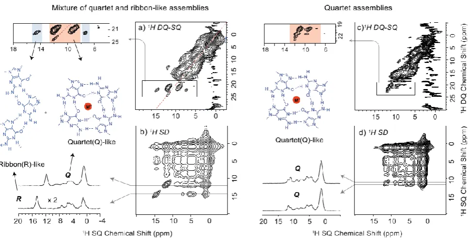
[image:7.612.135.469.404.680.2]Fast MAS NMR reveals the presence of quartet and ribbon-like assemblies in the solid state: Next to the
corresponding 1H solution-state NMR spectra, Figure 2 also presents 1H fast magic-angle spinning (MAS, 75 kHz)
solid-state NMR spectra of dried powders prepared by evaporation from the corresponding solution (see Supporting
Information, section 1b, for further details). While the resolution in the solid-state NMR spectra is much poorer
than in solution, distinct peaks can be resolved, such that clear differences between the solution- and solid-state
NMR high ppm chemical shifts can be identified. Moreover, the solid-state 1H NMR spectra show evident changes
that depend on the concentration and nature of the ionic species. Specifically, solid-state 1H NMR spectra of
GaceC10KPic 8:1 and 8:2 and GaceC10KI 8:1 complexes (Figures 2a, 2b and 2e) exhibit peaks at 12.1 and 14.8
ppm that are characteristic of both quartet and ribbon-like assembles, respectively: this is investigated further by
two-dimensional solid-state MAS NMR spectroscopy below. A line shape fitting analysis of one-pulse NMR spectra
quantified the Quartet/Ribbon (Q/R) ratios as: 65/35, 84/16 and 64/36 (error estimated as ±5%) wt/wt for the
GaceC10KPic 8:1, 8:2 and KI 8:1 complexes, respectively (see Figure S5). Note that for the GaceC10KEth 8:1
complex (Figure 2h), where only ribbon-like self-assembly is observed in the solid state, there is a clear broadening
at ~12 ppm that we assign to ribbon-like self-assembly also in solution.
The 1H double-quantum (DQ) single-quantum (SQ) MAS NMR experiment is a powerful probe of
proton-proton proximities in the solid state.[22] Specifically, 1H NMR chemical shifts are markedly sensitive to
intermolecular NH…N and NH…O hydrogen bonding interactions which interconnect Hoogsteen faces, such that
structurally different assemblies (herein quartet and ribbon-like) exhibit distinct 1H DQ-SQ spectral patterns,
facilitating a direct identification of quartet and ribbon-like assemblies.[9c, 23] For example, 1H DQ-SQ correlation
NMR spectra have been presented for guanosine dihydrate (G.2H2O),[23b] isopropylidine-guanosine (Gace)[23c] and
3’, 5’-dipropanoyl deoxyguanosine dG(C3)2,[23a] where there are crystal structures exhibiting different types of
ribbon-like self-assembly. A 1H DQ-SQ correlation spectrum has also been presented for a guanosine borate
hydrogel in the presence of K+ ions, whereby a 1H DQ “signature” (Figure 1 of Ref.[9c]) of stacked G-quartets
exhibits a NH auto peak due to inter-quartet stacking.
Although complexing GaceC10 with 0.125 equiv. of KPic and Sr(Pic)2 salts resulted in the formation of G4
assemblies in chloroform, only SrPic)2 fully retained G4 assemblies in the solid state. A two-dimensional 1H
DQ-SQ correlation NMR spectrum of the GaceC10KPic 8:1 complex is presented in Figure 4a. The 1H DQ peaks at
δDQ equal to 22.6 ppm (14.8 + 7.8) and 21.2 ppm (11.9 + 9.3) are assigned to intramolecular H-H proximities of
NH1 and one of the NH2 protons in ribbon-like and quartet assemblies, respectively.[9c, 23] Moreover, as noted
above,[9c] the NH1-NH1 autocorrelation peak at 23.8 (11.9 + 11.9) ppm is indicative of inter-quartet stacking. A 1H
NOESY-like spin-diffusion NMR spectrum of the GaceC10KPic 8:1 complex shown in Figure 4b clearly reveals
the co-existence of both quartet and ribbon-like arrangements. Specifically, no cross peaks are observed between
even for spectra recorded for four different mixing times between 10 ms and 319 ms (see Figure S6). Thus, this
spin-diffusion MAS NMR data proves that the quartet and ribbon-like peaks correspond to separate microcrystals
within the powdered sample. Likewise, no cross peaks between quartet and ribbon-like assemblies are observed in
a 1H spin-diffusion NMR spectrum for the GaceC10KI 8:1 complex (see Figure S6). While the 1H DQ and spin
diffusion data of the KPic complex reveal the existence of separate quartet and ribbon-like peaks, analogous spectra
for a SrPic)2 complex showed only peaks corresponding to quartets within a single phase as indicated by a 1H
spin-diffusion NMR spectrum (see Figure 4d). We note that the formation of micro-twinning structures such as those
[image:9.612.67.552.246.495.2]observed in metallohelicates/helicenes[24] is not ruled out by the present study.
Figure 4. Two-dimensional solid-state 1H (850 MHz, 75 kHz MAS) NMR spectra of a GaceC10KPic 8:1 complex (left) and a
GaceC10SrPic)2 8:1 complex (right). (top) DQ-SQ correlation spectra recorded using 1 τr of BABA recoupling [19]and
(bottom) NOESY-type spin-diffusion spectra (recorded with a mixing time of 106 ms) depicting the co-existence of quartet and ribbon-like assemblies for the GaceC10KPic complex and only quartet assembly for the GaceC10SrPic)2 complex. The base
contour level is at (a) 0.6%, (b) 0.03%, (a) 0.8% and (d) 0.5% of the maximum peak height.
Figure 5 presents 1H DQ-SQ correlation NMR spectra of GaceC10 in the absence and in the presence of K+
ions. G-quartet based 1H DQ spectral patterns are observed in Figure 5 for the GaceC10KPic 8:2 and 8:4,
GaceC10KI 8:1, 8:2 and 8:4 and GaceC10KEth 8:2 and 8:4 complexes. For the GaceC10KEth 8:1 complex, the
observation of two sets of 1H DQ peaks for the NH resonance at 14.8 ppm is likely due to proximity to both a NH2
Figure 5. Two-dimensional 1H (850 MHz, 75 kHz MAS) DQ-SQ correlation solid-state MAS NMR spectra of GaceC10 alone
(top left) and GaceC10 complexes with K+ ions, recorded using 1 τ
r of BABA recoupling.[19]Top: GaceC10, GaceC10KPic
8:2 and 8:4 complexes; middle: GaceC10KI 8:1, 8:2 and 8:4 complexes, and bottom: GaceC10KEth 8:1, 8:2 and 8:4 complexes.
Combining these observations from solution- and solid-state studies, it can be inferred that: (1) solvent plays
an important role in stabilizing the lipophilic G-quartets when a smaller amount of potassium ions, typically less
than 0.5 equiv. is used; (2) in the solid-state, up to at least 0.5 equiv. of potassium ions (depending on binding
the kinetic and thermodynamic stability of G4 assemblies including the nature of solvent, concentration, pH and
anion complexation: For example, Davis and co-workers showed that organic anions bridge individual G-quartets,
much like clips holding the exteriors of G4 assemblies,[12b, 14a] while Meijer and co-workers showed that solvent
polarity regulates the formation of G4-assemblies.[14e] In the solid state, consideration of X-ray diffraction structures
of analogous lipophilic G4 assemblies[12b, 14a, e] suggests that 0.125 equivalent of cations ought to be sufficient for
the formation of a lipophilic G-quadruplex. Taken altogether, these observations encouraged us to examine the
reversibility of quartet-ribbon interconversions during the dissolution process.
A dissolution NMR experiment demonstrates that quartet-ribbon interconversion is reversible: Reversibility
is a key feature in supramolecular chemistry. G-derivatives that respond to external stimuli such as light,
concentration, specific reagents, cation and anion binding are of considerable interest for designing switchable
assemblies.[9a, 14d, 25] To probe interconversion between quartet and ribbon-like assemblies, powdered samples of
GaceC10KPic 8:1 were suspended in known quantities of CHCl3 and the dissolution process was monitored
through a combined solution-state/solid-state NMR approach (Figure 6). We note that such a combined solution-
and solid-state NMR approach has previously enabled the investigation of labile chiral supramolecular ion pairs[26]
and monitored the crystallization process in small molecules.[27]
Figure 6. Single-pulse 1H (850 MHz) MAS NMR spectra (left) depicting the solid-state to solution-state transformation of
GaceC10KPic 8:1 in CHCl3. (a) 0.6 mg of powdered complex with no added solvent recorded using a JEOL 1 mm rotor at
[image:11.612.181.435.380.662.2]50 kHz MAS, Q/R is 74/26, wt/wt. (c) 0.5 mg complex in 5 l CHCl3 (0.25 M) recorded using a 1.3 mm rotor at 15 kHz MAS,
Q/R is 86/14 wt/wt. (d) 0.25 mg complex in 5 l CHCl3 (0.125 M) recorded under static conditions in a 1.3 mm rotor. A cartoon
representation (right) depicting interconversion of a mixture of stacked quartet (Q) and ribbon(R)-like structures into sandwiched quartet (Q) structures upon a solid-state to solution-state transformation.
For the GaceC10KPic 8:1 complex, Figure 6 presents 1H MAS spectra recorded during a dissolution
experiment: the solid/solution ratio was 1 mg/ 2.5 l, 0.5 mg/ 5 l and 0.25 mg/ 5 l for Figures 6b, 6c and 6d,
respectively. Note that the 1H MAS NMR spectra were recorded at different MAS frequencies as necessary to
achieve good spectral resolution at the varying solid/solution ratios. Specifically, MAS frequencies of 75, 50 and
15 kHz were used for the dried powder, 1 mg sample in 2.5 l chloroform (1 molar solution) and 0.5 mg of sample
dissolved in 5 l of chloroform (250 millimolar solution), respectively. A line shape fitting analysis showed that
the Q/R ratio (error estimated as ±5%) changes from 65/35 (Figure 6a), to 74/26 (Figure 6b), to 86/14 wt/ wt (Figure
6c), finally resulting in purely quartet-like assemblies in chloroform, 0.125 M (Figure 6d). This dissolution
experiment clearly exemplifies the reversibility of quartet-ribbon interconversions. The doubling of NH1 peaks
between 11 ppm to 12 ppm in Figure 6c is likely due to the formation of a kinetically-labile species, namely the
C4-symmetric octamer, as was similarly observed for the 1H solution-state NMR spectra of the GaceC10KPic 8:2 and
8:4 complexes shown in Figures 2b and 2c. Note the peak with a high 1H chemical shift of 16.3 ppm in Figure 6c:
we hypothesise that this corresponds to a ribbon-like intermediate mode of assembly, with distinct ribbon-like
structures having previously been observed.[23b, 28] Considering all the solid-state NMR results presented in this
paper, we emphasise that only solid-state NMR can provide this unique structural insight – by comparison, broad
spectral features are observed in powder X-ray diffraction patterns, as shown for example for the GaceC10KPic
8:1 complex in Figure S4.
Conclusions
To summarise, we have systematically investigated formation of distinct supramolecular assemblies for a long
alkyl chain G derivative in chloroform and in the solid state. Solution-state CD and NMR studies revealed the
formation of G-quartets in the presence K+ ions. Increasing the K+ ion concentration from 0.125 to 0.5 equiv.
triggered the formation of a kinetically labile C4-symmetric octamer that then slowly dissociated into a stable
D4-symmetric octamer, as monitored by time-course NMR spectroscopy. GaceC10KPic complexes exhibit relatively
strong CD signals, compared to KEth and KI complexes, suggesting a more tightly bound complex for KPic. By
comparison, 0.125 equiv. of Sr2+ ions induced the formation of a stable and long-lived hexadecamer species in
chloroform. Solution-state PFG NMR data clearly showed that picrate anion strongly binds to the GaceC10Sr2+
complex, but weakly binds to the GaceC10K+ complex as revealed by measurable differences in the extracted
explained by the stronger ion-dipole interactions between the oxygen atoms of Hoogsteen faces and the doubly
charged Sr2+ ion.
Chemical intuition tells us that the adoption of a particular self-assembled structure as driven by the formation
of specific intermolecular hydrogen bonds in solution would be expected to lead to the persistence of the same mode
of self-assembly in the solid state. Intriguingly, this work reveals that this is not the case for the supramolecular
assembly exhibited by a guanosine derivative in solution and in the solid state, with there being a subtle interplay
between competing hydrogen-bonding interactions and solvent and complexation effects that depend on both
concentration and the actual cation and anion. Importantly, for dried powders prepared by evaporation of the specific
solvents used, we have demonstrated the co-existence of quartet and ribbon-like supramolecular entities for
GaceC10KPic 8:1 and 8:2, and GaceC10KI 8:1 complexes. In future work, it could be interesting to see how
changes to the protocols for preparing the dry solids (e.g., change of solvent or slurrying) affect the observed
supramolecular self-assembly. The observations in this work were enabled by the ability of 1H solid-state NMR to
“view” distinct intermolecular hydrogen-bonding interaction, with the unusual mixed assemblies having been
quantitatively characterised in the solid state by fast MAS 1H DQ and spin-diffusion NMR experiments. Our work
has further shown that ribbon-quartet interconversion can be followed in a dissolution experiment, demonstrating
reversibility upon a solid- to solution-state transition and vice versa.
In conclusion, by employing a combined solution- and solid-state NMR approach, it could be inferred that,
alongside the crucial role played by solvent effects,[14e] higher salt concentration (typically 0.5 equiv. K+ ions) is
required to retain the structural integrity, and hence functionality of G-quadruplexes in the solid state as compared
to in solution. Otherwise, the solution- to solid-state transition leads to mixed self-assembly. This showcasing of
combination of solution- and solid-state NMR reveals it to be a powerful approach for studying the formation of
various other supramolecular assemblies in solution, gel and in the solid state.
Supporting information: Full experimental details including preparation of GaceC10 complexes, solution-state
time course NMR and DOSY spectra, CD spectra, powder X-ray diffraction data, solid-state NMR line shape
analysis, 1H DQ-SQ and spin-diffusion MAS NMR spectra. Experimental data for this study is provided as a
supporting dataset from WRAP, the Warwick Research Archive Portal at http://wrap.warwick.ac.uk/***.
Acknowledgements: We acknowledge funding from EPSRC (EP/K003674/1). The UK 850 MHz solid-state NMR
Facility used in this research was funded by EPSRC and BBSRC, as well as the University of Warwick including
via part funding through Birmingham Science City Advanced Materials Projects 1 and 2 supported by Advantage
West Midlands (AWM) and the European Regional Development Fund (ERDF). We thank Dr Nikola Chmel
di Farmacia, Biotecnologie e Scienze Motorie-University of Bologna for a “Tesi all’estero” grant. S. M. thanks the
European Community for the financial support through the project EC FP7 ICT-MOLARNET (318516)
Keywords
NMR spectroscopy; Self-assembly; Supramolecular Chemistry; fast magic-angle spinning (MAS); 2D
double-quantum (DQ) spectroscopy
References
[1] a) T. Aida, E. W. Meijer and S. I. Stupp, Science 2012, 335, 813-817; b) T. R. Cook, Y.-R. Zheng and P. J. Stang, Chem. Rev. 2013, 113, 734-777; c) L. Nicole, C. Laberty-Robert, L. Rozes and C. Sanchez, Nanoscale 2014, 6, 6267-6292; d) J.-M. Lehn, Angew. Chem. Int. Ed. 1988, 27, 89-112.
[2] a) S. M. Douglas, H. Dietz, T. Liedl, B. Hogberg, F. Graf and W. M. Shih, Nature 2009, 459, 1154-1154; b) H. Yang, C. K. McLaughlin, F. A. Aldaye, G. D. Hamblin, A. Z. Rys, I. Rouiller and H. F. Sleiman, Nat. Chem. 2009, 1, 390-396; c) X. Du, J. Zhou, J. Shi and B. Xu, Chem. Rev. 2015, 115, 13165-13307.
[3] a) J. W. Shim, Q. Tan and L.-Q. Gu, Nucleic Acids Res. 2009, 37, 972-982; b) X. Hou, W. Guo, F. Xia, F.-Q. Nie, H. Dong, Y. Tian, L. Wen, L. Wang, L. Cao, Y. Yang, J. Xue, Y. Song, Y. Wang, D. Liu and L. Jiang, J. Am. Chem. Soc.
2009, 131, 7800-7805; c) D. Y. Zhang and G. Seelig, Nat. Chem. 2011, 3, 103-113; d) B. G. Rusu, F. Cunin and M. Barboiu, Angew. Chem. Int. Ed. 2013, 52, 12597-12601; e) F. Zhang, J. Nangreave, Y. Liu and H. Yan, J. Am. Chem. Soc.
2014, 136, 11198-11211; f) E. Aznar, M. Oroval, L. Pascual, J. R. Murguía, R. Martínez-Máñez and F. Sancenón, Chem. Rev. 2016, 116, 561-718; g) A. Zhang and C. M. Lieber, Chem. Rev. 2016, 116, 215-257.
[4] a) M. Gellert, M. N. Lipsett and D. R. Davies, Proc. Natl. Acad. Sci. U. S. A. 1962, 48, 2013-2018; b) G. N. Parkinson, M. P. H. Lee and S. Neidle, Nature 2002, 417, 876-880; c) S. Burge, G. N. Parkinson, P. Hazel, A. K. Todd and S. Neidle,
Nucleic Acids Res. 2006, 34, 5402-5415.
[5] a) T. M. Schmeing and V. Ramakrishnan, Nature 2009, 461, 1234-1242; b) V. Ramakrishnan, Angew. Chem. Int. Ed.
2010, 49, 4355-4380.
[6] a) S.-T. D. Hsu, P. Varnai, A. Bugaut, A. P. Reszka, S. Neidle and S. Balasubramanian, J. Am. Chem. Soc. 2009, 131, 13399-13409; b) N. Q. Do, K. W. Lim, M. H. Teo, B. Heddi and A. T. Phan, Nucleic Acids Res. 2011, 39, 9448-9457; c) A. Wong and G. Wu, J. Am. Chem. Soc. 2003, 125, 13895-13905; d) G. Wu, A. Wong, Z. H. Gan and J. T. Davis, J. Am. Chem. Soc. 2003, 125, 7182-7183; e) M. R. Chierotti and R. Gobetto in Solid-State NMR Studies on Supramolecular Chemistry, John Wiley & Sons, Ltd, Chichester, 2012; f) H. Fenniri, G. A. Tikhomirov, D. H. Brouwer, S. Bouatra, M. El Bakkari, Z. Yan, J.-Y. Cho and T. Yamazaki, J. Am. Chem. Soc. 2016, 138, 6115-6118.
[7] a) S. Balasubramanian, L. H. Hurley and S. Neidle, Nat. Rev. Drug Discov. 2011, 10, 261-275; b) G. W. Collie and G. N. Parkinson, Chem. Soc. Rev. 2011, 40, 5867-5892; c) C. K. Kwok and S. Balasubramanian, Angew. Chem. Int. Ed. 2015,
54, 6751-6754.
[8] a) J. T. Davis, Angew. Chem. Int. Ed. 2004, 43, 668-698; b) J. T. Davis and G. P. Spada, Chem. Soc. Rev. 2007, 36, 296-313.
[9] a) N. Sreenivasachary and J. M. Lehn, Proc. Natl. Acad. Sci. 2005, 102, 5938-5943; b) G. M. Peters, L. P. Skala, T. N. Plank, B. J. Hyman, G. N. M. Reddy, A. Marsh, S. P. Brown and J. T. Davis, J. Am. Chem. Soc. 2014, 136, 12596-12599; c) G. M. Peters, L. P. Skala, T. N. Plank, H. Oh, G. N. Manjunatha Reddy, A. Marsh, S. P. Brown, S. R. Raghavan and J. T. Davis, J. Am. Chem. Soc. 2015, 137, 5819-5827; d) G. M. Peters and J. T. Davis, Chem. Soc. Rev. 2016, 45, 3188-3206.
[10] C. Arnal-Hérault, A. Pasc, M. Michau, D. Cot, E. Petit and M. Barboiu, Angew. Chem. Int. Ed. 2007, 46, 8409-8413. [11] a) D. Sen and W. Gilbert, Biochemistry 1992, 31, 65-70; b) M. Lu, Q. Guo and N. R. Kallenbach, Biochemistry 1992,
31, 2455-2459; c) T. C. Marsh, J. Vesenka and E. Henderson, Nucleic Acids Res. 1995, 696-700; d) A. Calzolari, R. Di Felice, E. Molinari and A. Garbesi, App. Phys. Lett. 2002, 80, 3331-3333; e) G. I. Livshits, A. Stern, D. Rotem, N. Borovok, G. Eidelshtein, A. Migliore, E. Penzo, S. J. Wind, R. Di Felice, S. S. Skourtis, J. C. Cuevas, L. Gurevich, A. B. Kotlyar and D. Porath, Nat. Nano 2014, 9, 1040-1046.
[13] a) F. van Leeuwen, W. Verboom, X. D. Shi, J. T. Davis and D. N. Reinhoudt, J. Am. Chem. Soc. 2004, 126 16575-12581; b) R. Ida and G. Wu, Chem. Commun. 2005, 4294-4296; c) V. Gubala, D. De Jesus and J. M. Rivera, Tetrahedron Lett. 2006, 47,, 1413-1416.
[14] a) X. D. Shi, K. M. Mullaugh, J. C. Fettinger, Y. Jiang, S. A. Hofstadler and J. T. Davis, J. Am. Chem. Soc. 2003, 125, 10830-10841; b) S. Pieraccini, S. Masiero, O. Pandoli, P. Samori and G. P. Spada, Org. Lett. 2006, 8, 3125-3128; c) L. Ma, M. Iezzi, M. S. Kaucher, Y. F. Lam and J. T. Davis, J. Am. Chem. Soc. 2006, 128, 15269-15277; d) M. Martin-Hidalgo and J. M. Rivera, Chem. Commun. 2011, 47, 12485-12487; e) D. Gonzalez-Rodriguez, J. L. J. van Dongen, M. Lutz, A. L. Spek, A. Schenning and E. W. Meijer, Nat. Chem. 2009, 1, 151-155; f) M. García-Arriaga, G. Hobley and J. M. Rivera, J. Org. Chem. 2016, 81, 6026-6035.
[15] J. L. Sessler, M. Sathiosatham, K. Doerr, V. Lynch and K. A. Abboud, Angew. Chem. Int. Ed. 2000, 39, 1300–1303. [16] P. M. Marcos, J. R. Ascenso, M. A. P. Segurado, R. J. Bernardino and P. J. Cragg, Tetrahedron 2009, 65, 496-503. [17] B. Zhang, Z. Cui and L. Sun, Org. Lett. 2001, 3, 275-278.
[18] S. Hayashi and K. Hayamizu, Bull. Chem. Soc. Jpn. 1991, 64, 685-687.
[19] a) W. Sommer, J. Gottwald, D. E. Demco and H. W. Spiess, J. Magn. Reson., Ser. A 1995, 113, 131-134; b) I. Schnell, A. Lupulescu, S. Hafner, D. E. Demco and H. W. Spiess, J. Magn. Reson. 1998, 133, 61-69.
[20] a) M. S. Kaucher, Y. F. Lam, S. Pieraccini, G. Gottarelli and J. T. Davis, Chem.—Eur. J. 2005, 11, 164-173; b) S. Lena, G. Brancolini, G. Gottarelli, P. Mariani, S. Masiero, A. Venturini, V. Palermo, O. Pandoli, S. Pieraccini, P. Samori and G. P. Spada, Chem.—Eur. J. 2007, 13, 3757-3764.
[21] T. Evan-Salem, L. Frish, F. W. B. van Leeuwen, D. N. Reinhoudt, W. Verboom, M. S. Kaucher, J. T. Davis and Y. Cohen, Chem.—Eur. J. 2007, 13, 1969-1977.
[22] a) S. P. Brown and H. W. Spiess, Chem. Rev. 2001, 101, 4125-4156; b) S. P. Brown, Prog. Nucl. Magn. Reson. Spectrosc. 2007, 50, 199-251; c) S. P. Brown, Solid State Nucl. Magn. Reson. 2012, 41, 1-27; d) M. Baias, A. Lesage, S. Aguado, J. Canivet, V. Moizan-Basle, N. Audebrand, D. Farrusseng and L. Emsley, Angew. Chem. Int. Ed. 2015, 54, 5971-5976; e) J. Xu, V. V. Terskikh, Y. Chu, A. Zheng and Y. Huang, Chem. Mater. 2015, 27, 3306-3316; f) J. A. Fernandes, M. Sardo, L. Mafra, D. Choquesillo-Lazarte and N. Masciocchi, Cryst. Growth Des. 2015, 15, 3674-3683; g) M. R. Hansen, R. Graf and H. W. Spiess, Chem. Rev. 2016, 116, 1272-1308.
[23] a) A. L. Webber, S. Masiero, S. Pieraccini, J. C. Burley, A. S. Tatton, D. Iuga, T. N. Pham, G. P. Spada and S. P. Brown,
J. Am. Chem. Soc. 2011, 133, 19777-19795; b) G. N. M. Reddy, A. Marsh, J. T. Davis, S. Masiero and S. P. Brown,
Cryst. Growth Des. 2015, 15, 5945-5954; c) G. N. M. Reddy, D. S. Cook, D. Iuga, R. I. Walton, A. Marsh and S. P. Brown, Solid State Nucl. Magn. Reson. 2015, 65, 41-48.
[24] R. Krämer, J.-M. Lehn, A. De Cian and J. Fischer, Angew. Chem. Int. Ed. 1993, 32, 703-706.
[25] a) A. Ciesielski, S. Lena, S. Masiero, G. P. Spada and P. Samorì, Angew. Chem., Int. Ed. 2010, 49, 1963-1966; b) S. Lena, P. Neviani, S. Masiero, S. Pieraccini and G. P. Spada, Angew. Chem., Int. Ed. Engl. 2010, 49, 3657-3660; c) S. Masiero, S. Pieraccini and G. Piero Spada in Self-assembly of Lipophilic Guanosines: Switching between Different Assemblies, The Royal Society of Chemistry, 2013, pp. 28-39.
[26] G. N. M. Reddy, R. Ballesteros-Garrido, J. Lacour and S. Caldarelli, Angew. Chem. Int. Ed. 2013, 52, 3255-3258. [27] C. E. Hughes, P. A. Williams and K. D. M. Harris, Angew. Chem. Int. Ed. 2014, 53, 8939-8943.
Table of Contents


![Figure 5. Two-dimensional 1H (850 MHz, 75 kHz MAS) DQ-SQ correlation solid-state MAS NMR spectra of GaceC10 alone (top left) and GaceC10 complexes with K+ ions, recorded using 1 τr of BABA recoupling.[19] Top: GaceC10, GaceC10KPic 8:2 and 8:4 complexes;](https://thumb-us.123doks.com/thumbv2/123dok_us/9479532.454210/10.612.60.554.79.569/figure-dimensional-correlation-spectra-complexes-recorded-recoupling-complexes.webp)

