Original Article
Long non-coding RNA Xist promotes progression of
non-small-cell lung cancer (NSCLC) by modulating
miR-103a and MAP3K3 pathway
Bin You*, Yili Fu*, Xin Li, Jingbai Miao, Bin Hu
Department of Thoracic Surgery, Beijing Chao-Yang Hospital, Capital Medical University, Beijing, China. *Equal contributors.
Received August 31, 2016; Accepted October 22, 2016; Epub February 1, 2017; Published February 15, 2017
Abstract: Background: Non-small-cell lung carcinoma (NSCLC) is the most common type of lung cancer with poor prognosis despite advent of newer treatment modalities. The role of long non-coding RNA (lncRNA)-Xist in can-cer including NSCLC has already been established. Hence, understanding the underlying mechanism of action of lncRNA-Xist in NSCLC might provide basis for novel drug targets. Objective: In this study, we explored the underlying molecular mechanism through which lncRNA-Xist promotes NSCLC progression. Materials and methods: In the hu-man lung cancer cell line A549, cell proliferation and percentage of apoptotic cells were measured by MTT assay
and flow cytometry. Western blot analysis was done to explore the expression level of MAP3K3. Real-time PCR was
used for RNA analysis. LncRNA-Xist silencing was done by infecting the A549 cells by lentivirus encoding shRNA
which targeted the lncRNA-Xist. Results: The results showed a significant increase in the expression of lncRNA-Xist
while a decrease in the miRNA103a expression (P<0.05). Silencing of lncRNA-Xist led to suppression of cell prolifer-ation and induction of apoptosis (P<0.05). Also, lncRNA-Xist knockdown led to significant increase in expression of
miRNA-103a, which in turn caused suppression of A549 cell proliferation (P<0.05). Finally, lncRNA-Xist knockdown
and miR-103a overexpression independently down-regulated MAP3K3 pathway. Conclusion: lncRNA-Xist promotes
progression of NSCLC by down-regulating the expression of miRNA-103a which can suppress cell proliferation and
induce apoptosis and by up-regulating MAP3K3 pathway.
Keywords: Non-small-cell lung carcinoma, long non-coding RNA-Xist, MiRNA-103-a, MAP3K3 pathway
Introduction
Lung cancer is one of the most common cause MAP3K3 pathway of cancer-related deaths in men whereas in women it stands second. About 14% of all new cancers are lung cancers. Lung cancer is responsible for about one out of 4 cancer deaths; cancer deaths due to lung can-cer exceeds those due to colon, breast, and prostate cancer combined [1]. Non-small-cell lung carcinoma (NSCLC) is the most frequent type of lung carcinoma accounting for about 85% of lung cancer varieties. Despite recent progress in treatment modalities for NSCLC, the 5-year survival rate is still very low (less than 18%) [2].
Noncoding RNAs (ncRNAs) belong to a parti- cular variety of RNA transcripts which do not translate into protein. Other less commonly
used terms are non-protein-coding RNA (npc- RNA), non-messenger RNA (nmRNA), functi- onal RNA (fRNA), etc. There are mainly two classes of ncRNA namely housekeeper ncRNAs (like rRNA, tRNA, snRNA, etc), and regulated ncRNAs [3]. The regulated ncRNAs are further categorized by the length of the nucleotide (nt); short ncRNAs, with <200 nt, and long ncRNAs (lncRNAs) with >200 nt.
Iyer MK et al, in his recent study published that there are approximately 60,000 ncRNAs, of which more than 68% are lncRNAs [4]. lncRNAs are associated with the process of tumorigenesis which affects tumor initiation, progression, and metastasis through modula-tion of oncogenic and tumor suppressing path-ways [2, 3, 5-7].
sev-hed that the expression of XIST is increased by many folds in certain cancers like gliob- lastoma [13], breast [14], and ovarian cancer [15]. Furthermore, these findings are sup- ported by the in vivo study in which tumor growth was suppressed in XIST knockdown nude mice and showed an improved survival of the mice [16]. Despite these findings, the underlying mechanism through which lncRNA-Xist promotes NSCLC are yet to be established unequivocally [5].
MicroRNAs (miRNAs) are single stranded non coding endogenous RNA molecules with about 19-25 nucleotides. miRNAs bind to comple-mentary recognition sequences of mRNA and can regulate the expression of mRNA targets. It has been established that each miRNA can regulate the expression of more than one RNA and vice versa [16-18] and has been estim- ated to target more than 30% of the protein-coding genes. Several biological processes like cell differentiation, proliferation, apoptosis, and metabolism are regulated by miRNAs [19]. Of the different types of miRNA, miR-103a has been associated with different types of can- cers such as pancreatic, bladder, nasopharyn-geal, colorectal, lung cancers, etc [20].
Mitogen-activated protein kinase (MAPK) path-ways are implicated in various processes like cellular proliferation, differentiation, migration, and apoptosis [21]. MAPK pathways consist of three-tiered kinase modules; MAPK is acti-vated by a mitogen-actiacti-vated protein kinase kinase (MAPKK), which is in turn activated by mitogen-activated protein kinase kinase kin- ase (MAP3K3, MAPKKK), located on the long arm of chromosome 17 (17 q) (23). Abnormali- ties in the MAPK signaling pathways resulted in both development and progression of cancer [20-22].
(24).
Cell proliferation assay
Cell metabolic activity was assessed using MTT (3-(4, 5-dimethylthiazol-2-yl)-2, 5-diphenyl tetrazolium proliferation assay [23]. On each day of the MTT assay, 100 μL of cells was taken from each of the culture media and placed, in triplicate, in a 96-well plate Fifty micrograms of MTT was added to each well and this mix was incubated for 4 hours at 37°C. At the end of the incubation period, 100 μL of 0.04 N HCl in 2-propanol was mixed thoroughly into each well. Plates were read with a Molecular Devices microplate reader (Sunnyvale, CA) at a wave-length of 570 nm, with a background reading at 650 nm subtracted. Triplicate readings for each sample were averaged.
MicroRNA transfection
miRNA precursors were purchased from Am- bion (Austin, TX). Synthetic miR-103a mimic and scrambled negative control RNA were pur-chased from Genepharma (Shanghai, China). Cells were seeded in 6-well plates and were transfected with Lipofectamine 2000 (Invitro- gen) with approximately 70% cell confluency [24]. In each well, equal amount (100 pmol) of either miR-103a mimic, or the scrambled negative control RNAs was used. The effici- ency of down-regulation of overexpression of miR-103a was evaluated by reverse transcrip-tion polymerase chain reactranscrip-tion (RT-PCR).
Estimation of lncRNA-Xist expression: infection with lentivirus encoding short hairpin RNA
pre-[26]. Immunoblotting were carried out with primary antibodies (anti-MAP3K3, anti-beta-actin, Cell Signaling). The proteins were detect-ed by enhancdetect-ed chemiluminescence (ECL-plus, Amersham Pharmacia Biotech).
RNA extraction and quantitative real-time PCR analysis
Total RNA was extracted with TRIzol reagent (Invitrogen, Grand Island, NY). RNA (500 ng) was polyadenylated and reverse transcribed to cDNA using an NCode miRNA First-Strand cDNA synthesis kit (Invitrogen) [27]. cDNA was used as the template for real-time PCR FastStart Universal SYBR green Master (Roche) with the universal reverse primer provided in the kit. Real-time PCR was performed on Applied Biosystems real-time detection system (Applied Biosystems), and the thermocycling parameters were 95°C for 3 min and 40 cycles of 95°C for 15 s followed by 60°C for 30 s. Each sample was run in triplicate and was normalized to U6 snRNA levels (U6 primers 5’-CTTCGGCAGCACATATACT-3’ [forward] and 5’- AAAATATGGAACGCTTCACG-3’ [reverse]). Melt- ing curve analysis was performed to confirm the specificity of the PCR products. The repli-cates were then averaged, and the fold induc-tion was determined by a ΔΔCT-based fold change calculation.
Statistical analysis
Statistical analysis was performed using SPSS 19.0 (IBM Corporation, Armonk, NY, USA). The pared by GenePharma (Shanghai, China). A549
cells were kept in 6-well plates at a density of 1×10⁴ cells/well and incubated for 12 h at
37°C. The culture medium was then replaced with fresh DMEM, and the cells were immedi-ately infected with shRNA lentiviruses at a mul-tiplicity of infection (MOI) of one (2 μL). After 24 h of growth at 37°C, the medium was replaced with fresh medium; the cells were cultured for 96 h at 37°C. The knockdown efficiency at the mRNA level was assessed using quantitative RT-PCR.
Apoptosis assay by Annexin V-FITC/PI-staining
Cells (3×10⁶) were stained using the FITC
Annexin V/Dead Cell Apoptosis Kit (V13242, Invitrogen) according to the manufacturer’s instructions [25]. Stained cells were diluted in Annexin V-binding buffer (Invitrogen). Then the suspended cells were used to perform flow cytometry. Annexin V-FITC/PI-stained cells were analyzed using a BD FACS Calibur flow cytome-ter (BD Biosciences, Heidelberg, Germany). In total 10,000 cells were analyzed per measure-ment. Data was analyzed using FlowJo 10.0.7 software (TreestarInc, Ashland, US).
Western blot analysis
[image:3.612.94.517.75.231.2]The cells were washed 2 times with PBS and then lysed with 1×SDS loading buffer (50 mM Tris-Cl, pH 6.8, 100 mM DTT, 2% SDS, 10% glycerol, and 0.1% bromophenol blue) as the whole-cell sample. The protein samples were subjected to SDS-PAGE gel electrophoresis
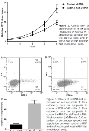
analysis (Figure 3A) was carried out to mea- sure apoptosis. It was found that the percent-age of cells undergoing apoptosis in lncRNA-Xist knockdown cells was significantly higher compared to the normal cells (Figure 3A-C).
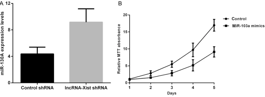
lncRNA-Xist suppression mediates the prolif-eration of human lung cancer A549 cells by regulating the expression of miR-103a
In the lncRNA-Xist knockdown cells, miR-103a expression, measured by real-time PCR, was found to be significantly higher in comparison to control cells (Figure 4A). Furthermore, MTT assay, conducted to determine cell prolifera-tion after ectopic expression of miR-103a mim-ics (miR103a overexpression) into A549 cells, data were represented as mean ± SD. Different
groups were analyzed using one way ANOVA.
P-values <0.05 was considered to be statisti-cally significant.
Results
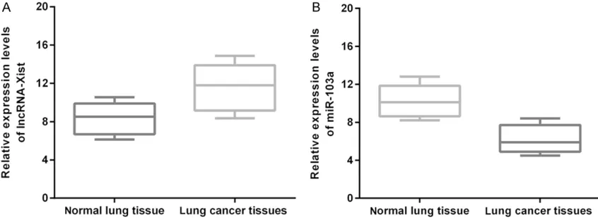
Expression of lncRNA-Xist and miR-103a ex-pression in human lung cancer cells
RNA was isolated from the surgically resected lung cancer specimens and from the neighbor-ing normal lung tissues (N=60). The expression levels of lncRNA-Xist and miR-103a were mea-sured by real-time PCR as described above. The expression of lnc-RNA XIST was significantly up-regulated in human lung cancer cells compared
Figure 3. Effects of lncRNA-Xist ex-pression on cell apoptosis. A: Flow cytometry data on apoptosis in control shRNA A549 cells. B: Flow cytometry data on apoptosis in control lncRNA-Xist shRNA (lncRNA- Xist knockdown) A549 cells. C: Com-parison of percentage apopotic cell population between control shRNA and lncRNA-Xist shRNA (lncRNA-Xist knockdown) cells.
As described earlier shRNA-mediated silencing of lnc- RNA-Xist was done by in- fecting the A549 cells by lentivirus infection and then cell proliferation was det- ermined using MTT absor-bance. It was found that cell proliferation was signi- ficantly suppressed in the human lung cancer cells when compared to that in the normal lung tissue (Figure 2).
lncRNA-Xist suppression induces apoptosis of A549 cells
[image:4.612.90.385.70.500.2]R et al, has established that lncRNA PVT1 worsens the prognosis of gastric cancer by promoting the proliferation of cancerous cells through regulating p15 and p16 [28]. Similarly, two studies, which were conducted, indepen-dently by two different groups of researchers (Yang MH, et al and JI Q, et al) concluded that lncRNA MALAT1 promotes disease progres- sion in colorectal cancer patients [29, 30]. Similarly, few other studies have supported the progression of cancers by lncRNA (hepatocel-lular and bladder cancer) [31, 32].
revealed significant inhibition of A459 cells proliferation compared to the control group (Figure 4B).
lncRNA-Xist suppression and miR-103a over-expression down-regulate the over-expression of MAP3K3
Western blot analysis was conducted to exam-ine the expression levels of MAP3K3 after shR-NA-mediated silencing of lncRNA-Xist (Figure 5A) and ectopic expression of miR-103a
mim-Figure 4. lncRNA-Xist regulated lung cancer cell proliferation by mediating miR-103a. A: Comparison of expression of miR-103a between control shRNA and lncRNA-Xist knockdown A549 cells. B: Comparison of proliferation of A549 cells between control shRNA and miR-103a mimics A549 cells.
Figure 5. lncRNA-Xist expression regulated the MAP3K3 signal pathway in lung cancer cells. A: Comparison of expression of MAP3K3 between control shRNA and in lncRNA-Xist knockdown A 549 cells: Western blot analysis. Ac
-tin acted as an internal control. B: Comparison of expression of MAP3K3
between control shRNA and in lncRNA-Xist knockdown A 549 cells. C:
Com-parison of expression of MAP3K3 between control shRNA and in miR103 a mimics A 549 cells: Western blot analysis. Actin acted as an internal con
-trol. D: Comparison of expression of MAP3K3 between control shRNA and in
miR103 a mimics A 549 cells.
ics (Figure 5C) in A549 cells. As shown in the respective figures (Figure 5A-D), the ex- pressions of MAP3K3 in both the lncRNA-Xist knockdown cells and overexpressing miR-103a cells were significantly down-regulated when com-pared to the control group.
Discussion
[image:5.612.95.519.74.230.2]the expression of MAPK pathway [36]. The role of micro RNAs (miRs) as tumor suppressor genes has already been described in different cancers [37-39]. Shin SS, et al, have descri- bed the role miR-106a in suppression of pro-gression of bladder cancer [38]. In our study we found that overexpression of miR-103a has led to the suppression of A549 cell line and down-regulation of the MAPK pathway. Simi- larly, Shin SS, et al, also found that overexpres-sion miR-106a led to tumor suppresoverexpres-sion in bladder cancer cells through suppression of ERK/MAPK pathway. Again, Wang X, et al found that miR 335 is associated with suppression of bladder cancer through reduced expression of MAPK1 [37].
In this study, we have explored the underlying mechanism through which lncRNA-Xist pro-motes disease progression in NSCLC. In the human lung cancer cell line A549, the expres-sion of lncRNA-Xist was increased significantly whereas the expression of miR-103a was dras-tically decreased (Figure 1A and 1B). To deduce the effects of lncRNA-Xist on disease progres-sion in lung cancer cells we measured both pro-liferation and apoptosis in lncRNA-Xist knock-down A549 cells. It was found that shRNA mediated silencing of lncRNA-Xist in A549 cell line led to favorable outcomes where disease progression decreased significantly (Figure 2) while percentage of apoptotic cells significantly increased (Figure 3B and 3C).
Furthermore, in the lncRNA-Xist knockdown A549 cells the expression miR-103a was sig-nificantly increased (Figure 4A) and overex-pression of miR-103a led to supoverex-pression of proliferation of A549 cells to a significantly (Figure 4B). These results suggested prolifera-tion of lncRNA-Xist knockdown A459 cells was suppressed by overexpression of miR-103a. kinases are the most thoroughly studied
mem-bers of the MAPK pathway [21].
The MAPK pathway is already known to be associated with a number of cellular functions like cell proliferation, differentiation, survival, and finally death [34]. Any kind of aberrations in the functioning in the MAPK pathway have been associated in the pathogenesis of num-ber of diseases like Alzheimer’s disease (AD), Parkinson’s disease (PD), amyotropic lateral sclerosis (ALS) and finally various cancers. It has already been established that over activa-tion of the JNK and P38 pathways are associ-ated with neurodegenerative diseases like AD, PD, and ALS whereas the ERK pathway is associated with the different steps in cancer progression such as stimulation of prolifera-tion, migraprolifera-tion, and invasion of the malignant cells [34].
Several studies have established that the degree of transcription of lnc RNA is strongly associated with cell differentiation and res- ponsible for the development of metastasis [35, 36]. Studies have also described the striking similarity between fetal tissue develop-ment and progression of cancer in terms of overexpression of fetal oncogenes under both the circumstances. Similar to the above find-ings, it is not surprising that lnc RNAs are also overexpressed in cancer cells in comparison to normal cells as lnc RNA transcription is enhanced in fetal tissue [35].
[7] Ye LC, Chen T, Zhu DX, Lv SX, Qiu JJ, Xu J, Yuan
FL and Wei Y. Downregulated long non-coding
RNA CLMAT3 promotes the proliferation of colorectal cancer cells by targeting regulators of the cell cycle pathway. Oncotarget 2016; [Epub ahead of print].
[8] Lin L, Gu ZT, Chen WH and Cao KJ. Increased
expression of the long non-coding RNA ANRIL promotes lung cancer cell metastasis and cor-relates with poor prognosis. Diagn Pathol 2015; 10: 14.
[9] Yang Q, Xu E, Dai J, Liu B, Han Z, Wu J, Zhang
S, Peng B, Zhang Y and Jiang Y. A novel long
noncoding RNA AK001796 acts as an onco -gene and is involved in cell growth inhibition by resveratrol in lung cancer. Toxicol Appl Phar-macol 2015; 285: 79-88.
[10] Hu T and Lu YR. BCYRN1, a c-MYC-activated long non-coding RNA, regulates cell metasta-sis of non-small-cell lung cancer. Cancer Cell Int 2015; 15: 36.
[11] Wu Y, Liu H, Shi X, Yao Y, Yang W and Song Y.
The long non-coding RNA HNF1A-AS1 regu-lates proliferation and metastasis in lung adenocarcinoma. Oncotarget 2015; 6: 9160-9172.
[12] Bertozzi D, Iurlaro R, Sordet O, Marinello J, Zaf-faroni N and Capranico G. Characterization of
novel antisense HIF-1α transcripts in human
cancers. Cell Cycle 2011; 10: 3189-3197. [13] Yao Y, Ma J, Xue Y, Wang P, Li Z, Liu J, Chen L,
Xi Z, Teng H and Wang Z. Knockdown of long
non-coding RNA XIST exerts tumor-suppressive functions in human glioblastoma stem cells by up-regulating miR-152. Cancer Lett 2015; 359: 75-86.
[14] Salvador MA, Wicinski J, Cabaud O, Toiron Y, Finetti P, Josselin E, Lelièvre H, Kraus-Berthier
L, Depil S and Bertucci F. The histone deacety-lase inhibitor abexinostat induces cancer stem cells differentiation in breast cancer with low Xist expression. Clin Cancer Res 2013; 19: 6520-6531.
[15] Ren C, Li X, Wang T, Wang G, Zhao C, Liang T,
Zhu Y, Li M, Yang C and Zhao Y. Functions and mechanisms of long noncoding RNAs in ovari-an covari-ancer. Int J Gynecol Covari-ancer 2015; 25: 566-9.
[16] Dong L, Xin Z, He R, Tang R, Ping L, He Q and Gang C. MiR-133a is downregulated in non-small cell lung cancer: a study of clinical
sig-nificance. Eur J Med Res 2015; 20: 50.
[17] Weber DG, Casjens S, Johnen G, Bryk O, Raiko I, Pesch B, Kollmeier J, Bauer TT and Brüning T.
Combination of MiR-103a-3p and mesothelin improves the biomarker performance of malig-nant mesothelioma diagnosis. PLoS One 2014; 9: e114483-e114483.
[18] Du L, Schageman JJ, Irnov, Girard L, Hammond SM, Minna JD, Gazdar AF and Pertsemlidis A.
Finally, it was found that lnc RNAXIST knock-down and miR-103a overexpression led to decreased expression of MAP3K3 in A549 cells (Figure 5A-D, respectively).
The role of lncRNA as an early diagnostic tool in different types of cancer has already been established. The expression of lncRNA not only aids in diagnosing but also acts as a prognostic factor in varied types of cancer [5, 8].
In conclusion, our study demonstrated that overexpression of lnc RNAXIST in the A549 cells promotes disease progression by up-regu-lation of MAP3K3 pathway and at the same time, is responsible for the decreased expres-sion of miR-103a which independently sup-presses the proliferation and down-regulation of MAP3K3 pathway in cancerous A549 cells.
Understanding the underlying mechanism of lncRNA in NSCLC might provide the basis for development of novel drug targets in near future.
Disclosure of conflict of interest
None.
Address correspondence to: Bin Hu, Department of Thoracic Surgery, Beijing Chao-Yang Hospital, Capital Medical University, 8, Gongti South Road, Chaoyang District, Beijing 100020, China. E-mail: [email protected]
References
[1] Cancer UK. Lung cancer Key Stats. 2014.
[2] Zappa C and Mousa SA. Non-small cell lung cancer: current treatment and future advanc-es. Transl Lung Cancer Res 2016; 5: 288-300. [3] Soudyab M, Iranpour M and Ghafourifard S.
The role of long non-coding RNAs in breast cancer. Arch Iran Med 2016; 19: 508-517. [4] Iyer MK, Niknafs YS, Malik R, Singhal U, Sahu
A, Hosono Y, Barrette TR, Prensner JR, Evans JR and Zhao S. The landscape of long noncod-ing RNAs in the human transcriptome. Nat Genet 2015; 47: 199-208.
[5] Tantai J, Hu D, Yang Y and Geng J. Combined
identification of long non-coding RNA XIST and
HIF1A-AS1 in serum as an effective screening for non-small cell lung cancer. Int J Clin Exp Pathol 2015; 8: 7887-7895.
[6] Zhao Y, Zhang X and Zhang W. Functions and
[21] Kim EK and Choi EJ. Pathological roles of MAPK signaling pathways in human diseases.
Biochim Biophys Acta 2010; 1802: 396-405. [22] Dhillon AS, Hagan S, Rath O and Kolch W. MAP
kinase signalling pathways in cancer. Onco-gene 2007; 26: 3279-3290.
[23] Nasonburchenal K, Allopenna J, Bègue A,
Stéhelin D, Dmitrovsky E and Martin P. Target-ing of PML/RARalpha is lethal to retinoic acid-resistant promyelocytic leukemia cells. Blood 1998; 92: 1758-1767.
[24] Mraz M, Chen L, Rassenti LZ, Ghia EM, Li H,
Jepsen K, Smith EN, Messer K, Frazer KA and Kipps TJ. miR-150 influences B-cell receptor
signaling in chronic lymphocytic leukemia by regulating expression of GAB1 and FOXP1. Blood 2014; 124: 84-95.
[25] Brauchle E, Thude S, Brucker SY and
Schenke-layland K. Cell death stages in single apoptotic
and necrotic cells monitored by Raman micro-spectroscopy. Sci Rep 2014; 4: 4698-4698. [26] Leung WK and Sung JY. p53-independent pRB
degradation contributes to a drug-induced apoptosis in AGS cells. Cell Res 2005; 15: 695-703.
[27] Motiño O, Francés DE, Mayoral R, Castrosán-chez L, Fernándezvelasco M, Boscá L, Gar-cíamonzón C, Brea R, Casado M and Agra N. Regulation of MicroRNA 183 by cyclooxygen-ase 2 in liver is DEAD-Box heliccyclooxygen-ase p68 (DDX5) dependent: role in insulin signaling. Mol Cell Biol 2015; 35: 2554-2567.
[28] Kong R, Zhang EB, Yin DD, You LH, Xu TP, Chen WM, Xia R, Wan L, Sun M and Wang ZX. Long
noncoding RNA PVT1 indicates a poor progno-sis of gastric cancer and promotes cell prolif-eration through epigenetically regulating p15 and p16. Mol Cancer 2015; 14: 82.
[29] Yang MH, Hu ZY, Xu C, Xie LY, Wang XY, Chen SY
and Li ZG. MALAT1 promotes colorectal cancer
cell proliferation/migration/invasion via PRKA
kinase anchor protein 9. Biochim Biophys Acta 2015; 1852: 166-174.
non-protein-coding RNA up-regulated in
blad-der carcinoma and embryo, influencing cell
growth and promoting invasion. FEBS Lett 2008; 582: 1919-1927.
[33] Dong C, Davis RJ and Flavell RA. MAP kinases in the immune response. Annu Rev Immunol 2002; 20: 55-72.
[34] Roux PP and Blenis J. ERK and p38 MAPK-acti -vated protein kinases: a family of protein ki-nases with diverse biological functions. Micro-biol Mol Biol Rev 2004; 68: 320-344.
[35] Huang JL, Ren TY, Cao SW, Zheng SH, Hu XM, Hu YW, Lin L, Chen J, Zheng L and Wang Q.
HBx-related long non-coding RNA DBH-AS1 promotes cell proliferation and survival by
acti-vating MAPK signaling in hepatocellular carci -noma. Oncotarget 2015; 6: 33791-33804. [36] Li R, Zhang L, Jia L, Duan Y, Li Y, Bao L and Sha
N. Long non-coding RNA BANCR promotes pro-liferation in malignant melanoma by regulating
MAPK pathway activation. PLoS One 2014; 9: e100893.
[37] Wang X, Wu G, Cao G, Chen X, Huang J, Jiang X
and Hou J. MicroRNA-335 inhibits bladder can-cer cell growth and migration by targeting mito-gen-activated protein kinase 1. Mol Med Rep 2016; 14: 1765-70.
[38] Shin SS, Park SS, Hwang B, Kim WT, Choi YH, Kim WJ and Moon SK. MicroRNA-106a sup -presses proliferation, migration, and invasion
of bladder cancer cells by modulating MAPK
signaling, cell cycle regulators, and Ets-1-medi-ated MMP-2 expression. Oncol Rep 2016; 36: 2421-9.
[39] Fu X, Zhang W, Su Y, Lu L, Wang D and Wang H.