warwick.ac.uk/lib-publications
A Thesis Submitted for the Degree of PhD at the University of Warwick
Permanent WRAP URL:
http://wrap.warwick.ac.uk/112025
Copyright and reuse:
This thesis is made available online and is protected by original copyright.
Please scroll down to view the document itself.
Please refer to the repository record for this item for information to help you to cite it.
Our policy information is available from the repository home page.
The protein bodies o f the endosperm of the seeds of the castor oil
plant Ricinus cannon is contain a variety of storage proteins,
along with two lectins, ricin and Ricinus communis agglutinin.
Ricin is a heterodimer consisting of an A chain which is toxic to
cell-free translation systems, and a B chain w h ich has galactose-binding
activity; the thole molecule is toxic to cells and animals by
virtue of the ability o f the A chain to enzymatically inactivate
ribosomes after it crosses the cell membrane, this latter being
achieved after binding of the molecule to cell surfaces by the
B chain. The agglutinin consists of two ricin-like species linked
non-covalently, is divalent, and is not significantly toxic to cells.
Previous work o n the synthesis of the lectins indicated diet
each subunit had its o w n precursor, each being cotranslationally
segregated and glycosylated, and assembly of the lectins was thought
to occur after transport to the protein bodies. However, the
putative B chain precursor was far larger than the mature B chains,
and further protein-based evidence Indicated that It contains both
A and B chain sequences. The former A chain precursor was
Identified as an albumin which contalmlnated lectin preparations
used for raising antisera.
The work reported here confirms that the putative B chain
precursor contains A a n d B chain sequences, by mesns of d m cloning
of cDNA complementary t o lectin-specific mRNA. Clones of nearly
full length have been obtained and sequenced, and the precursor is
shown to have an N-terminal signal sequence, which is followed b y the
A chain sequence, and then by the B chain sequence. A linker of
12 amino acids is shown to be present beoaaen the o*o chains.
Sequences corresponding to both lectins are reported, and the
similarities and differences between them are discussed. The sequences
are placed into context b y comparison with other plant nucleotide
and protein sequences.
Literature on the castor bean lectins is reviewed, and the
uses of the clones are discussed, with special reference to their
CONTENTS
Acknowledgements
Declaration
List of figures
Abbreviations
Chapter 1: Introduction
1A IB 1C ID IE IF
Erasable 1
Isolation and structure of castor b ean lectins 7
Synthesis of the lectins
Biological functions of the lectins
1D1 General features
1D2 Properties of the A chains
1D3 Properties of the B chains
IDA Entry of rlcin into cells
Uses of castor been lectins
Castor b e e n lectin cENA cloning
Chapter 2t Materials and Methods
2A Materials
2B General methods
2B1 Growth and harvesting of castor beans
2B2 Growth end maintenance of E coll
2B3 Preparation of competent cells 1 transformation
2B4 Extraction and purification of plasmid ENA
2BS Restriction enzymes and mapping
2B6 In vitro protein synthesis snd lmnunoprecipltatlon
2B7 End-labelling of ENA f r ^ m n t s
2C1 Neutral agarose gels
2C2 Alkaline agarose gels
2C3 Formamide agarose gels
2C4 SDS-Polyacrylamide gels
2C5 DNA sequencing gels
2C6 Elution of ENA from gels
2D Extraction and fractionation of castor bean nRNA
2E Construction of cENA library In pBR322
2E1 Synthesis of double-stranded cENA
2E2 Construction of recombinants
2F Screening of the cENA library
2F1 Differential hybridisation
2F2 Oligomer hybridisation
2F3 Translation of hybridisation-selected nRNA
2G Subcloning into pUC8
2H M13 cloning and dldeaxy sequencing
2H1 Cloning into M13
2H2 Dldeaxy sequencing
21 Msxsm and Gilbert sequencing
211 Preparation of ENA frapoants
212 Modification and cleavage reactions
2J Primer extension
2J1 Cloning of the primer
2J2 Primer extension reaction
2K Nucleic acid transfer and hybridisation
2K1 Southern blotting
2K2 Northern blotting
61 61 62 62 62 .3 64 66 66 67 69 69 70 72
74 i
Chapter 3t Results and Discussion 87
3A Isolation and charactarlsation of nRNA 87
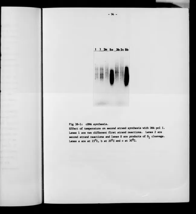
3B cDNA synthesis 93
3C Construction of clonss 98
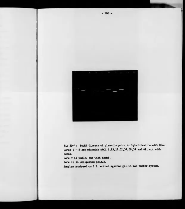
3D Screening of ths cENA library 100
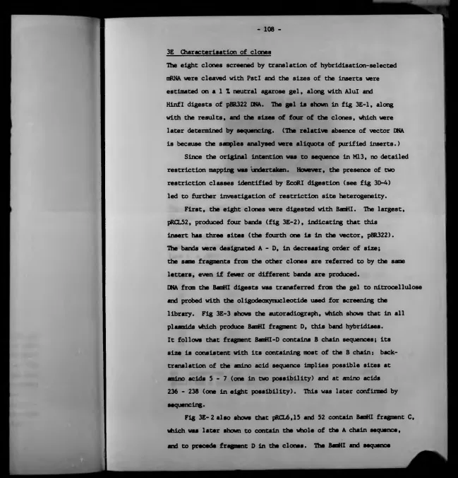
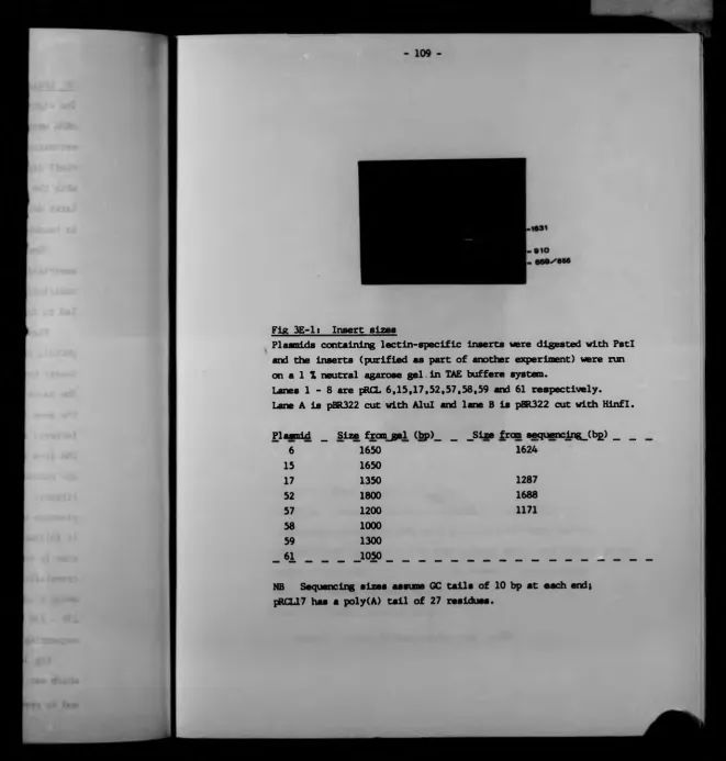
3E Characterisation o f clonss 108
3F Subcloning into pUC8 115
3G Sequencing: Introduction 119
3G1 Suanary of results 119
3G2 Ml 3 cloning and dldeaxy sequencing 126
3G3 Msxam it Gilbert sequencing 129
co chunk m y Supervisor, r>rJH L o #d, foe «Hotinoel help,
3H The preproricin sequence 135
31 Ths preprosgglutinin sequence 137
3J Analysis of protein sequences 139
3J1 Comparison with protein-determined sequences 139
3J2 Comparison of deduced aaino acid sequences 149
3J3 The signal and litter peptides 158
3K Analysis of nucleotide sequences 165
3K1 Comparison o f prepro-lectin sequsnces 165
3K2 Base compoeltion, dinucleotides and codon usags 168
3K3 The 5' non-coding region 173
3K4 The 3' non-coding region 185
3L Primer extension 193
3M Northern blotting 195
Chapter 4i Final Discussion 199
Appendix It
Appendix 2i
References
Computer p r o g r a m m e
Restriction data
213
237
ACKNOWLEDGEMENTS
I wish to thank m y Supervisor, Dr JM Lord, for continual help,
advice and encouragement, and Dr L M Roberts and many others at
Warwick for Information, suggestions and materials, and to
Professor H Woodland for performing the oocyte injections.
I am indebted to Celltech Ltd for the provision, free of charge,
of the oligodeaocynucleotlde with which the cDNA library was screened,
and especially to Dr T Harris and Dr M Bodmer, for two weeks spent
at Celltech learning M13 cloning and dldeaxy sequencing.
M y thanks also to the Agriculture and Food Research Council
for the grant which supported this work.
I am very grateful to m y wife Helen for moral support and tolerance,
and for assistance with the manuscript, and to m y mother for
- ii
The conclusions reached In this Thssis are m y own, and are baaed on
the experiments reported, on many discussions with m y Supervisor,
Dr JM Lord, with Dr L M Roberts, and others, and on published
works (opp cit).
The experiments were performed In the Department o f Biology,
University of Bradford (first year) and the Department of
Biological Sciences, Uhlverslty of Warwick. They were carried
out by myself, though Dr Roberts provided much ssslstance with
cDNA synthesis; Dr Lord performed the experiments described in
fig 3A-2, and Professor H Woodland carried out the oocyte Injections
end 1 shelling.
List of figures (continued)
F i g ___ T i t l e ____________________________________________________ .Pjg£ .
3G-2 Map of plasmid pRQ.17 124
3G-3 Junction region in pRCT.17 125
3G-4 Extant of M13 d o n a libraries 127
3G-5 Maxam & Gilbert strategy: clone pRCL6 131
3G-6 Maxam Si Gilbert strategy: d o n a pRCL17 132
3G-7 Maxam Si Gilbert strategy: clone pRCL52 133
3G-8 Maxam Si Gilbert strategy: clone pRCL57 134
3H-1 Preproricin cDNA aequance 136
31-1 Preproagglutinin cDNA sequence 138
3J-1 Amino acid compositions and molecular weights 140
3J-2 Differences between rlcin cDNA and Funatsu sequences 141
3J-3 Plot of 'genuine' differences between two rlclns 143
3J-4 Amino acid sequences of prepro-lectins 150
3J-5 Amino a d d differences between the lectins 151
3J-6 A chain hydropathy plots 154
3J-7 B chain hydropathy plots 155
3J-8 Hydropathy difference plots 156
3J-9 Signal sequence hydropathy plot 159
3J-10 Plant signal sequence cleavage sites 161
3J-11 Plant precursor protein processing sites 163
3K-1 Prepro-lectln cDNA sequences 166
3K-2 Plant ofiNA base caapoeltlons 169
3K-3 Preproricin dinucleotlde frequencies 170
3K-4 Preproricin codon usage 171
3K-5 Preproricin cDNA S' end: sequencing gel 174
3K-6 Plant a«NA S' non-codirg regions 178
Liât of figures (continued)
Fi£ _ _ T i t l e __________________________________________________
3K-8 Plant mRNA Initiation environments: selected sequences
3K-9 Plant nRNA termination environments
3K-10 Bases flanking plant termination codons
3L-1 Primer extension
3M-1 Northern blot of castor bean mRNA 196
3N-1 Oocyte injection products 198
4-1 Homology of conA and favin 203
4-2 B chain halves: Funatsu data 204
4-3 B chain halves: Ricin cDNA data 205
4-4 B chain halves: Ricin hydropathy plots 207
4-5 B chain halves: Agglutinin cDNA data 208
4-6 Putative B chain signal sequence 210
Sodimi dodecyl sulphate
Standard saline citrate
1
-CHAPTER 1: INTRODUCTION
1A Preamble
The endosperm cells of Che castor bean plane Ricinos communis
• contain protein bodies in « M c h a variety o f proteins are stored
prior to germination. These include the nitrogen-rich albunins,
the 11 S globulins, and two lectins: the toocic ricin, and the
related agglutinin RCA (Ricinos comnunls agglutinin) (1,2).
The toxic and medical actions of the beans have been known
since ancient times, and were used in classical Greek and
Sanskrit medicine (3); the oil Is noted for its purgative effects.
More recently, attention has focussed on the anti-tumour
properties of ricin (4), and as recently as 1984 a report was
published describing clinical trials using ricin in the treatment
of terminal cancer (3). However, since ricin is a lectin,
and consequen tly displays relatively non-specific binding to cell
surfaces, most cancer work is now directed towards the exploitation
of conjugates betwe en ricin and antibodies. It is hoped that
such conjugates will have specificity for tuaour cells, and
trill represent the ultimate development of Ehrlich's 'magic bullet’
concept (6).
Ballnt (7) has collected descriptions of over 700 cases of
ricin poisoning in man, though by far the m o s t notorious case
is that of Georgi Markov, a Bulgarian expatriate who vns
assassinated by the injection from an umbrella of a
ricin-impregnated metal ball in 1978, near Waterloo Bridge (8).
The possibility of ricin poisoning on a vastly larger scale
beans (9). All work on rlcin In Bulgaria Is apparently
classified (10).
Considerations of such possibilities have led to controversy
regarding more contemporary approaches to the study of ricln and
other toxic proteins: a letter published in Nature (11)
advocated the outright banning of cloning of toxin nucleotide
sequences, though a later correspondent pointed out that large
supplies of diphtheria toxin are available without cloning (12).
It Is of note that the anti-cloning letter was from an employee
of the Cetus Immune Corporation of California - eight months
later, Cetus published the sequence of cloned diphtheria
toxin (13). This latter sequence has been expressed In E coll
(14,15,16), and a number of other toxin DNA sequences have also been
cloned. Examples are bee venom melittln (17), cholera toxin (18,19),
E coll heat-labile enterotoocln (20,21) and Pseudomonas
exotoxin (22). Cetus hold a patent on the E coll toxin, and
claim expression (23).
Th e extreme toxicity of ricin Is Indicated b y its LDj q values:
one estimate gave 12 pg per kg in mice, while more recent
determinations give 2.7 pg per kg In mice and 1.75 pg per kg
in dogs (see ref 5). Fodstad et al (5) believe that the maxlnun
does tolerable b y m a n is some 23 pg per aq m, given Intravenously.
Doses o f 1 ng/ml of ricln are adequate to Inhibit the activity
of cell-free protein synthesising eye tame, and it is believed
diet a single molecule can kill a cell (24).
T h e castor bean plant is a native o f tropical Africa, bu t
3
-European varieties tend to grow as small bushes - often a bare stem
carries just a few leaves at the top - and flower only with
difficulty. Tropical specimens can grow rapidly to a height of
30 - AO feet (23): It Is sometimes named the 'miracle tree',
one of which was used by G o d to teach Jonah the virtue of
mercy (26). Etymologically, the name Rlcinua probably derives
' from the Hebrew kikar, which means 'round', via the Greek
kikinos (3).
A brief history of scientific work on rlcin follows, and
is condensed from Balint's thorough review (7).
The proteinaceous nature of the toxic component of castor
beans was noted in 1878 b y Ritthausen, and confirmed In 1887
by Dixon. In that year, Stlllmark named this component rlcin.
The first detailed description of human rlcin poisoning was
published in 1899 b y Muller. The resistance of rlcin to
proteolytic enzymes was noted around the turn of the century, and
was long t-hr.ight- to be related to the protease activity attributed
to It - as late as 1973, Funatsu's group described experiments
demonstrating this function (27). Klein Is no longer believed
to have proteolytic activity (eg ref 28).
These early experiments were performed using crude extrects
of castor beans; better preparations were obtained in 1903
by Osborne. In 1913 Robert im proved the methodology b y die
introduction of amaonlua sulphate precipitation, and in 1923
Karrer Introduced inorganic adsorbents. Affinity chromatography
on aephaross, exploiting the galactose specificity of rlcin,
was first used In 1963, b y Dirhelmar. This is now die most
The toxicity of rlcin was for many years attributed to
a haamagglutlnatlng activity, but with the advent of more
discriminating fractionation techniques, the toxic and
agglutinating activities were assigned to two different proteins,
referred co as rlcln and Ricinus ccnmunia agglutinin respectively.
- At the same time, die existence of multiple forms of the lectins
became apparent - for example, Schone, in 1958, obtained five
bands on paper electrophora tograms. Most contemporary workers
report two riclns and two agglutinins (eg refs 29,30), though
occasionally more are reported (31). It w a s shown in the early
1970s that both the toxin and the agglutinin are composed of
two dissimilar types of subunit - the agglutinin being essentially
a dimer of ricln. Although the A chains an d the B chains of
the two proteins cross-react immunologically (though A chains
and B sera do not react, and vice versa), they must clearly be
different, in order to account far the differences in structure,
toxicity and sugar specificity. The toxic and sugar-binding
activities of the proteins have been assigned to different
subunits, the A chains being toxic, while the B chains have
lectin activity. Although the agglutinin A chain is toxic to
cell-free systems, its activity is only a fraction of that of
the ricln A chain, and the whole protein is virtually non-toxic
to animals. It is likely that this is d u e to haamagglutlnation
brought about only b y the agglutinin, and a failure of its A chain
to penetrate oell membranes.
The sequen ces of both chains of rlcin have been determined,
by Funatsu's group in the late 1970s (32 - 36), though only
partial sequences are available for the agglutinin (31).
N-terminal sequences of the subunits of b o t h proteins show
4
-The toxicity of ricin was for many years attributed to
a haemagglutinatlng activity, but with the advent of more
discriminating fractionation techniques, the toxic and
agglutinating activities were assigned to two different proteins,
referred Co as ricin and Rlcinua conmunla agglutinin respectively.
- At the same time, die existence of multiple forms of ths lectins
became apparent - for example, Schone, in 1958, obtained five
bands on paper electrophoretograms. Most contempo rary workers
report two ricins and two agglutinins (eg refs 29,30), though
occasionally more are reported (31). It was shown in d m early
1970s that both the toxin and d m agglutinin are composed of
two dissimilar types o f subunit - the agglutinin being essentially
a dimer of ricin. Although the A chains and the B chains of
the two proteins cross-reset Immunological 1 y (though A chains
and B sera do not react, and vice versa), they must clearly b e
different, in order to account for the differences in structure,
toxicity and sugar specificity. The toxic and sugar-binding
activities of the proteins have been assigned to different
subunits, d m A chains being toxic, while the B chains have
lectin activity. Although d m agglutinin A chain is toxic to
cell-free systams, its activity is only a fraction of that of
the ricin A chain, and the whole protein is virtually non-toxic
to animals. It is likely that this is due to haamagglutination
brought about only by the agglutinin, and a failure of its A chain
to panetrate cell mambranss.
Ths ssqusnces of both chains o f ricin have been determined,
b y Funatsu's group in the late 1970s (32 - 36), though only
partial sequences are available for d m agglutinin (31).
N-terminal sequences of the subunits of both proteins show
a variety of other plant proteins, in terns of the structural
distribution of the toxic and sugar-binding activities, and the
mechanism of toxicity. For example the jequirity bean (Abrus
precatoriua) contains a toxin, abrin, with the same sugar
specificity and toxicity as ricin, along with an agglutinin
which is related to abrin in the same way as the castor bean
agglutinin is related to ricin (3). A number of other examples
are known, and several plants contain proteins which resemble
the A chains of abrin and rlcin, but lack sugar-binding properties
(and B chains). For a recent review of plant toxin proteins,
see reference (37), in which all such proteins known up to 1982
are listed. More such proteins are regularly discovered, for
example volkensin, found in June 1984 (38).
It is curious that all the highly toxic two-chain lectins
have galactose as their sugar specificity - including ricin, abrin,
modeccin, viscumin and volkensin. This may imply that only
cell surface glycoproteins and glycoliplds bearing terminal
galactose residues are able to transport such molecules into the
interior of the cell.
A number of bacteria also contain toxic proteins which separate
their call-binding and enzymatic activities onto different
polypeptide chains - these include diphtheria toxin (39), E coll
heat-labile enterotaxin (20), Pseudomonas exotoxin A (40), pertussis
toxin (41), cholera toxin (42) and tetanus toxin (43). Of these,
diphtheria toxin and exo toxin A interfere with protein synthesis; all
catalyse ADP-ribosylatlon of cell components.
The synthesis and intracellular assembly of the castor bean
6
-proposed Chet the A and B chains have separate precursors» on the
basis of detecting two precursors w i t h anti-agglutinin sera.
However» Butterworth & Lord (45) compared the Immunological
properties and enzymatic cleavage products of both precursors
with the mature lectin subunits» and found that both chains are
contained within a single precursor. The other, smaller» precursor
was Identif ied as a pre-albumin. This is believed to copurify
with the lectins (in camnarclal preparations as well as those
carried out In die laboratory) - and since it is an extremely
allergenic protein, antisera raised against the agglutinin
contain high anti-albumin titres.
A fairly large number of plant proteins appear to be
synthesised by comparable mechanisms» Including a variety of
lectins and storage proteins. Examples are also known in animals,
such as insulin, and more are emerging.
Interest in ricin continues to flourish, not only because
of its awn complex and perplexing nature, but also because of
its great potential for l mamotaxins. This report describes die
application of recombinant ENA methods to the castor bean lectlnet
cENA clones fbr both are described, along with their nucleotide
sequences, and the future uses of these clones will be discussed.
Note that a isobar of reviews of the extensive literature
on ricin are available. Including reference 46 (1982), other
articles in die same volume, reference 7 (1974), reference 3 (1976).
IB Isolation and Structure of the Lectins
The lectins are generally isolated and purified by adsorption
chromatography o n columns containing materials such as CM-cellulose
followed by affinity chromatography o n sepharose. The properties
of the proteins In the various fractions may be assayed by
hemagglutination, toxicity and gel electrophoresis (30,31,45,49).
Multiple forms of the lectins are usually reported, the properties
and number of vrtiich appear to depend on the variety of beans
In use. Although at least 21 varieties of the castor oil plant
are recognised (7), very fe w workers specify which they use.
At best, the beans are described as 'small', grown in Texas or
Japan, or as 'large', grown In Thailand (50). The beans used In
this laboratory are probably a heterogeneous mixture, and they vary
in size and patterning.
Funatsu's group reports one toxin (ricin D) from large
beans (51) and two (ricins D and E) from small Japanese beans (52);
Lin & Li (30) report two toxins and two agglutinins from small
beans, and Olsnes et al (49) also find two forms of each lectin,
though they do not specify bean size. Cawley et al (28,31) find
three ricins and two agglutinins, again from undescribed beans.
Toxins and agglutinins may be distinguished by elution from
CM-cellulose at different salt concentrations (49) or by successive
elution from sepharose columns with N-acetylgalactosanlne and
galactose respectively (31,45). The molecular weights of the two
lectins, as determined by sedimentation analysis, gel filtration
and gel electrophoresis, generally fall within the ranges
54 - 65 kOel for ricins, and 110 - 140 kDal for agglutinins
(30,31,45,49,53,54), though Nicolson points out (29) that
estimates depend on the conditions used In the
The molecular weight of rlcln, calculated from the complete
sequence data, la 62,057 Dal, Including carbohydrate (33,35).
There la reaaon to believe that this estimate la in error, since
evidence to be presented Indicates that the sugars are more
complex than Funatsu supposed, and the sequences to be reported
here imply that those of Funatsu may contain errors.
Funatsu investigated the similarities and differences between
ricins D and E (52): in terms of molecular weight, N-terminal
amino acids, toxicity and agglutinating activity, the two proteins
were very similar, though they had different Isoelectric points,
amino acid compositions, affinities for sepharose, and taxlcities
for certain malignant cells.
It la likely that the castor bean lectins represent a family
of related proteins, differing within and between varieties.
Differences between the primary sequences, while probably small,
must nonetheless account for the quite gross differences in
quaternary structure of toxins and agglutinins. The existence
of gene families in plants la well documented! for example
there are believed to be over 100 genes for rain in Zea mays (55).
Bolling of the lectlns in the presence of SD6 reduces the size
of the agglutinin to about that of rlcln, while rlcln Itself
la unchanged (28). Along with the evidence of the similarity of
the subunits between the two, this supports the idea that the
agglutinin contains two rlcln-like heterodimers linked non-covalently
While the mechanisms which hold the two dimers together are unclear,
It has been suggested that proton donation or sulphydryl -catalysed
Reduction of the agglutinin prior to dissociation with SOS produces
bands which correspond to dimers of two A chains, and of two B chains:
the non-covalent forces holding the ricin-like heterodimers
together may thus act both on the A chain half of the molecule,
and on the B chain half (56). This is presented in schematic form:
AjBj RSËüStinn ^ + ^ SOS..,. ^ + ^
Although studies on viscumin indicate that tetrameric forms
comparable to Ricinus communis agglutinin form by aggregation
of dimers at high concentration (57), such concentration-dependent
agglutination of ricin is not observed to an extent sufficient
SffiSÊ From Ref (45)
Ricin A chain 29.5 - 33.0 32.0 (34.0)
Ricin B chain 32.0 - 34.7 34.7
R C A A' chain 27.5 - 33.0 32.0 (34.0)
RC A B' chain 33.0 - 37.0 36.0
Figures in brackets correspond to ricir^ and R C A ^ the less
abundant variants found in reference (45).
It is often observed that ricin and RCA share an A chain
of similar length, while the B chains differ (29-31,45,52), though
other workers have found the opposite (49,54). However,
isoelectric points determined for the subunits of the two proteins
found differences for both chains (31). When the B chains are of
different lengths, the ricin chain is generally 1300 - 3000 Dal
smaller than that of RCA. The significance of these
differences may sometimes lie in the systems used for measurement
(see reference 29), or may reflect heterogeneity between seeds.
One group (31) suggested that the agglutinin B chain may have
an extra 30 or so residues at the C-tarminus (the N-terminus
amino acids were Identical), and deglycosylation of B chains with
EndoH (45) indicated that the agglutinin B chain remained larger
titan the ricin B chain. However, the significance of this is
unclear, since in vivo synthesis o f the lectins in tits presence of
tunicamycin, which blocks N-glycoaylation, produces only two bands.
One of these corresponds to A chains, while the other corresponds
to B chains, implying that they m a y be of very similar size
(JM Lord, personal casmunicatlon). The structural model
currently accepted for the two proteins using size data from
gosaccharid e
A Chain 32,000
B Chain 34,700
qgglu tin in
Hj. A' Chain 32,000
B' Chain 36,000
Structuras o f ricin and RCA.
[image:26.693.17.663.14.703.2]The two c*stor been lectins ere related inmunologically - that
Is, the two A chains cross-reect, as do the two B chains (3),
though sera specific to each protein have been made (59).
The A' chain apparently lacks antigenic determinants present
on the ricln A chain (54). T h e chains are also related by peptide
mapping (29,54) and by amino acid composition (29,30,54,59-61).
For both pairs of chains, comparison of N-terminal sequences
reveals great similarity: the first 9 amino acids of the
A chains, and die first 19 o f the B chains are Identical (31).
Although Gurtlsr & Horstmann (54) report similarities
between the A and B chains o f ricln, and Olsnes at al (61)
claim relationships based on amino acid compositions, the
A and B chains do not cross-reset Immunologically (3).
(Calculation of SOQ values f r o m the amino acid compositions
of the ricln A and B chains, using data derived from the published
sequences, yields a value of 585. This large result does
not Imply a d o e s relationship between the two (62).)
Olsnes suggests that the chains ma y be folded differently
in the n o proteins (63).
Both chains of both proteins are glycosylated (30,54), and
all bind conA (30). Abrin la only glycosylated on the B cheln (46).
Funatau's group has shown that the ricln A chain contains one
carbohydrate group, attached t o residue lusher 10 (asparagine),
while the B chain carries two c arbohydrate groups, attached
to aaparaglne residues 93 and 133 ( 33,35). The sequences around
these sites are characteristic of N-glycoeyl ation sites in proteins
(64,65). The compositions of the carbohydrate moieties have
bean determined (35,61), and a r e consistent with the classical
work (JM Lord, personal communication) Indicates that both
lectins contain fucose: both A chains and die B chain of the
agglutinin can be labelled In vivo with trltiated fucose,
though the sugar is not incorporated into the ricin B chain.
Although the detailed structures of the castor bean lectin
oligosaccharides have not been elucidated, other examples of
fucoaylation indicate that this sugar is likely to be attached to
the proximal N-acetylglucosamine unit of the classical
N-linked oligosaccharide (65-67). The ricin B chain may
contain xylose (B Faxwell, personal communication), as has
been reported for Phaaeolua vulgaris phytohaamagglutlnin (68),
and lima bean'lectin (69). However, these latter two proteins
contain fucose as wall as xylose.
It is noteworthy that fucoaylation generally results in
Insensitivity to cleavage b y EndoH, as is the case with pineapple
stem bromelain (67) and Phaaeolua phytohaemagglutlnln (70),
but this appears not to occur with the RCA B* chain (JM Lord,
personal communication). Such sensitivity of a fucosylated
protein to EndoH has also been reported for rat alpha^-antitrypein (66) >
in the presence of swslnsonins (an inhibitor of lysosomal
alpha-memosidase II), a partially processed product was
obtained. This contained fucose attached to the proximal
N-acetyl glucosamine, and was EndoH-sensltive - the completed
R C A B' chain m a y perhaps resemble this product.
The complete ssdno acid sequences of the A and B chains of
ricin 0 have been determined by Funatsu's group (32-36), and are
presented in fig IB-2. Other workers have sequenced parts of the
a » chains, mainly at the N-termlni. Bull e t al (50) sequenced
14
1 to ■to
cart
41 so 00
0«
111 ISO too
»•» 170 IDO tM
M l
0 0 S V 1 T L n o 210
241 . 1 » MO M S
Ali-dû)*
i " r * * 1" to to
A 0 » C N 0 0 t 0 I * A I V 0 0 0 0 I C I ’ C K40
41
S H T Q A H Q L T L K O O H T I O S H O K C L T T V O Y O S O V Y V H I Y 0 c *
• « M
t aa t t a o o i i o h r o t i i h
t o o 110 I M
A r t
t i t t M 140
t o t I TS
M l i t o M O
Ml too MO
« • I J l Y M . O 0 H D M I 0 l . l l O r
Fig IB-2
Aolno acid — guanea» of cha A «id B chaina o f riela 0, as
74 residues, o n of thich w a s not Ldontifiad. Apart fran this ana,
and ona glutamlne/glutamic a c i d uncertainty, the results ware in
B chain, however, a number o f differences ware noted. These will
be discussed in detail In t h e Results and Discu ssion section, where
it will be shown that the results of Bull at al are in closer
agreement with the sequences deduced from the cDNA d o n a s than with
the Funatsu sequences. Bull e t el point out that these differences
are likely to represent heterogeneity between seed types - Funatsu
uses large beans, whereas Bull et al use small beans. However,
mistakenly transposed two residues.
Cawley et al (31) obtained N-terminal sequences for both chains
of ricin and of the agglutinin, and found diet both were
extrem e ly similar:
A chain
Ricin (34)* I F P K Q Y P I I N F T T A G A T V Q
R C A (31) I F P K Q Y P I X(X)F T Y A 0 A(T)V Q
*The A chain sequence of r i cin is that of FUnatsu, since Cawley et al
determined only the first s e ven residues, which are Identical to
those of ftaatsu*
Residues underlined in t h e • chain were not identified by
Cawley e t el, so the results o f Ihnetsu's group haws bean inserted.
complete accord with those o f FUnatsu's group. In the case of the
for one of the differences, they suggest that FUnatsu's group
â-Çhain
Ricin (31) A D V £ M D P E P I V R I V G R N G L
16
-The sequence of ths rleln A chain reveals d u e it contains
two cysteine residues. Funatsu (34) has shown that cysteine - 257
Is Involved in the disulphide linkage w i t h the B chain, the other
cysteine not being involved in any disulphide bridge. This residue
is apparently extremely Inert, perhape because of its very
hydrophobic environment (34). The B chain containa nine cysteine
residues, which were shown to participate in four intrachain
disulphide bonds, and one interchain bond. Thua, cysteine - 4 of the
[image:31.686.23.650.15.699.2]B chain is linked to cysteine - 257 of the A chain (35).
Fig IB-3 show« the orientations of the intrachain bonds in the
B chain.
The interchain bond is apparently more susceptible to
reduction than the intrachain bonds (61), and has been examined
in more detail by Lappl at al (71). They found that the bond
required soma 50 times as much 2-mercaptoethanol to reduce it in
native rlcln than in the SD6-danacurad protein, suggesting that
the bond is in the hydrophobic interior of the molecule.
Oithloerythritol, normally a more efficient reducing agent, was
less effective than 2-mercapcoethanolt it is a larger molecule,
and is presumably unable to gain access Co a buried disulphide bond.
A preliminary X-ray analysis was unable to define the position of
the bond unambiguously (72).
Good X-ray diffraction data for rlcln are not yet available.
Although rlcln crystals were obtained as long ago as 1964 ( 73),
these were too small for analysis. Larger crystals made more
recently (53,74) were unstable on irradiation. Villafranca fc
Robertus in 1961 (72) obtained better results, finding that rlcln
- 20
bm made for ricin and • variety of other proteins, laeluding
the plant toxins from species other then Abrus. the sir^le
-chain ribosome inhibitors, such as gslonin, and other lactlna.
One report suggests that two lectins from Momordice char anti a
m a y be homologous to ricin, starting at residue 9 of the A chain (78).
Although s number of gaps must b e included to show the homology
to full effect, 12 out of 30 residues c a n be aligned (the 3 0 residues
represent the N-terminal 27 which were sequenced, plus 3 gaps
relative to the ricin A chain):
It D V S F R L S G A D P R S - - - Y G M F I K D L R N A L P F
t i i • i i • i i i i i i i
lit D V N F D L S T A T A K T ---Y T K F I E D F R A T L P F
• t it i i i i i i i i
Rt I I N F T - - T A G A - T V Q S Y T N F I - - - R A V R G R
I “ Momordlca charantia lectin I.
II - Momordlca charantia lectin II.
R “ R icin A chain from residue 9.
Lectins I and II are identified as an agglutinin and a toxin respectively
(78); both are apparently dimers linked b y disulphide bonds, all
chaine being approximately 26 kDel in size. SoQ values determined
from the amino acid compositions d o not Indicate e d o s e
relationship with ricin (comparison of either Honordlca lectin with
ricin gives e value of 930, whereas comparison of the two M omotdlca
lectins with each other gives 330, which m a y imply homology. Values
were calculated as in ref 62; see List of Abbreviations, p vil).
Th e leek of iaeunological cross -reactivity between the
castor bean lectins and these other proteins does not absolutely
rule cu t the possibility of sequen ce relationships - p h a s e d i n and
O n the ocher hand, broad sCruccural and functional elmilarlclea
do not guarantee evolutionary re 1 atlonahlpa: if a given function
la required by a g r oup of otherwlae dlatently related planta,
each ma y evolve Its own protein to fulfil that need.
Such convergent evolution may indeed produce proteins which
superficially appear to be related, but in sequenoe terms m a y be
entirely distinct.
21
-p iuo » U n
Ida/ ''/ignoa
euriMxl e*w<Lc i.-sn, with th* crtwpoptf mmm?
Ot '-¡fee veil. # fuKMtti’. p o i n ?xoc«tn b<>‘t fr*< •
TWe &£&•* F*»e*ai*o* ve* cen'¿stive ijr Identified *s * * «h*if
pr««wr*rr wfciis «she eetHer '-we ham» w ¿n h «fei
precursor >>n tb* ’ ■■■■¡ms . •s.v.-re-. 4<*).
1C Synth— la of the lectins
The lectins axe synthesised during seed development, first
appearing about 20 days after pol1inatlon (80), corresponding to
the first stages of taste formation (81). It la not known
- whether the developmental regulation la mediated at the
transcriptional or translational level, b u t translatable nRNA
only appears at this time (81,82). Certain other storage proteins
I n plants have bean shown to be developmental ly regulated by
alteration of transcriptional activity (83,84).
An Initial report describing the synthesis of the castor bean
lectins (44) Indicated that the A and B chains are derived from
separate precursor at In vivo labelling of endosperm tissue,
as well as In vitro translation of mRNA, was shown to produce two
proteins of molecular weights 33.5 kOel and 59 kOal, which
were reactive with, antisera directed against the agglutinin.
Addition of dog pancreas membranes to the in vitro system
resulted in die appearance of a group of proteins of 66 - 69 kDal.
These were prestiesri to derive from the 59 kDal species by cleavage
o f a signal sequence and b y glycoeylatlon. Both apparent
precursors.were cotrsnslatlonally segregated. O n digestion of
the larger group with EndoH, the multiple forms were replaced
b y a single b « d of 57 kDal, Implying that the signal sequence
is some 2 kDal l a else. In vivo pulse-chase experiments
demonstrated the d isappearance of the larger precursor from the
endoplasmic reticulum fraction, with the concomitant a r m sail at ion
o f the mature subunit polypeptides In the protein body fraction (45)
T h e larger precursor was tentatively Identified as a B chain
These Idantlflcations «ere not confirmad b y ingr other amane at
this stage.
The anomalously high molecular «tight of thè putativa B chain
precursor led to furtber workt tí» A and B aubunits of both
lactina «ere purifled and antlbodlaa «era ralead agalnat tham (43).
Both A and B chain tara recognieed tha 59 kDal precursor and ita
glycosylated derivativas, «hile neither raactad «idi tha putativa
A chain pracursor. This suggasts that tha largar molécula containa
sequences corraeponding to both chaina, «hila tha amaller one is
a contaminant. To support this, partial protaolyala o f tha
aubunits with papain (45) showad that similar pattarne «ara
obtained with all A-type aubunits, and that a compari son of tha
producta obtained frcm thè agglutinin A' chain and f r c m the
putativa A chain pracursor «ere grossly different. This lattar
protain «as than idantif led b y ite la eunoraactivity w i t h
antisera ralead agalnat castor baan 2 S albumine (45).
It is suggestad that thesa albuedns copurlfy in amali quantitlea
with the lactins uniese vary caraful aeparatione of die A and B
chaina are p e rfomad, and, as highly imeainganir proteine (85),
gansrate diaproportlon ataly larga amounte of antibodies In aera.
Tha problam was also an countera d uslng ccaaasrcially preparad
sera. Tha complete amino acid aequance of an 11 kDal castor baan
Storage protain has baan determinad (86), and ita amino acid
atrongly auggasts that lt is a maabar of the 2 S albundn
group. Zt is a hatsrodlmsr o f 4 kDal and 7 kDal aubunits lihked
b y a disulphlde bridge i if lt is a produci o f tha 32 kDal
24
-U » pulse-chase s t u d l u already nenCloned Indicate that
the caetor b e a n lectins are synthesised on the endoplasmic
reticulum membrane; in support of this is the exclusive
location there of the enzymes responsible for glycosylation (44).
Although cereal protein bodlea often have ribosomes attached to
-than (87,88), this is not observed in other plants (89), and is
unlikely to b e the case in castor beans.
It has n o t bean demonstrated that the lectins pass through
the Golgi appar atus, though their glycosylated nature implies
that they do - this organelle is characteristically the site of
m odification o f core sugars (90,91). Similarly, the mode of
transport of t h e lectins is ill-defined. Sucrose density gradient
fractionation o f castor bsan endosperm yields a smooth membrane
fraction distinct from the endoplasmic reticulun (JM Lord,
personal c o m n i c a t t o n ) , which m a y represent transport vesicles.
Phaaaolus vulgaris lectin has been shown to pass from the Golgi
apparatus to t h e protein bodies in 'dense vesicles' (92).
A. number o f other hstsrodimeric plant proteins are known
to b e synthesised as single-chain precursors, including
ps a legtanin (93), pea saad lectin (94), r apeased nap in (93), and
others. Examples are also known in animals, the best known of which
is insulin (96), and a m a h e r of final microvillar enzymes
ate assaabled b y cleavage of a single precursor (97).
ID Biological functions of rlcln and RCA
1D1 General features
As will be discussed, the two chains of the lectin molecules are
responsible for different facets of the function of the whole
molecule. Thus, the B chain functions as a lectin, attaching the
dimer to cell surfaces and, in the case of ricin, promotes the
transfer of the A chain to the cytosol. The A chain of ricin
then enzymatically inactivates ribosomes, leading to cell death.
The agglutinin A' chain is also capable of inactivating
ribosomes, but since It does not gain access to the cell interior,
this Is presumably not biologically important.
Many plants contain toxic proteins which, like rlcln, act by
inhibition of protein synthesis. A recent review (37) shows that
they can be grouped into two classes. First, the ricin-like
proteins, which have two chains, and secondly the single-chain
proteins, in which the chain Is analogous to the rlcln A chain.
The range of plants containing one or other class of protein Is wide,
including cereals, legumes, Euphorbiaceae, Solanaceae, etc.
They are present in various parts of the plants, Including leaves,
seeds and stems, though in most cases information on the
subcellular localisation Is sadly lacking. In castor beans, the
toxin end the agglutinin are sequestered in protein bodies (1,80),
and a similar situation Is likely to occur with the other proteins
-In order to protect the cell from its own toxins. However, as will
be discussed, the sensitivity of a given cell to its awn
The mechanisms of action have in most cases not been determined,
but it is probable that most will be very similar to ricin, abrin
and modeccln, the best characterised of the toxins. For example,
one single chain ribosome inhibitor, gelonin, is known to inhibit
ribosomes by the same mechanism as ricin (98).
Although one study (99) found that of 27 lectins, only 6
were highly toxic (including one from the roe o f the fish
Rutilus rutilus), most lectins are considered toxic in high
concentrations (100), and it has been suggested that toxicity
should be considered a general property of lectins (99).
A variety of other organisms contain toxic proteins which,
like the plant toxins, display structural segregation of
toxic and binding functions. Most notable among these are
bacteria (for example, diphtheria toxin, cholera toxin, pertussis
toxin), though others have comparable toxins.
Historically, ricin was long considered to be a proteolytic
enzyme - o n this bas i s ,.Funatsu (27,101) suggested a role in the
degradation of storage protein during germination. This may now be
discounted as ricin is no longer considered to b e a protease (eg 28).
The most obvious possible biological role o f ricin is as
a protective agent, preventing predation either b y grazing
animals, or by burrowing insects, and against infections
(see reference 102 for a discussion of plant defence). However,
the large quantities present (103) might argue for a role as a
storage protein. Of course, there is no reason why, once a
plant's basic storage requirements are fulfilled by other appropriate
proteins, other useful proteins should not be co-opted for this purpose.
-1D2 Properties of the A chains
The A chains are enzymatic inhibitors of protein synthesis, acting
on the 60 S subunit of eukaryotic ribosomes. Prokaryotic ribosomes
are highly resistant (104), though one study found that a partial
tryptic digest of ricin inhibited a cell-free system from E coli
(105). Animal ribosomes are more sensitive than those from plants:
the concentration of ricin required to inhibit plant ribosomes is
some 23,000 times that needed for animal ribosomes (106).
Castor bean ribosomes have been reported to be less sensitive
to ricin than those of other plants (107): resistance of plant
ribosomes to their own toxins or single-chain inhibitors has been
observed i n other systems (108,109). However, one study (106)
found that castor bean ribosomes were as sensitive as other plant
ribosomes, but Stirpe (108) points out that in these experiments
soluble factors were provided b y a wheatgerm extract, which might
contain tritin - the single chain inhibitor of this tissue.
Resistance of plant ribosomes to toxins from the same plant may
well be a general feature.
Reports of sensitivity of mitochondria to ricin are in
conflict (110,111): the general resistance of prokaryotic ribosomes
suggests that they should also be resistant. Nuclear protein
synthesis also appesrs to be resistant to ricin, though since this
system is relatively ill-defined, its significance is unclear (111).
0ns report (112) suggests that protozoan ribosomes may be
28
-Although ricin la notoriously the toxic lectin from castor beans,
the A chain of the agglutinin is also toxic to cell-free systems:
Harley & Bee vers (106) found that in plant systems SO 1 inhibition
of protein synthesis required some 5 - 1 0 times the amount of
RCA A' chain as was needed with ricin A chain. Other workers
have obtained similar results (31,59).
It is believed that the ricin A chain can inactivate
ribosomes at a rate of some 1,500 per minute per A chain (113),
confirming the catalytic nature of its action. The effect can
be stopped at any time by the addition of anti - A-chain serun.
The extreme toxicity of ricin, whereby a single molecule can,
under appropriate conditions, kill a cell (24), m a y be partly
accounted for by the probability that only one or two
ribosomes per polysome may need to be affected. Once these have
been inactivated, they would stop migrating along the nRNA,
thus blocking all other ribosomes in the polysome (114).
The Involvement of the 60 S subunit hss been Indicated
by reconstitution experiments: rlcln-treated ribosomes mixed
with untreated soluble fsctors (EF-1 end EF-2) d i d not support
protein synthesis, while the reverse experiment produced a
competent system (115). Similarly, when 40 S and 60 S subunits
were separetely treated and reconstituted with untreated partners,
proteins were synthesised except when ricln-treated 60 S subunits
were involved (115,116). Also, subunits from ricln-treated cells
were mixed with those from untreated cells: 40 S subunits
were active from treated cells, but 60 S subunits were not (117).
This latter is direct evidence thst the 60 S subunit is the tsrget
There la evidence that rlcln inhibits the Initiation phase
of protein synthesis: a reduction in the amount of mRNA
associated with polysomes was observed after treatment of
cell-free systems with abrin (which acts In the same way as
rlcln) (118). The results indicated inhibition of binding of the
40 S subunit to the 60 S subunit In the formation of the 80 S
Initiation complex.
More evidence exists for the effect o f rlcln on elongation.
Addition of A chain to cell-free systems, in the presence of
aurin tricarboxylic acid (an inhibitor of initiation)
produced no change in the polysome profile (119,120) - thus, ricin
does not result in premature termination o f protein synthesis by
polysome degradation. However, polysomes are eventually broken
down in vivo (46).
Ricin appears to inhibit the EF-1 - catalysed ability of the
ribosome to hydrolyse GTP (115), and decreases the binding of
amlnoacyl-tRNA (121,48). Some authors have not observed this
(122,123) - It has been suggested that this reflects excessively high
concentrations of EF-1 in their reactions, protecting the
ribosomes from rlcln (46).
Peptidyltransferase activity, also associated with the
60 S subunit. Is unaffected, as Is evidenced by the failure of ricin
to decrease the incorporation of puromycin (115,122). This
antibiotic binds to the ribosomal A site, and the growing
polypeptide chain la transferred onto it b y peptidyl transferase
(123). Since the resulting puramycyl-peptide Is no longer
e substrate for translocation to the P site, It detaches from
30
-Many reports point to the translocation step as the major
site of ricin action. Binding of EF-2 to the ribosome Is
decreased (122,124), and GTP, GDP and their non-hydrolysable
analogues are also prevented from binding (125-128). Again, the
concentration of EF-2 appears to be Important, possibly because
prebound EF-2 protects the site of action against ricin, or because
modification of the ribosome does not completely abolish the
affinity of the ribosome for EF-2, merely diminishing it.
Prebound aninoacyl-tSNA also protects against ricin (see 46).
The incompleteness of the Inhibition of EF-2 binding, along with
the partial reversibility of ricln-mediated inactivation by
high concentrations of magnesium ions (118,129) or high EF-2
concentrations (130) has been interpreted to suggest that
In ricin-treated ribosomes, same functional groups are spatially
disturbed, and that under the conditions described, these changes
can be reversed (46).
The exact site on the 60 S subunit which Is affected by ricin
Is unknown. The effect o n the EF*2 - dependent GTPase activity
led one group to examine the effects of ricin on a group of
rlbosomal proteins as follows (131). Prokaryotic GTPase Is
associated with proteins L7 and L12, and these can be selectively
removed by washing with aomonium chloride and ethanol. Untreated
and treated eukaryotic ribosomes were thus washed, and assayed
by reconstitution: the proteins which were removed were able to
complement untreated rlbosomal cores, but not treated cores.
Analysis of the proteins removed Indicated that at least 10 were
Involved, and n o differences were detected In those from treated
ribosomes. This Is not unexpected, since the effect appears to
be o n the ribosomal cores, and it was not shown that they
the action of colicin E3 (104) and with that of alpha-sarcin (132).
Thus, an 8 S complex was released from the 60 S subunit by washing
with EDTA, and this contained the 5 S rRNA and one protein.
Neither of these species was decreased in size by ricin, even though
the GTPase and ATPase activities associated with the 8 S complex
were both inhibited (115). Houston (130) searched for
dephosphorylation of the major phosphoprotein of the 60 S subunit,
but failed to detect this. It has also been suggested that,
since ricin A chain can act in simple buffer solutions without
added cofactors, it may function as a hydrolytic enzyme (46).
In contrast, an elongation inhibitor from wheatgerm requires
added ATP and tRNA (133).
The N-terminal 18 amino acids were removed from the ricin
A chain with nagarase, which had no effect on the toxicity (134).
Since the carbohydrate of the A chain is attached within this region,
it is clear that this is not involved in the toxicity.
Peptides obtained from partial hydrolysis have also been shown to
be toxic in cell-free systems, though insufficient characterisation
was carried out to localise the active site upon the A chain (see 46).
In sunmary, although the broad location of the ricin A chain
target is known, the details are proving elusive. It is
possible that ricin affects a ribosomal component in a
particularly subtle way - and that the component is as yet
1D3 Properties of the B chains
The B chains of both lectins confer upon them the ability to
bind to the surfaces of a wide variety of cell types (see 46).
This binding is prevented by the presence of lactose or
galactose, as is discussed below. Thus, binding is to the
oligosaccharide moieties of glycoproteins and glycolipids
-treatment of cells with neuraminidase, which removes terminal
sialic acid, thus revealing subterminal sugars, often increases
the number of binding sites (46).
The sugar specificities of both lectins are similar in that
both bind terminal galactose residues - for example galactose,
lactose and melibiose. However, the ricin B chain is also able
to bind N-acetylgalactosamine (29,49). The agglutinin binds fucose
more strongly than does ricin (49). The importance of the
terminal location of the galactose is indicated by the stronger
binding of galactose than of lactose (135). Ricin equally
binds alpha- and beta-linked sugars, but the agglutinin binds
beta-linked sugars more strongly (29). The inhibition of agglutination
by both lectins by rhamnose has been interpreted to imply that the
important attribute of the binding sugars is the arrangement of the
hydroxyl groups at the C-l, C-2 and C-4 positions (numbered as in
galactose) (29). However, it has been suggested that rhamnose binds
at a site other than the galactose-binding site, because its
effect on the circular dichroism spectrum was grossly different
from those of other hapten sugars (75).
The biologically meaningful targets of the lectins, as far as
binding to cell surfaces is concerned, are oligosaccharides
linked either to proteins or to lipids. One study has
investigated the binding of both lectins to a variety of glycopeptides
tightly than did ricln, while the association constants for
tribranched sugars were Indistinguishable (all these sugars
were attached to core mannose structures). In contrast,
ricin bound oligosaccharides consisting of
galactosyl-N-acetylgalactosamine - seryl moieties same 2 - 1 0 fold better
than RCA, and the affinity depended on the spatial organisation
of the disaccharides along the peptide backbone. This report
also noted that the addition of sialic acid residues to the
terminal sugars generally reduced binding, while removal of
terminal galactose, leaving terminal N-acetylglucosamine,
abolished binding. Only ricin was able to bind to structures
having terminal N-acetylgalactosamine. Thus, the results
obtained with simple sugars are consistent with those obtained
with biologically more significant haptens.
The number of saccharide binding sites on each B chain has
been the subject of seme disagreement: equilibrium dialysis seems
usually to point to one site per chain (49,137), but the X-ray
structure (72) implies two binding sites, one "more highly
occupied" than the other. A number of workers (eg 60)
point out that since ricin is able to agglutinate cells, albeit
weakly, it should have two sites. Funatsu (60) also considers
the existence of two sugar specificities for ricin (that is, for
galactose and for N-acetylgalactosamine) to imply two sites.
Perhaps in the case of the agglutinin, the single observed
specificity indicates that two sites exist, and are identical.
A study of sugar binding by microcalorimetry (135) found that there
may be one or two sites - if there is only one, then it is an
extended one, able to bind more than one sugar. This is
a prominent groove (72).
The location of the sites on the B chains have not yet been
determined; although the duplicate nature of the B chain (72)
might indicate conservation of sequences surrounding the sites,
it is just as likely that the sites are formed by the
' apposition of different parts of the polypeptide chain, so that
conserved residues are not necessarily reflected in conserved
sequences. Attempts to identify the binding site have involved
the modification of specified amino acids in the chain, followed
by assay of sugar binding. In one study, modification of
charged amino groups, and (separately) modification of tyrosine
residues, resulted in the inhibition of agglutination by RCA (138);
the tyrosine results have been repeated for ricin (75,139).
Analysis of these results implicate two lysine residues and one
tyrosine. Other modifications, to tryptophan, arginine
and carbaxy-group - containing residues, had no effect on
binding (138).
The involvement of the sugar moieties attached to the B chains
in their binding functions have been ruled out, by demonstration
of the ability of RCA with conA attached to its oligosaccharides
to bind lactose (137), and oxidation of mannose residues with
periodate did not affect binding activity (140).
The binding of sugars to the lectins appears to cause
conformational changes: alterations in the circular dichroism spectra
were reported (75), indicating that binding resulted in the
alteration of the environment mainly of tyrosine residues
exposed at the molecule's surface. These changes were more
galactose caused changes involving the burying of tryptophan
residues which were otherwise partially exposed (141).
In the castor bean lectins, tryptophan residues were more
buried to start with, and no change was seen. Conformational
changes on sugar binding have been observed with several other
lectins (142).
Spectral and immunological evidence indicates that release
of the A chain from the B chain, at least in abrin, results
in a conformational change in the A chain (143). Such a change
might facilitate the interaction of either the A or the B chain,
or both, with membrane components, and could be an obligate step
in the uptake of the A chain. Lappi et al (71) suggested that a
conformational change might: occur prior to dissociation of the chains,
that is, on binding to cell surfaces, which might be required
to allow reduction of the otherwise quite stable interchain