R E S E A R C H
Open Access
Insertion of a chimeric retrotransposon
sequence in mouse
Axin1
locus causes
metastable kinky tail phenotype
Zhuqing Wang
1, Hayden McSwiggin
1, Simon J. Newkirk
3, Yue Wang
1, Daniel Oliver
1, Chong Tang
1, Sandy Lee
1,
Shawn Wang
1, Shuiqiao Yuan
1, Huili Zheng
1, Ping Ye
2,3, Wenfeng An
3and Wei Yan
1,4,5*Abstract
Background:Transposable elements (TEs) make up > 50% of the human genome, and the majority of retrotransposon insertions are truncated and many are located in introns. However, the effects of retrotransposition on the host genes remain incompletely known.
Results:We report here that insertion of a chimeric L1 (cL1), but not IAP solo LTR, into intron 6 ofAxin1using CRIPSR/ Cas9 induced the kinky tail phenotype with ~ 80% penetrance in heterozygousAxincL1mice. Both penetrant (with kinky tails) and silent (without kinky tails)AxincL1mice, regardless of sex, could transmit the phenotype to subsequent generations with similar penetrance (~ 80%). Further analyses revealed that a longerAxin1transcript isoform containing partial cL1-targeted intron was present in penetrant, but absent in silent and wild type mice, and the production of this uniqueAxin1 transcript appeared to correlate with altered levels of an activating histone modification, H3K9ac.
Conclusions:The mechanism forAxincL1mice is different from those previously identified in mice with spontaneous retrotransposition of IAP, e.g.,AxinFuandAvy, both of which have been associated with DNA methylation changes. Our data suggest thatAxin1locus is sensitive to genetic and epigenetic alteration by retrotransposons and thus, ideally suited for studying the effects of new retrotransposition events on target gene function in mice.
Keywords:Retrotransposon, CRISPR/Cas9, LINE-1, IAP, MaLR, Alternative splicing, Histone modification, DNA methylation, Epigenetic inheritance
Background
Transposable elements (TEs) make up > 50% of the human genome [1]. The vast majority of human TEs are retrotran-sposons, which replicate via a RNA-based process termed retrotransposition [2]. Based on sequence organization, retrotransposons are further classified into LTR (long terminal repeat) and non-LTR retrotransposons. LTR retrotransposons are also called endogenous retroviruses (ERVs), which display ongoing insertional activities in mice but not in humans [3]. Among them, mammalian apparent LTR retrotransposon (MaLR) elements are the most
abundant in both human and mouse genomes although they are no longer replicating [3–6]. On the other hand, IAP (intracisternal A-type particle) is one of a few LTR retrotransposon families that remain active in the mouse genome [3–6]. Non-LTR retrotransposons include long interspersed elements (LINEs) and short interspersed elements (SINEs). LINE-1 (L1) sequences are abundant (~ 17% of the human genome) and have been identified as the only active and autonomous mobile element in the human genome [2, 3]. L1 consists of four components: a 5′ untranslated region (UTR) that serves as a promoter, a 3′ UTR containing a polyadenylation signal, an ORF1 (open reading frame 1) encoding an RNA binding protein with nucleic acid chaperone activity, and a conserved ORF2 protein that harbors reverse transcriptase and endonucle-ase activities [2]. In addition to self-mobilization, L1 proteins can also copy other RNAs into a new locus via
© The Author(s). 2019Open AccessThis article is distributed under the terms of the Creative Commons Attribution 4.0 International License (http://creativecommons.org/licenses/by/4.0/), which permits unrestricted use, distribution, and reproduction in any medium, provided you give appropriate credit to the original author(s) and the source, provide a link to the Creative Commons license, and indicate if changes were made. The Creative Commons Public Domain Dedication waiver (http://creativecommons.org/publicdomain/zero/1.0/) applies to the data made available in this article, unless otherwise stated.
* Correspondence:[email protected]
1Department of Physiology and Cell Biology, University of Nevada School of
Medicine Center for Molecular Medicine, Room 207B 1664 North Virginia Street MS/0575, Reno, NV 89557, USA
4Department of Obstetrics and Gynecology, University of Nevada, Reno
School of Medicine, Reno, NV 89557, USA
several distinct pathways. SINEs, such as human Alu and SVA (SINE-VNTR-Alu) elements, hijack the L1 retrotran-sposition machinery and have successfully proliferated in the human genome [7–9]. Although not as efficient, non-TE transcripts can also be copied, forming processed pseudogenes [10,11]. The sequence downstream to a full-length L1 can be mobilized to new locations via 3′ trans-duction [12–15]. Indeed, a study of 244 cancer patients has revealed that almost 25% of patients have 3′transductions of L1 sequence [16]. Chimeric or hybrid sequences can be generated when L1 reverse transcriptase switches tem-plates [17–19]. The vast majority of TEs in the genome are truncated or rearranged, leaving behind 3′ fragments of L1 s or single (“solo”) LTRs of ERVs, in which ORFs critical for TE replication are lacking [2,3,20,21]. Moreover, most of the TE insertions described to date in cancers are intronic or intergenic [22,23]. It remains to be investigated the extent to which TE insertions affect the expression of their host coding genes and genomic activities near the insertion sites.
It is difficult to study retrotransposition and its effects on gene expression because retrotransposon sequences are widespread in the genome and often integral parts of the introns of coding genes [22]. One approach is to fol-low the fate of de novo insertions that are launched from engineered donor L1 transgenes. In this regard, several cell and mouse models have been generated to study the effects of L1 retrotransposition by tagging human or mouse L1 s with intron-disrupted retrotransposition reporters [24–29]. This approach has indeed provided important insights into L1 retrotransposition activities in various cell lines and tissues [26–31]. In several cell types (e.g. mouse embryonic stem cells, rat neuronal progenitor cells, human embryonic carcinoma and other cancer derived cell lines), the newly integrated L1 s are efficiently silenced by epigenetic marks, such as DNA methylation, histone deacetylation or H3K9me3 (H3 Lys9 trimethylation) [26–29]. In mouse models, when propagated through the germline, the retrotransposed sequences exert a graded influence on the flanking gen-omic sequences at the level of DNA methylation, creat-ing “sloping shores” around the hypomethylated CpG island in germ cells [32]. A limitation of this approach is that both the site and the length of insertions are unpre-dictable. So it is impossible to compare the effect of different retrotransposon sequences on flanking genes. A complementary approach is to study the effect of spontaneous insertional mutagenesis by endogenous ret-rotransposition events in mice [33] and in humans [34]. However, these insertions are fixed, some insertions of retrotransposons in these loci may have no discernable phenotype, and therefore, the effects of these insertions remain to be elucidated. We sought to find some DNA loci that could result in discernable phenotypes to study
the effects of L1 retrotransposition. Interestingly, two of most studied mouse models involve spontaneous LTR retro-transposon (e.g. IAP) insertions. The first is AxinFu (Axin-Fused
) mice, in which a 5.1-kb IAP retrotransposon is inserted in antisense orientation into intron 6 of Axin1, causing a kinky tail phenotype [35]. The second case isAvy (agouti viable yellow) mice, which show variable yellow agouti coat color phenotypes and is, similar to AxinFu, caused by a 5.1-kb IAP insertion in antisense orientation into the pseudoexon 1A of agouti (A) locus [36]. In both cases, DNA methylation levels of the IAP retrotransposon appear to inversely correlate with the severity of the pheno-type. Additionally, both the DNA methylation patterns and phenotypes can be transmitted to subsequent generations in a metastable manner [37–40]. To study the effects of retro-transposed sequences, we attempted to generate mutant mice carrying either a shorter version of IAP LTR (e.g., a solo LTR) or a chimeric L1 (cL1) at the same sites inAxin1 andA (agouti)loci using the CRISPR/Cas9 technology [41– 43]. Surprisingly, we failed to recapitulate the phenotypes when the solo LTR of IAP was inserted into the same two loci as those in theAxinFuandAvymice. Of interest, we did observe kinky tail phenotype when the cL1 was inserted into intron 6 ofAxin1(termedAxincL1). Moreover, we found that the molecular mechanisms underlying the kinky tail pheno-type were different betweenAxincL1andAxinFumice.
Results
yellow agouti coat color phenotype was found in either of the founder (F0) or 21 F1 mice when an antisense IAP solo LTR, which is the same as that identified inAvymice [37,
39, 40], was inserted into theA(agouti) locus (Additional file1: Figure S1B and supplemental notes).
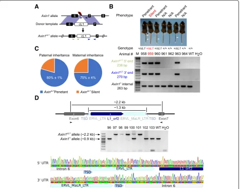
Next, we tested whether insertion of other repetitive se-quences can induce the kinky tail phenotype. We generated a repetitive sequence, called chimeric L1 (cL1) herein, con-sisting of a partial Orf2 of L1 and an LTR of MaLR, and inserted it into intron 6 of Axin1using the CRISPR/Cas9 (Fig. 1a and d and Additional file1: supplemental notes). To represent a retrotransposed sequence, we also included 6 bp target site duplications (TSDs) and 44 bp 5′extra nu-cleotides in the cL1 donor construct (Fig. 1d and Add-itional file 1: supplemental notes). We chose to use this specific chimeric L1 to mimic retrotransposition in vivo for the following reasons: First, such a chimeric sequence may result from template switching or transduction during ret-rotransposition, which is a pervasive phenomenon in both human and mouse genomes [17–19]. Second, the 762 bp Orf2 of L1 (Fig. 1d and Additional file 1: supplemental notes), which harbors a partial Z-motif and a partial reverse transcriptase domain, is highly conserved among different L1 families [44–46]. When aligning the Orf2 sequence against the mm10 genome with BLAT [47], one exact match was found on chromosome 3:76892981–76,893,742 and 70 other hits showed > 80% sequence identity (Add-itional file2: Table S1). Moreover, the MaLR elements are the most abundant LTR retrotransposon sequences in both human and mouse genomes [3]. When aligning the LTR of the MaLR to the mouse genome mm10, 20 perfect matches were found and over 200 other hits showed > 96% sequence identity (Additional file 3: Table S2). Therefore, insertion of the two conserved regions (L1 Orf2 and MaLR LTR) of TEs into the genome allows for studying the com-bined genetic/epigenetic impact at this locus. Finally, both TE fragments have no retrotransposition capability, which is further confirmed by our assays for DNA copy number variation (Additional file1: Figure S1C).
We obtained 3 founders (F0) carrying the cL1 insertion, which was confirmed by Sanger sequencing of the long-range PCR products containing the full-length cL1 (Fig.1d). One of the three founders showed a strong kinky tail phenotype, while the other two had normal tails, des-pite the same genotype (Axin1+/cL1). By further breeding the F0 s with WT mice, we obtained F1 heterozygous mice. Intercrossing F1 heterozygous mice produced WT, heterozygous and homozygous F2 mice at the Mendelian ratio (Fig.1d). All homozygous (Axin1cL1/cL1) mice showed kinky tails and also displayed neuronal abnormalities char-acterized by motor discordances (e.g., spinning with shaky heads and imbalance), whereas ~ 80% of the heterozygous (Axin1+/cL1) mice showed the kinky tail phenotype and the remaining heterozygous mice had normal tails (Fig. 1 b
and c). These results suggest that a chimeric L1/MaLR se-quence, rather than IAP solo LTR, can cause the kinky tail phenotype once inserted into intron 6 ofAxin1in mice.
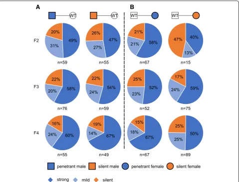
Stable transmission of the kinky tail phenotype with a fixed penetrance across multiple generations
AxinFu (Axin1+/Fu) mice showed kinky tails with highly variable severity, and the penetrant AxinFu mice (with strong or mild kinky tails) produced more penetrant off-spring compared to those silent ones (without kinky tails) [38]. This phenotype can be transmitted to the next gener-ation in a metastable manner, and the phenotypic variability correlates with the methylation status of the IAP retrotran-sposed into intron 6 in the offspring [38]. To examine whether the kinky tail phenotype induced by cL1 also displays a similar variability in phenotypic severity, we conducted breeding experiments to test the transmission of the phenotype through either paternal or maternal germline across three generations. Heterozygous AxincL1 (Axin1+/cL1) penetrant and silent male F2 s were bred with WT females, and ~ 80% of theAxincL1offspring (F3 s) were penetrant mice (Fig.2a). Further breeding of the penetrant and silent F3 and F4AxincL1 males and females with WT controls led to F4 and F5 AxincL1offspring displaying the kinky tail phenotype with similar penetrance (~ 80%) (Fig.
2a). The phenotypic penetrance stayed the same (at ~ 80%) across all three generations when the cL1 insertion was propagated through either the paternal or the maternal germline (Fig.1c and Fig.2). Among the penetrantAxincL1 mice, the ones with stronger kinky tails accounted for ~ 70% across all three generations, regardless of paternal or maternal inheritance (Fig.2a and b), suggesting the kinky tail phenotype can be stably inherited transgenerationally as long as the cL1 insertion exists.
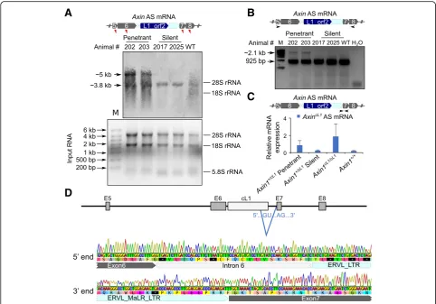
The kinky tail phenotype inAxincL1mice is caused by an aberrantly splicedAxin1transcript
Northern blot results, our RT-PCR analyses using primers specific to exons 5 and 8 also found an alternative splicing event in penetrant, but not in silentAxincL1mice (Fig.3b). Sanger sequencing of the longer isoform revealed that part of the cL1 (L1-MaLR) sequence was included in the aber-rant Axin1 transcript, which was spliced at the canonical GU-AG splicing site, in penetrant mice (Fig.3d and Add-itional file 1: supplemental notes). We further designed specific primers for the alternatively spliced Axin1 tran-script. qPCR confirmed that the alternative splicedAxin1 transcript is exclusively expressed in penetrant AxincL1
mice (Fig. 3c). Taken together, these data strongly suggest that the kinky tail phenotype in theAxincL1mice is induced by an aberrantly spliced Axin1 transcript due to intron retention of cL1.
Altered H3K9ac modification, rather than DNA methylation changes, correlates with the aberrantly splicedAxin1mRNA
Despite the same genotype (Axin1+/cL1), only ~ 80% of AxincL1mice express the aberrant transcripts with par-tial cL1 retention. Therefore, epigenetic mechanisms
A
B
C
D
Fig. 1Generation ofAxin1cL1mice using CRISPR/Cas9 and phenotypic characterization. a Schematics showing the strategy for generatingAxincL1mice. The
red lightning bolt represents the gRNAs used to target the reverse strands of the genomic DNA. The black arrows show the position of internal primers in theAxin1+allele, whereas the blue and the light green arrows indicate those for amplifying the 5′and 3’ends of theAxin1cL1allele, respectively. The
expected size of PCR products is indicated in the same color in the lower panels ofb.bImage of a representative litter of seven F5 mice derived from a F4 silentAxincL1(Axin1+/cL1) female mouse bred with a WT male, including three penetrant (white arrows pointing to the kinked regions in the tails), one
silentAxincL1mouse and three WT (Axin1+/+) littermates (Upper panel). Genotyping results of these mice are shown in lower panels and the positions of
the three sets of primers used are marked ina.cPie charts showing the distribution of penetrant (with kinky tails) vs. silent (without kinky tails)AxincL1
(Axin1+/cL1) mice in an outbreeding scheme (Axin1+/cL1× WT) across three generations. Data are represented as means ± SEM (n= 353 for paternal
transgenerational inheritance, andn= 425 for maternal transgenerational inheritance).dConfirmation of cL1 insertion into intron 6 ofAxin1. Long-range PCR was used to amplify fragments derived fromAxincL1(~ 2.2 kb) and WT (~ 0.9 kb) alleles (upper and middle panels), and the PCR products were
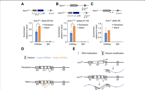
are likely involved. Given that DNA methylation and histone modifications of the IAP LTR sequences in AxinFu mice have been correlated with the variable phenotypic severity [38, 54], we first examined DNA methylation of cL1 and its flanking regions in both penetrant and silentAxincL1mice. Surprisingly, bisulfite sequencing showed that DNA methylation patterns were not significantly altered between penetrant and silent AxincL1 mice (Additional file 1: Figure S3 A and B). Additionally, no major changes in DNA methylation were found between the two groups by both methylated DNA immunoprecipitation (MeDIP) of 5-methylcyto-sine (5mC) and HhaI restriction enzyme (RE) digestion, which cleaves unmethylated GCGC site specifically, followed by qPCR (MeDIP-qPCR and RE-qPCR) (Add-itional file1: Figure S3C). Taken together, these results
suggest that the aberrant alternative splicing of Axin1 transcript is not due to altered DNA methylation. Given that histone modifications (e.g. H3K9ac) affect alternative splicing [55–57], and H3K9ac and H4K20me3 marks have been associated with proper splicing of intron 6 of Axin1 [54], we performed chromatin immunoprecipitation followed by qPCR (ChIP-qPCR) to examine H3K9ac levels (Fig. 4 a-c) at the AxincL1 locus. Levels of H3K9ac, a histone mark for open chromatin structure, were much higher at the cL1 insertion site in the silent than in the pene-trant mice (Fig. 4 a and b). These data suggest that a reduction in H3K9ac levels on the cL1 insertion site and its neighboring regions may affect splicing, lead-ing to the production of a longer transcript contain-ing cL1 (Fig. 4 d).
A
B
Fig. 2Transgenerational inheritance of the kinky tail phenotype inAxincL1(Axin1+/cL1) mice.aPaternal transgenerational inheritance of the kinky tail phenotype amongAxincL1mice. Male penetrant (left panel) and silent (right panel)AxincL1mice were bred with wild type (WT) females, and the percentage of strong (dark blue) or mild (light blue) kinky and silent (orange)AxincL1offspring, as well as the number of mice counted are indicated (Note that WT pups were excluded from the analyses).bMaternal transgenerational inheritance of the kinky tail phenotype among
Discussion
Mutant mice with variable yellow agouti coat color and kinky tail phenotypes were first reported 82 and 57 years ago, respectively [58, 59]. It was not until ~ 20 years ago that these phenotypes were correlated with spontaneous retrotransposition of IAPs in the mouse A(agouti) and Axin1 loci, respectively [35, 36]. However, validation by inserting the IAP into these loci to recapitulate the pheno-types in different strains of mice has not been reported. Moreover, identification of a locus that is sensitive to retro-transposition, and tends to produce a visually discernable phenotype (e.g., kinky tails, coat color changes, etc.) as a result of functional disruptions would be ideal for investi-gating the effects of retrotransposition in vivo. To this end, we generated a number of mouse lines by inserting various repetitive sequences into exactly the same genomic loca-tion in eitherA(agouti)orAxin1locus as that reported in
AvyorAxinFumice [37–40]. Interestingly, we found that in-sertion of IAP solo LTR induced no phenotypes, whereas insertion of a composite cL1 sequence into Axin1 locus caused the kinky tail phenotype, which can be transmitted faithfully across multiple generations. These findings indi-cate that intronic retrotransposition events do not neces-sarily cause disruptions in the host genes leading to discernable phenotypes and that the effects of retrotran-sposition depend on sequence context and organization. Indeed, previous studies have shown that heterozygotes of 3 spontaneous mutations inAxin1 gene, includingAxinFu (AxinFused), AxinKi (AxinKinky) andAxinKb(AxinKnobby),all display the kinked tail phenotype, yet heterozygotes of a transgenic line called AxinTg1 showed no phenotype [35]. Furthermore, AxinFu homozygotes are viable, whereas AxinKi, AxinKb andAxinTg1 homozygotes die around em-bryonic days 8–10 [35]. It is highly likely that the variable
A
B
C
D
Fig. 3A longerAxin1transcript isoform containing partial cL1 is exclusively expressed in the penetrantAxincL1mice.aA representative Northern blot showing that the wild type transcript (~ 3.8 kb) was detected inAxincL1mice, both penetrant and silent, as well as wild type mice, whereas a longer transcript (~ 5 kb) was present only in penetrantAxincL1mice (middle panel). Red triangles indicate the relative positions of the probes used in the upper panel, and the total RNA inputs are shown in the lower panel.bRT-PCR detection of the longer transcript isoform unique to penetrantAxincL1
phenotypes among these strains reflect the positional ef-fects of different insertions, e.g., theAxinTg1mice contain a ~ 600 bp transgene replacing exon 2, whereasAxinFu and AxinKbcontain an IAP insertion in intron 6 and exon 7, re-spectively [35]. The lack of phenotype in mice carrying an insertion of IAP solo LTR into intron 6 ofAxin1or pseudo exon 1A ofagouti (A)loci is consistent with a recent report [21] showing that IAP LTR rarely displays promoter activ-ity in vivo. Given that the IAP solo LTR sequence used was a part of the full-length IAP identified in Avyand AxinFu mice, the negative finding hints that other parts of the full-length IAP sequences may contain certain hidden fea-tures (e.g., subtle sequence variations and/or RNA modifi-cations), which are required for functional disruption of the host genes and consequently the induction of the kinky tail or variable yellow coat color phenotypes.
Although insertions of full-length IAP or cL1 into intron 6 ofAxin1locus all induced the kinky tail phenotype, the
underlying mechanisms appear to be different. In AxinFu, IAP insertion into intron 6 compromises Axin1 gene expression by producing a truncated transcript, which is inversely correlated to DNA methylation status [38]. In contrast, in our AxincL1 mice, while the inserted cL1 sequence displays neither promoter activities in vitro nor DNA methylation changes in vivo, production of the aber-rant transcript resulting from the retention of cL1 appears to correlate with significantly reduced levels of H3K9ac. Supporting our findings, reduced H3K9ac has been shown to cause alternative exon retention in Ncam(Neural cell adhesion molecule) due to decreased RNA polymerase processivity [56, 57]. Moreover, H3K9ac is also signifi-cantly more enriched in the IAP LTR of theAxinFulocus in embryos sired by penetrant males than those by silent males [54]. A recent study [53] reports that MaLR LTRs function as splicing donors rather than splicing acceptors, which is consistent with our data showing that the MaLR
A
B
C
D
E
Fig. 4Reduced H3K9ac levels at the cL1 insertion site in penetrantAxincL1mice.aChIP-qPCR analyses of H3K9ac levels using primers specific to the exon 6 splicing site of theAxin1cL1allele. Arrows indicate relative locations of the primers used for ChIP-qPCR analyses (upper panel). Data are
presented as means ± SEM,n= 3, *p< 0.05.bChIP-qPCR analyses of H3K9ac levels using primers specific to the exon 7 splicing site of theAxin1cL1and
Axin1+alleles. Arrows indicate relative locations of the primers used for ChIP-qPCR analyses (upper panel). Data are presented as means ± SEM,n= 3, *p< 0.05.cChIP-qPCR analyses of H3K9ac levels using primers specific to the exon 6 splicing site of theAxin1+allele. Arrows indicate relative locations of the
primers used for ChIP-qPCR analyses (upper panel). Data are presented as means ± SEM,n= 3.dSchematic illustration showing the effect of reduced H3K9ac levels on splicing. Briefly, higher H3K9ac levels ensure correct splicing, which excludes cL1 from the transcript, whereas with lower H3K9ac levels, the cL1 tends to be retained and included in the transcript.eComparison of the molecular mechanisms underlying the kinky tail phenotype between
AxinFuandAxincL1mice. The kinky tail phenotype inAxinFumice results from a shorter transcript isoform initiated from intron 6, and the phenotypic severity is inversely correlated with DNA methylation status, whereas the kinky tail phenotype inAxincL1mice is caused by a longer transcript isoform with cL1
LTR in the cL1 serves as a splicing donor. While associa-tions between H3K9ac levels and aberrant splicing have been established [55–57], the underlying mechanism re-mains elusive. InAxin1cL1mice, the longer splicing variant containing partial chimeric L1 sequence possesses several premature termination codons (PTCs), which are well known to cause transcript degradation via the nonsense mRNA decay (NMD) pathway [60–62]. However, our Northern blot results revealed that the longer transcript, which is unique to the penetrantAxincL1mice, was nearly as abundant as the shorter wild-type one, suggesting that the splicing variant does not undergo NMD-mediated degradation. Therefore, it is highly likely that the longer splicing variant is translated into a mutant form of AXIN1 with a truncated C terminus lacking DIX domain, as compared to wild-type AXIN1. Unfortunately, we have not been able to identify a commercial antibody that could detect wild-type AXIN1 correctly (~92kD protein). Pro-duction of good AXIN1 antibodies and generation of a mouse model over-expressing the splicing variant/mutant AXIN1 lacking DIX domain would provide the ultimate evidence supporting the cause-effect relationship between the splicing variant/mutant AXIN1 without DIX domain and the kinky tail phenotype in the future. Together, our data suggest that the Axin1 locus is sensitive to genetic and epigenetic alterations caused by retrotransposition and thus, can serve as an ideal genomic location for study-ing the effects of retrotransposition on host gene expres-sion and activities of nearby genome. With advancement of the CRISPR/Cas9 technology, TEs of interest can easily be inserted into theAxin1locus and the effects of various TEs onAxin1and nearby genome can be analyzed in vivo. Transgenerational epigenetic inheritance of the vari-able yellow agouti coat color and kinky tail phenotypes in Avyand AxinFu mice is of great interest although the underlying mechanism remains elusive. In AxinFu mice, the variable DNA methylation levels of IAP inversely correlate with the severities of the kinky tail phenotype, and penetrant mice tend to produce more penetrant off-spring [38]. DNA methylation undergoes two waves of reprogramming during fertilization and germ line specification [63] and IAP seems to be resistant to these reprogramming events [38], which may explain the transgenerational inheritance of the phenotypes in Avy and AxinFu mice. However, in AxincL1 mice, the kinky tail phenotype occurs as long as the cL1 insertion is present, and the penetrance is fixed at ~ 70–80%. There-fore, the kinky phenotype most likely represent a genetic phenomenon at first glance, and the stable inheritance of this phenotype across multiple generations inAxincL1 mice appears to be a simple genetic, rather than an epi-genetic, transmission, i.e., a cL1 insertional mutation causes the phenotype in each generation. However, partial penetrance (70–80%) of the phenotype can only
be explained by an epigenetic mechanism. Our data have linked H3K9ac to the aberrant splicing events, but it re-mains unknown how such an alteration in histone modi-fications causes aberrant splicing at a rate of 70–80% rather than 100%.
In summary, we show that insertion of a chimeric L1 into intron 6 of Axin1 affects histone modification pat-terns on cL1 and its neighboring regions, leading to the production of an aberrant Axin1 transcript correlated with the kinky tail phenotype. This mechanism is differ-ent from that previously iddiffer-entified in mice with spontan-eous IAP retrotransposition (e.g., AxinFuand Avy mice), which results from DNA methylation changes. Axin1 locus may serve as an ideal genomic location for study-ing the effects of new retrotransposition events on target gene function in mice in vivo.
Conclusions
Despite their widespread distribution in the human gen-ome, effects of retrotransposons on their host genes and nearby genome have not been exhaustively investigated in vivo. Here, we show that insertion of a chimeric L1 into intron 6 of Axin1 locus in mice could induce the kinky tail phenotype due to the production of an aber-rantly spliced transcript isoform, which is associated with altered histone modifications rather than DNA methylation changes. Together with previous reports, our data strongly suggest thatAxin1is an ideal locus for studying the effects of retrotransposition on host gene expression and nearby genome activities in vivo.
Methods
Animal use and care
All the mice used in this study were on C57Bl/6 J back-ground, and housed under specific pathogen-free condi-tions in a temperature- and humidity- controlled animal facility at the University of Nevada, Reno.
The gRNAs, Cas9 mRNA and donor DNA template were microinjected into mice zygotes of FVB/NJ × C57BL/6 J background for Axin1 locus knock-in and C57BL/6 J for A locus knock-in. The genomic DNA of founder mice from tail tips or ear snips were extracted for PCR-based genotyping. Founder mice were out-crossed with C57BL/6 J WT to obtain heterozygous F1 (Axin+/IAP or Axin+/cL1). For AIAP, F1 s were outcrossed with C57BL/6 J WT to obtain heterozygous F2 s, and coat color was recorded. For AxinIAP, F1 s were out-crossed with C57BL/6 J WT to obtain heterozygous F2 s, and tail phenotype was examined. ForAxincL1, F1 s were outcrossed with C57BL/6 J WT to obtain heterozygous F2 s, and tail phenotype was recorded. Penetrant and si-lent heterozygous F2 s, F3 s, and F4 s were further out-crossed with C57BL/6 J WT to obtain the breeding data across multiple generations. Primers used for all the constructs are listed in Additional file4: Table S3.
Mouse genotyping
Mouse tail or ear snip samples were lysed in a lysis buffer containing 40 mM NaOH (221465, Sigma Aldrich) and 0.2 mM EDTA (46–034-CI, Corning) for 1 h at 95 °C, followed by neutralization with the same volume of the neutralizing buffer containing 40 mM Tris-HCl (15567027, Thermo Fisher Scientific). PCR-based genotyping of the AxincL1, AxinIAP and AIAPwas conducted using the GoTaq Green master mix (M7123, Promega) or Platinum™ SuperFi™ Green PCR Master Mix (12359010, Thermo Fisher Scien-tific). Primers used for genotyping are listed in Additional file4: Table S3.
Dual luciferase assay
Different fragments of the repetitive sequences were ampli-fied from donor templates and then used to replace the SV40 early promotor that drives the expression of the Renillaluciferase-coding sequence in the psiCHECK-2 plas-mid (C8021, Promega). HEK293 cells were transfected with psiCHECK-2 containing the different fragments from the re-petitive sequences using Lipofectamine 2000 (11668019, Thermo Fisher Scientific) in a 24-well cell culture plate (CLS3527-100EA, Corning). After 24 h, cells were lysed and used for the Dual Luciferase Assay (E1910, Promega) ac-cording to the manufacturer’s instructions. The psiCHECK-2 and psiCHECK-2 vectors with deletion of the SV40 early promotor of the Renilla luciferase-coding sequence were used as positive and negative controls, respectively.Renilla luciferase signals were normalized toFireflyluciferase signals to correct the transfection efficiency. Primers used for all the constructs are listed in Additional file4: Table S3.
DNA, RNA extraction and cDNA synthesis
DNA and RNA were extracted from kidneys and tail snips from penetrant and silent mice using the Quick-DNA Plus
Kits (D4074, Zymo Research) and mirVana miRNA Isola-tion Kit (AM1560, Thermo Fisher Scientific), respectively, according to the manufacturer’s instructions. Briefly, kid-ney or tail samples were homogenized in 600μL of Lysis/ Binding Buffer with homogenizer (D1000, Benchmark), followed by centrifugation to remove cell debris. The supernatant was passed through a column, in which the genomic DNA was retained, whereas the RNA got eluted. For genomic DNA extraction, the column containing genomic DNA was treated with a genomic lysis buffer at room temperature for 10 min, followed by washing with a DNA Pre-Wash Buffer once and a g-DNA Wash Buffer twice. The genomic DNA was eluted with nuclease-free water and stored at −80 °C for further use. For RNA extraction, 60μL of miRNA Homogenate Additive was added into the flow-through followed by incubation on ice for 10 min. The mixture was subjected to Phenol: Chloro-form RNA extraction, and total RNA was isolated accord-ing to the manufacturer’s instructions. cDNA synthesis was performed using SuperScript II Reverse Transcriptase (18064014, Thermo Fisher Scientific) with random primers. qPCR and long-range PCR were performed using the Fast SYBR Green Master Mix (4385612, Thermo Fisher Scientific) and PrimeSTAR GXL DNA Polymerase (R050B, TaKaRa), respectively. Primers used for qPCR and long-range PCR are listed in Additional file4: Table S3.
Bisulfite sequencing
Genomic DNA samples were bisulfite-converted using the EZ DNA Methylation-Gold™ Kit (D5005, Zymo Re-search). PCR was performed using the TaKaRa EpiTaq™ HS enzyme (for bisulfite-treated DNA) (R110B, TaKaRa), which is more tolerant to dUTP-containing templates, with Tm at 55 °C for 40 cycles. PCR products were li-gated into the pGEM®-T Easy Vector (A1360, Promega) for Sanger sequencing. Primers used for bisulfite sequen-cing are listed in Additional file4: Table S3.
Methylated DNA immunoprecipitation-qPCR (MeDIP-qPCR)
room temperature for 2 h. Then the strip wells were washed with the wash buffer three times. The antibody-enriched DNA was eluted with a DNA release buffer con-taining proteinase K and purified with columns. qPCR was performed to identify DNA methylation levels. Primers used for MeDIP-qPCR are listed in Additional file
4: Table S3.
Northern blot
Northern blot analyses were performed using a Northern-Max® Kit (AM1940, Thermo Fisher Scientific) and a Biotin Chromogenic Detection Kit (K0662, Thermo Fisher Scien-tific) following the manufacturer’s instructions. Briefly, RNA extracted from kidney was mixed with 3 volumes of a formaldehyde-containing loading dye followed by denatur-ation at 65 °C for 15 min. The denatured RNA was the fractionated through 1 × MOPS Gel Running Buffer (1% denaturing gel with a voltage of 140 V for 30 min). Then the RNA was transferred onto a BrightStar®-Plus Positively Charged Nylon Membrane (AM10100, Thermo Fisher Scientific) using Novex™ Semi-Dry Blotter (SD1000, Thermo Fisher Scientific) in 1× TBE buffer with a voltage of 20 V for 30 min. After transfer, the membrane was rinsed 1× Gel Running Buffer, then crosslinked in Spectrolinker™ XL-1500 UV crosslinker (Spectronics Corporation) followed by baking at 80 °C for 15 min. The crosslinked membrane was prehybridized at 65 °C in a preheated ULTRAhyb Buffer in a roller bottle in a hybridization oven at 42 °C for 30 min, followed by incubation with 10pM bio-tinylated probe (IDT) in the ULTRAhyb Buffer at 42 °C overnight. After rinsing with 1 × Blocking/Washing Buffer for 5 min three times at room temperature, the membrane was blocked with 1 × Blocking Buffer for 30 min in a shaker at room temperature. Following blocking, the membrane was incubated with Streptavidin-AP conjugate for 1 h at room temperature, then washed with 1 × Blocking/Wash-ing Buffer for 5 min three times and 1× detection buffer for 10 min. Then the membrane was incubated with freshly prepared NBT/BCIP Substrate Solution at room temperature in the dark. 2 h later, the reaction was stopped by rinsing with double deionized water. Probes used for Northern blot are listed in Additional file4: Table S3.
Chromatin immunoprecipitation followed by quantitative PCR (ChIP-qPCR)
ChIP-qPCR was performed as previously described [64]. Briefly, tail snips were lysed on ice for 30 min in 600μl of buffer 1 plus detergents [15 mM Tris-HCl (pH 7.5) (1556 7027, Thermo Fisher Scientific), 60 mM KCl (P217–500, Fisher Scientific), 5 mM MgCl2(BP214–500, Fisher
Scien-tific) and 0.1 mM EGTA (O2783–100, Fisher Scientific), 0.3 M sucrose (freshly added) (0335-5KG, Amresco), 10 mM DTT (freshly added) (GE17–1318-02, GE Healthcare), 0.25% (volume/volume) NP-40 (NP40S-500ML, Sigma
Aldrich) and 0.5% (weight/volume) sodium deoxycholate (freshly prepared) (D6750-100G, Sigma Aldrich)]. Then 600μl of MNase buffer [85 mM Tris-HCl, pH 7.5, 3 mM MgCl2, 2 mM CaCl2 (C7902-500G, Sigma Aldrich) and 0.3
M sucrose (freshly added)] was added into the lysed solu-tion. The mixture was aliquoted into 200μl per tube to ob-tain sufficient digestion, followed by Micrococcal Nuclease (M0247S, NEB) digestion at 37 °C in a thermomixer for 5 min and then terminated by adding 2ul of 0.5 M EDTA (46–034-CI, Corning) and incubation on ice for 5 min. The digested sample was then centrifuged at 15,000×g for 10 min at room temperature to remove cell debris, followed by adding protease inhibitors to the chromatin. 200μl of the mixture was saved as input DNA. After preclearing of the chromatin with blocked protein G beads (10004D, Thermo Fisher Scientific), 3μl of H3K9ac antibody (ab4441, Abcam) was added into the precleared chromatin and incubated at 4 °C overnight. Then the chromatin was incubated with blocked protein G beads at 4 °C for 4 h, followed by washing with wash buffer A (50 mM Tris-HCl (pH 7.5), 10 mM EDTA and 75 mM NaCl (BP358–10, Fisher Scientific)) once and wash buffer B (50 mM Tris-HCl (pH 7.5), 10 mM EDTA and 125 mM NaCl) twice. Then the chromatin was eluted by resuspending in 150μl of elution buffer (1% (weight/volume) SDS (L4509-500G, Sigma Aldrich) in TE) at 25 °C in a thermomixer twice. The eluted chromatin was then subjected to RNase A (EN0531, Thermo Fisher Scientific) and proteinase K (P8107S, NEB) digestion followed by phenol/chloroform extraction of DNA. The pull-down DNA and input DNA were used for qPCR using the Fast SYBR Green Master Mix (4385612, Thermo Fisher Scientific). Primers used for ChIP-qPCR are listed in Additional file4: Table S3.
Statistical analysis
All data were presented as mean ± SEM, and statistical differences were assessed by the Two-sample t test un-less stated otherwise. p< 0.05 was considered as signifi-cant differences.
Additional files
Additional file 1:Figure S1, S2, S3 and Supplemental notes.
Generation ofAxinIAPandAIAPfounder mice and copy number variation
assays forAxincL1mice (Figure S1); promoter activity analyses (Figure S2);
DNA methylation levels around cL1 in penetrant and silentAxincL1mice
(Figure S3); and annotated sequences of retrotransposons used in this study (Supplemental notes). (PDF 5204 kb)
Additional file 2: Table S1.Orf2 BLAT results. (XLSX 52 kb)
Additional file 3: Table S2.MaLR BLAT results. (XLSX 48 kb)
Additional file 4: Table S3.Oligos used in this study. (XLSX 11 kb)
Abbreviations
5mC:5-methylcytosine;Avy: Agouti viable yellow;AxincL1:Axin1+/cL1;
AxinFu:AxinFused;AxinKb:AxinKnobby;AxinKi:AxinKinky; BLAT: BLAST-like alignment
immunoprecipitation; cL1: Chimeric L1; CRIPSR: Clustered regularly interspaced short palindromic repeats; ERV: Endogenous retroviruse; IAP: Intracisternal A-type particle; L1: LINE-1; LINE: Long interspersed element; LTR: Long terminal repeat; MaLR: Mammalian apparent LTR retrotransposon; MeDIP: Methylated DNA immunoprecipitation; ORF: Open reading frame; qPCR: Quantitative PCR; RE: Restriction enzyme; SINE: Short interspersed elements; SVA: SINE-VNTR-Alu; TE: Transposable element; TSDs: Target site duplications; UTR: Untranslated region; WT: Wild type
Acknowledgements
Not applicable.
Funding
This work was supported by grants from the NIH (P30GM110767, HD071736 and HD085506 to WY; R21HD080143 and P50GM107632 to WA), the Markl Faculty Scholar Fund (to WA) and the Templeton Foundation (PID: 50183 to WY).
Availability of data and materials
All data generated or analyzed during this study are included in this published article and its supplementary information files.
Authors’contributions
WY and ZW designed the research. ZW, HM, SJN, YW, DO, CT, SL, SW, SY, PY and HZ performed the experiments. WA contributed reagents and protocols, and edited the manuscript; all analyzed the data; WY and ZW wrote the manuscript. All authors read and approved the final manuscript.
Ethics approval and consent to participate
The animal use protocol was approved by the Institutional Animal Care and Use Committee (IACUC) of the University of Nevada, Reno (protocol number 00494).
Consent for publication
Not applicable.
Competing interests
The authors declare that they have no competing interests.
Publisher’s Note
Springer Nature remains neutral with regard to jurisdictional claims in published maps and institutional affiliations.
Author details
1Department of Physiology and Cell Biology, University of Nevada School of
Medicine Center for Molecular Medicine, Room 207B 1664 North Virginia Street MS/0575, Reno, NV 89557, USA.2Avera McKennan Hospital and
University Health Center, Sioux Falls, SD 57108, USA.3Department of Pharmaceutical Sciences, South Dakota State University, Brookings, SD 57007, USA.4Department of Obstetrics and Gynecology, University of Nevada, Reno
School of Medicine, Reno, NV 89557, USA.5Department of Biology, University
of Nevada, Reno, Reno, NV 89557, USA.
Received: 19 January 2019 Accepted: 21 April 2019
References
1. de Koning AP, Gu W, Castoe TA, Batzer MA, Pollock DD. Repetitive elements may comprise over two-thirds of the human genome. PLoS Genet. 2011; 7(12):e1002384.
2. Kazazian HH Jr, Moran JV. Mobile DNA in health and disease. N Engl J Med. 2017;377(4):361–70.
3. Mager DL, Stoye JP. Mammalian endogenous retroviruses. Microbiol Spectr. 2015;3(1):MDNA3-0009-2014.
4. Akagi K, Li J, Stephens RM, Volfovsky N, Symer DE. Extensive variation between inbred mouse strains due to endogenous L1 retrotransposition. Genome Res. 2008;18(6):869–80.
5. Nellaker C, Keane TM, Yalcin B, Wong K, Agam A, Belgard TG, Flint J, Adams DJ, Frankel WN, Ponting CP. The genomic landscape shaped by selection on transposable elements across 18 mouse strains. Genome Biol. 2012;13(6):R45.
6. Mouse Genome Sequencing C, Waterston RH, Lindblad-Toh K, Birney E, Rogers J, Abril JF, Agarwal P, Agarwala R, Ainscough R, Alexandersson M, et al. Initial sequencing and comparative analysis of the mouse genome. Nature. 2002;420(6915):520–62.
7. Dewannieux M, Esnault C, Heidmann T. LINE-mediated retrotransposition of marked Alu sequences. Nat Genet. 2003;35(1):41–8.
8. Hancks DC, Goodier JL, Mandal PK, Cheung LE, Kazazian HH Jr.
Retrotransposition of marked SVA elements by human L1s in cultured cells. Hum Mol Genet. 2011;20(17):3386–400.
9. Raiz J, Damert A, Chira S, Held U, Klawitter S, Hamdorf M, Lower J, Stratling WH, Lower R, Schumann GG. The non-autonomous retrotransposon SVA is trans-mobilized by the human LINE-1 protein machinery. Nucleic Acids Res. 2012;40(4):1666–83.
10. Esnault C, Maestre J, Heidmann T. Human LINE retrotransposons generate processed pseudogenes. Nat Genet. 2000;24(4):363–7.
11. Wei W, Gilbert N, Ooi SL, Lawler JF, Ostertag EM, Kazazian HH, Boeke JD, Moran JV. Human L1 retrotransposition: cis preference versus trans complementation. Mol Cell Biol. 2001;21(4):1429–39.
12. Moran JV, DeBerardinis RJ, Kazazian HH Jr. Exon shuffling by L1 retrotransposition. Science. 1999;283(5407):1530–4.
13. Pickeral OK, Makalowski W, Boguski MS, Boeke JD. Frequent human genomic DNA transduction driven by LINE-1 retrotransposition. Genome Res. 2000;10(4):411–5.
14. Goodier JL, Ostertag EM, Kazazian HH Jr. Transduction of 3′-flanking sequences is common in L1 retrotransposition. Hum Mol Genet. 2000;9(4):653–7. 15. Solyom S, Ewing AD, Hancks DC, Takeshima Y, Awano H, Matsuo M,
Kazazian HH Jr. Pathogenic orphan transduction created by a nonreference LINE-1 retrotransposon. Hum Mutat. 2012;33(2):369–71.
16. Tubio JMC, Li Y, Ju YS, Martincorena I, Cooke SL, Tojo M, Gundem G, Pipinikas CP, Zamora J, Raine K, et al. Mobile DNA in cancer. Extensive transduction of nonrepetitive DNA mediated by L1 retrotransposition in cancer genomes. Science. 2014;345(6196):1251343.
17. Buzdin A, Ustyugova S, Gogvadze E, Vinogradova T, Lebedev Y, Sverdlov E. A new family of chimeric retrotranscripts formed by a full copy of U6 small nuclear RNA fused to the 3′terminus of l1. Genomics. 2002;80(4):402–6. 18. Symer DE, Connelly C, Szak ST, Caputo EM, Cost GJ, Parmigiani G, Boeke JD.
Human l1 retrotransposition is associated with genetic instability in vivo. Cell. 2002;110(3):327–38.
19. Gilbert N, Lutz-Prigge S, Moran JV. Genomic deletions created upon LINE-1 retrotransposition. Cell. 2002;110(3):315–25.
20. Thompson PJ, Macfarlan TS, Lorincz MC. Long terminal repeats: from parasitic elements to building blocks of the transcriptional regulatory repertoire. Mol Cell. 2016;62(5):766–76.
21. Kazachenka A, Bertozzi TM, Sjoberg-Herrera MK, Walker N, Gardner J, Gunning R, Pahita E, Adams S, Adams D, Ferguson-Smith AC. Identification, characterization, and heritability of murine metastable Epialleles: implications for non-genetic inheritance. Cell. 2018;175(5): 1259–71.e13.
22. Burns KH. Transposable elements in cancer. Nat Rev Cancer. 2017;17(7):415–24. 23. Elbarbary RA, Lucas BA, Maquat LE. Retrotransposons as regulators of gene
expression. Science. 2016;351(6274):aac7247.
24. Ostertag EM, DeBerardinis RJ, Goodier JL, Zhang Y, Yang N, Gerton GL, Kazazian HH Jr. A mouse model of human L1 retrotransposition. Nat Genet. 2002;32(4):655–60.
25. An W, Han JS, Wheelan SJ, Davis ES, Coombes CE, Ye P, Triplett C, Boeke JD. Active retrotransposition by a synthetic L1 element in mice. Proc Natl Acad Sci U S A. 2006;103(49):18662–7.
26. Muotri AR, Chu VT, Marchetto MC, Deng W, Moran JV, Gage FH. Somatic mosaicism in neuronal precursor cells mediated by L1 retrotransposition. Nature. 2005;435(7044):903–10.
27. Garcia-Perez JL, Morell M, Scheys JO, Kulpa DA, Morell S, Carter CC, Hammer GD, Collins KL, O'Shea KS, Menendez P, et al. Epigenetic silencing of engineered L1 retrotransposition events in human embryonic carcinoma cells. Nature. 2010;466(7307):769–73.
28. Kannan M, Li J, Fritz SE, Husarek KE, Sanford JC, Sullivan TL, Tiwary PK, An W, Boeke JD, Symer DE. Dynamic silencing of somatic L1 retrotransposon insertions reflects the developmental and cellular contexts of their genomic integration. Mob DNA. 2017;8:8.
30. Kano H, Godoy I, Courtney C, Vetter MR, Gerton GL, Ostertag EM, Kazazian HH Jr. L1 retrotransposition occurs mainly in embryogenesis and creates somatic mosaicism. Genes Dev. 2009;23(11):1303–12.
31. Newkirk SJ, Lee S, Grandi FC, Gaysinskaya V, Rosser JM, Vanden Berg N, Hogarth CA, Marchetto MCN, Muotri AR, Griswold MD, et al. Intact piRNA pathway prevents L1 mobilization in male meiosis. Proc Natl Acad Sci U S A. 2017;114(28):E5635–E44. 32. Grandi FC, Rosser JM, Newkirk SJ, Yin J, Jiang X, Xing Z, Whitmore L, Bashir
S, Ivics Z, Izsvak Z, et al. Retrotransposition creates sloping shores: a graded influence of hypomethylated CpG islands on flanking CpG sites. Genome Res. 2015;25(8):1135–46.
33. Maksakova IA, Romanish MT, Gagnier L, Dunn CA, van de Lagemaat LN, Mager DL. Retroviral elements and their hosts: insertional mutagenesis in the mouse germ line. PLoS Genet. 2006;2(1):e2.
34. Hancks DC, Kazazian HH Jr. Roles for retrotransposon insertions in human disease. Mob DNA. 2016;7:9.
35. Vasicek TJ, Zeng L, Guan XJ, Zhang T, Costantini F, Tilghman SM. Two dominant mutations in the mouse fused gene are the result of transposon insertions. Genetics. 1997;147(2):777–86.
36. Duhl DM, Vrieling H, Miller KA, Wolff GL, Barsh GS. Neomorphic agouti mutations in obese yellow mice. Nat Genet. 1994;8(1):59–65.
37. Morgan HD, Sutherland HG, Martin DI, Whitelaw E. Epigenetic inheritance at the agouti locus in the mouse. Nat Genet. 1999;23(3):314–8.
38. Rakyan VK, Chong S, Champ ME, Cuthbert PC, Morgan HD, Luu KV, Whitelaw E. Transgenerational inheritance of epigenetic states at the murine Axin (Fu) allele occurs after maternal and paternal transmission. Proc Natl Acad Sci U S A. 2003;100(5):2538–43.
39. Waterland RA, Jirtle RL. Transposable elements: targets for early nutritional effects on epigenetic gene regulation. Mol Cell Biol. 2003;23(15):5293–300. 40. Rosenfeld CS, Sieli PT, Warzak DA, Ellersieck MR, Pennington KA, Roberts RM.
Maternal exposure to bisphenol a and genistein has minimal effect on a (vy)/a offspring coat color but favors birth of agouti over nonagouti mice. Proc Natl Acad Sci U S A. 2013;110(2):537–42.
41. Cong L, Ran FA, Cox D, Lin SL, Barretto R, Habib N, Hsu PD, Wu XB, Jiang WY, Marraffini LA, et al. Multiplex Genome engineering using CRISPR/Cas systems. Science. 2013;339(6121):819–23.
42. Wang Z, Lee S, Oliver D, Yuan S, Tang C, Wang Y, Zheng H, Yan W. Prps1l1, a testis-specific gene, is dispensable for mouse spermatogenesis. Mol Reprod Dev. 2018;85(10):802–4.
43. Oliver D, Yuan S, McSwiggin H, Yan W. Pervasive genotypic mosaicism in founder mice derived from Genome editing through pronuclear injection. PLoS One. 2015;10(6):e0129457.
44. Sookdeo A, Hepp CM, McClure MA, Boissinot S. Revisiting the evolution of mouse LINE-1 in the genomic era. Mob DNA. 2013;4(1):3.
45. Mathias SL, Scott AF, Kazazian HH Jr, Boeke JD, Gabriel A. Reverse transcriptase encoded by a human transposable element. Science. 1991; 254(5039):1808–10.
46. Clements AP, Singer MF. The human LINE-1 reverse transcriptase:effect of deletions outside the common reverse transcriptase domain. Nucleic Acids Res. 1998;26(15):3528–35.
47. Kent WJ. BLAT--the BLAST-like alignment tool. Genome Res. 2002;12(4):656–64. 48. Goodier JL, Kazazian HH Jr. Retrotransposons revisited: the restraint and
rehabilitation of parasites. Cell. 2008;135(1):23–35.
49. Erwin JA, Marchetto MC, Gage FH. Mobile DNA elements in the generation of diversity and complexity in the brain. Nat Rev Neurosci. 2014;15(8):497–506. 50. Chen J, Rattner A, Nathans J. Effects of L1 retrotransposon insertion on
transcript processing, localization and accumulation: lessons from the retinal degeneration 7 mouse and implications for the genomic ecology of L1 elements. Hum Mol Genet. 2006;15(13):2146–56. 51. Li J, Kannan M, Trivett AL, Liao H, Wu X, Akagi K, Symer DE. An
antisense promoter in mouse L1 retrotransposon open reading frame-1 initiates expression of diverse fusion transcripts and limits
retrotransposition. Nucleic Acids Res. 2014;42(7):4546–62. 52. Li J, Akagi K, Hu Y, Trivett AL, Hlynialuk CJ, Swing DA, Volfovsky N,
Morgan TC, Golubeva Y, Stephens RM, et al. Mouse endogenous retroviruses can trigger premature transcriptional termination at a distance. Genome Res. 2012;22(5):870–84.
53. Franke V, Ganesh S, Karlic R, Malik R, Pasulka J, Horvat F, Kuzman M, Fulka H, Cernohorska M, Urbanova J, et al. Long terminal repeats power evolution of genes and gene expression programs in mammalian oocytes and zygotes. Genome Res. 2017;27(8):1384–94.
54. Fernandez-Gonzalez R, Ramirez MA, Pericuesta E, Calle A, Gutierrez-Adan A. Histone modifications at the blastocyst Axin1(Fu) locus mark the heritability of in vitro culture-induced epigenetic alterations in mice. Biol Reprod. 2010; 83(5):720–7.
55. Luco RF, Pan Q, Tominaga K, Blencowe BJ, Pereira-Smith OM, Misteli T. Regulation of alternative splicing by histone modifications. Science. 2010; 327(5968):996–1000.
56. Naftelberg S, Schor IE, Ast G, Kornblihtt AR. Regulation of alternative splicing through coupling with transcription and chromatin structure. Annu Rev Biochem. 2015;84:165–98.
57. Schor IE, Rascovan N, Pelisch F, Allo M, Kornblihtt AR. Neuronal cell depolarization induces intragenic chromatin modifications affecting NCAM alternative splicing. Proc Natl Acad Sci U S A. 2009;106(11):4325–30. 58. Reed SC. The inheritance and expression of fused, a new mutation in the
house Mouse. Genetics. 1937;22(1):1–13.
59. Dickies MM. A new viable yellow mutation in the house mouse. J Hered. 1962;53:84–6.
60. Kurosaki T, Maquat LE. Nonsense-mediated mRNA decay in humans at a glance. J Cell Sci. 2016;129(3):461–7.
61. Conti E, Izaurralde E. Nonsense-mediated mRNA decay: molecular insights and mechanistic variations across species. Curr Opin Cell Biol. 2005;17(3):316–25. 62. Wilkinson MF. A new function for nonsense-mediated mRNA-decay factors.
Trends Genet. 2005;21(3):143–8.
63. Yan W. Potential roles of noncoding RNAs in environmental epigenetic transgenerational inheritance. Mol Cell Endocrinol. 2014;398(1–2):24–30. 64. Hisano M, Erkek S, Dessus-Babus S, Ramos L, Stadler MB, Peters AH.