CC S.. =
BY NC ND
http://doi.org/10.5114/bta.2019.90239
Modeling of the phospholipid vesicular nanostructure
formation process
LESIA AVDIEIEVA1*, OLEKSANDR CHUNIKHIN2
1 Institute of Engineering Thermophysics, NAS of Ukraine, Kyiv, Ukraine
2?.V. Palladin Institute of Biochemistry, NAS of Ukraine, Kyiv, Ukraine
Abstract
This study presents the results of experimental studies and mathematical modeling of the process of vesicular structure formation from phospholipids under the influence of mechanisms of the discrete impulse input of energy (DIIE). The possibility of using this method for increasing the productivity of the process of obtaining vesicles from phospholipids is shown. Moreover, the use of the properties of lipid nanostructures obtained by the DIIE method for the composition of products of special therapeutic nutrition is proposed. The DIIE effect was realized in a flowing rotary-pulsation apparatus of a cylindrical type. The effectiveness of using this type of equipment and the high level of mechanical and physicochemical effects on the dispersed system with phospho-lipids are established. The results of the studies of the effect of certain regime parameters (such as initial tem-perature, material concentration, and angular velocity of the rotor) on the analysis of the aqueous suspension of phospholipids on the size distribution of the formed particles are presented. The heat-technological parameters of the process were selected for obtaining particles having an average diameter of up to 500 nm. The possibility of predicting the properties of the formed phospholipids structures obtained by the proposed treatment in a wide range of regime parameters is shown. For the simulation, a hybrid functional Petri net was used, which made it possible to combine the initial thermal technological conditions of the process (such as temperature and material concentration) and the characteristics of the process equipment (such as type of DIIE activator and rotor speed) in the mathematical model.
Key words: discrete-pulse input of energy, dispersion, lipid vesicular nanostructures, mathematical modeling,
hybrid functional Petri nets
Introduction
Nanotechnology provides materials with fundamen-tally new useful properties. These materials provide an opportunity for the active development of various techni-cal areas such as energy, ecology, information techno-logy, engineering, biotechno-logy, and medicine. At present, all over the world, development of nanotechnology belongs to priority areas and is based on the analysis of the re-sults of significant data sets of theoretical and experi-mental studies using mathematical modeling. An integra-ted approach allows to purposefully manage the techno-logical process of the structural formation of
nanoma-terials and to ensure rational conditions for its imple-mentation (Tibbals, 2010; Geckeler and Nishide, 2010; Tiwari and Tiwari, 2017; Naito et al., 2018).
In aqueous solutions, under the influence of the energy introduced into the system, phospholipids mole-cules form closed bilayer structures (vesicles) (Alipour et al., 2017; Yang et al., 2018; Berg et al., 2012). These nanostructures have attracted considerable interest because of their unique properties, namely, the possibi-lity of solubilization of substances of different nature and their direct transportation into biological cells. The ve-sicle shell protects the active substance from the
ence of negative external factors, has an affinity for cell membranes, and is biodegradable; therefore, it signifi-cantly increases the positive effect of the use of this form of nanopreparations (McClements and Gumus, 2016; Vance and Vance, 2008; Eliaz, 2011; Berg et al., 2012; Vitkova and Petrov, 2013).
Currently, vesicular structures are obtained from phospholipids using methods such as ultrasonic pro-cessing, filtering, injection methods and high-pressure homogenization (Tibbals, 2010). These methods enable obtaining nanostructures with predetermined proper-ties; however, they have several disadvantages such as low productivity and considerable energy consumption (Avdieieva, 2011). Consequently, using phospholipid nanostructures is limited to basic research as a model of cell membranes, as well as the production of drugs for diagnosis and treatment in medical and pharmaceutical industries (McClements and Gumus, 2016; Nii and Ishii, 2005; Eliaz, 2011; Vitkova and Petrov, 2013). Further-more, the use of phospholipids vesicles in the food in-dustry and agriculture to obtain nanopreparations with increased efficiency seems very promising (Mozafari et al., 2006, Sanguansri, 2006; Cushen et al., 2012; Tsai, 2016). The increased interest in the potential of phos-pholipid vesicles and the expansion of their range of applications requires the presence of highly productive equipment and development of energy-efficient indus-trial technology for producing new products (Sanguansri and Augustin, 2006; Avdieieva, 2011; Cushen et al., 2012; Berekaa, 2015; Tsai, 2016).
The solution to the problem of reducing energy in-tensity and increasing the productivity of processes in developing industrial technology for producing lipid nanostructures is achieved using equipment that imple-ments the mechanisms of discrete impulse input of energy (DIIE). The DIIE principle was proposed in the early 1980s as a generalizing method of directional, local, and intensive use of concentrated energy in com-pressible vapor-liquid heterogeneous disperse systems. Subsequently, there has been considerable research on its successful use in various industries (Dolinskyi, 2008). The DIIE principle determines the paths of the con-version of the continuously and randomly distributed energy introduced in the apparatus into short-duration high-power pulses, which are discretely distributed in the working volume of the system being processed. During the practical implementation of the DIIE, the
phenomena that occur in the working volume of a dis-persed system include explosive boiling up, as well as hydrodynamic, acoustic, and steam cavitation. Moreover, DIIE initiates the targeted use of powerful dynamic ef-fects with accompanying high-frequency oscillations, spherical shock waves, and cumulative microstructures. Thus, a high level of intensification of the mass transfer and hydromechanical processes is achieved with a mini-mal waste of energy, thus maintaining high quality mate-rial processing (Alipour et al., 2017; Yang et al., 2018; Nii and Ishii, 2005; Sanguansri and Augustin, 2006; Cu-shen et al., 2012; Tsai, 2016). To date, a number of devi-ces operating on this principle (such as emulsifiers, rotary pulsation apparatus, pulsators and cavitators) and a large number of technologies allowing to intensify va-rious technological processes have been developed (Do-linskyi, 2008). Such devices are highly efficient that allow to obtain a multifactorial pulse effect on such types of dispersed systems as “liquid-liquid”, “liquid-solid”, or “gas-liquid.” The DIIE principle has been successfully used for the processes of mixing, crushing, emulsifying, and homogenizing to significantly increase the total con-tact surface of the components and increase the homoge-neity and stability of the disperse system (Dolinskyi, 2008).
While obtaining phospholipid vesicles, the process of structure formation and the properties of the formed vesicles was controlled by thermodynamic and hydro-dynamic parameters of the process, i.e., pressure, tem-perature, flow rate, angular velocity of the rotor, dura-tion of treatment, as well as the composidura-tion of phospho-lipids (Nii and Ishii, 2005; Berg et al., 2012; Tsai, 2016). Therefore, the properties of the particles obtained de-pend largely on the characteristics of the equipment (such as power, angular velocity, and volume), techno-logical parameters of processing of the dispersed system (such as pressure, concentration, and temperature), as well as the qualitative and quantitative composition of phospholipids.
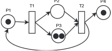
P1 T1
P2
P3
T2 P4
Materials and methods
Materials
Dry soy lecithin Solaye (Solaye, EU) with a total phospholipids content of 97% was used. This phospho-lipid preparation has been approved for use in the food industry, has the properties to form persistent micro-emulsions in oil-in-water systems, and has been characte-rized by a high total content of phosphatidylcholine (35.4%) and phosphatidyl ethanolamine (21.8%), both of which are required for forming a closed vesicle mem-brane (Vance and Vance, 2008; Dyńska-Kukulska and Ciesielski, 2012).
Methods
1. A flow-type rotary-pulsation apparatus of cylindrical type, which consists of two stators and a rotor with 24 slit-shaped holes, developed at the Institute of Technical Thermophysics of the National Academy of Sciences of Ukraine was used for this study (Dolin-skyi, 2008; Avdieieva, 2011). The nominal value of the angular velocity of the rotor is 314 rad/s. The processing was carried out with three cycles of pas-sage of the material through the working bodies of the apparatus.
2. The particle size distribution function was investi-gated using dynamic light scattering on a laser pho-ton-correlation spectrometer “ZetaSizer–3” Malvern Instruments, UK (Merkus, 2009; McNeil, 2011). The recording and statistical processing of the changes in the scattering intensity in the particle water suspen-sion (n = 1.33) were performed ten times for 10 min at +22EC at a scattering angle of 90EC. The obtained results were processed using the PCS-Size mode v1.61 software using the CONTIN algorithm. 3. Under the conditions of a multifactor experiment, it is
difficult to obtain a result over the entire range of va-riations in the parameters of the process. Depending on the conditions of their formation in the whole range of variability of the parameters of the technological process, the determination of quantitative indicators of lipid vesicles (size and internal volume), was perfor-med using mathematical modeling (Nagasaki et al., 2007; Koch et al., 2011). This approach allowed us to consider a large number of physical factors and pro-vide a prediction of the behavior of a dispersed system in a wide range of operating parameters.
The Cell yllustrator v.3 software environment (Hu-man Genome Center, University of Tokyo, Japan) was selected as a tool for modeling the mechanism of forma-tion of vesicular phospholipid nanostructures, in which hybrid functional Petri nets are the basic modeling tool (Nagasaki et al., 2007; Koch et al. 2011). The advantages of hybrid functional Petri nets as modeling tools are as follows:
C the possibility of a structural display of the state of the system that is modeled, along with the processes that occur in the system;
C the possibility of quantitative modeling of states and processes of three types at the same time: discrete, continuous, and associative (generatrix);
C the possibility of considering the activating, inhibi-ting, and catalytic effects using special type of bonds. Petri nets are a mathematical abstraction for repre-senting discrete and continuous distributed systems gra-phically represented as a bipartite oriented multigraph with markers (marked directed graph), which has two types of vertices: positions P and transitions T. Positions can be either empty or marked and they determine the state of the network while transitions show the action (process).
The operation of the Petri net depends on the se-quential execution of transitions and the corresponding recalculation of the number of “resource” in the posi-tions (Fig. 1)
Fig. 1. Schematic of the Petri net
The efficiency of inclusion (Xls) is one of the most important indicators of lipid vesicular preparations, which characterizes the amount of the substance in the middle of the vesicle. This indicator can be calculated using the following formula (Merkus, 2009):
6 ) ) ( (
10 )
(
2 2
27 3
⋅ ⋅ − +
⋅ ⋅ ⋅ − ⋅
= −
Mr h D D
S N h D C
X A
35 30 25 20 15 10 5 0
[%]
1 10 100 1000 10000
d [nm]
1 2 3 4
25
20
15
10
5
0
1 10 100 1000 10000
[%]
d [nm]
1 2 3 4
where C is the concentration of phospholipids, mg/ml;
D is the diameter of the vesicle, A; h is the monolayer thickness, A; NA is the Avogadro number, and S is the area of the phospholipid molecule in the monolayer, !2;
and Mr is the molar mass of the phospholipid.
Results
Size is the most important property of lipid vesicles (Cevc, 1993; Vance and Vance, 2008; Tsai, 2016; Vitkova and Petrov, 2013). Our task was to obtain, because of the proposed treatment, primarily large single-layer particles with an average size of 400–500 nm, all of which have a large internal volume and are characterized by persi-stence during the entire shelf life of the food product.
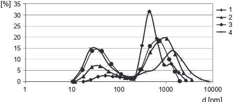
Figure 2 shows the results of the studies of samples with a concentration of lecithin in the range from 0.5% to 7.5% obtained at a temperature of 42 ± 2EE.
Fig. 2. Distribution of particle sizes (diameter) of the tested samples obtained at temperature of 42 ± 2EE with concentra-tions of phospholipids: sample 1 – 0.5%; sample 2 – 2.5%;
sample 3 – 5%; sample 4 – 7.5%
Experimental results showed that in all the samples that were obtained and tested, the particles had a bi-modal distribution of diameter from 10–15 nm to 450–500 nm and from 300 to 2.0 nm and above. These results may indicate the absence of micelles and the presence of large single-layer and multi-layer vesicles (Evans and Skalak, 1980; Vance and Vance, 2008; Naito et al., 2018). In a sample with a 0.5% concentration of phospholipids, large particles were predominant, 82.5% of which had sizes ranging from 300 to 3.0 μm. Increa-sing the concentration of phospholipids to 2.5% led to a decrease in the overall dispersion range with small and large single-layer vesicles with sizes of up to 1000 nm forming 83% of the sample size. In a sample with a 5% concentration of phospholipids, the dispersion range of
the particles was further narrowed and large single-layer vesicles with sizes of up to 1000 nm formed more than 86% of the sample size. The average particle diameter of this sample was 440 nm, and the studies of samples with a phospholipid concentration of 7.5% showed an increase in the number of small single-layer vesicles with sizes up to 100 nm to 46%; however, there was a significant ex-pansion of the dispersion range up to 3.0 μm. This may indicate deterioration of the conditions of dispersion and significant aggregation of particles (Evans and Skalak, 1980; Vance and Vance, 2008). Thus, the most rational possibility was to use a 5% concentration of phospho-lipids in the dispersed system.
Next, we studied the effect of temperature processing parameters on the size of the particles formed. Figure 3 shows the results of experimental studies of samples with 5% concentration of phospholipids obtained at dispersion temperatures ranging from 20 to 80EE.
Fig. 3. Particle size distribution of samples obtained at a phos-pholipid concentration of 5% and a temperature of dispersion in ranges: sample 1 – 20EE; sample 2 – 42EE; sample 3 – 60EE;
sample 4 – 80EE
30 25 20 15 10 5 0
[%]
1 10 100 1000 10000
d [nm]
1 2 3
1400 1300 1200 1100 1000 900 800 700 600 500 400 300 200 100 0
d(a
) [
nm
]
c [%]
0 2 4 6 8 10 12 1 2 3 4
y = 18.42x 141.6x + 889.52!
y = 7.242x 64.15x + 612.82!
y = 4.247x 40.63x + 517.32!
y = 4.377x 33.4x + 378.22!
60EE (sample 3) was characterized by a three-modal tribution of particles and an increase in the range of dis-persion to 7.0 μm, which may indicate the aggregation of particles after dispersion. Note that sample 4 had a bi-modal particle distribution with 54.4% of particles having sizes larger than 1 μm. Thus, for further experimenta-tion, the temperature dispersion range of 42 ± 2EE was used.
By using a belt transmission, the angular velocity of the DIIE activator was successively increased from 314 to 472 and 628 rad/s. Figure 4 shows the results of ex-perimental studies of the effect of the angular velocity of the rotor in the course of DIIE-processing on the size of the formation of particles. During production, the samp-les with a phospholipid concentration of 5%, obtained at dispersion temperatures of 42 ± 2EE, were used.
Fig. 4. Particle size distribution of the obtained samples with a concentration of 5% at a dispersion temperature of 42 ± 2EE
with an angular speed of the rotor: sample 1 – 314 rad/s, sample 2 – 472 rad/s, sample 3 – 628 rad/s
The results show an increase in the impact on the material being processed with an increase in the angular velocity of rotation of the rotor for this type of DIIE-activator. Therefore, at a speed equal to 314 rad/s, parti-cles with a size of up to 500 nm formed 70% of the dis-persion of the entire system, i.e., an increase in speed to 472 rad/s led to an increase in the percentage of such particles to 86.7%. A further increase in speed up to 628 rad/s led to the simultaneous sticking of small particles into conglomerates as well as the grinding of large particles. Consequently, the number of particles with a size of up to 500 nm slightly decreased and ac-counted for 80% of the dispersity of the entire system. The results show that increasing the angular velocity of the rotor to 472 rad/s and 628 rad/s led to an improve-ment in the dispersion conditions and a larger number of particles in the specified range up to 500 nm were
obtained. However, these conditions led to a significant increase in energy consumption for material processing, as well as required additional strengthening of certain parts to preserve the durability and operability of the equipment.
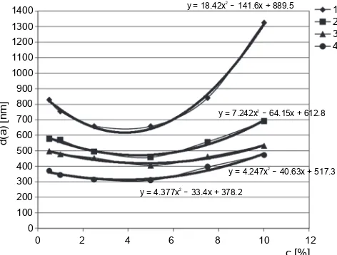
To analyze the obtained results and to formulate ge-neral laws of the influence of the thermal parameters on the change in the size of vesicular nanostructures during the process of DIIE processing, the expected value of the particle size was calculated. Figure 5 shows the re-sults of the calculation of the dependence of the average particle diameter on the concentration of phospholipids at different temperatures of dispersion with an angular velocity of 314 rad/s.
Fig. 5. The dependence of the average particle diameter, obtai-ned with an angular velocity of 314 rad/s, on the concentration of phospholipids at the following temperatures of DIIE treat-ment: sample 1 – 20EC; sample 2 – 42EC; sample 3 – 60EC;
sample 4 – 80EC
which were obtained at higher temperatures of the homogenization process, a change in the concentration of the material led to less significant changes in the average particle size, but the general tendencies re-mained. The lowest diameter was observed after dis-persing the lipid material at 5% concentration. A further increase in concentration (by 2 times) from 5 to 10% led to a gradual increase in the diameter of 1.3–1.5 times.
An increase in the homogenization temperature to 42EC (sample 2) led to the transfer of the dispersion with phospholipids from the solid crystalline state to the liquid crystal state and weakening of the intermolecular bonds. This in turn led to a decrease in the average dia-meter of the particles formed (Cevc, 1993; Hanahan, 1997; Vance and Vance, 2008). A further increase in the temperature of the process led to a further weakening of the bonds between the molecules of the lipid material; therefore, the average diameter of the particles obtained decreased, and the efficiency of homogenization impro-ved. Thus, for all samples tested, increasing the tem-perature of the process to 60EC (sample 3) and 80EC (sample 4) led to an increase in its efficiency and a de-crease in the dispersion of the system.
Thus, the analysis of the results obtained indicates and confirms the possibility of controlling the process of the formation of vesicles from phospholipids under the conditions of application of the DIIE mechanisms using rational heat-engineering regime parameters (Avdieieva, 2011).
Mathematical modeling
Modeling the mechanism of formation and self-orga-nization of vesicular phospholipid nanostructures under the DIIE influence was aimed at systematically integra-ting in the mathematical model both the initial techno-logical parameters of the material (such as temperature and concentration) and the characteristics of the equip-ment used (such as DIIE-activator and rotation frequen-cy rotor). Based on the results of experimental studies of the average diameter of lipid vesicles on the condi-tions and modes of the technological process of their production, partial functional dependencies were obser-ved. As approximating functions, polynomials from the second to the fifth degree were used.
The approximating dependences of the average dia-meter (Dav) of lipid vesicles on the temperature (i) and the concentration of phospholipids (c) at different
an-gular speeds of rotation of the rotor (ω) are shown in the equations 2–4:
at ω = 3,000 rpm = 314 rad/s
Dav = (67 × 10!4 × t2! 0.8951 t + 33.536) c2!
+ (0.041 t2! 5.865 t + 241.59) × c! 8.2877 t + 1018 (2) at ω = 4,500 rpm = 472 rad/s:
Dav = (4 × 10!5 × t3! 53 × 10!4t2! 0.2074 t + 5.5137) c2! + (5 × 10!4t3! 0.0729 t2 + 3.108 t + 49.963) c + + (18 × 10!4t! 0.2547 t2 + 9.4619 t + 392.96)
(3) at ω = 6,000 rpm = 628 rad/s:
Dav = (4 × 10!5 × t3! 84 × 10!4t2! 0.5627 t! 6.8177) c2! + (6 × 10!4t3! 0.1034 t2 + 6.7957 t! 81.854) c + + (3 × 10!4t! 0.0571 t2 + 3.4706 t + 349.81)
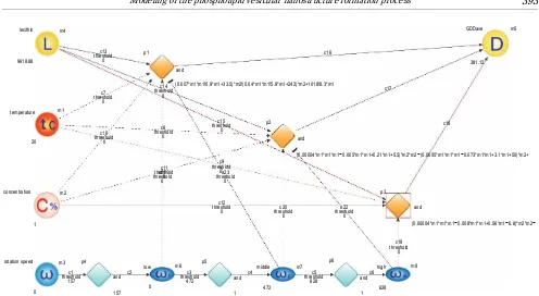
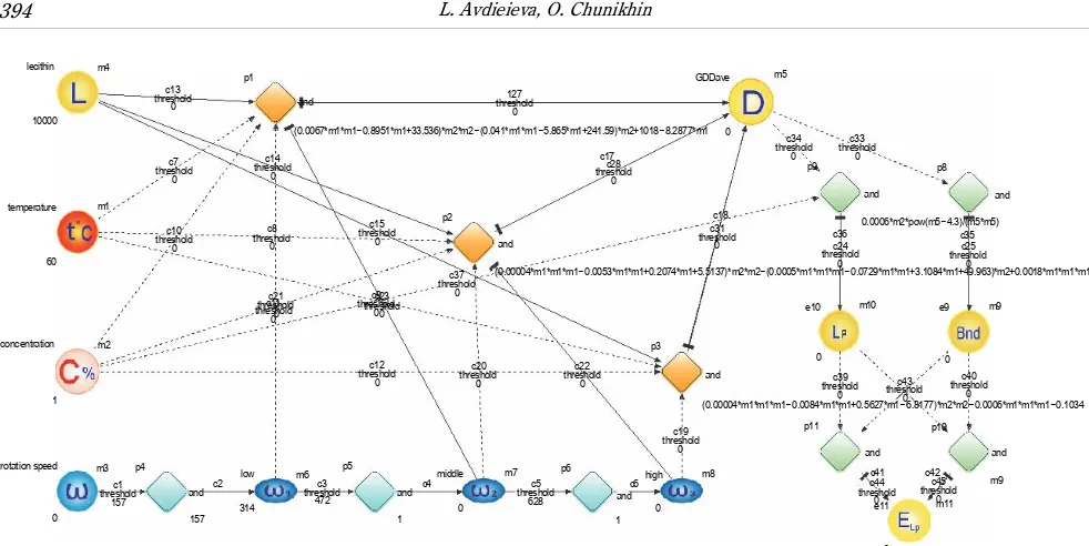
(4) The results of the studies of partial functional depen-dences of the average diameter of lipid vesicles on the parameters of the technological process made it possible to parameterize the Petri net. Moreover, using Petri net, a graph-analytical model was built, which allows calcula-ting the average diameter of lipid nanostructures obtai-ned because of the processing of a lipid suspension de-pending on the rotor speed of the DIIE-activator, and the concentration and the temperature of the starting ma-terial (Fig. 6). Thus, equations 2–4 parameterized the p1–p3 transitions, respectively, which made it possible to obtain the numerical value of the vesicles average dia-meter “GDDave” at the m5 position.
Table 1 shows the examples of calculating the dia-meter of phospholipid vesicles according to the results of studies obtained experimentally (De) and analytically (Dm), using Petri nets, at a temperature t = 42 ± 2EC, material concentration (c) from 0.5 to 10% and rotor an-gular speeds (ω) of 314, 472, or 628 rad/s. The absolute error of the simulation was calculated using the formula:
Δ = |Dm ! De|, nm, and the relative modeling error was calculated using the formula: δ = (Δ / De) × 100%.
lecithin m4
c13 threshold
0 9618.88
p1
and
c7 threshold
0
c14 threshold
0
c16
GDDave m5
381.12
c17
c18 (0.007*m1*m1 0.9*m1+33.5)*m2 (0.04*m1*m1 5.9*m1+242)*m2+1018 8.3*m1! ! ! !
temperature m1
20
c10 threshold
0
c8 threshold
0
c15 threshold
0
p2
and
(0.00004*m1*m1*m1 0.005*m1*m1+0.21*m1+5.5)*m2*m2 (0.0005*m1*m1*m1 0.073*m1*m1+3.1*m1+50)*m2+! ! !
concentration m2
c11 threshold
0 c21 threshold
0
c23 threshold
0 c9 threshold
0
c12 threshold
0 thresholdc20 0
c22 threshold
0
p3
and
c19 threshold
0
c5 threshold
628 c1
threshold 157
c3 threshold
472
(0.00004*m1*m1*m1 0.008*m1*m1+0.56*m1 6.8)*m2*m2! ! !
rotation speed m3 p4
c2 low m6
p5
middle m7 p6 high m8
c6 and
628 1 472
c4 and 0
1 and
157 0
1
Fig. 6. Graph-analytical model of calculation of the average diameter of lipid vesicles with Petri nets at the concentration of 1%, temperature of 20EC, speed of rotor of 628 rad/s
Table 1. The diameter of the phospholipid vesicles obtained experimentally and analytically using Petri nets
Temperature
[EC] Rotor speed[rad/s] Concentration[%]
Model diameter
[nm]
Experimental diameter
[nm]
Absolute error
[nm]
Relative error
[%]
42
314
0.5 603 575 28 4.87
2.5 517 496 21 4.23
5 427 441 14 3.17
10 722 689 33 4.79
472
0.5 456 441 15 3.40
2.5 301 302 1 0.33
5 226 225 1 0.44
10 369 350 19 5.43
628
0.5 386 380 6 1.58
2.5 299 314 15 4.78
5 234 245 11 4.49
10 312 330 18 5.45
Efficiency of inclusion estimation
Setting the initial data for lipid vesicles: the monolayer thickness h = 40 and A = 4 nm, and the area of the phospholipid molecule in the monolayer was S = 60 A2,
i.e., the molar mass of the phospholipid Mr = 1000 g/mol and lipid concentration (%) after simple transformations.
We then an obtained an expression for the dependence of the inclusion efficiency on the diameter of the vesicles in nanometers (Dnm) and lipid concentrations (C%):
2 2
3 4
) 4 (
) 4 ( 10 6
− +
− ⋅ ⋅
= −
nm nm
nm ls
D D
D X
lecithin m4 p1 c13 threshold 0 10000 and temperature 60 m1 c7 threshold 0 c14 threshold 0 c10 threshold 0 c8 threshold 0 c15 threshold 0 c21 thresholdc110 threshold0 c23 threshold 0 c9 threshold 0 concentration m2 c37 threshold 0 p2 and 127 threshold 0 c28 threshold 0 c17 GDDave
(0.0067*m1*m1 0.8951*m1+33.536)*m2*m2 (0.041*m1*m1 5.865*m1+241.59)*m2+1018 8.2877*m1! ! ! ! 0 m5 p9 p8 and c33 threshold 0 c34 threshold 0 and
0.0006*m2*pow(m5 4.3)/(m5*m5)!
c18
c36 c35
c31 threshold
0 thresholdc24 0
c25 threshold
0
(0.00004*m1*m1*m1 0.0053*m1*m1+0.2074*m1+5.5137)*m2*m2 (0.0005*m1*m1*m1 0.0729*m1*m1+3.1084*m1+49.963)*m2+0.0018*m1*m1*m1! ! !
1 rotation speed 0 m3 p4 c1 threshold 157 and 157 c2 low 314 m6 p5 c3 threshold 472 c12 threshold 0 c20 threshold 0 c22 threshold 0 c5 threshold 628 c19 threshold 0 p3 and e10 m10 0 1 and c4 middle 0 m7 p6 and 1 c6high 0 m8 e9 m9 0 p11 p10 and and 0 m9 e11 m11 c41 c42 c44 threshold 0 c45 threshold 0 c39 threshold 0 c40 threshold0 c43 threshold 0
(0.00004*m1*m1*m1 0.0084*m1*m1+0.5627*m1 6.8177)*m2*m2 0.0006*m1*m1*m1 0.1034! ! ! !
Fig. 7. The calculation of the vesicles average diameter and the effective integration of polar substances using Petri net at the concentration of 1%, temperature of 20EC, speed of rotor of 628 rad/s
However, there is a theoretical limit to the effective-ness of the inclusion, which is determined by the ratio of the internal volume of the vesicles and its total volume for a given diameter and thickness of the lipid bilayer. Con-sidering the lipid vesicle as an ideal ball with a diameter
D and an internal cavity with a diameter (D! 2h), it is easy to determine the ultimate inclusion efficiency Xt:
(1 2 )3
D h
Xt = − (6)
Note that the limits of the application of dependences (1) and (5) are completely determined by expression (6). Therefore, for vesicles with a diameter of > .200 nm for medium and high concentrations of phospholipids, it is necessary to compare the calculated values of the effi-ciency of inclusion with the theoretical limit. Moreover, when considering single-layer vesicles, it is advisable to limit the diameter to 600 nm.
The developed mathematical model was supplemen-ted by dependences that allow us to calculate the in-clusion efficiency indicator. For this purpose, the corres-ponding positions and transitions were added to the existing structure (Fig. 7).
Thus, dependences (5) and (6) parameterized the p9 and p8 transitions, respectively, which makes it possible to obtain the calculated values of the inclusion efficiency at position m10 ("Lp") and the marginal efficiency at inclusion at position m9 ("Bnd"). The final value of the
efficiency of inclusion of substances in the vesicles in the position m11 ("+Lp") was defined as the minimum value between the calculated and limiting values of the effecti-veness of the inclusion. The automatic selection of the minimum value was ensured by the corresponding thre-sholds established between positions m9 and m10 and transitions p10 and p11.
The work of the Petri net comprises two stages. The first one calculates the values of the average diameter of the “GDDave“ vesicles depending on the angular velo-city of the rotation of the DIIE-activator, the tempera-ture, and the concentration of the starting material. In the second, on the basis of the average diameter of the vesicles and the concentration of lipids, the values of the effectiveness of the inclusion of substances in the vesi-cles (“ELP”) are calculated.
The obtained results of calculation of the vesicles ave-rage diameter and the effective integration of polar sub-stances using the Petri net (concentration of 1%, tem-perature of 20EC, speed of rotor of 628 rad/s) agree well with the results of experimental studies (Fig. 5).
Conclusions
pre-sented. Depending on the heat-engineering parameters and modes for DIIE treatment, the study and analysis of changes in the properties of nanostructures made it pos-sible to choose rational indicators for obtaining lipid vesicles with certain physical properties.
The mathematical model created using hybrid func-tional Petri nets made it possible to determine the pe-culiarities of the formation of vesicular phospholipid nanostructures under the influence of DIIE effects and to obtain the value of the average diameter of vesicles and the efficiency of incorporation in a wide range of process parameters without additional costs for obtai-ning vesicles and experimental measurement of their size using complex technical means. The advantage of the proposed approach is the possibility of obtaining values of the average diameter of lipid vesicles both in the “nodes” of tabulated values and in the whole range of variability of the process parameters.
References
Alipour E., Halverson D., McWhirter S. et al. (2017) Phospho-lipid bilayers: stability and encapsulation of nanoparticles. Ann. Rev. Phys. Chem. 68: 261–283.
Avdieieva L.I. (2011) Energy-efficient technology for the pro-duction of phospholipid nanostructures. Industr. Heat Eng. 33(8): 139–143 (in Ukrainian).
Berg J.M., Tymoczko J.L., Stryer L. (2012) Biochemistry, 5th ed. New York: W.H. Freeman.
Cushen M., Kerry J., Morris M., Cruz-Romero M., Cummins E. (2012) Nanotechnologies in the food industry – Recent developments, risks and regulation. Trends Food Sci. Technol. 24(1): 30–46.
Dolinskyi A.A. (2008) Heat-mass transfer and hydrodynamics in steam-liquid dispersive environments. Kiev: Sci. Opin. (in Russian).
Dyńska-Kukulska K., Ciesielski W. (2012) Methods of extra-ction and thin-layer chromatography determination of phospholipids. Rev. Anal. Chem. 31(1): 43–56.
Eliaz N. (2011) Applications of electrochemistry and nano-technology in biology and medicine I. New York: Springer Science + Business Media, LLC.
Geckeler K.E., Nishide H. (2010) Advanced nanomaterials. Weinheim: Wiley-VCH Verlag GmbH & Co. KGaA. Koch I., Reisig W., Schreiber F. (eds.) (2011) Modeling in
sy-stems biology. The Petri net approach. Springer. McClements D., Gumus C. (2016) Natural emulsifiers –
bio-surfactants, phospholipids, biopolymers, and colloidal par-ticles: molecular and physicochemical basis of functional performance. Adv. Colloid Interface Sci. 234: 3–26. McNeil S.E. (2011) Characterization of nanoparticles intended
for drug delivery. Humana Press.
Merkus H.G. (2009) Particle size measurements. Fundamen-tals, practice, quality. Springer.
Nagasaki M. et al. (2007) Foundations of systems biology. Using cell illustrator and pathway databases. Springer. Naito M., Yokoyama T., Hosokawa K., Nogi K. (2018)
Nano-particle technology handbook, third edition. Elsevier. Nii T., Ishii F. (2005) Encapsulation efficiency of water-soluble
and insoluble drugs in liposomes prepared by the micro-encapsulation vesicle method. Intern. J. Pharmaceut. 298(1): 198–205.
Sanguansri P., Augustin M.A. (2006) Nanoscale materials de-velopment – a food industry perspective. Trends Food Sci. Technol. 17(10): 547–556.
Tibbals H.F. (2010) Medical nanotechnology and nanomedi-cine. CRC Press.
Tiwari A., Tiwari A. (2017) Bioengineered nanomaterials. CRC Press Published.
Tsai W. (2016) Liposomal microencapsulation using the con-ventional methods and novel supercritical fluid processes, Trends Food Sci. Technol. 55: 61–71.
Vance J.E., Vance D. (2008) Biochemistry of lipids, lipopro-teins and membranes, 5th ed.
Vitkova V., Petrov A.G. (2013) Lipid bilayers and membranes: material properties. [in:] Advances in planar lipid bilayers and liposomes, vol. 17, Academic Press, Burlington: 89–138.