Mahdiyeh Sarraf Razavi1, Mehdi Tehranidoost1,2,3, Farnaz Ghassemi4*, Parivash Purabassi4, Athena Taymourtash4
Research Paper:
Emotional Face Recognition in Children
With Attention Deficit/Hyperactivity Disorder: Evidence
From Event Related Gamma Oscillation
Introduction: Children with attention-deficit/hyperactivity disorder (ADHD) have some impairment in emotional relationship which can be due to problems in emotional processing. The present study investigated neural correlates of early stages of emotional face processing in this group compared with typically developing children using the Gamma Band Activity (GBA). Methods: A total of 19 children diagnosed with ADHD (Combined type) based on DSM-IV classification were compared with 19 typically developing children matched on age, gender, and IQ. The participants performed an emotional face recognition while their brain activities were recorded using an event-related oscillation procedure.
Results: The results indicated that ADHD children compared to normal group showed a significant reduction in the gamma band activity, which is thought to reflect early perceptual emotion discrimination for happy and angry emotions (P<0.05).
Conclusion: The present study supports the notion that individuals with ADHD have some impairments in early stage of emotion processing which can cause their misinterpretation of emotional faces.
A B S T R A C T
Key Words: Emotional face recognition, Event-Related Oscillation (ERO), Gamma band activity, Attention Deficit Hyperactivity Disorder (ADHD)
Article info:
Received: 03 Feb. 2017
First Revision: 29 Feb. 2017 Accepted: 11 Jul. 2017
1. Department of Neurosciences and Addiction Studies, School of Advanced Technologies in Medicine, Tehran University of Medical Sciences, Tehran, Iran. 2. Department of Psychiatry, School of Medicine, Tehran University of Medical Sciences, Tehran, Iran.
3. Research Center for Cognitive and Behavioral Sciences, Tehran University of Medical Sciences, Tehran, Iran. 4. Department of Biomedical Engineering, Amirkabir University of Technology, Tehran, Iran.
* Corresponding Author:
Farnaz Ghassemi, PhD
Address: Department of Biomedical Engineering, Amirkabir University of Technology, Tehran, Iran. Tel:+98 (912) 3260661
E-mail: [email protected]
Citation:Sarraf Razavi, M., Tehranidoost, M., Ghassemi, F., Purabassi, P., & Taymourtash, A. (2017). Emotional Face Rec-ognition in Children With Attention Deficit/Hyperactivity Disorder: Evidence From Event Related Gamma Oscillation. Basic and Clinical Neuroscience, 8(5):419-426. https://doi.org/10.18869/NIRP.BCN.8.5.419
:
: https://doi.org/10.18869/NIRP.BCN.8.5.419
Use your device to scan and read the article online
CrossMark
1. Introduction
DHD is a common neurodevelopmental disorder characterized by inattentiveness
and hyperactivity/impulsivity (American
Psychiatric Association, 2013). Individu
-als with ADHD -also show problems in social and emo
-tional functions, including the effective assessment of the emotional state of others. It is important to set the
adaptive behavior of human facial expressions in social
interactions (Cadesky, Mota, & Schachar, 2000;Corbett
& Glidden, 2000). Based on the evidence, frontotem-poral-posterior and fronto striatal cerebellar systems are involved in emotional functions. These regions may contribute to impairments of emotional recognition in
ADHD (Corbett & Glidden, 2000;Dickstein, Bannon,
Xavier Castellanos, & Milham, 2006; Durston, Van Belle, & De Zeeuw, 2011).
However, neural studies to investigate these impair -ments are rare. Results of recent studies revealed that gamma band activity (GBA) in electroencephalography (EEG) as well as Event-Related Potentials (ERP) play
an important role in evaluation of higher cognitive pro
-cesses such as attention, memory, language, and emotion (Fell et al., 2001;Keil et al., 2001;Sebastiani, Simoni, Gemignani, Ghelarducci, & Santarcange-lo, 2005;Mat -sumoto, Ichikawa, Kanayama, Ohira, & Iidaka, 2006; Martini et al., 2012). Several studies report that the gam -ma band activity at 40 Hz oscillation is a correlate of
selective attention (Lutzenberger, Pulvermüller, Elbert,
& Birbaumer, 1995;Keil et al., 2001;Keil, Stolarova, Moratti, & Ray, 2007;Martini et al., 2012).
Also results of studies indicate that an increase in gamma band activity at 40 Hz frequency is associated
with the tuning of novelty processing by negative emo
-tions (Singer, 1995;Garcia-Garcia, Yordanova, Kolev,
Domínguez-Borràs, & Escera, 2010). Interestingly, re
-sults of Janik, Rezlescu, & Banissy (2015) study indi
-cates that modulating occipital gamma with 40 Hz us
-ing transcranial Alternat-ing Current Stimulation (tACS)
enhances facial anger perception. Balconi and Pozoli
(2009) showed an increased gamma band activity in re -sponse to emotions (happiness, sadness, fear, and anger) compared with neutral stimuli. Keil et al., (2007) found
an increase in synchrony of very early cortical oscilla
-tions at 20–35 Hz aversive visual stimuli.
Müller, Gruber, & Keil (2000) showed an increased gamma band activity (30±50 Hz) for negative valence over the left temporal region as compared to the right one and a lateral shift towards the right hemisphere for
positive valence. Martini et al., (2012) found that un
-pleasant images compared to neutral visual stimuli, elic
-ited an increase of gamma power (30–45 Hz) at 200 ms and 850 ms after stimulus presentation.
Time course and specific topography of affective gam
-ma band activity modulations during cognitive stimuli
demonstrate discrimination between early and late pro
-cessing stages. Basar (2012) reported possibility of 3-4
phase/time-locked gamma responses in 28–45 Hz fre
-quency window during presentation of stimuli. Accord
-ing to their explanation, the early response, starts at 100 ms, in the primary occipital cortex which is probably the
direct response over the short pathway via lateral genicu
-late nucleus and reflects the early visual processing.
According to Keil et al., (2007), the early gamma response could be sensory in origin and sensitive to simple features
associated with emotions. Balconi and Pozilli (2009) report
-ed an increasing early gamma band response for emotional stimuli between 150 ms and 250 ms. On the other hand, according to Basar (2012), late processing stage, starting
around 300 ms, can be associated with the conscious per
-ception and discrimination of the emotional stimuli. Ac
-cording to Polich (2007) study, time window 300-500 ms onset stimuli is modulated by allocation of attention, initial memory storage, and processing of cognitive tasks.
Martini et al., (2012) found two peaks of weak acti -vation at high gamma frequencies (65–80 Hz) between 200 and 400 ms after stimulus onset; it is interpreted to be less sensitive to image features and more dependent on the conceptual processing of the stimulus identity. Balconi and Lucchiari (2008) reported higher GBA for emotional and neutral stimuli and this increase was more pronounced between 250 and 350 ms. And finally, time window around 400 to 800 ms reflect more strategic
high-level processes that require the conscious aware
-ness such as decision making, response criterion, and deep and elaborate processing of emotion (Schupp et al., 2004;Williams et al., 2007). Accordingly, these findings
show an important role of gamma oscillations in evaluat
-ing early and late stages of cognitive stimuli.
On the other hand, abnormalities in gamma band re
-sponse and phase synchronization have been shown to
be related to various neurological and psychiatric dis
-orders such as schizophre-nia, Alzheimer, and ADHD (Matsumoto et al., 2006; Basar & Guntekin, 2013). Gross et al., (2012) reported reduced gamma oscillations
in emotional face perception in Autism Spectrum Dis
-order. Studies show that ADHD patients have lower ab
-solute and/or relative gamma power in comparison with
age-matched healthy controls (Cited in Basar & Gun
-tekin, 2013). Few studies indicate impaired gamma band responses related to cognitive stimuli in ADHD patients
compared to healthy controls (Yordanova, Banaschews
-ki, Kolev, Woerner, & Rothenberger, 2001;Lenz et al., 2008; Lenz et al., 2010). Lenz et al., (2010) reported
ADHD group (11-17 years old) showed no differentia
-tion between known and unknown stimuli during appli
-cation of forced-choice–reaction task. However, normal
group revealed increased evoked gamma response fol
-lowing familiar stimuli compared to new images.
Based on the mentioned studies, gamma band activity could reflect the characteristics of emotional integration or emotional utilization processes in individuals with
ADHD. To our knowledge and according to review ar
-ticles by Basar and Guntekin (2013) and Güntekin and
Başar (2014), our study is the first one that evaluates
emotional face recognition in children with ADHD. We expected that gamma band activity would be diminished
for emotional faces in these patients compared to typi
-cally developing children during stages of facial emo
-tional recognition. The present study aimed to gain better understanding of the neurobiological basis of children with ADHD in emotional face processing.
2. Methods
2.1. Participants
Nineteen boys, aged between 7 and 11 years (Mean±SD
age: 9.21(1.13) years) diagnosed with ADHD were com
-pared with 19 typically developing ones (9.73±1.04 years) matched with age, sex, and years of education. The control group was recruited from elementary schools in Tehran. Children with ADHD were recruited from patients referred
to a child and adolescent psychiatric clinic. The individu
-als with ADHD were diagnosed according to DSM-IV (Statistical Manual of Mental Disorders, Fourth edition) (American Psychiatric Association, 2013). The patients were diagnosed as combined type and were drug naive.
Conners’ Parent Rating Scale-Revised (CPRS-R, short version) was administered to the participants to confirm the diagnosis of ADHD and determine severity of their symptoms. If the T-scores of CPRS-R subscales were above 65, the participant was excluded from the normal group. The two groups were right-handed, had corrected to normal visual acuity. Intelligence Quotient (IQ) of all participants were evaluated according to the WISC-R IQ test (ADHD group: 106±4.36, Control group: 122±10.71) (Table 1).
2.2. Task and stimuli
A compilation of 6 Caucasian faces (3 females and 3 males) in JPG format expressing happy, angry, sad, and neutral expressions were collected from Cohn Kanade AU-coded Facial Expressions Database (Kanade, Cohn, & Tian, 2000). Luminance and contrast of all images were equivalent across stimuli using Photoshop (version 7). The photos were in black and white and positioned within a rectangular frame (261×365 pixel array). The pictures were presented for 2000 ms at the center of the monitor screen and instantly replaced by a white fixation point in the light gray background (1024×768 pixels). The inter stimulus interval (ISI) was 1400±100 ms. The task was designed using Eevoke software (version 3.1).
The main task included 1 practice and 5 experimental
blocks. Each experimental block comprised with 48 tri
-als; 4 emotions (anger, happiness, sadness, and neutral) of each faces (3 female and 3 males) that repeated two times. Thus, there were 60 repeats of each expression in a random way. We defined four buttons for each facial expressions procedure (anger, happiness, sadness, and neutral) on a joy stick.
All participants were invited to the laboratory of EEG recording. The parents completed a consent form before
starting the examination. During the EEG session, par
-ticipants were seated in a comfortable chair in a dimly lit room 60 cm from a 17-inch LG computer screen. The participants were asked to look at the center of the screen during the recordings, if possible without making eye
movements, and to blink only during the intervals. To en
-sure that they attend to stimuli, they were monitored by camera in another control room during task performance. All children were instructed to press on the button for each expression when they recognized the target stimuli during each trial (Stimuli presentation until the end of fixation).
2.3. Electrophysiological recording and analysis
Continuous EEG signals were recorded by 64 Ag/
AgCl electrodes mounted in an electrode cap (Waveg
-uard, ANT, Netherlands) according to the international 10-20 standard and additional intermediate positions in
neuropsychology laboratory of Payam-e Noor Univer
-sity. ASA 4.7.1 software was used for data acquisition. Electrode impedances were maintained below 10 kΩ. The sampling rate was 512 Hz. A 50-Hz notch filter of the recording system, eliminated the line noise during signal acquisition. Event-related oscillations data were analyzed offline using MATLAB R2013a software.
Raw data were filtered with a band-pass filter of 0.1 to 80
Hz and referenced to the mastoids average. The eye move
-ment artifacts were canceled using the independent com
-ponent analysis. In addition, the remaining artifacts with deflection amplitudes of ±100 μV from the baseline were
eliminated (primarily through automatic artifact reduc
-tion). Artifact-free EEG recordings were then segmented into epochs ranging from 200 ms prestimulus to 800 ms poststimulus. Each channel baseline epoch was corrected
by prestimulus average voltage subtraction. Several stud
-ies reported that the lower frequenc-ies of Gamma band os
-cillation (about 40 Hz), modulated with emotional stimuli indicated attentional processing and discrimination of the
emotional stimuli (Keil et al., 2001;Keil et al., 2007;Mar
-tini et al., 2012;Herrmann, Munk, & Engel, 2004).
by concatenating a specific time window from all clean epochs. Then we estimated the signal power spec-trum in frequency band of 35–45 HZ for three different time
windows based on literature (Martini et al., 2012) and
following current data analysis: 0–250 ms as early pro
-cessing (can be sensitive to simple features associated with emotions (Keil et al., 2007)), 250–500 ms as late processing (can indicate allocation of attention, initial
memory storage the processing of cognitive tasks (Pol
-ich, 2007)), and 500–750 ms as later ones (can reflect
more strategic high-level processes that require the con
-scious awareness and deeper and elaborate processing
of emotion (Schupp et al., 2004;Williams et al., 2007)).
Based on the previous studies, F3, F4, C3, C4, P3, P4, O1, O2 electrodes were used for the statistical analysis (Ramos-Loyo, González-Garrido, Sánchez-Loyo, Me -dina, & Basar-Eroglu, 2009).
2.4. Statistical analysis
Frequency-band measures were statistically analyzed using repeated-measure analysis of variance (ANOVA)
with the following core factors: facial expression (hap
-piness, anger, sadness, and neutral), site (area) (ante
-rior [F3, F4], central [C3, C4], occipital [O1, O2], and parietal [P3, P4]), side (lateralization) (left [F3, C3,
P3, O1], and right [F4, C4, P4, O2]) as the within sub
-jects factors, and groups (patients and controls) as the between subject factor. Greenhouse Geisser correction was used for the degrees of freedom. In the next step, paired t-test were used to break down between main effects such as site and independent sample t test were
used to break down between-subject and interaction ef
-fects such as facial expression×group. Throughout the experiment, P<0.05 were considered significant.
3. Results
We carried out repeated-measure ANOVA (facial expression×site×side×group [4×4×2×2]) for three time windows separately. In the first time window (0–250 ms), this analyses revealed a significant main effect of
group (F1, 36=12.36, P=0.001; partial η2=0.155), and a
significant main effect of site (F2.1, 76.26=3.78, P=0.02;
partial η2=0.013). Also the results showed a signifi
-cant interaction effect of site×group (F2.1, 76.26=3.79,
P=0.02; partial η2=0.059), and a significant interac
-tion effect of facial expression×group (F2.1, 77.47=4.68,
P=0.01; partial η2=0.069). In the second (250–500 ms) and third (500–750 ms) time windows, repeated-measure ANOVA revealed only main effect of group which showed smaller gamma band activity in ADHD group compared to healthy control one (P<0.05). We
observed no significant main effect or interaction ef
-fect in terms of other factors of side, site, face expres
-sion, and group (P>0.05) in the second and third time windows. Therefore, our results focused on first time window that indicated early facial emotion processing.
Table 1. Clinical and demographic characteristics.
Diagnosis
F P
TD (n=19) ADHD (n=19)
Mean SD Mean SD
Age, y 9.68 1.05 9.15 1.16 0.1 0.7
Full-scale IQ 122 10.7 106 4.73 10 0.003
Conners’-oppositional 47 6.7 77 6.1 1.38 0.2
Conners’ -inattentive 47 6.2 75 10.4 3.75 0.03
Conners’ -hyperactive 48 8.8 76 4.7 2.003 0.04
Conners’ -ADHD index 45 5.6 78 7.8 4.23 0.04
Conner’s: Conners’ parent rating scale revised.
*
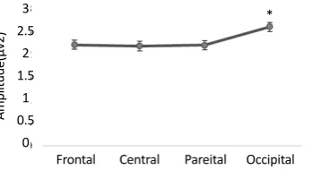
Figure 1. Mean (standard error) of gamma band activity in
different areas in response to facial expressions. 3
2.5 2 1.5
1 0.5
0
Amplitude
(µv2)
Also post hoc analysis showed that gamma oscillation measures were smaller in ADHD group (1.6±0.2) compared to normal group (2.9±0.2) in the first time window.
Follow-up paired t test showed greater gamma band activity in oc
-cipital site compared to frontal (P=0.05, t(37)=1.97), cen
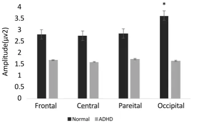
-tral (P=0.01, t(37)=2.47), and parietal (P=0.02, t(37)=2.28) areas in ADHD group (Figure 1). Follow-up independent t test revealed a significant lower gamma band activity in ADHD group compared to normal children in occipital
regions (F4.2, 36=14.8, P=0.001) (Figure 2). Also follow-up
independent t test showed a significant decrease for happi
-ness (F2.6, 36=6.63, P=0.01), (F3.2, 36=6.8, P=0.01) and anger
(F4.2, 36=13.7, P=0.001), (F6, 36=8.25, P=0.007) in ADHD
group compared to typically developing children in left and right occipital, respectively (Figure 3).
4. Discussion
This study aimed to compare the gamma band oscilla
-tions among patients with ADHD and healthy controls
during facial emotion recognition. We expected to ob
-serve emotion recognition deficits in ADHD children.
The results showed a significant (P<0.05) increased gam
-ma band activity within occipital regions during the first time window (0–250 ms) compared to other sites. Also, we observed a significant (P<0.05) reduction in gamma band activity in ADHD children compared to normal group in response to facial expressions within occipital
lobe. This study supported our hypothesis that individu
-als with ADHD were different from typically developing children during facial expression recognition, especially in early stage of facial emotion processing.
Consistent with our findings, several studies have shown a similar gamma-band increase in response to
emotional pictures with occipital distribution (Keil et
al., 2001;Keil et al., 2007;Garcia-Garcia et al., 2010). Balconi and Lucchiari (2008) as well as Aftanas, Var -lamov, Pavlov, Makhnev and Reva (2002) reported a more emotional compared to non-emotional stimuli in posterior distribution of gamma oscillations. The results of the current study revealed that gamma band activity modulated by emotions compared to neutral, only in first time interval (0–250 ms) indicating early stage of facial emotion recognition but not late stage. Consistent with the current results about early gamma response (<250
ms), Martini et al., (2012) revealed the increase of gam
-ma activity in the low gam-ma frequency band (30–45 Hz) for unpleasant picture compared to neutral visual stimuli at the shortest latencies (0–250 ms).
Also, Balconi and Pozoli (2009) showed an increased
gamma band activity in response to emotions com
-pared to neutral at 150–250 ms time interval with a peak at around 240 ms of latency. Keil et al., (2001) reported gamma band activity (30–45 Hz) at 80 ms
Figure 2. Mean (standard error) of the gamma band activity
in different sites in response to facial expressions. * 4
3.5 3 2.5
2 1.5
1 0.5
0
Frontal
Normal ADHD
Central Pareital Occipital
Amplitude
(µv2)
*
Figure 3. Mean (standard error) of the gamma band activity in left and right hemispheres (O1, O2) in response to facial
expres-sions among the ADHD and control groups.
* * *
Normal ADHD
Angry
01 02
Angry
Happy Neutral Sad Happy Neutral Sad
Amplitude
(µv2)
6
5
4
3
2
1
poststimulus enhanced in response to unpleasant stim -uli compared to neutral. These findings are consistent with our results that supported the role of early gamma
response in assessing early stage of facial emotion pro
-cessing. However, our findings did not reveal modula
-tion of gamma band activity by emo-tion compared to neutral during the late stage. Unlike the current results, Martini et al., (2012) reported an increased gamma band activity (lower gamma frequency) in response to emotions (unpleasant compared to neutral) at the time interval of 250–500 ms. Also, they found two peaks of weak activation at high gamma frequencies (65–85
Hz) at 200 ms and 850 ms after stimulus onset. Keil et
al., (2001) reported an increased gamma band activ -ity (46–65 Hz) in response to unpleasant compared to neutral pictures at 500 ms. In fact, we did not analyze the 45–65 Hz or 65–85 Hz interval, that may be the reason for inconsistent results.
Also, the current results showed a significant dimin
-ished evoked gamma band responses only to anger and happiness emotions compared to neutral ones during early stage of processing (time window 0–250 ms) within occipital regions in ADHD group compared
to healthy control ones. However, the current find
-ings did not reveal any difference between groups in response to sad faces. Behavioral studies showed that
ADHD children have deficits in emotional face recog
-nition, especially negative (fear, anger, sadness) ones
compared to healthy children (Singh et al., 1998;
Ca-desky et al., 2000; Corbett& Glidden, 2000, Dan & Raz, 2015). However, few behavioral studies did not report facial emotion recognition impairment (anger, happiness, sadness) in ADHD group compared to normal ones (Boakes, Chapman, Houghton, & West, 2007; Schwenck, et al., 2013).
Thus, the current results should be discussed with re
-spect to early visual processing. Unfortunately, the rele
-vant literature is scarce. Inconsistent with current results, William et al., (2008) reported adolescents with ADHD [(mean±SD) age: 13.79(2.33) years; range 8–17)] have been shown to display reduced occipital P1 component during all of expressions (fear, anger, sadness, disgust, happiness, or neutral) compared to normal group. The P1 component (in the ERP studies)is primarily involved in visual attention and initial sensory encoding (similar
to role of early gamma responses according to Basar,
2012), localized in bilateral occipital areas and fusiform
gyrus (Hillyard, Mangun, Woldorff, & Luck, 1995;
Ei-mer & Holmes, 2002).
Few studies report reduced P100 amplitude in occipital
regions in ADHD group (Barry et al., 2009;Nazari et al.,
2010), probably indicating dysfunction of early visual pathways, which provide sensory input to the amygdala
and may cause emotion recognition deficits. In the pres
-ent study, participants’ Mean(SD) age was 9.21(1.13)
years (age range was 7-11 years), which may be the rea
-son for inconsistent results especially with William et al., (2008). In this regard, further research with other age groups or other gamma band frequencies (for happy and angry faces recognition) are recommended.
These findings show the important role of occipital gamma oscillations in facial emotion perception. The
present study shows that children with ADHD have ab
-normality in brain function for early stage of emotional face recognition compared to normal children that can be caused by deficits in selective and sustained attention
and early visual processing. These findings might pro
-vide preliminary e-vidence for future planning of inter
-ventional approaches for children with ADHD.
Acknowledgments
This study is a part of first author PhD thesis in neuro
-science entitled Evaluation of brain function in recog-nizing emotional faces in children with attention-deficit /hyperactivity disorder (ADHD) compared with normal children using the Event Related Potentials (ERP). It was supported by a grant from Tehran university of Medical sciences. We thank Miss. Zamani and Dr.Rahmanian, supervisor of neuropsychology laboratory of Payame Noor University, and all children and their parents for their contribution to this study.
Conflict of Interest
All authors certify that this manuscript has neither been published in whole nor in part nor being considered for publication elsewhere. The authors have no conflicts of interest to declare.
References
American Psychiatric Association. (2013). Diagnostic and statisti-cal manual of mental disorders (DSM-5®). Missouri: American Psychiatric Pub.
Balconi, M., & Lucchiari, C. (2008). Consciousness and arousal effects on emotional face processing as revealed by brain oscil-lations: A gamma band analysis. International Journal of Psycho-physiology, 67(1), 41–6. doi: 10.1016/j.ijpsycho.2007.10.002
Balconi, M., & Pozzoli, U. (2009). Arousal effect on emotional face comprehension. Physiology & Behavior, 97(3-4), 455–62. doi: 10.1016/j.physbeh.2009.03.023
Barry, R. J., Clarke, A. R., & Johnstone, S. J. (2003). A review of electrophysiology in attention-deficit/hyperactivity disor-der: I. Qualitative and quantitative electroencephalography. Clinical Neurophysiology, 114(2), 171–83. doi: 10.1016/s1388-2457(02)00362-0
Barry, R. J., Clarke, A. R., Hajos, M., McCarthy, R., Selikowitz, M., & Dupuy, F. E. (2010). Resting-state EEG gamma activ-ity in children with Attention-Deficit/Hyperactivactiv-ity Disor-der. Clinical Neurophysiology, 121(11), 1871–7. doi: 10.1016/j. clinph.2010.04.022
Barry, R. J., Clarke, A. R., McCarthy, R., Selikowitz, M., Brown, C. R., & Heaven, P. C. L. (2009). Event-related potentials in adults with Attention-Deficit/Hyperactivity Disorder: An investigation using an inter-modal auditory/visual oddball task. International Journal of Psychophysiology, 71(2), 124–31. doi: 10.1016/j.ijpsycho.2008.09.009
Başar, E. (2012). Multiple oscillations and phase locking in hu-man gamma responses: An essay in search of eigenvalues. NeuroQuantology, 10(4). doi: 10.14704/nq.2012.10.4.603
Başar, E., & Güntekin, B. (2013). Review of delta, theta, alpha, beta, and gamma response oscillations in neuropsychiatric disorders. In E. Başar, C. Başar-Eroglu, A. O zerdem, P. M. Rossini, & G. G. Yener (Eds.). Supplements to Clinical Neuro-physiology (pp. 303–41). Cambridge, Massachusetts: Academic Press. doi: 10.1016/b978-0-7020-5307-8.00019-3
Boakes, J., Chapman, E., Houghton, S., & West, J. (2007). Fa-cial affect interpretation in boys with attention deficit/hy-peractivity disorder. Child Neuropsychology, 14(1), 82–96. doi: 10.1080/09297040701503327
Cadesky, E. B., Mota, V. L., & Schachar, R. J. (2000). Beyond words: how do children with ADHD and/or conduct prob-lems process nonverbal information about affect. Journal of the American Academy of Child & Adolescent Psychiatry, 39(9), 1160–7. doi: 10.1097/00004583-200009000-00016
Corbett, B., & Glidden, H. (2000). Processing affective stimuli in children with Attention-Deficit Hyperactivity Disorder. Child Neuropsychology (Neuropsychology, Development and Cognition: Section C), 6(2), 144–55. doi: 10.1076/chin.6.2.144.7056
Dan, O., & Raz, S. (2015). Response patterns to emotional faces among adolescents diagnosed with ADHD. Journal of Atten-tion Disorders. doi: 10.1177/1087054715606215
Dickstein, S. G., Bannon, K., Xavier Castellanos, F., & Milham, M. P. (2006). The neural correlates of attention deficit hy-peractivity disorder: An ALE meta-analysis. Journal of Child Psychology and Psychiatry, 47(10), 1051–62. doi: 10.1111/j.1469-7610.2006.01671.x
Durston, S., van Belle, J., & de Zeeuw, P. (2011). Differentiat-ing frontostriatal and fronto-cerebellar circuits in
Attention-Deficit/Hyperactivity Disorder. Biological Psychiatry, 69(12), 1178–84. doi: 10.1016/j.biopsych.2010.07.037
Eimer, M., & Holmes, A. (2002). An ERP study on the time course of emotional face processing. Neuroreport, 13(4), 427-31. doi: 10.1097/00001756-200203250-00013
Fell, J., Klaver, P., Lehnertz, K., Grunwald, T., Schaller, C., Elger, C. E., et al. (2001). Human memory formation is accompanied by rhinal–hippocampal coupling and decoupling. Nature Neu-roscience, 4(12), 1259-64. doi: 10.1038/nn759
Garcia-Garcia, M., Yordanova, J., Kolev, V., Domínguez-Borràs, J., & Escera, C. (2010). Tuning the brain for novelty detection under emotional threat: The role of increasing gamma phase-synchronization. NeuroImage, 49(1), 1038–44. doi: 10.1016/j. neuroimage.2009.07.059
Gross, E., El-Baz, A. S., Sokhadze, G. E., Sears, L., Casanova, M. F., & Sokhadze, E. M. (2012). Induced EEG gamma oscilla-tion alignment improves differentiaoscilla-tion between autism and ADHD group responses in a facial categorization task. Journal of Neurotherapy, 16(2), 78–91. doi: 10.1080/10874208.2012.677631
Güntekin, B., & Başar, E. (2014). A review of brain oscillations in perception of faces and emotional pictures. Neuropsychologia, 58, 33–51. doi: 10.1016/j.neuropsychologia.2014.03.014
Herrmann, C. S., Munk, M. H. J., & Engel, A. K. (2004). Cognitive functions of gamma-band activity: Memory match and utili-zation. Trends in Cognitive Sciences, 8(8), 347–55. doi: 10.1016/j. tics.2004.06.006
Hillyard, S. A., Mangun, G. R., Woldorff, M. G., & Luck, S. J. (1995). Neural mechanisms mediating selective attention. In M. S. Gazzaniga (Ed.). The Cognitive Neurosciences. Massa-chussets: MIT Press.
Janik, A. B., Rezlescu, C., & Banissy, M. J. (2015). Enhancing an-ger perception with transcranial alternating current stimula-tion induced gamma oscillastimula-tions. Brain Stimulation, 8(6), 1138– 43. doi: 10.1016/j.brs.2015.07.032
Kanade, T., Cohn, J. F., & Yingli Tian. (2000). Comprehensive data-base for facial expression analysis. Paper presented at the 4th IEEE International Conference on Automatic Face and Gesture Rec-ognition (Cat. No. PR00580). Grenoble, France, 28-30 March 2000. doi: 10.1109/afgr.2000.840611
Keil, A., Müller, M. M., Gruber, T., Wienbruch, C., Stolarova, M., & Elbert, T. (2001). Effects of emotional arousal in the cerebral hemispheres: A study of oscillatory brain activity and event-related potentials. Clinical Neurophysiology, 112(11), 2057–68. doi: 10.1016/s1388-2457(01)00654-x
Keil, A., Stolarova, M., Moratti, S., & Ray, W. J. (2007). Adap-tation in human visual cortex as a mechanism for rapid dis-crimination of aversive stimuli. NeuroImage, 36(2), 472–9. doi: 10.1016/j.neuroimage.2007.02.048
Lenz, D., Krauel, K., Flechtner, H. H., Schadow, J., Hinrichs, H., & Herrmann, C. S. (2010). Altered evoked gamma-band responses reveal impaired early visual processing in ADHD children. Neuropsychologia, 48(7), 1985–93. doi: 10.1016/j.neu-ropsychologia.2010.03.019
Luo, Q., Mitchell, D., Cheng, X., Mondillo, K., Mccaffrey, D., Holroyd, T., et al. (2008). Visual awareness, emotion, and gamma band synchronization. Cerebral Cortex, 19(8), 1896–904. doi: 10.1093/cercor/bhn216
Lutzenberger, W., Pulvermüller, F., Elbert, T., & Birbaumer, N. (1995). Visual stimulation alters local 40-Hz responses in hu-mans: an EEG-study. Neuroscience Letters, 183(1-2), 39–42. doi: 10.1016/0304-3940(94)11109-v
Martini, N., Menicucci, D., Sebastiani, L., Bedini, R., Pingitore, A., Vanello, N., et al. (2012). The dynamics of EEG gamma responses to unpleasant visual stimuli: From local activ-ity to functional connectivactiv-ity. NeuroImage, 60(2), 922–32. doi: 10.1016/j.neuroimage.2012.01.060
Matsumoto, A., Ichikawa, Y., Kanayama, N., Ohira, H., & Iida-ka, T. (2006). Gamma band activity and its synchronization reflect the dysfunctional emotional processing in alexithymic persons. Psychophysiology, 43(6), 533–40. doi: 10.1111/j.1469-8986.2006.00461.x
Müller, M. M., Gruber, T., & Keil, A. (2000). Modulation of in-duced gamma band activity in the human EEG by attention and visual information processing. International Journal of Psy-chophysiology, 38(3), 283–99. doi: 10.1016/s0167-8760(00)00171-9
Nazari, M. A., Berquin, P., Missonnier, P., Aarabi, A., Debatisse, D., De Broca, A., et al. (2010). Visual sensory processing deficit in the occipital region in children with attention-deficit/hy-peractivity disorder as revealed by event-related potentials during cued continuous performance test. Neurophysiologie Clinique/Clinical Neurophysiology, 40(3), 137–49. doi: 10.1016/j. neucli.2010.03.001
Pelc, K., Kornreich, C., Foisy, M.-L., & Dan, B. (2006). Recog-nition of emotional facial expressions in Attention-Deficit Hyperactivity Disorder. Pediatric Neurology, 35(2), 93–7. doi: 10.1016/j.pediatrneurol.2006.01.014
Polich, J. (2007). Updating P300: An integrative theory of P3a and P3b. Clinical Neurophysiology, 118(10), 2128–48. doi: 10.1016/j. clinph.2007.04.019
Ramos Loyo, J., González Garrido, A. A., Sánchez Loyo, L. M., Medina, V., & Basar-Eroglu, C. (2009). Event-related potentials and event-related oscillations during identity and facial emo-tional processing in schizophrenia. International Journal of Psy-chophysiology, 71(1), 84–90. doi: 10.1016/j.ijpsycho.2008.07.008
Schupp, H. T., Öhman, A., Junghöfer, M., Weike, A. I., Stock-burger, J., & Hamm, A. O. (2004). The facilitated processing of threatening faces: An ERP analysis. Emotion, 4(2), 189–200. doi: 10.1037/1528-3542.4.2.189
Schwenck, C., Schneider, T., Schreckenbach, J., Zenglein, Y., Gensthaler, A., Taurines, R., et al. (2013). Emotion recognition in children and adolescents with attention-deficit/hyperactiv-ity disorder (ADHD). ADHD Attention Deficit and Hyperactiv-ity Disorders, 5(3), 295–302. doi: 10.1007/s12402-013-0104-z
Sebastiani, L., Simoni, A., Gemignani, A., Ghelarducci, B., & San-tarcangelo, E. L. (2005). Relaxation as a cognitive task. Archives Italiennes De Biologie, 143(1), 1-12. doi: 10.4449/aib.v143i1.336
Singer, W. (1995). Visual feature integration and the temporal correlation hypothesis. Annual Review of Neuroscience, 18(1), 555–86. doi: 10.1146/annurev.neuro.18.1.555
Singh, S. D., Ellis, C. R., Winton, A. S. W., Singh, N. N., Leung, J. P., & Oswald, D. P. (1998). Recognition of facial expres-sions of emotion by children with Attention-Deficit Hyper-activity Disorder. Behavior Modification, 22(2), 128–42. doi: 10.1177/01454455980222002
Williams, L. M., Hermens, D. F., Palmer, D., Kohn, M., Clarke, S., Keage, H., et al. (2008). Misinterpreting emotional expres-sions in Attention-Deficit/Hyperactivity Disorder: Evidence for a neural marker and stimulant effects. Biological Psychiatry, 63(10), 917–26. doi: 10.1016/j.biopsych.2007.11.022
Williams, L. M., Kemp, A. H., Felmingham, K., Liddell, B. J., Palmer, D. M., & Bryant, R. A. (2007). Neural biases to cov-ert and ovcov-ert signals of fear: Dissociation by trait anxiety and depression. Journal of Cognitive Neuroscience, 19(10), 1595–1608. doi: 10.1162/jocn.2007.19.10.1595