R E S E A R C H
Open Access
Downregulating CD26/DPPIV by apigenin
modulates the interplay between Akt and
Snail/Slug signaling to restrain metastasis
of lung cancer with multiple EGFR statuses
Jer-Hwa Chang
1,2, Chao-Wen Cheng
3, Yi-Chieh Yang
3,4, Wan-Shen Chen
2, Wen-Yueh Hung
3, Jyh-Ming Chow
5,
Pai-Sheng Chen
6,7, Michael Hsiao
4, Wei-Jiunn Lee
8,9*and Ming-Hsien Chien
3,8,10*Abstract
Background:Metastasis rather than the primary cancer determines the survival of cancer patients. Activation of Akt plays a critical role in the epithelial-to-mesenchymal transition (EMT), the initial step in lung cancer metastasis. Apigenin (API), a flavonoid with a potent Akt-inhibitory effect, shows oncostatic activities in various cancers. However, the effects of API on metastasis of non-small cell lung cancer (NSCLC) remain unclear.
Methods:NSCLC cell lines with different epidermal growth factor receptor (EGFR) statuses and in vivo orthotopic bioluminescent xenograft model were employed to determine antitumor activity of API. Western blot and genetic knockdown by shRNA or genetic overexpression by DNA plasmids were performed to explore the underlying
mechanisms. The Cancer Genome Atlas (TCGA) database was used to investigate the prognosis of API-targeted genes.
Results:API was demonstrated to inhibit the migration/invasion of NSCLC cells harboring different EGFR statuses via suppressing the Snail/Slug-mediated EMT. Mechanistic investigations showed that CD26/dipeptidyl peptidase IV (DPPIV) was downregulated by API following suppressive interplay of Akt and Snail/Slug signaling to modulate the EMT and the invasive ability of NSCLC cells. CD26 expression was positively correlated with the invasive abilities of NSCLC cells and a worse prognosis of lung cancer patients. Furthermore, we observed that patients with CD26high/Akthigh tumors had the shortest recurrence-free survival times. In vivo, API drastically reduced the growth and metastasis of A549 xenografts through targeting CD26.
Conclusions:CD26 may be a useful biomarker for predicting NSCLC progression. API effectively suppressed lung cancer progression by targeting the CD26-Akt-Snail/Slug signaling pathway.
Keywords:Non-small cell lung cancer, Invasion, Metastasis, CD26/dipeptidyl peptidase IV, Akt, Snail, Slug, Apigenin
Background
Lung cancer is the leading cause of cancer-related mor-tality worldwide [1], because most patients have already progressed to an advanced stage or have developed dis-tant metastasis when diagnosed. More than 85% of lung cancers are histologically of non-small cell lung cancer (NSCLC) [2], and the prognosis of NSCLC patients with
metastatic tumors or at stage IV is very poor, with only a median survival time of 8~ 10 months [3]. Currently, chemotherapy, radiotherapy, and targeted therapy are treatment options for advanced NSCLC. The response rate of standard first-line chemotherapy (platinum-based combined with third-generation cytotoxic agents) of advanced lung cancers has significantly improved sur-vival times but with low short-term efficacy, high toxicity, and ultimately the development of drug resist-ance [4]. When chemotherapy is no longer effective, some targeted medicines such as gefitinib (an epidermal growth factor receptor (EGFR) tyrosine kinase inhibitor
* Correspondence:[email protected];[email protected]
8
Department of Medical Education and Research, Wan Fang Hospital, Taipei Medical University, 111 Hsing Long Road, Section 3, Taipei 11696, Taiwan 3Graduate Institute of Clinical Medicine, College of Medicine, Taipei Medical University, 250 Wu-Hsing Street, Taipei 11031, Taiwan
Full list of author information is available at the end of the article
(TKI)) can be used for advanced NSCLC patients with EGFR mutations (deletions in exon 19 and L858R in exon 21) to further prolong patient survival [5]. However, most patients eventually have no effective response to gefitinib, because cancer cells develop a second mutation, such as T790 M in the EGFR gene [6]. Therefore, searching for new drugs with high efficacy and low toxicity is urgently needed.
Tumor metastasis is a continuous multi-step process, and the epithelial-to-mesenchymal transition (EMT) is one of the most important mechanisms in the initiation and promotion of tumor metastasis [7]. In NSCLC, the EMT of cells was reported to promote metastasis and also determine chemoresistance [8] and insensitivity to EGFR inhibitors [9]. The serine-threonine protein kin-ase, Akt, was reported to play a crucial role in NSCLC invasion [10], but the underlying molecular mechanisms of NSCLC invasion mediated by the phosphatidylinositol 3-kinase (PI3K)/Akt signaling pathway is not completely understood. At present, the EMT is known to be a cellu-lar process subject to Akt kinase regulation. Activated Akt was shown to regulate several steps of the EMT, such as loss of cell-cell adhesion and polarization, mor-phological changes, induction of cell motility, and changes in the production of various proteins [11–13]. For example, Snail and Slug (Snail2), the most thor-oughly investigated EMT regulators in lung cancer, are reportedly regulated by activated Akt [14]. PI3K/Akt can inhibit the degradation of Snail and Slug by targeting glycogen synthase kinase (GSK)-3β or by directly upregulating Snail expression in different cancer types [15–17]. Actually, the PI3K/Akt signaling pathway which mediates the EMT process has garnered widespread atten-tion as a potential target for preventing and treating meta-static tumors. Therefore, investigating compounds with medicinal effects on Akt activation and the Snail family-mediated EMT should be a good strategy for NSCLC.
CD26, a 110-kDa type II transmembrane glycoprotein with dipeptidyl peptidase IV (DPPIV) activity in its extracellular domain, can cleave N-terminal dipeptides from polypeptides with an alanine or proline at the pen-ultimate position [18]. Previously, CD26 was shown to participate in T-cell biology as a marker of T-cell activa-tion or as a costimulatory molecule able to regulate sig-naling transduction pathways [19, 20]. Recently, CD26 was shown to play a critical role in cancer biology. For example, CD26 overexpression was associated with tumor aggressiveness in many cancer types such as as-trocytomas [21], lymphomas [22], urothelial carcinoma [23], colorectal cancer [24], and gastrointestinal stromal tumors [25]. For example, CD26-positive colorectal can-cer stem cells, which are mediators of the EMT, contrib-ute to the invasive phenotype and metastatic capacity
[24]. An in vivo study further showed that vildagliptin, a CD26 inhibitor, significantly suppressed metastasis of colorectal cancer [26]. These data emphasize the involvement of CD26 in cancer metastasis. So far, little information is known about the role of CD26 and its underlying mechanisms in regulating metastasis and invasion of NSCLC in vitro and in vivo.
Flavonoids are plentiful in fruits and vegetables and are a class of plant secondary metabolites with a ubiquitous phenolic structure. Recent cancer research studies have shown that flavonoids are highly promising compounds alone or in combination with other therapeutic agents against the growth and/or metastasis of different cancer cells in vitro and in vivo [27].Apigenin (API), 4′ ,5,7-trihy-droxyflavone, is one of the most common flavonoids and is abundantly present in various fruits, vegetables, and Chinese medicinal herbs [28]. API recently received much attention in cancer treatment, due to its potent anticancer activities in different cancer types in vitro and in vivo, including inducing apoptosis or autophagy, regulating the cell cycle, inhibiting migration/invasion, attenuat-ing drug resistance, and stimulatattenuat-ing immune re-sponses [29]. The PI3K/Akt signaling pathway was reported to play a critical role during most anticancer processes induced by API [29].
Although API was recently shown to inhibit prolifera-tion by targeting Akt in NSCLC cells harboring the wild-type EGFR [30], whether API has a broad impact on NSCLC with different EGFR mutation statuses, how API impacts the metastatic ability of NSCLC cells in vivo, and what the underlying mechanisms of API are in regulating Akt activity and modulating cell motility are still undefined. In the present study, we further investi-gated the antimetastatic effect of API on four NSCLC cell lines which harbor the wild-type (WT) or mutant EGFR and defined its underlying mechanisms in vitro and in an orthotopic xenograft model.
Methods Materials
(EGFR) were obtained from Cell Signaling Technology (Danvers, MA). Antibodies specific for vimentin and N-cadherin were purchased from BD Biosciences (San Jose, CA). Antibodies specific for presenilin-1 andβ-actin were obtained from Santa Cruz Biotechnology (Santa Cruz, CA). Polyvinylidene fluoride (PVDF) membranes for Western blotting were purchased from Bio-Rad (Hercules, CA). Un-less otherwise specified, other chemicals used in this study were purchased from Sigma Chemical (St. Louis, MO).
Cell lines and cell culture
The A549, H1975, and HCC827 NSCLC cell lines were purchased from American Type Culture Collection (ATCC, Manassas, VA), and a series of NSCLC cell lines, CL1–0, CL1–3, and CL1–5, in ascending order of invasiveness, were established in the National Health Research Institute laboratory [31]. PC9 cells were devel-oped by Lee and colleagues at National Cancer Center Hospital (Tokyo, Japan) [32]. All cells were maintained in RPMI 1640 supplemented with 10% FBS and 1% penicillin-streptomycin-glutamine. All cells were incu-bated at 37 °C in a humidified 5% CO2atmosphere.
Cell viability assay (MTS assay)
A549, CL1–5, HCC827, and H1975 NSCLC cells (5 × 10³) were seeded in 96-well plates, treated with various concentrations of API (5~ 80 μM) for 24 and 48 h, and then subjected to a cell-viability assay (MTS assay; Pro-mega, Madison WI) according to the manufacturer’s in-structions. Data were collected from three replicates.
Plate clonogenic assay
NSCLC cells were diluted and seeded at 1000 cells/well in six-well plates and incubated for 24 h. Subsequently, various concentrations (5~ 40 μM) of API were added for 24 h, and then continuously incubated in new fresh medium at 37 °C. After incubation for 7~ 10 days, cells were stained with crystal violet, and a colony was defined as consisting of more than 50 cells.
Transwell migration and invasion assays
Migration and invasion assays were performed according to our previous study [33]. Briefly, 3 × 10⁴cells were plated in a uncoated top chamber (24-well insert; pore size, 8 μm; Corning Costar, Corning, NY) for the transwell migration assay. The invasion assay used 4 × 10⁴ cells (A549 and HCC827) or 3 × 10⁴cells (CL1–5 and H1975) plated in a Matrigel (BD Biosciences, Bedford, MA)-coated top cham-ber. In both assays, cells which had been pretreated for 24 h with API (5~ 40μM) were plated in medium without serum or growth factors, and medium supplemented with 10% serum was used as a chemoattractant in the lower chamber. Cells that were allowed to migrate or invade for 24 h were fixed with methanol and stained with crystal
violet. The number of cells migrating through or invading through the membrane was counted under a light micro-scope (× 100 or × 200, three random fields per well).
Immunofluorescence (IF) microscopy
IF techniques were used to observe actin rearrangement after treatment of NSCLC cells with API or the vehicle. Cells were grown on coverslips and fixed in 4% parafor-maldehyde, permeabilized with 0.1% Triton X-100, and stained with Alexa Fluor 594 Phalloidin (Thermo Fisher Scientific, Rockford, IL) in the dark. Slides were exam-ined and photographed using a Zeiss Axiophot fluores-cence microscope (Carl Zeiss Microimaging, Gottingen, Germany). Nuclei were counterstained with 4′ ,6-diami-no-2-phenylindole (DAPI).
Preparation of total cell extracts and western blot analysis
Protein lysates were prepared as described previously [34]. A Western blot analysis was performed with indicated pri-mary antibodies and horseradish peroxidase-conjugated secondary antibodies. After washing, blots were incubated with the Western blotting reagent ECL (TOOLS, New Taipei City, Taiwan), and chemiluminescence was detected by the chemiluminescence imaging system, MultiGel-21 (TOP BIO, New Taipei City, Taiwan). Furthermore, the same blots were stripped by stripping buffer (TOOLS, New Taipei City, Taiwan) and reprobed with the β-actin antibody as an internal control.
Transient transfection of DNA and small interfering (si)RNA
siRNA for human CD26 (s4245) was obtained from Thermo Fisher Scientific. The pENTER-CD26 plasmid was obtained from Vigene Biosciences (Rockville, MD), and the myr-Akt, pLEX-Snail, and pCIneo-Slug plasmids were obtained from Dr. C.C. Chen and Dr. T.C. Kuo (National Taiwan University, Taipei, Taiwan). To knock down CD26 or overexpress CD26, Akt, Snail, or Slug, semiconfluent cultures of NSCLC cells in a 6-mm2Petri dish were transfected with 50 nM of siRNA using Gen-Mute™ siRNA Transfection Reagent (SignaGen Labora-tories, Gaithersburg, MD) or 3 μg of an empty or expression vector using Lipofectamine 3000 Transfec-tion Reagent (Invitrogen, Carlsbad, CA) for 6 h accord-ing to the manufacturer’s instructions. At 24 h after transfection, cells were analyzed for invasion/migration and expressions of CD26, Snail, Slug, and p-Akt.
Lentiviral production and infection
RNA preparation and reverse-transcriptase polymerase chain reaction (RT-PCR)
Messenger (m) RNA was isolated and amplified as de-scribed previously [35]. Primer sequences of CD26 were F: 5’-GAATGCCAGGAGGAAGGAATCT-3′and R: 5’-TAT
TCCACACTTGAACACGCCA-3′.
In vivo lung cancer orthotopic model
All animal experiments were performed under a protocol approved by the Institutional Animal Care and Use Com-mittee of Taipei Medical University. For the CD26 overex-pression and knockdown experiments in an orthotopic xenograft model, 5-week-old nonobese diabetic (NOD)-S-CID male mice were anesthetized with isoflurane and placed in the right lateral decubitus position; then the mock-luciferase, CD26-luciferase, or A549-sh-CD26-luciferase (sh-775 and sh-777) stable cell lines (106 cells) were resuspended in a 1:1 mixture of phosphate-buffered saline (PBS) and GFR-Matrigel and injected into the left lung parenchyma of a NOD-SCID mouse using a 30-gauge needle. After 7 days, the mice were randomized into six groups according to bioluminescence images taken using the Xenogen IVIS-Spectrum system (Caliper; Xenogen, CA), and treatment was initiated ac-cording to similar mean tumor sizes in each group. Subse-quently, mice were intraperitoneally (IP) administered 3 mg/kg API or the vehicle (10% DMSO in PBS) 5 days/ week. The day after API treatment, the mice were injected with D-luciferin and imaged for 1~5 min using this live im-aging device to monitor the tumor size and location in real time. After 28 days, A549-injected mice were sacrificed, and luciferase activities in the excised lungs were further determined using the In Vivo Imaging System (IVIS)-Spec-trum system. Mouse lungs were also fixed, sectioned, and stained with hematoxylin and eosin (H&E).
Statistical analysis
Values are presented as the mean ± standard deviation (SD). The statistical analysis was performed using Statis-tical Package for Social Science software, vers. 16 (SPSS, Chicago, IL). Data were analyzed using Student’s t-test when two groups were compared. A one-way analysis of variance (ANOVA) followed by Tukey’s post-hoc test was used to analyze three or more groups. Statistical analyses of the correlation between CD26 and the inva-siveness of NSCLC cells were performed using Spear-man rank correlations. Differences were considered significant at a 95% confidence interval (p< 0.05).
Results
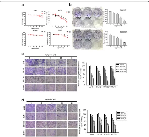
API suppresses migration, invasion, and colony formation of human NSCLC cells harboring different EGFR statuses Recent reports indicated that API can inhibit the in vitro proliferation and motility of human A549 NSCLC cells
which harbor the WT EGFR [30]. To further investigate the broad therapeutic potential of API against NSCLC, we examined the effects of API on cell migration and in-vasion of NSCLC cells harboring different EGFR statuses including the exon 19 deletion (del E746-A750) and L858R/T790 M. First of all, four NSCLC cell lines in-cluding A549 and CL1–5 (WT EGFR), HCC827 (del E746-A750), and H1975 (L858R/T790 M) were treated with various concentrations (0~ 80μM) of API for 24 or 48 h to determine the cytotoxic effects of API. An MTS analysis indicated that only at the highest concentration of 80μM, API partially suppressed or did not alter the via-bility of these NSCC cell lines after 24 h of treatment, compared to that of the controls (Fig.1a). In contrast to short-term treatment (24 h), the colony-formation assay showed that long-term treatment (7 days) with API sig-nificantly inhibited the colony-forming abilities of A549, CL1–5, HCC 827, and H1975 cells even at lower concen-trations of 10~20 μM (Fig. 1b, Additional file 1: Figure S1). These results indicated that 24-h treatment with API (0~ 40 μM) exhibited very low cytotoxicity in all tested NSCLC cells, but long-term treatment with API efficiently inhibited the in vitro tumorigenicity of these cells. To fur-ther determine the effects of short-term API treatment on the cell motility of NSCLC cells, transwell-migration and Matrigel-invasion assays were performed. As shown in Fig. 1c and d, after 24 h of treatment of A549, CL1–5, HCC827, and H1975 cells with API (0~ 40 μM), the mi-gratory and invasive abilities of these cells were all concentration-dependently suppressed, suggesting that even non-toxic concentrations of API can abolish the cell motility of these NSCLC cells.
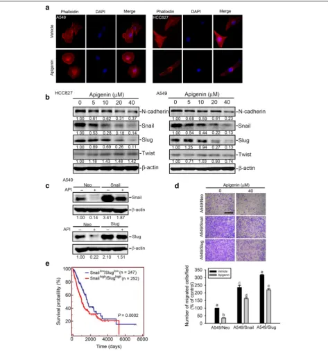
API inhibits the snail family-regulated EMT and invasive ability of NSCLC
The EMT and extracellular matrix (ECM) degradation are two key steps in cancer metastasis and were also shown to play critical roles in regulating the invasion and migration of NSCLC [36,37]. Previous studies indi-cated that actin remodeling is an important upstream regulator of the EMT in metastatic cancer cells [38].
transcription factors, Snail and Slug, were all concentra-tion-dependently downregulated after treating cells with API (0~ 40 μM) for 24 h (Fig.2b). These data suggested that downregulating Snail family members (Snail and Slug) might be critical for the API-mediated suppression of the EMT in EGFR WT and EGFR mutant NSCLC cells. Next, to determine whether Snail family members play critical roles in API-modulated cell motility, we overex-pressed Snail or Slug in A549 cells to reverse the inhibi-tory effect of API on the Snail family (Fig.2c). Meanwhile,
shorter survival times compared to patients with tumors exhibiting low expression levels of Snail and Slug (Fig.2e). These data suggest that downregulating Snail and Slug is crucial for the API-mediated inhibition of EMT progres-sion and cell invaprogres-sion, and those high levels of Snail and Slug predicted a poor prognosis in patients with NSCLC.
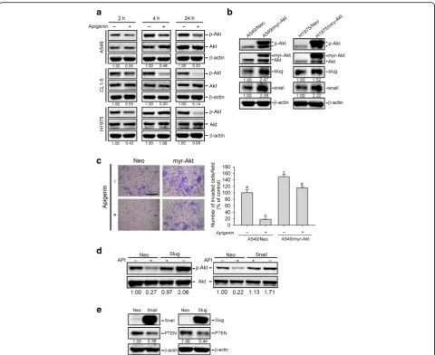
An interplay between Akt activation and expressions of snail family members is involved in API-mediated inhibition of cell motility
Previous studies indicated that the Akt signaling path-way plays a crucial role in most API-mediated anticancer processes and increases the stability and transcription of Snail family members during the EMT of cancer cells [15–17,29]. Herein, we found that API inhibited activa-tion of Akt at indicated time points (2, 4, and 24 h) in A549, CL1–5, and H1975 cells (Fig. 3a). Moreover, we further overexpressed the constitutively activated Akt, myr-Akt, in A549 and H1975 cells and found that over-expressing myr-Akt significantly increased Snail and Slug expression levels in both cells (Fig. 3b). Functionally, we observed that overexpressing activated Akt significantly reversed API-induced inhibition of invasion in A549 cells (Fig.3c). These data suggest that API suppression of Snail family-mediated cell motility is attributable to its capacity to inhibit Akt activation. In contrast to Akt-mediated Snail and Slug expressions, Snail was also reported to induce the EMT through activating the Akt pathway in NSCLC [39]. Our results showed that overexpression of Snail and Slug could reverse the API-mediated suppression of Akt activation (Fig.3d). Moreover, overexpression of Snail and Slug also inhibited expression of the PI3K/Akt pathway suppressor, PTEN (Fig. 3e), suggesting that API may in-hibit Snail family members to maintain PTEN expression and further suppress the PI3K/Akt pathway. Taken to-gether, these data suggest that a regulatory network be-tween Akt activation and Snail family expressions exists in API-mediated inhibition of cell motility.
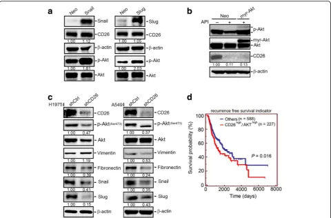
Alterations of protease levels in API-treated NSCLC cells Recent findings indicated that several proteases, including MMP-2, MMP-3, MMP-9, presenilin 1, and CD26, are promoters and mediators of EMT processes in different cancer types [24, 40, 41]. We next screened the effect of API on these proteases and found that MMP-3 and CD26 were considerably downregulated in CL1–5 cells after API treatment (Fig.4a). In a further check of the inhibitory ef-fect of API on proteases in other NSCLC cell lines (A549 and H1975), Western blot data showed that only CD26 was consistently suppressed by API in all tested NSCLC cells (Fig.4b, Additional file1: Figure S2). Moreover, re-sults showed that API concentration-dependently sup-pressed CD26 expression at concentrations of 10~ 80μM in CL1–5 cells (Fig. 4b). In addition to the protein level,
CD26 mRNA expression was also inhibited by API in A549, CL1–5, and H1975 cells (Fig.4c). These data sug-gest that inhibition of CD26 by API may be a general phenomenon in NSCLC cells. To further determine the functional role of CD26 in NSCLC, we investigated the correlation between the CD26 expression and the cell in-vasive ability. We first detected expression levels of CD26 in a set of NSCLC cell lines (A549, CL1–0, CL1–3, CL1– 5, HCC827, PC9, and H1975) using Western blotting and an RT-PCR (Fig.4d). Next, the invasive abilities of these cell lines were further evaluated, and we found a variety of invasive abilities, with A549 and H1975 cells exhibiting relatively high invasive abilities (Fig. 4e). Moreover, we observed that H1975 and A549 cells also expressed rela-tively high CD 26 protein and mRNA levels among these cell lines (Fig.4d). We further combined results from Fig. 4d and eand found a significant correlation (correlation coefficient r= 0.773, p= 0.042) between CD26 protein levels and the invasive abilities of these cell lines (Fig.4f). Next, to determine whether CD26 modulates the cell invasive ability, we knocked-down CD26 in A549, H1975, and CL1–5 cells. The knockdown efficiency of CD26-specific siRNA was detected by a Western blot ana-lysis (Fig. 4g, left panel). After knocking-down CD26 in these cells, we found their invasive abilities were all signifi-cantly attenuated compared to control cells (Fig.4g, right panel). In comparison, overexpression of CD26 signifi-cantly increased the cell-invasive abilities of poorly inva-sive CL1–0 cells and also reversed the API-mediated suppression of the invasive ability of CL1–0 cells (Fig.4h). In the clinic, we analyzed CD26 gene expression data obtained from TCGA and found that CD26 expression was inversely correlated with recurrence-free survival of lung cancer patients (Fig.4i). These results suggested that CD26 may play a critical role in the API-regulated invasive ability of NSCLC cells and may be associated with the poor prognosis of patients with lung cancer.
CD26 is an upstream regulator involved in API-regulated Akt activation and the EMT in NSCLC cells
significantly decreased Akt activation and Snail, Slug, and fibronectin expressions, and the knockdown efficiency of CD26-specific shRNA was also shown here. Similar inhibi-tory effects of CD26 shRNA on Akt activation and Snail/ Slug expression were also observed in CL1–5 cells (Add-itional file1: Figure S3). From TCGA database, we observed that patients with CD26high/Akthigh lung tumors had the shortest recurrence-free survival times compared to the CD26high/Aktlow, CD26low/Akthigh, and CD26low/Aktlow groups (Fig.5d). Taken together, these results revealed that
CD26 may be vital in mediating the Akt-Snail/Slug-induced EMT in NSCLC cells, and API can target this pathway. Clinical data indicated that upregulation of CD26 and Akt may be critical events in promoting lung cancer progression.
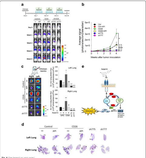
Significant anticancer effects of API via targeting CD26 in an A549 orthotopic graft model
To investigate the role of CD26 in tumor progression and in API-mediated anticancer activity in vivo, we Fig. 3Interplay between Akt activation and Snail family expression is involved in apigenin (API)-mediated inhibition of cell motility.aAkt
established an orthotopic lung tumor-bearing model by transplanting luciferase-tagged cells, A549-mock-lucifer-ase, A549-CD26-luciferA549-mock-lucifer-ase, or A549-shCD26-luciferase (sh-775 and sh-777), into NOD-SCID mice and allowed them to become established for 7 days before initiating treatment. A549–1-Luc orthotopic graft mice were IP-administered API or the vehicle 5 days/week. The ef-fects of API administration or CD26 knockdown and overexpression on tumor growth and metastasis were monitored by bioluminescence imaging. A schematic timeline of this experimental design and setup is shown in upper panel of Fig.6a. In vivo photon emission detec-tion revealed that both API treatment and CD26 knock-down in A549 cells attenuated tumor growth compared to the control group (Fig.6a, b). The tumorigenic ability was promoted in CD26-overexpressing A549 cells com-pared to control A549 cells and significantly reversed the antitumor growth effect of API (Fig. 6a, b). Similar to the in vivo data, after the mice were sacrificed, ex vivo
imaging of their lungs respectively showed lower and higher photon intensities in A549/shCD26- and A549/ CD26-injected mice compared to control mice, in both orthotopic tumors (left lung) and metastatic tumors (right lung) (Fig. 6c). The growth- and metastasis-inhibitory ef-fects of API were also reversed in cancer cells overexpress-ing CD26 (Fig. 6c). Moreover, histological data also confirmed that mice bearing A549/shCD26 tumors or re-ceiving API treatment all developed fewer metastatic nod-ules (as indicated by a black arrow) in the right lung compared to control mice (Fig. 6d). Taken together, these results revealed that API treatment suppressed the in vivo metastasis of NSCLC through targeting CD26.
Discussion
therapy and chemotherapy, higher invasive abilities, and acquisition of stem cell features [42]. Recently, API was used as a traditional medicine for its anticancer activities and low toxicity to normal cells. In both in vitro and in vivo models, API was shown to suppress tumor growth and metastasis in various cancer types. The antiprolifera-tive activities involve inducing apoptosis and autophagy, modulating the cell cycle and stemness, stimulating an immune response, and enhancing chemosensitivity [29]. Moreover, API was also shown to inhibit migration, in-vasion, and metastasis of various cancer cells through suppressing the EMT or modulating ECM-degrading en-zymes [29]. However, compared to other tumor types, little information on the effects of API on the migration and invasiveness of NSCLC cells is available. Until now, only one report indicated that API can inhibit the in vitro cell motility of NSCLC A549 cells by targeting Akt-mediated MMP-9 expression [30]. Our present re-sults showed that API inhibited the migratory and inva-sive abilities of a series of NSCLC cell lines harboring WT (A549 and CL1–5) or different mutant EGFR sta-tuses (HCC827 and H1975). Moreover, our results showed that API-mediated downregulation of MMP-2 and MMP-9 was only observed in A549 cells, but not in other NSCLC cell lines (CL1–5 and H1975), suggesting that inhibition of MMP-2 and MMP-9 by API might be cell-type specific. In contrast to MMPs, our study identi-fied that CD26/DPPIV plays a critical role in regulating the invasive abilities of several NSCLC cell lines (A549, CL1–5, CL1–0, and H1975) and can be downregulated by API treatment in these cell lines. Moreover, the anti-metastatic effect of API and the metastasis-promoting effect of CD26 were also observed in a human A549 xenograft model. These results suggested that suppres-sion of CD26 might be a general phenomenon in API-regulated cell motility of NSCLC cells.
CD26 is a multifunctional cell-surface glycoprotein with intrinsic DPPIV activity, and it is widely expressed in most cell types. Cell-surface proteases participate in
cancer progression and malignant transformation by fa-cilitating tumor cell invasion and metastasis [43]. Clin-ical studies of urothelial carcinoma, thyroid cancer, colorectal cancer, and gastrointestinal stromal tumors indicated that CD26 expression is associated with distant metastasis or recurrence after resection [23, 25, 44]. Similar to previous findings, we also observed that lung cancer patients with a high CD26 expression level had significantly worse recurrence-free survival than those with a lower level. Pang et al. recently identified that CD26-positive colon cancer cells were associated with enhanced invasiveness and chemoresistance, which might be due to EMT induction [24]. Our results showed abundant CD26 expression in highly invasive NSCLC cell lines (A549, H1975, and CL1–5), but low CD26 expression in the poorly invasive CL1–0 cell line and found a significant correlation between CD26 pro-tein levels and the invasive abilities of a set of NSCLC cell lines (A549, CL1–0, CL1–3, CL1–5, HCC827, PC9, and H1975). A similar correlation between CD26 ex-pression and invasive abilities was also reported in T-cell malignancies [45]. Moreover, CD26 knockdown in NSCLC cells caused decreases in the invasive abilities and EMT-related markers (Snail, Slug, and fibronectin). Based on these results, we suggest that the CD26 level is a potential marker for predicting the invasive ability of NSCLC cells and the prognosis of lung cancer patients, and CD26 may regulate invasion of cells through EMT induction.
According to the oncogenic role of CD26 in cancers, CD26 itself appears to be a novel therapeutic target. For example, an anti-CD26 monoclonal antibody or CD26 inhibitor treatment was shown to inhibit growth and in-vasiveness against several tumor types such as renal cell carcinoma and colon cancer [26, 46]. Recently, several flavonoids, particularly luteolin, API, and resveratrol, were shown to inhibit the in vitro enzyme activity of CD26 [47]. Our present study further demonstrated that API also suppressed the protein and mRNA expressions (See figure on previous page.)
of CD26 and the EMT-mediated cell invasion in several NSCLC cell lines (A549, CL1–5, and H1975). These re-sults suggested that inhibition of CD26 might be a gen-eral phenomenon in API-regulated cell motility of NSCLC cells. Moreover, our studies also showed that API treatment attenuated growth and metastasis via tar-geting CD26 in an A549 orthotopic graft model. Taken together, our data suggest that the ability of API to in-hibit cell invasiveness might be attributable to its cap-acity to suppress CD26 expression followed by inhibiting the EMT, and API has potential value in clinical applica-tions for advanced NSCLC. Furthermore, as CD26 was reported to be a possible stem cell marker that induced the EMT in colon cancer [24], the role of CD26 and the effect of API on the stemness of NSCLC will be further investigated in the future.
Transcription factors of the Snail family (Snail and Slug) were associated with EMT progression during lung cancer metastasis [42]. Our results showed that protein levels of Snail family members exhibited dramatic decreases after API treatment of NSCLC cells. PI3K/Akt was reported to play a crucial role in regulating expressions of Snail family members through multiple mechanisms. For example, activation of PI3K/Akt can phosphorylate GSK-3β to promote GSK-3β degradation and further maintain the stability of Snail and Slug [14]. Moreover, activation of Akt can induce an increase of nuclear factor (NF)-κB sub-unit p65, further leading to increased Snail transcrip-tion [48]. Our present study demonstrated that API treatment could suppress Akt activation in several NSCLC cell lines (A549, CL1–5, and H1975). More-over, overexpressing activated Akt in A549 cells induced upregulation of Snail and Slug and reversed API-mediated inhibition of the invasive ability, suggesting that Akt inhibition by API is a general phenomenon in NSCLC cells and may be the main cause for the API-mediated suppression of Snail family-induced cell motility. However, the effects of API on GSK-3β phosphorylation and p65 expression in NSCLC cells need to be further investigated in the future. Moreover, Akt activation was recently defined as a convergent feature of acquired EGFR TKI resistance, and increased p-Akt levels were observed in clinical specimens obtained from EGFR-mutant NSCLC patients who acquired EGFR TKI resistance [49, 50]. Combined treatment of Akt inhibitor with EGFR TKI had shown to exhibit the synergistic growth inhibitory effect in several EGFR TKI-resistant NSCLC models [49]. From the inhibitory effect of API on invasion and colony formation of NSCLC cells, our data also showed that EGFR mutant cells, HCC827 and H1975, were more sensitive to API treatment compared to the EGFR WT cells, A549 and
CL1–5. Among these NSCLC cells, TKI-resistant H1975 cell was the most sensitive cells to API treat-ment. In addition to Akt, our study further demonstrated that API also suppressed the expression of p-EGFR in EGFR mutant cells (Additional file1: Figure S4), suggest-ing the combination of API and EGFR TKI for the treat-ment of TKI-resistant NSCLC cells is worthy of further investigation.
As to upstream factors of PI3K/Akt signaling in cancer cells with EMT progression, transforming growth factor (TGF)-βwas reported to act as an important inducer to stimulate the Akt-mediated EMT. TGF-β was reported to activate Akt directly or through upregulating hyaluro-nan synthase (HAS) expression, promoting CD44/EGFR expression and co-localization, and subsequently activat-ing Akt in NSCLC cells [14, 51]. The effect of API on TGF-β expression and its downstream signaling mole-cules will be further investigated in NSCLC cells. In addition to TGF-β, the present study showed that knockdown of CD26 also suppressed Akt activation in NSCLC cells, suggesting that CD26 suppression by API might contribute to API-mediated inhibition of the Akt-induced EMT in NSCLC cells. Recent studies indi-cated that expression of CD26 in T cell lines leads to in-creased stromal-cell-derived factor (SDF)-1-α-mediated invasion through inducing PI-3 K/Akt pathways. Moreover, CD45 was reported to be associated with CD26 to regulate CD26’s enhancement of invasion [45]. Hence, the roles of SDF-1-α and CD45 in CD26-mediated invasive abilities of NSCLC and the effect of API on SDF-1-α and CD45 warrant further study in our future work.
Conclusion
The present data demonstrated that API treatment of NSCLC cells which harbor the WT or mutant EGFR can suppress CD26 expression and the interplay of down-stream targets, p-Akt and Snail/Slug, resulting in inhibition of the EMT-mediated invasive ability; the mechanism is schematically illustrated in Fig.6e. More-over, our study showed that increased CD26 expression was correlated with worse prognoses in lung cancer pa-tients and higher invasive abilities of NSCLC cells. Fur-thermore, we also demonstrated the antimetastatic effect of API and the metastasis-promoting effect of CD26 in a human A549 xenograft model. Our present findings strongly support API being used as a potential prevent-ive/therapeutic agent for managing NSCLC, and CD26 could be a useful biomarker for predicting NSCLC progression.
Additional file
Additional file 1:Figure S1.Effect of apigenin (API) on colony formation in CL1–5 and H1975 non-small cell lung cancer (NSCLC) cells. Figure S2.Effects of apigenin (API) on changes in different proteases in A549 and H1975 cells following 24-h treatment with API.Figure S3. Knockdown of CD26 suppresses Akt activation and the epithelial-to-mesenchymal transition (EMT) in CL1–5 cells.Figure S4.Apigenin (API) inhibits phosphorylation of the epidermal growth factor receptor (EGFR) in EGFR-mutant HCC827 and H1975 cells. (DOCX 431 kb)
Abbreviations
API:Apigenin; DPPIV: Dipeptidyl peptidase IV; ECM: Extracellular matrix; EGFR: Epidermal growth factor receptor; EMT: Epithelial-to-mesenchymal transition; GSK-3: Glycogen synthase kinase 3; MMP: Matrix metalloproteinase; NSCLC: Non-small cell lung cancer; PI3K: Phosphatidylinositol 3-kinase; PTEN: Phosphatase and tensin homologue; TKI: Tyrosine kinase inhibitor; WT: Wild-type
Acknowledgments
We would like to thank Dr. Tsang-Chih Kuo (National Taiwan University, Taipei, Taiwan) for the gift of the Snail- and Slug-overexpressing plasmids. We also give our thanks to Dr. Ching-Chow Chen from National Taiwan Uni-versity for providing myr-Akt plasmid.
Funding
This study was supported by a grant (106TMU-WFH-13) from Wan Fang Hospital, Taipei Medical University (to J.-H. Chang and M.-H. Chien). This study was also supported by the TMU Research Center of Cancer Translational Medicine from The Featured Areas Research Center Program within the framework of the Higher Education Sprout Project by the Ministry of Education (MOE) in Taiwan (to M.-H. Chien).
Availability of data and materials
All data generated or analyzed during this study are included in this published article and its additional files.
Authors’contributions
W-JL, J-HC, and M-HC designed and conceived the study. W-SC, W-YH, P-SC, and Y-CY performed the in vitro experiments and acquired the data. W-JL and W-SC performed the in vivo studies and analyzed data from the animal model. H, J-MC, and Y-CY analyzed clinical data from patients. C-WC, M-HC and J-M-HC wrote the manuscript. All authors read and approved the final manuscript.
Ethics approval and consent to participate
All animal experiments were carried out in accordance with guidelines of a protocol approved by the Taipei Medical University Animal Ethics Research Board.
Consent for publication Not applicable.
Competing interests
The authors declare that no competing interests exist.
Publisher’s Note
Springer Nature remains neutral with regard to jurisdictional claims in published maps and institutional affiliations.
Author details
1Department of Internal Medicine, School of Medicine, College of Medicine, Taipei Medical University, Taipei, Taiwan.2Division of Pulmonary Medicine, Department of Internal Medicine, Wan Fang Hospital, Taipei Medical University, Taipei, Taiwan.3Graduate Institute of Clinical Medicine, College of Medicine, Taipei Medical University, 250 Wu-Hsing Street, Taipei 11031, Taiwan.4Genomics Research Center, Academia Sinica, Taipei, Taiwan. 5Division of Hematology and Medical Oncology, Department of Internal Medicine, Wan Fang Hospital, Taipei Medical University, Taipei, Taiwan. 6Institute of Basic Medical Sciences, College of Medicine, National Cheng Kung University, Tainan, Taiwan.7Department of Medical Laboratory Science and Biotechnology, College of Medicine, National Cheng Kung University, Tainan, Taiwan.8Department of Medical Education and Research, Wan Fang Hospital, Taipei Medical University, 111 Hsing Long Road, Section 3, Taipei 11696, Taiwan.9Department of Urology, School of Medicine, Taipei Medical University, Taipei, Taiwan.10TMU Research Center of Cancer Translational Medicine, Taipei Medical University, Taipei, Taiwan.
Received: 2 July 2018 Accepted: 8 August 2018
References
1. Siegel R, Naishadham D, Jemal A. Cancer statistics, 2012. CA Cancer J Clin. 2012;62(1):10–29.
2. Chen Z, Fillmore CM, Hammerman PS, Kim CF, Wong KK. Non-small-cell lung cancers: a heterogeneous set of diseases. Nat Rev Cancer. 2014;14(8): 535–46.
3. Fossella F, Pereira JR, von Pawel J, Pluzanska A, Gorbounova V, Kaukel E, Mattson KV, Ramlau R, Szczesna A, Fidias P, et al. Randomized, multinational, phase III study of docetaxel plus platinum combinations versus vinorelbine plus cisplatin for advanced non-small-cell lung cancer: the TAX 326 study group. J Clin Oncol. 2003;21(16):3016–24.
4. Reck M, Heigener DF, Mok T, Soria JC, Rabe KF. Management of non-small-cell lung cancer: recent developments. Lancet. 2013;382(9893):709–19. 5. Wu JY, Shih JY, Chen KY, Yang CH, Yu CJ, Yang PC. Gefitinib therapy in
patients with advanced non-small cell lung cancer with or without testing for epidermal growth factor receptor (EGFR) mutations. Medicine. 2011; 90(3):159–67.
6. Mukohara T, Engelman JA, Hanna NH, Yeap BY, Kobayashi S, Lindeman N, Halmos B, Pearlberg J, Tsuchihashi Z, Cantley LC, et al. Differential effects of gefitinib and cetuximab on non-small-cell lung cancers bearing epidermal growth factor receptor mutations. J Natl Cancer Inst. 2005;97(16):1185–94. 7. Talbot LJ, Bhattacharya SD, Kuo PC. Epithelial-mesenchymal transition, the
tumor microenvironment, and metastatic behavior of epithelial malignancies. Int J Biochem Mol Biol. 2012;3(2):117–36.
8. Xiao D, He J. Epithelial mesenchymal transition and lung cancer. J Thorac Dis. 2010;2(3):154–9.
9. Thomson S, Buck E, Petti F, Griffin G, Brown E, Ramnarine N, Iwata KK, Gibson N, Haley JD. Epithelial to mesenchymal transition is a determinant of sensitivity of non-small-cell lung carcinoma cell lines and xenografts to epidermal growth factor receptor inhibition. Cancer Res. 2005;65(20):9455–62.
11. Kim D, Kim S, Koh H, Yoon SO, Chung AS, Cho KS, Chung J. Akt/PKB promotes cancer cell invasion via increased motility and metalloproteinase production. FASEB J. 2001;15(11):1953–62.
12. Irie HY, Pearline RV, Grueneberg D, Hsia M, Ravichandran P, Kothari N, Natesan S, Brugge JS. Distinct roles of Akt1 and Akt2 in regulating cell migration and epithelial-mesenchymal transition. J Cell Biol. 2005; 171(6):1023–34.
13. Grille SJ, Bellacosa A, Upson J, Klein-Szanto AJ, van Roy F, Lee-Kwon W, Donowitz M, Tsichlis PN, Larue L. The protein kinase Akt induces epithelial mesenchymal transition and promotes enhanced motility and invasiveness of squamous cell carcinoma lines. Cancer Res. 2003;63(9):2172–8. 14. Xu W, Yang Z, Lu N. A new role for the PI3K/Akt signaling pathway in the
epithelial-mesenchymal transition. Cell Adhes Migr. 2015;9(4):317–24. 15. Liu ZC, Wang HS, Zhang G, Liu H, Chen XH, Zhang F, Chen DY, Cai SH, Du J.
AKT/GSK-3beta regulates stability and transcription of snail which is crucial for bFGF-induced epithelial-mesenchymal transition of prostate cancer cells. Biochim Biophys Acta. 2014;1840(10):3096–105.
16. Hong KO, Kim JH, Hong JS, Yoon HJ, Lee JI, Hong SP, Hong SD. Inhibition of Akt activity induces the mesenchymal-to-epithelial reverting transition with restoring E-cadherin expression in KB and KOSCC-25B oral squamous cell carcinoma cells. J Exp Clin Cancer Res. 2009;28:28.
17. Kim JY, Kim YM, Yang CH, Cho SK, Lee JW, Cho M. Functional regulation of Slug/Snail2 is dependent on GSK-3beta-mediated phosphorylation. FEBS J. 2012;279(16):2929–39.
18. Havre PA, Abe M, Urasaki Y, Ohnuma K, Morimoto C, Dang NH. The role of CD26/dipeptidyl peptidase IV in cancer. Front Biosci. 2008;13:1634–45. 19. Fox DA, Hussey RE, Fitzgerald KA, Acuto O, Poole C, Palley L, Daley JF, Schlossman SF, Reinherz EL. Ta1, a novel 105 KD human T cell activation antigen defined by a monoclonal antibody. J Immunol. 1984;133(3):1250–6. 20. Torimoto Y, Dang NH, Vivier E, Tanaka T, Schlossman SF, Morimoto C.
Coassociation of CD26 (dipeptidyl peptidase IV) with CD45 on the surface of human T lymphocytes. J Immunol. 1991;147(8):2514–7.
21. Stremenova J, Krepela E, Mares V, Trim J, Dbaly V, Marek J, Vanickova Z, Lisa V, Yea C, Sedo A. Expression and enzymatic activity of dipeptidyl peptidase-IV in human astrocytic tumours are associated with tumour grade. Int J Oncol. 2007;31(4):785–92.
22. Bauvois B, De Meester I, Dumont J, Rouillard D, Zhao HX, Bosmans E. Constitutive expression of CD26/dipeptidylpeptidase IV on peripheral blood B lymphocytes of patients with B chronic lymphocytic leukaemia. Br J Cancer. 1999;79(7–8):1042–8.
23. Liang PI, Yeh BW, Li WM, Chan TC, Chang IW, Huang CN, Li CC, Ke HL, Yeh HC, Wu WJ, et al. DPP4/CD26 overexpression in urothelial carcinoma confers an independent prognostic impact and correlates with intrinsic biological aggressiveness. Oncotarget. 2017;8(2):2995–3008.
24. Pang R, Law WL, Chu AC, Poon JT, Lam CS, Chow AK, Ng L, Cheung LW, Lan XR, Lan HY, et al. A subpopulation of CD26+ cancer stem cells with metastatic capacity in human colorectal cancer. Cell Stem Cell. 2010;6(6):603–15.
25. Yamaguchi U, Nakayama R, Honda K, Ichikawa H, Hasegawa T, Shitashige M, Ono M, Shoji A, Sakuma T, Kuwabara H, et al. Distinct gene expression-defined classes of gastrointestinal stromal tumor. J Clin Oncol. 2008;26(25):4100–8. 26. Jang JH, Baerts L, Waumans Y, De Meester I, Yamada Y, Limani P, Gil-Bazo I,
Weder W, Jungraithmayr W. Suppression of lung metastases by the CD26/ DPP4 inhibitor Vildagliptin in mice. Clin Exp Metastasis. 2015;32(7):677–87. 27. Sak K. Cytotoxicity of dietary flavonoids on different human cancer types.
Pharmacogn Rev. 2014;8(16):122–46.
28. Shukla S, Gupta S. Apigenin: a promising molecule for cancer prevention. Pharm Res. 2010;27(6):962–78.
29. Yan X, Qi M, Li P, Zhan Y, Shao H. Apigenin in cancer therapy: anti-cancer effects and mechanisms of action. Cell Biosci. 2017;7:50.
30. Zhou Z, Tang M, Liu Y, Zhang Z, Lu R, Lu J. Apigenin inhibits cell proliferation, migration, and invasion by targeting Akt in the A549 human lung cancer cell line. Anti-Cancer Drugs. 2017;28(4):446–56.
31. Chu YW, Yang PC, Yang SC, Shyu YC, Hendrix MJ, Wu R, Wu CW. Selection of invasive and metastatic subpopulations from a human lung
adenocarcinoma cell line. Am J Respir Cell Mol Biol. 1997;17(3):353–60. 32. Lee YC, Saijo N, Sasaki Y, Takahashi H, Sakurai M, Ishihara J, Hoshi A, Chen
KM, Hamburger AW. Clonogenic patterns of human pulmonary
adenocarcinoma cell lines (PC-9, PC-13 and PC-14) and how they influence the results of test for chemosensitivity to cisplatin in the human tumor clonogenic assay. Jpn J Clin Oncol. 1985;15(4):637–44.
33. Yang SF, Lee WJ, Tan P, Tang CH, Hsiao M, Hsieh FK, Chien MH. Upregulation of miR-328 and inhibition of CREB-DNA-binding activity are critical for resveratrol-mediated suppression of matrix metalloproteinase-2 and subsequent metastatic ability in human osteosarcomas. Oncotarget. 2015;6(5):2736–53.
34. Lee WJ, Hsiao M, Chang JL, Yang SF, Tseng TH, Cheng CW, Chow JM, Lin KH, Lin YW, Liu CC, et al. Quercetin induces mitochondrial-derived apoptosis via reactive oxygen species-mediated ERK activation in HL-60 leukemia cells and xenograft. Arch Toxicol. 2015;89(7):1103–17.
35. Chien MH, Lee WJ, Hsieh FK, Li CF, Cheng TY, Wang MY, Chen JS, Chow JM, Jan YH, Hsiao M, et al. Keap1-Nrf2 interaction suppresses cell motility in lung adenocarcinomas by targeting the S100P protein. Clin Cancer Res. 2015;21(20):4719–32.
36. Mahmood MQ, Ward C, Muller HK, Sohal SS, Walters EH. Epithelial mesenchymal transition (EMT) and non-small cell lung cancer (NSCLC): a mutual association with airway disease. Med Oncol. 2017;34(3):45. 37. Ming SH, Sun TY, Xiao W, Xu XM. Matrix metalloproteinases-2,−9 and tissue
inhibitor of metallo-proteinase-1 in lung cancer invasion and metastasis. Chin Med J. 2005;118(1):69–72.
38. Shankar J, Nabi IR. Actin cytoskeleton regulation of epithelial mesenchymal transition in metastatic cancer cells. PLoS One. 2015;10(3):e0119954. 39. Liu CW, Li CH, Peng YJ, Cheng YW, Chen HW, Liao PL, Kang JJ, Yeng MH.
Snail regulates Nanog status during the epithelial-mesenchymal transition via the Smad1/Akt/GSK3beta signaling pathway in non-small-cell lung cancer. Oncotarget. 2014;5(11):3880–94.
40. Li P, Lin X, Zhang JR, Li Y, Lu J, Huang FC, Zheng CH, Xie JW, Wang JB, Huang CM. The expression of presenilin 1 enhances carcinogenesis and metastasis in gastric cancer. Oncotarget. 2016;7(9):10650–62.
41. Stallings-Mann ML, Waldmann J, Zhang Y, Miller E, Gauthier ML, Visscher DW, Downey GP, Radisky ES, Fields AP, Radisky DC. Matrix metalloproteinase induction of Rac1b, a key effector of lung cancer progression. Sci Transl Med. 2012;4(142):142ra195.
42. Shih JY, Yang PC. The EMT regulator slug and lung carcinogenesis. Carcinogenesis. 2011;32(9):1299–304.
43. Hakomori S. Aberrant glycosylation in tumors and tumor-associated carbohydrate antigens. Adv Cancer Res. 1989;52:257–331.
44. Lam CS, Cheung AH, Wong SK, Wan TM, Ng L, Chow AK, Cheng NS, Pak RC, Li HS, Man JH, et al. Prognostic significance of CD26 in patients with colorectal cancer. PLoS One. 2014;9(5):e98582.
45. Havre PA, Abe M, Urasaki Y, Ohnuma K, Morimoto C, Dang NH. CD26 expression on T cell lines increases SDF-1-alpha-mediated invasion. Br J Cancer. 2009;101(6):983–91.
46. Inamoto T, Yamochi T, Ohnuma K, Iwata S, Kina S, Inamoto S, Tachibana M, Katsuoka Y, Dang NH, Morimoto C. Anti-CD26 monoclonal antibody-mediated G1-S arrest of human renal clear cell carcinoma Caki-2 is associated with retinoblastoma substrate dephosphorylation, cyclin-dependent kinase 2 reduction, p27(kip1) enhancement, and disruption of binding to the extracellular matrix. Clin Cancer Res. 2006;12(11 Pt 1):3470–7. 47. Fan J, Johnson MH, Lila MA, Yousef G, de Mejia EG. Berry and Citrus
phenolic compounds inhibit dipeptidyl peptidase IV: implications in diabetes management. Evid Based Complement Alternat Med. 2013; 2013:479505.
48. Julien S, Puig I, Caretti E, Bonaventure J, Nelles L, van Roy F, Dargemont C, de Herreros AG, Bellacosa A, Larue L. Activation of NF-kappaB by Akt upregulates snail expression and induces epithelium mesenchyme transition. Oncogene. 2007;26(53):7445–56.
49. Jacobsen K, Bertran-Alamillo J, Molina MA, Teixido C, Karachaliou N, Pedersen MH, Castellvi J, Garzon M, Codony-Servat C, Codony-Servat J, et al. Convergent Akt activation drives acquired EGFR inhibitor resistance in lung cancer. Nat Commun. 2017;8(1):410.
50. Nukaga S, Yasuda H, Tsuchihara K, Hamamoto J, Masuzawa K, Kawada I, Naoki K, Matsumoto S, Mimaki S, Ikemura S, et al. Amplification of EGFR wild-type alleles in non-small cell lung Cancer cells confers acquired resistance to mutation-selective EGFR tyrosine kinase inhibitors. Cancer Res. 2017;77(8):2078–89.
51. Li L, Qi L, Liang Z, Song W, Liu Y, Wang Y, Sun B, Zhang B, Cao W. Transforming growth factor-beta1 induces EMT by the transactivation of epidermal growth factor signaling through HA/CD44 in lung and breast cancer cells. Int J Mol Med. 2015;36(1):113–22.
well as PI3K/AKT-independent pathways during prostate cancer progression. Cell Adhes Migr. 2015;9(4):255–64.
53. Escriva M, Peiro S, Herranz N, Villagrasa P, Dave N, Montserrat-Sentis B, Murray SA, Franci C, Gridley T, Virtanen I, et al. Repression of PTEN phosphatase by Snail1 transcriptional factor during gamma radiation-induced apoptosis. Mol Cell Biol. 2008;28(5):1528–40.
54. Uygur B, Abramo K, Leikina E, Vary C, Liaw L, Wu WS. SLUG is a direct transcriptional repressor of PTEN tumor suppressor. Prostate. 2015;75(9):907–16.
55. Lin Y, Kang T, Zhou BP. Doxorubicin enhances snail/LSD1-mediated PTEN suppression in a PARP1-dependent manner. Cell Cycle. 2014;13(11):1708–16. 56. Perez-Ramirez C, Canadas-Garre M, Molina MA, Faus-Dader MJ,
Calleja-Hernandez MA. PTEN and PI3K/AKT in non-small-cell lung cancer. Pharmacogenomics. 2015;16(16):1843–62.
57. Nam E, Park C. Maspin suppresses survival of lung cancer cells through modulation of Akt pathway. Cancer Res Treat. 2010;42(1):42–7. 58. Chang JH, Lai SL, Chen WS, Hung WY, Chow JM, Hsiao M, Lee WJ, Chien