Multiplex Reverse Transcriptase-PCR Assay for Typing and
Subtyping of Influenza A (H5 & H9) Virus in Iran
Esmaeil Saberfar
*1, Mohammad M. Forghani-Fard
2and Mirlatif Mosavi
21
Dept. of Microbiology and Biological Centre, Faculty of Medicine,Baqiyatallah (a.s.), University of Medical Sciences, Tehran; 2Dept. of Biology, Shahed University, Tehran, Iran
Received 29 March 2006; revised 16 September 2006; accepted 17 October 2006
ABSTRACT
Background: Avian influenza virus (AIV) infection is a major cause of bird or human mortality and morbidity, therefore the rapid identification of the virus is of important clinical and epidemiological
implication. Methods: A multiplex Reverse Transcriptase PCR (RT-PCR) was optimizedfor the detection of
influenza A virus and the H5 and H9 subtypes. The influenza type A specific primers were directed to the region of the influenza A matrix gene that is conserved among most type A influenza viruses. The H5 and H9 primers were directed to H5 and H9 hemagglutinin (HA) gene regions that are conserved among H5 and H9 subtypes. The selected primer sets were used in the RT-PCR for simultaneous detection of matrix, H5 and H9
responding specific sequences in a multiplex format. Results: Three reaction conditions were optimized
which include: i) RT-PCR typing using matrix gene primers for five subtypes of flu A (H1, H3, H5, H7 and H9), ii) RT-PCR subtyping for H5 and H9 subtypes, and iii) multiplex subtyping of H5 and H9. In this study, the multiplex RT-PCR was applied to 147 cloacal and tracheal swabs of clinical poultry cases with similar
influenza symptoms. Conclusions: These results suggest that multiplex RT-PCR assay can be a useful test
for rapid detection and subtyping of AIV in clinical samples. Iran. Biomed. J. 11 (2): 69-74, 2007
Keywords: Influenza, Hemagglutinin (HA), H9, Multiplex Reverse Transcriptase PCR (RT-PCR), Subtyping
INTRODUCTION
nfluenza A virus, a member of the Orthomyxoviridae family, infects swine, horses, seals, and a large number of birds as well as human. This virus contains a genome composed of eight single-stranded, negative-sense RNA segments that encode 10 different viral proteins. Two envelope glycoproteins, the hemagglutinin (HA) and NA proteins are the major viral antigens that induce protective antibodies following infection. Both proteins have highly variable sequences that give rise to variable antigenicity. There are 16 different HA subtypes and 9 different NA subtypes of the virus [1, 2]. Each of the hemagglutinin (H1-H16) subtypes are found in birds though only three of them have found in human viruses (H1-H3), two subtypes in pigs (H1, H3) and two subtypes in
horses (H3, H7) [3].
Host enzyme proteolytic cleavage of the HA protein is a prerequisite for the infectivity of Avian
influenza virus (AIV). The outcome of an AIV infection depends on the virus subtypes and on host [4]. Three influenza A virus subtypes (H1N1, H2N2, H3N2) with hemagglutinin and neuraminidase gene segments of avian origin have been associated with pandemic outbreaks and annually recurring disease in humans in the past century. In 1997, influenza A viruses of the H5N1 subtype were isolated from
patients in the Hong Kong area [5, 6]. In 1999,
H9N2 viruses were also isolated from patients with influenza-like illnesses [7, 8]. The transmission of influenza viruses H9N2 which containing H5N1-like internal genes to humans emphasizes the need for better understanding the occurrence and molecular
epidemiology of these viruses. The H9N2 virus
subtype was first isolated from Turkeys in 1996, when the virus was associated with mild respiratory disease. In Asia, long-term surveillance in live poultry markets in Hong Kong (1975-1985) detected H9N2 influenza viruses in apparently healthy ducks. Since the early 1990's, H9N2 influenza viruses have
I
become widespread in domestic chickens in Asia. Results of the studies suggested that the H5N1 viruses infected humans in 1997, caused by reassortment which occurred between the H9N2 and H5N1 viruses. Surveillance studies in 1997 indicated that two subtypes of influenza viruses were cocirculating, raising the possibility of genetic exchange between these viruses after coinfection of the same host [9].
Since there is a greater risk for these subtypes to become highly pathogenic, it is important to identify them specifically in surveillance programs. In this study, we describe a multiplex reverse transcriptase PCR (RT-PCR) for rapid screening of clinical samples for simultaneous detection of type A influenza virus and H5 and H9 subtypes.
MATERIALS AND METHODS
Viruses. Virus strain used:
A/Turkey/England/50-92/91 (H5N1), A/Turkey/England/7732/66 (H5N9), (H7N7). A/Tehran/49/2001(H1N1), A/Tehran/82/79 (H3N2), A/Chicken/Iran/11T/99 (H9N2). The H5N1, H5N9 and H7N7 antigens were originally obtained from the Veterinary Laboratories Agency in the United Kingdom. Dr. T. Mokhtari from the National Influenza Centre, Tehran University of Medical Sciences (Iran) was kindly provided the H1N1 and H3N2 human influenza viruses. The A/Chicken/Iran/11T/99 (H9N2) influenza antigen was obtained from Razi Vaccine and Serum Research Institute (Karaj, Iran), isolated previously from outbreak among poultry in Iran. Hemagglutination (HA) titers of the viruses ranged from 512 to 1024 HA Unit, when tested according to the methods as described previously [10].
RNA extraction from virus stocks. RNA
purification was performed using the RNX™-Plus Kit (CinnaGen, Iran) according to the manufacturer instructions. Briefly, 100-150 µl viral suspension
(egg-fluid, clinical specimens and water control) was mixed with 1 ml RNX and left for at least 5 min at 4ºC. After the addition of 200 µl chloroform and mixing, the liquid was clarified by centrifugation at 12,000 ×g at 4ºC for 15 min. The supernatant was transferred into a new tube and mixed with an equal volume of isopropanol followed by centrifugation at 12,000 ×g at 4ºC for 15 min. The pellet was washed with 1 ml 70% ethanol. Finally, RNA was eluted by 50 µl of 1 mM RNase free EDTA.
Reverse transcription. An influenza virus matrix
gene-specific PCR primer set for a region conserved in all type A influenza virus matrix genes; in addition, H5 and H9-specific primer sets for conserved regions of the H5 and H9 hemagglutinin gene sequences were used for RNA isolation and RT-PCR detection with single primer sets or by multiplex RT-PCR as described in [11] (Table1). The oligonucleotide primers were commercially synthesized (MWG, Germany). The 20-µl reaction for each gene (M and HA) contained 5 µl of extracted RNA, 4 µl of 5-times reverse transcriptase buffer, 2 µl dNTP (2.5 mM each of four dNTPs), 1 µl (10 pmol/µl) of forward primer, 0.5 µl (40 unit/µl) of RNase inhibitor and 1 µl (40 unit/µl) of M-muLV
RT and 6.5 µl of water mix separately for each
subtype. Reverse transcription was carried out at 42ºC for 45 min followed by incubation at 70ºC for 10 min.
PCR. PCR was carried out for both genes of each
subtype according to the following conditions: the 30 µl reaction contained 10 µl of cDNA, 0.5 µl (2.5 mM each of four dNTP), 2 µl (10 pmol) of each primer, 3 µl of 10-times PCR-buffer, 1 µl (10 mM)
MgCl2 and 0.25 µl (5 unit/µl) of Taq DNA
polymerase enzyme and 13.75 µl of water. Wide ranges of cycling conditions were tested. After an initial denaturation at 94ºC for 5 min, three-step PCR cycling protocol was used for the matrix gene primer set as follows: 94ºC for 1 s, 54ºC for 1 s and
Table 1. The oligonucleotides used.
Size Location
Sequences Primers
Specificity
101 24-47
124-100 AGA TGA GTC TTC TAA CCG AGG TCG
TGC AAA AAC ATC TTC AAG TCT CTG MF
MR Influenza A virus
152 1504-1525
1655-1635 ACG TAT GAC TAT CCA CAA TAC TCA G
AGA CCA GCT ACC ATG ATT GC H5F
H5R Avian H5
190 477-485
656-645 AAG GGC TTT CAC CGA AGA G
CCC ATT CTC ATT ACT GCT TCT H9F
H9R Avian H9
72ºC for 3 s for 35 cycles were followed by a final primer extension step at 70ºC for 3 min. The H5 PCR cycling conditions were the same as those for the matrix gene except that annealing and extension temperatures were used for 3 and 5 s, respectively. A three-step cycling protocol was used for the H9-specific PCR as follows: 94ºC for 3 s, 54ºC for 5 s and 72ºC for 8 s for 35 cycles. RT-PCR was performed with the BIOTECH-MWG thermocycler (Germany). RNA purification, RT-PCR set-up, run and agarose gel electrophoresis were performed in separated rooms. Negative reagent controls were included in each assay. No contamination was detected at any time.
Multiplex RT-PCR. Multiplex RT-PCR was
carried out in 50 µl reaction (total volume) for HA gene of H5 and H9 subtypes. Cycling conditions were the same as those for the H9-specific PCR except that a 54ºC annealing temperature was used for 30 cycles.
Sequence analysis. The PCR products were
analysed by sequencing (MWG, Germany). As a first step, the products were cleaned using a PCR purification kit (Fermentas, USA). Sequencing was performed in both the sense and anti-sense direction with primers MF and MR for the matrix gene and the RT-PCR primers H5F, H5R (H5) and H9F, H9R (H9), respectively for the HA gene (Table1). Sequencing data were aligned with matrix and HA influenza sequences from NCBI database to assess homology.
Detection of PCR product by gel electrophoresis.
PCR products (10 µl) were analysed by agarose gel electrophoresis, using a 2% agarose in 1× TBE buffer. Amplified products were visualized by ultraviolet light transillumination after staining with 0.1 µg/ml ethidium bromide. A 50 base pair ladder was used as a molecular weight marker.
RESULTS
The nucleotide sequences of matrix gene are conserved in all subtypes of influenza A viruses. To identify these viruses by RT-PCR, we used two primers based on consensus sequences of matrix
genes of Influenza viruses [12]. The matrix RT-PCR
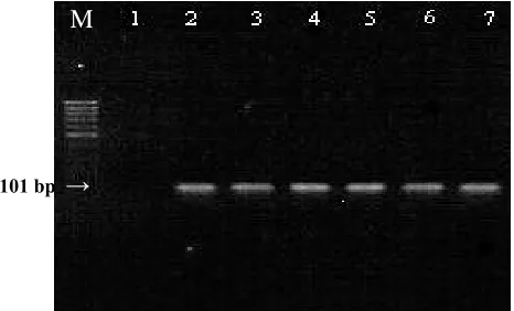
(primer MF and MR) was able to amplify a 101-bp fragment of the matrix gene from a series of different influenza A strains (H1, H3, H5, H7 and H9) as demonstrated in Figure 1. To test the
Fig. 1. Identification of Influenza type A virus by RT-PCR. The M gene RT-PCR was applied to a panel of influenza A strains representing various subtypes. Lane M, size markers (100 bp ladder, CinnaGen, Iran) lane 1, H2O control and lanes 2-7, 10 µl of RT-PCR products obtained by using primers specific to M genes of the viruses H1N1, H3N2, H5N1, H5N9, H7N7, and H9N2. The size of RT-PCR M gene product is indicated by arrow.
sensitivity of the matrix RT-PCR, serial ten-fold dilutions of virus pools of known titer were RNA purified and PCR amplified. Three different avian strains were tested namely A/turkey/England/50-92/91 (H5N1), A/Turkey/England/7732/66 (H5N9), and A/Chicken/Iran/11T/99 (H9N2) with 50%
end-point dilution of 10-5, 10-5.4, and 10-7, respectively.
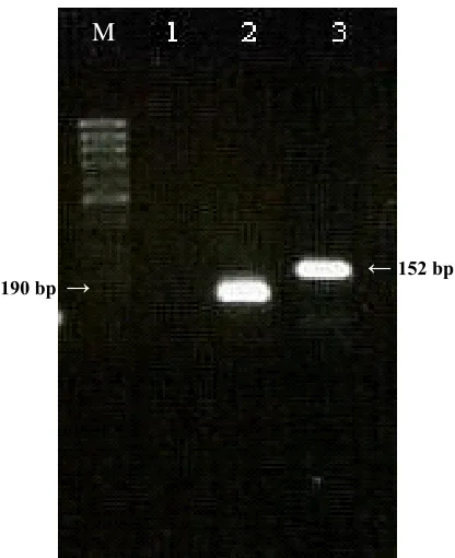
The specificity of the primers for the detection of influenza A was examined using RNA from other human and avian viral species. The matrix RT-PCR detected influenza A virus not only in avian strains but also in other animal species including human strains (H1N1 and H3N2). RNA isolated from influenza B and C viruses did not yield amplified products. The HA RT-PCR (primer H5F, H5R) for the detection of H5 influenza A subtypes amplified a band of 152 bp (Fig. 2). A sharp band of the expected size was obtained from H5 strains tested and no PCR product was amplified from non-H5 influenza subtypes (data not shown). The H5 origin was verified by sequencing.
The HA RT-PCR (primer H9F, H9R) for the detection of H9 influenza A subtypes amplified a band of 190 bp. A single band of the expected size was detected from avian H9 strain (Fig. 2) and HA sequence of H9 subtype origin verified by sequencing too (data not shown). The multiplex RT-PCR was tested for its specificity for viral targets (influenza A H5 and H9) by adding each of the influenza virus primer pairs. No mispriming was observed when primer sets were present with both influenza A H5 and H9 template. A product of the expected size was obtained for each viral template
101 bp →
M
Fig. 2. Identification and subtyping of avian influenza viruses in cloacal and tracheal swabs samples. Lane M, size markers (50 bp ladder, CinnaGen, Iran) lane 1, H2O control and lanes 2 and 3, ten µl of RT-PCR products obtained by using primers specific to HA genes of the H5N1 and H9N2 viruses product was applied on a 2% gel. The sizes of RT-PCR HA gene product are indicated by arrows.
by the multiplex RT-PCR with primer set present (Fig. 3). The specific products could be clearly separated and identified on a 2% agarose gel. The multiplex RT-PCR was performed on both avian and tracheal swabs for detection of H9 and H5 subtype. Twenty nine out of 98 (29%) cloacal swabs and 11 out of 49 (22%) tracheal swab samples contained influenza A virus H9 subtype. No detectable PCR products were seen for influenza A virus H5 subtype. Nucleic acid extraction and multiplex RT-PCR amplification from 26 human clinical samples (nasopharyngeal aspirate or nose and throat swabs)
did not show any PCR product.
DISCUSSION
Multiplex PCR capable of amplifying various regions of chromosomal DNA with a precise detection and typing of several bacterial pathogens has been described previously [11, 12]. The use of RT-PCR for the detection of influenza viruses is not new; several strategies of RT-PCR have been used to detect influenza A viruses [13, 14] or to
distinguish between influenza A, B or C viruses [15, 16], Subtype-specific primers have been used to differentiate H1 virus from H3 virus or to differentiate N1 virus from N2 virus [15, 17].
Since the rate of evolution for influenza virus HA gene is substantially higher than other genes, the sequences of these genes do change over time [3], therefore, it may be necessary to update the primer sequences as needed to compensate for genetic drift. In this study, we describe a multiplex RT-PCR method capable of distinguishing the matrix genes of all influenza type A viruses, and HA genes of avian H5 and H9 subtypes, which were used to screen viruses from clinical samples isolated in Iran [18].
To diagnose influenza, culture confirmation is a well-established laboratory technique and remains as the standard method in most clinical laboratories. Culture conformation is a combination of cell culture-based virus isolation (VI) and subsequent immunostaining; however, the requirements of maintaining tissue culture cells and infectious virus particles represent drawbacks in obtaining timely and consistent results [19]. Moreover, VI can detect only live virus; therefore the viruses inactivated during shipping or by disinfectants (which may be present in environmental samples) may be detected
Fig. 3. Subtyping of avian influenza viruses in cloacal and tracheal swabs samples. Lane M, size markers (50 bp ladder, CinnaGen, Iran) lane 1, H2O control and lanes 2, ten µl of Multiplex RT-PCR products obtained by using primers specific to HA genes of the H5N1 and H9N2 viruses. The sizes of RT-PCR HA gene products are indicated by arrows.
190 bp → ←152 bp
M
M
←190 bp ←152 bp
by RT-PCR. In addition, all influenza viruses may not readily adapt to growth detectable titers in
embryonated
chicken eggs within two passages.This may explain why some samples are RT-PCR positive and VI negative [20]. We have optimized a multiplex RT-PCR as a rapid alternative for AIV
detection and subtyping to VI in
embryonated
chicken eggs. Further to the advantage of the speed, sensitivity and ease of multiplex RT-PCR method, the risks of handling infectious materials encountered with VI method are reduced. However, we are the first to optimize multiplex RT-PCR that were able to detect type A influenza and differentiate the H5 and H9 subtypes simultaneously.
The multiplex RT-PCR was performed on both avian and tracheal swabs for detection of H9 and H5 subtypes. Twenty nine out of 98 (29%) cloacal swabs and 11 out of 49 (22%) tracheal swab samples contained influenza A virus H9 subtype. We found that by RT-PCR, 40 out of the 127 (31%) swab samples contained avian influenza and all of them were H9 subtypes. These samples were collected from chickens of 14 different farms in Iran in 2002-2004. This finding suggests a current endemic of H9 AIV in the chicken population of Iran. In fact, we have isolated a total of 40 AIV from chicken in Iran in 2002-2004 and all of them were H9 and none of them was H5 subtypes.
Therefore, the multiplex RT-PCR can be a reliable and accurate method for large scale screening and detection of type A influenza virus and the avian H5 and H9 subtypes simultaneously.
ACKNOWLEDGEMENTS
The authors thank Dr. T. Mokhtari Azad at the National Influenza Centre, Tehran Medical Science University (Iran) and Dr. M. Farshian at Reference Laboratory of Veterinary Organization of Iran (Tehran) for providing human and AIV. This work was supported by a grant from the Ministry of Higher Education of Iran.
REFERENCES
1. Berinstein, A., Bruce, S. and Suarez, D.L. (2002)
Heteroduplex mobility assay for detection of new avian influenza virus variants. Avian Disease 46: 393-400.
2. Munster, V.,Anders, W., Bestebroer,T.M., Herfst, S., Derek, S., Rimmelzwaan, G., Björn, O. and Osterhaus, M. (2005) Characterization of a novel influenza A virus hemagglutinin subtype (H16) obtained from black-headed gulls. J. Virol. 79: 2814-2822.
3. Brown, E.G. (2000) Influenza virus genetics.
Biomed. Pharmacother 54: 196-209.
4. Munch, M., Neilson, K., Handberg, S. and Jorgensen,
P.H. (2001) Detection and subtyping (H5 & H7) of avian type A influenza viruses by RT-PCR and PCR-ELISA. Arch. Virol. 146: 87-97.
5. Guan, Y., Shortridge, K.F., Krauss, S. and Webster,
R.G. (1999) Molecular characterization of H9N2
influenza viruses; were they the donors of the "internal" genes of H5N1 viruses in Hong Kong? Proc. Natl. Acad. Sci. (USA) 96: 9363-9367.
6. Zhou, N., Shortridge, K.F., Claas, E.J., Krauss, S.L.
and Webster, R.G. (1999) Rapid evolution of H5N1 influenza viruses in chickens in Hong Kong. J. Virol. 73: 3366-3374.
7. Peiris, M., Yuen, K.Y., Leung, C.W., Chan, K.H.,
Lai, R.W., Orr, W.K. and Shortridge, K.F. (1999) Human infection with influenza H9N2. Lancet 354: 916-917.
8. Nikolai, V., Kaverin, I., Rudneva, A., Ilyushina,
N.A., Liptov, A.S., Kruss, S. and Webster, R.G. (2004) Structural difference among heamagglutinin of influenza A viruses subtypes are reflected in their antigenic architector. J. Virol. 78: 240-249.
9. Peiris, M., Yam, W., Chan, K., Ghose, P. and
Shortridge, K.F. (1999) Influenza A H9N2: Aspect of laboratory diagnosis. J. Clin. Microbiol. 37: 3426-3427.
10. Saberfar, E., Marschall, M., Mohammadi, H., Fayas,
A. and Meier-Ewert, H. (2001) A sensitive neutralization assay for influenza C viruses based on the acetyl esterase activity HEF glycoprotein. Iran. Biomed. J. 1: 27-32.
11. Mahoney, J.B., Luinestra, M., Tyndall, S.W., Sellars, S. and Chernesky, M. (1995) Multiplex RT-PCR for detection of Chlamydia trachomatis and Neisseria gonorrhoea in genitourinary specimens. J. Clin. Microbiol. 33: 7049-3053.
12. Stockton, J.S., Ellis, S., Sauille, M., Clewley, J.P. and Zambon, M.C. (1999) Multiplex PCR for typing and subtyping influenza and respiratory syncytial viruses. J. Clin. Microbiol. 9: 2990-2995.
13. Cherian, T., Bobo, L., Steihoff, M.C., Karron, R.A.
and Yolken, R.H. (1994) Use of PCR-enzyme immunoassay for identification of influenza A virus matrix RNA in clinical samples negative for cultivable virus. J. Clin. Microbiol. 32: 623-628.
14. Atmar, R.L., Baxter, B.D., Dominguez, E.A. and
Taber, L.H. (1996) Comparison of reverse transcription-RT-PCR with tissue culture and other rapid diagnostic assay for detection of type A influenza virus. J. Clin. Microbiol. 34: 2604-2606.
15. Wright, K.E. (1995) Typing and subtyping of influenza viruses in clinical samples by PCR. J. Clin. Microbiol. 33: 1180-1184.
16. Zou, S. (1997) A practical approach to genetic
screening for influenza virus variants. J. Clin. Microbiol. 35: 2623-2627.
17. Gavin, P.J. and Thomson, R.B. (2003) Review of
rapid diagnostic tests for influenza. Clin. Appl. Immunol. Rev. 4: 151-172.
18. Lee, M.S., Chang, P.C., Shien, J., Cheng, M. and
Shieh, H.P. (2001) Identification and subtyping of
avian influenza viruses by reverse transcription-PCR. J. Virol. Method 97: 13-22.
19. Sheau-Mei, C., Vainionpa, R., Zhao, P., Fenglan, L.,
Hu, A., Forrest, B. and Rappaport, R. (2004) Detection of influenza B in clinical specimens: comparison of high through put RT-PCR and culture conformation. Virus Res. 103: 85-90.
20. Spakeman, E., Denis, A., Senne, T.S., Myers, L., Lindsey, P. and Lohman, K. (2002) Development of real time RT-PCR assay for type A influenza viruses and the avian H5 and H7 heamagglutinin subtypes. J. Clin. Microbiol. 31: 3256-3260.