125
Obesity: An Introduction and Evaluation
Paras Gupta*, Sandeep Tyagi, Minky Mukhija, Arminder Singh Saini, Rohit Goyal,
Pyare Lal Sharma
Department of Pharmacology,
I.S.F. College of Pharmacy, Moga, Punjab, India.
*Corresponding author: [email protected]
ABSTRACT:
Obesity is a pathological condition in with excess body fat. It is a chronic disorder with
complex interaction between genetic and environmental factors. It is being
characterized by high cholesterol, fatty acid levels, Insulin desensitization; high blood
pressure; and excessive adipose mass accumulation. Currently more than 1 billion
adults are overweight and at least 300 million of them are clinically obese. It is defined
by body mass index and further evaluated by both percentage body fat and total body
fat. Obesity is a risk to many secondary conditions like cardiovascular disorder, insulin
pathological resistance, retinopathy, neuropathy and cancer. Various factors
modulating the development of obesity are age, sex, smoking, growth hormone level,
skeletal muscle metabolism. Experimental models used to evaluate obesity are high fat
diet, high cafeteria diet, hypothalamic lesions, goldthioglucose induced obesity,
monosodium glutamate induced obesity. Non-human primates, Spontaneously obese
rats, Obesity due to natural allele defects in mice, V-Genetic variants in the human
Uncoupled Protein-1 gene and Viral induce obesity are also preferred.
Keywords: Obesity, Mechanisms, Co-morbidities, Worldwide prevalence, Evaluation.
INTRODUCTION:
Obesity is a pathological condition in which excess body fat accumulated, leading
adverse effects on health and life expectancy. [1] It is a chronic disorder with complex
interaction between genetic and environmental factors. It characterized by high
cholesterol, fatty acid levels; imbalance in metabolic energy; insulin desensitization;
lethargy, gallstones; high blood pressure; shortness of breath; emotional and social
problems; and excessive adipose mass accumulation with hyperplasia and hypertrophy.
[2] Pathological obesity is associated with several secondary commodities like heart
126
[1] It is most commonly caused by a combination of excessive dietary calories, lack of
physical activity, and genetic susceptibility. Evidence to support this view is that some
obese people eat little yet gain weight due to slow metabolic rate. [3,4] The primary
treatment for obesity are dieting and physical exercise. To supplement this, or in case of
failure, anti-obesity drugs may be taken to reduce appetite or inhibit fat absorption. In
severe cases, surgery is performed or an intragastric balloon is placed to reduce
stomach volume and/or bowel length, leading to earlier satiation and reduced ability to
absorb nutrients from food [5,6].
Worldwide prevalence:
Obesity is one of the leading preventable causes of death worldwide. [7] Currently more
than 1 billion adults are overweight and at least 300 million of them are clinically
obese. Current obesity levels range from below 5% in China, Japan and certain African
nations, to over 75% in urban Samoa. Childhood obesity is already epidemic in some
areas and on the rise in others. An estimated 17.6 million children under five yr. age
are estimated to be overweight worldwide. According to the US Surgeon General, in the
USA the number of overweight children has doubled and the number of overweight
adolescents has trebled since 1980. The prevalence of obese children aged 6-to-11 years
has more than doubled since the 1960s. Obesity prevalence in youths aged 12-17 has
increased dramatically from 5% to 13% in boys and from 5% to 9% in girls between
1966-70 and 1988-91 in the USA. The problem is global and increasingly extends into
the developing world: In Thailand the prevalence of obesity in 5-to-12 year olds children
rose from12.2% to 15-6% in just two years. Obesity accounts for 2-6% of total health
care costs in several developed countries. The true costs are undoubtedly much greater
as not all obesity-related conditions are included in the calculations. [8] In the United
States obesity is estimated to cause an excess 111,909 to 365,000 death per year, while
1 million (7.7%) of deaths in the European Union are attributed to excess weight. [9,10]
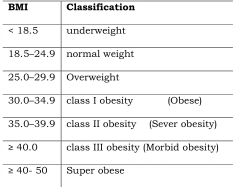
Classification:
Obesity is a medical condition in which excess body fat has accumulated to the extent
that it may have an adverse effect on health. [11] It is defined by body mass index
(BMI) and further evaluated in terms of fat distribution via the waist–hip ratio and total
cardiovascular risk factors. [12] BMI is closely related to both percentage body fat and
total body fat. [13] In children a healthy weight varies with age and sex. Obesity in
children and adolescents is defined not as an absolute number but in relation to a
127
95th percentile. The reference data that these percentiles are based on are from 1963 to
1994, and thus have not been affected by the recent increases in weight. [7,14,15]
BMI is calculated by dividing the subject's mass by the square of his or her height,
typically expressed either in metric or US "customary" units)
Metric: BMI = kilograms / meters2; US customary and imperial: BMI = lb *703
/ in2)where lb is the subject's weight in pounds and in is the subject's height in inches
(Table 1).
The most commonly used definitions, established by the World Health Organization in
1997 and published in 2000 provide the values listed in the table at right. [2] The
surgical literature breaks down "class III" obesity into further categories whose exact
values are still disputed. [16] As Asian populations develop negative health
consequences at a lower BMI than Caucasians, some nations have redefined obesity;
the Japanese have defined obesity as any BMI greater than 25 [17] while China uses a
BMI of greater than 28. [18]
Table 1: Classification of obesity based on BMI
BMI Classification
< 18.5 underweight
18.5–24.9 normal weight
25.0–29.9 Overweight
30.0–34.9 class I obesity (Obese)
35.0–39.9 class II obesity (Sever obesity)
≥ 40.0 class III obesity (Morbid obesity)
≥ 40- 50 Super obese
Mechanisms:
Obesity is majorly responsible for metabolic dysfunction involving lipid and glucose. It
also facilitates secondary complications like cardiac, liver, intestinal, pulmonary,
endocrine, and reproductive dysfunctionings. The provoked inflammatory,
insulin-resistant, hypertensive, and thrombotic-promoting adipokines, which are atherogenic
are counter-balanced by anti-inflammatory and anti-atherogenic adipocyte hormones
128
actions of leptin and resistin are pro-atherogenic. [19] It occurs due to imbalance
between food intake and energy expenditure. Possible involvement of NPY (Neuro
Peptide Y), MCH (Melanocortin hormone), AGRP (Agouti gene related peptide), Orexin-A
and –B, Galanin, α-MSH (α-Melanin stimulating hormone), CRF (corticotrophin
releasing hormone), CART (caffeine and amphetamine releasing hormone),
Glucagon-like peptide-1 (GLP-1), CCK (coli cysto kine), 5-HT (5-Hydroxy triptamine), insulin, and
leptin found to occur during regulation of food intake. [20,21] Moreover, it also
contributes to immune dysfunction from the effects of its inflammatory adipokine
secretion; and the worsening of metabolic syndrome. Molecular and genetic studies of
animal models have identified numerous genes that may cause or contribute to the
development of obesity. They have also provided significant insight into the peripheral
and central regulating cascades like (i) Peripheral: insulin, leptin, gheralin, CCK, 5- HT
[20] and (ii) Central: NPY, AGRP, α-MSH, Orexin, CART, MCH [21] that control energy
intake and expenditure. [Fig 1] Genetic studies of families and populations have
generated useful information on genes and mutations associated with or linked to
obesity, body fat distribution, and other relevant phenotypes. [22]
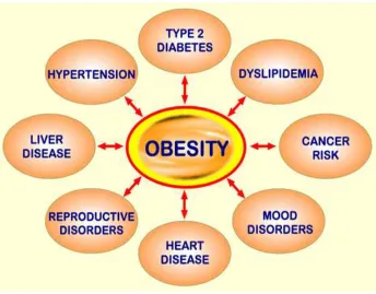
129
Co-morbidities associated with obesity:Obesity increases the risk of several physical and mental conditions. The co-morbidities
are most commonly shown in metabolic syndrome, which includes: diabetes mellitus
(type 2), high blood pressure, high blood cholesterol, and high triglyceride levels. [23]
Complications are either directly caused by obesity or indirectly related through
mechanisms sharing a common cause such as a poor diet or a sedentary lifestyle.
Excess body fat underlies 64% of cases of diabetes in men and 77% of cases in women.
[24] Health consequences fall into two broad categories: those attributable to the effects
of increased fat mass (such as osteoarthritis, obstructive sleep apnea, social
stigmatization) and those due to the increased number of fat
cells (diabetes, cancer, cardiovascular disease, non-alcoholic fatty liver disease). [24,25]
Increases in body fat alter the body's response to insulin, potentially leading to insulin
resistance. It also creates a inflammatory state, Shoelson SE and a
pro-thrombotic state. [24,26] Large-scale American and European studies have found that
mortality risk is lowest at a BMI of 22.5–25 kg/m2 in non-smokers and at 24–
27 kg/m2 in current smokers, with risk increasing along with changes in either
direction. [27,28] The risk of obesity with higher co-morbidities are as follows.
Cardiology - ischemic heart disease, angina and myocardial infarction, congestive
heart failure, high blood pressure, abnormal cholesterol levels, deep vein
thrombosis and pulmonary embolism. [1]
Endocrinology - Diabetes mellitus, polycystic ovarian syndrome,
menstrual disorders, infertility, complications during pregnancy, birth defects and
intrauterine fetal death. [1]
Neurology - Stroke, neuralgia parenthetical, migraines, carpal tunnel syndrome,
dementia, idiopathic intracranial hypertension and multiple sclerosis. [29]
Psychiatry - Depression in women and social stigmatization. [1]
Rheumatology and Orthopaedics - Gout, poor mobility, osteoarthritis and low back
pain. [30]
Gastrointestinal – Gastroesophageal reflux disease, fatty liver disease and
130
Respirology - Obstructive sleep apnea, obesity hypoventilation syndrome, asthma
and increased complications during general anesthesia [Fig 2]. [1]
Fig 2: Co-Morbidities associated with Obesity
Factors modulating obesity:
Age - Childhood obesity is a risk factor for adulthood obesity, Body fat content
increases during adulthood, the maximal rates of overweight and obesity attained from
55 to 65 yr. [31]
Sex - Women have more body fat. The differences in prevalence of obesity vary in
populations or among ethnic groups. [31]
SES - More obese in high people SES (Socionomic status) classes and in poor countries,
and obese in low SES classes and in rich countries. [31]
Energy intake - Overfeeding causes weight gain and leads to obesity. [31]
Dietary fat intake - Dietary fat is related to prevalence of overweight in ecologic
131
RMR - A low body mass and composition adjusted RMR (Resting metabolic rate) is a
risk factor for weight gain, but some reports reveal that the, Overweight and obese
people have higher absolute RMR. [31]
Physical activity (PA) level - A low level of PA is a risk factor for weight gain, Regular
PA contributes to weight loss and weight maintenance. [31]
GH level - Low GH level is a risk factor for weight gain. [31]
Insulin sensitivity - Obese are often insulin resistant and hyperinsulinemic. [31]
Sex - Obese men often have low androgen levels, Obese women often have high
androgen levels with further elevation on ACTH stimulation. [31]
Skeletal muscle (SM) metabolism - SM type I fibre type proportion is not affected by
obesity, SM type IIb fibre type proportion is often elevated in obesity, SM oxidative
enzyme markers are inversely related to obesity, SM LPL activity is low during obesity.
[31]
Smoking: It attributol is associated with a lower body weight; Cessation increases body
weight in most people. [31]
EVALUATION OF OBESITY
1. Diet induced obesity
Obesity can be induced in rats by offering a diet containing corn oil and condensed milk
special diet contains Purina Rodent Chow, corn oil and condensed milk, resulting in a
composition of 14.7% protein, 44.2% carbohydrate, 15.8% lipid, 2.5% fiber, 1.2%
vitamin mixture, and 19% water. Body weight and food intakes are measured, and diet
replaced, every 3 to 4 days. Obesity is developed in 2–3 months. [32]
2. Hypothalamic obesity
Hyperphagia in rats has been reported after hypothalamic lesions. [33,34] Due to the
occurrence of hypothalamic lesions, the desensitization of leptin and insulin receptor
present in the hypothalamus, takes place. Moreover, it is also attributed with the over
132
3. High Fat DietLard or saturated oil added to diet takes 8 weeks to develop obesity most Commonly
Used model HFD contains (32.6% Protein, 33% Fat , 30% Carbohydrate, Normal chow,
Lard, Casein, cholesterol, Vitamins, minerals, Yeast powder, Methionine, Nacl). [35]
The induction with HFD treatment causes increased free fatty acid, LDL, cholesterol,
adipocyte differentiation.
4. Gold-thio-glucose induced obesity
Intraperitoneal or intramuscular injection of gold thio glucose induces obesity in mice.
The effect is related to destruction of hypothalamic and extra-hypothalamic areas of the
brain. [36]
5. Monosodium glutamate-induced obesity
Adiposity can be induced in mice by repeated subcutaneous injections of
monosodium-L-glutamate at an early stage of life. [37]
6. Spontaneously obese rats
The occurrence of spontaneous obesity has been reported in several strains of rats.
WBN/KOB RAT - Spontaneous hyperglycaemia, glucosuria and glucose intolerance
have been observed in aged males of an inbred Wister strain, named the WBN/Kob rat.
[38,39,40] These animals exhibit impaired glucose tolerance and glucosuria at 21 weeks
of age. Obvious decreases in the number and size of islets are found already after 12
weeks of age. Fibrinous exudation and degeneration of pancreatic tissue are observed in
the exocrine part, mainly around degenerated islets and pancreatic ducts in 16 weeks
old males.
7. Obesity due to natural allele defects in mice
The ‘new age’ of obesity research seemingly began with the isolation of genes that cause
spontaneous Mundelein obesity in mice. Most of the mouse monogenic obesity genes
were discovered in mice with spontaneous mutations. More recently, investigators have
used random mutagenesis [X-ray mutagenesis, chlorambucil, ethylnitrosourea (ENU)]
tomanipulate the mouse genome and thus search for obesity genes. X-ray mutagenesis
and chlorambucil cause chromosomal rearrangements and deletions. In this model
133
8. Non-human primatesSeparation of the primate and rodent lineages is a relatively ancient event (65–85
million years ago. In contrast, the separation of the Hominoidea (humans and the other
great apes) and the Cercopithecoidea (The Old World monkeys) occurred relatively
recently (about 25 million years ago. So, Old World monkeys (such as macaques,
rhesus monkey and baboons) may provide a genetically more appropriate model for
studying human obesity. It has been demonstrated that 10–15% of captive macaque
and rhesus monkeys develop age-related obesity when maintained on a relatively
low-fat (10% of energy) ad libitum diet. [41] Interestingly, the reduced loco motor activity
arising from caging appears not to be a key factor contributing to obesity in monkeys.
9. V–Genetic variants in the human UCP1 gene
The role of BAT and UCP1 in thermo genesis and body weight regulation has pointed to
UCP1 as a candidate gene in the search for genetic variants and its relationship with
some human obesity phenotypes. The human UCP1 gene was cloned, sequenced and
mapped to the long arm of chromosome 4 (q31) (123), allowing identification of a first
genetic variant of the UCP1 gene. A BclI polymorphic site was identified at bp –3826
upstream of the TATA box of the UCP1 promoter. This polymorphism resulted from an
A(r)G point mutation. and Obesity in the result of UCP 1 gene mutation. [42]
10. Viral induced obesity
Canine distemper virus- Canine distemper virus is a morbillivirus and is antigenically
related to measles. However, CDV primarily infects dogs and other wild mammals, not
humans. [43] Experimental intracerebral injection of CDV produced enlarged fat cells, a
twofold increase in body weight (63.7 ± 1.5 vs. 33.1 ± 0.8 g) and decreased levels of
catecholamine among infected mice. No brain lesions were found. Did further studies
on mice and CDV. They found that 30% of the infected mice had hyperinsulinemia,
which was then followed by the development of obesity. It was also observed that viral
particles had a tendency to accumulate in the hypothalamus, which controls hunger,
among many other bodily functions. No significant changes in blood glucose levels were
seen. However, leptin receptor expression levels were found to be decreased. Further,
observed that there were decreased levels of prepro-melanin concentrating hormone
precursor (ppMCH) mRNA, which is found to be involved in energy regulation, in all
surviving obese mice. Decreased levels of ppMCH result in an increased food intake
134
Adenovirus-37 and adenovirus-5- Two other human adenoviruses, Ad-37 and Ad-5,
have been identified as causing obesity in chickens and mice. Ad-37 was shown to
increase body fat and visceral fat in chickens. In contrast to Ad-36, Ad-37 did not
reduce serum cholesterol levels; rather it increased serum cholesterol by 50% in
infected chickens. The mechanism by which Ad-37 causes obesity is believed to be
somewhat similar to how Ad-36 works in that it affects pre adipocyte differentiation.
Infection with Ad-5 was found to increase body fat by 300% in infected mice compared
with controls. Serum lipids were not measured in the study so there is no way of
knowing how levels changed in response to infection. [44]
Borna disease virus- Borna disease virus, a non-segmented negative-stranded RNA
virus, infects a broad range of warm-blooded animals from birds to primates. Infection
causes movement and behavioral disturbances reminiscent of some neuropsychiatric
syndromes. The virus has not been clearly linked to any human disease. However, an
association between infection with the virus and selected neuropsychiatric disorders
has been suggested. Gosztonyi and Ludwig first described obesity caused by BDV in
rats. They observed inflammation of the hypothalamus, hyperplasia of pancreatic islets,
and elevated glucose and triglyceride levels in infected rats. The development of obesity
in BDV-infected rats depends on different factors such as age of infection, virus strain
and rat strain. Later research done by comparative effects of different strains of BDV on
obesity, they suggested that BDV causes obesity by damaging the hypothalamus with
the accumulation of viral particles, which is similar to what is seen in the mechanism of
CDV infection. [45]
CONCLUSION:
The review may conclude that the obesity is a multifactorial disease and it is
characterized by extra fat accumulation, increased BMI. It is occurred due to imbalance
in energy expenditure and food intake. The assessment can be done using fat diet,
genetic and viral induced obesity models. Hence the evaluation and search of new
therapeutic strategies are demanded to prevent this world wide co-morbidity.
REFERENCES:
1. Haslam D.W., James W.P. "Obesity". Lancet.2005; 366: 1197–209.
2. Kushner R.F., Bessesen D.H. Treatment of the obese patient. Endocrinolog.2009;
135
3. Gupta S.K., Khandelwal M., Pancholi S.S., Gupta M.K., Khinchi M.P. Obesity-
Challenges and treatment: a review. IJRAP. 2010; 1(2): 350-357.
4. Adams J.P., Murphy P.G. "Obesity in anaesthesia and intensive care". Br J Anaesth.
2000; 85: 91–108.
5. Imaz I., Martínez-Cervell C., García-Alvarez E.E., Sendra-Gutiérrez J.M.,
González-Enríquez J. "Safety and effectiveness of the intragastric balloon for obesity. A
meta-analysis". Obes Surg.2008; 18: 841–6.
6. Shah Y.R., Sen D.J., Patel R.N., Patel J.S., Patel A.D., Prajapati P.M. Liposuction: A
Remedy From Obesity. Int. J. Drug Dev. Res. 2011; 3(1): 14-30.
7. Grill H., Ginsberg A., Seeley R., Kaplan J. Brainstem application of melanocortin
receptor ligands produces long-lasting effects on feeding and body weight. J
Neurosci., 1998; 18: 10128 –35.
8. Puska P., Stahl T. Health in all policies-the Finnish initiative: background,
principles and current issues. Annu Rev Public health. 2010; 31: 315-328.
9. Woods S., Lotter E., McKay L., Porte D. Chronic intracerebroventric- ular infusion of
insulin reduces food intake and body weight of ba- boons. Nature.1979; 282: 503–5.
10.Zhang Y., Proenca R., Maffei M., Barone M., Leopold L., Friedman J.M. Positional
cloning of the mouse obese gene and its human homologue. Nature.1994; 372: 425–
32.
11.Diament A.L., Fisler J.S., Warden C.H. Obesity alleles in mice and humans. Obesity
Reviews.2003; 4: 249–255.
12.Sweeting H.N. "Measurement and definitions of obesity in childhood and
adolescence: A field guide for the uninitiated". Nutr J.2007; 6: 32.
13.Gray D.S., Fujioka K. "Use of relative weight and Body Mass Index for the
determination of adiposity". J Clin Epidemiol.1991; 44: 545–50.
14.Flegal K.M., Ogden C.L., Wei R., Kuczmarski R.L., Johnson C.L. "Prevalence of
overweight in US children: comparison of US growth charts from the Centers for
Disease Control and Prevention with other reference values for body mass
index". Am. J. Clin. Nutr.2001; 73: 1086–93.
15.Baskin D.G., Wilcox B., Figlewicz D.P., Dorsa D.M. Insulin and insulin- like growth
factors in the CNS. Trends Neurosci. 1988; 11: 107–11.
16.Sturm R. "Increases in morbid obesity in the USA." Public Health.2005; 121: 492–6.
17.Kanazawa M., Yoshiike N., Osaka T., Numba Y., Zimmet P., Inoue S. "Criteria and
classification of obesity in Japan and Asia-Oceania". Asia Pac J Clin Nutr.2002; 8:
S732–S737
18.Bei-Fan Z. Cooperative Meta-Analysis Group of Working Group on Obesity in China.
136
certain related diseases in Chinese adults: study on optimal cut-off points of body
mass index and waist circumference in Chinese adults". Asia Pac J Clin Nutre.2002;
8: S685–93.
19.Matsuzawa Y., Funahashi T., Kihara S., Shimomura I. Adiponectin and metabolic
syndrome. Arterioscler Thromb Vasc Biol.2004; 24: 29-33.
20.Bernardis L.L., Bellinger L.L. The lateral hypothalamic area revisited, ingestion
behavior. Neurosci Biobehav Rev.1996; 20: 189– 287.
21.Travers S., Norgren R. Gustatory neural processing in the hindbrain. Annu Rev
Neurosci.1987; 10: 595–632
22.Krude, H., Biebermann, H., Luck, W., Horn, R., Brabant, G., Gruters, A. Nat.
Genet.1998; 19: 155–157.
23.Grundy S.M. "Obesity, metabolic syndrome, and cardiovascular disease". J. Clin.
Endocrinol. Metab.2004; 89: 2595–600
24.Shekharappa K.R., Johncy S., Mallikarjuna P.T., Vedavathi K.J., Jayarajan M.P.
Correlation between body mass index and cardiovascular parameters in obese and
non-obese in different age groups. Int J Biol Med Res. 2011; 2(2): 551-555.
25.Bray G.A. "Medical consequences of obesity". J. Clin. Endocrinol. Metab.2004; 89:
2583–9.
26.Dentali F., Squizzato A., Ageno W. "The metabolic syndrome as a risk factor for
venous and arterial thrombosis". Semin. Thromb. Hemost.2009; 35: 451–7
27.Baura G.D., Foster D.M., Porte D., Kahn S.E., Bergman R.N., Cobelli C., Schwartz
M.W. Saturable transport of insulin from plasma into the central nervous system of
dogs in vivo: a mechanism for regulated insulin delivery to the brain. J Clin
Invest.1993; 92: 1824– 30.
28.Unger J., McNeil T.H., Moxley R.T.I., White M., Moss A., Livingstone J.N.
Distribution of insulin receptor-like immunoreactivity in the rat forebrain.
Neuroscience.1989: 31: 148–57
29.Wall M. "Idiopathic intracranial hypertension (pseudotumor cerebri)". Curr Neurol
Neurosci Rep.2008; 8: 87–93.
30.Molenaar E.A., Numans M.E., Van-Ameijden E.J., Grobbee D.E. "[Considerable
comorbidity in overweight adults: results from the Utrecht Health Project]" (in
Dutch; Flemish). Ned Tijdschr Geneeskd.2008; 152: 2457–63
31.Bouchard C. In: Physical activity and obesity. Bouchard, C. Human Kinetics.
Champaign IL.2000
32.Rothwell N.J., Saville M.E., Stock M.J. Effects of feeding a “cafeteria” diet on energy
balance and diet-induced thermogenesis in four strains of rats. J Nutr.1982; 112:
137
33.Leibowitz S.F., Hammer N.J., Chang K. Hypothalamic paraventricular nucleus
lesions produce overeating and obesity in the rat. Physiol Behav.1981; 27: 1031–
1040.
34.Verlaeten O., Griffond B., Khuth S.T., Giraudon P., Akaoka H., Belin M.F., Fellmann
D., Bernard A. Down regulation of melaninconcentrating hormone in virally
induced obesity. Mol Cell Endocrinol.2001; 181: 207–219
35.Srinivasan K., Viswanad B., Asrat L., Kaul C.L., Ramarao P. Combination of high-fat
diet-fed and low-dose streptozotocin-treated rat: A model for type 2 diabetes and
pharmacological screening. Pharmacological Research. 2005; 52: 313–320
36.Perry J.H., Liebelt R.A. Extra-hypothalamic lesions associated with gold-thioglucose
induced obesity. Proc Soc Exp BiolMed.1961; 106: 55–57.
37.Olney J.W. Brain lesions, obesity, and other disturbances in mice treated with
monosodium glutamate. Science.1969; 164: 719–721
38.Koizumi M., Shimoda I., Sato K., Shishido T., Ono T., Ishizuka J., Toyota T., Goto Y.
Effects of CAMOSTAT on development of spontaneous diabetes in the WBN/Kob
rats. BiomedRes.1989; 1: 45–50
39.Nakama K., Shichinohe K., Kobayashi K., Naito K., Ushida O., Ya-suhara K., Zobe
M. Spontaneous diabetes-like syndrome in WBN/Kob rats. Acta Diabetol Lat.1985;
122: 335–342
40.Tsichitani M., Saegusa T., Narama I., Nishikawa T., Gonda T.A. New diabetic strain
of rat (WBN/Kob) Laboratory Animals.1985; 19: 200–207
41.Kemnitz J.W. Obesity in macaques: spontaneous and induced. Adv Vet Sci Comp
Med.1984; 28: 81–114
42.Cassard-Doulcier A.M., Bouillaud F., Chagnon M., Gelly C., Dionne F.T., Oppert
J.M., Bouchard C., Chagnon Y., Ricquier D. The Bcl I polymorphism of the human
uncoupling protein (ucp) gene is due to a point mutation in the 5¢-flanking region.
Int J Obes Relat Metab Disord.1996; 20: 278–279
43.Dhurandhar N. Contribution of pathogens in human obesity. Drug News Perspect.
2004; 17: 307–313.
44.So P.W., Herlihy A., Bell J. Adiposity induced by adenovirus 5 inoculation. Int J
Obes. 2005; 29: 603–606
45.Herden C., Herzog S., Richt J.A., Nesseler A., Christ M., Failing K., Frese K.
Distribution of Borna disease virus in the brain of rats infected with an