This is an open access journal, and articles are distributed under the terms of the Creative Commons Attribution-Non Commercial-ShareAlike 4.0 License, which allows others to remix, tweak, and build upon the work non-commercially, as long as appropriate credit is given and the new creations are licensed under the identical terms.
© 2018 Journal of Advanced Pharmacy Education & Research | Published by SPER Publication
1
Early outcome of fallot repair with preservation of the pulmonary
valve annulus versus transannular patch
Mohamed Ezzeldin Azzam
*, Ahmed Sultan Mohamed, Hesham Abdelfattah Shawky, Tarek Ahmed
Nossier, Alaa Mohamed Omar, Michael Atef Morcos, Mahmoud Gamaleldin Ali
Cardiothoracic surgery Department Kasr Ainy Medical School, Cairo University, Egypt
Correspondence: Mohamed Ezzeldin Azzam Cardiothoracic Surgery Department Kasr Ainy Medical School, Cairo University, Egypt, Email: [email protected]
ABSTRACT
Background: Pulmonary valve incompetence following transannular patch repair of tetralogy of Fallot, results in long term morbidity and mortality. Pulmonary valve preservation (PVP) has recently gained recognition even in repair of patients with Z score -3 or less. The aim of the study was to evaluate the results with pulmonary valve preservation versus transannular patch in selected patients with tetralogy of Fallot, according to pulmonary valve annulus Z score. Methods: This was a prospective, comparative study that enrolled a total of 50 patients, 25 within each group. Results: There was no significant difference in age, sex, BSA and oxygen saturation between 2 groups, median Z score in PV preservation was -3.22 while in TAP, it was -3.89 (P value=0.001), mean cross clamp time in PV preservation 48.76±11.65 (P value =0.0001), mean mechanical ventilation in PVP 10.2± 4.50 Hr (P value = 0.005), mean ICU stay in PVP was 4.76 ± 2.0 days (P value=0.03), median RVOT PG in PVP was 30(15-50) mmhg while in TAP, it was 27 (15-76) mmhg (P value > 0.05), the degree in PV regurgitation in group A was none/mild in 20 patients (80%) and moderate in 5 patients ( 20%), in group B, it was non/mild in 6 patients (24%), moderate in 8 patients (32%) and severe in 11 patients (44%) (P value= 0.01). There was one case of mortality in group A, and 3 in group B (P value> 0.05).b Conclusion: PV annulus preservation technique should be adopted in almost all patients with TOF due to its superior outcome in maintaining a competent pulmonary valve.
Keywords: Tetralogy of fallot, pulmonary valve annulus, transannular patch, z score
Introduction
For the first time, the anatomic features of what is now termed tetralogy of Fallot (TOF) was described by[1]. Then, [2] published his findings describing the four features of the congenital cardiac anomaly that bears his name: infundibular pulmonic stenosis, ventricular septal defect, and dextroposition of the aorta and right ventricular (RV)hypertrophy. These days, TOF repair is a standard practice in numerous pediatric heart focuses and can be accomplished with a low surgical hazard[2].
However, the use of a transannular patch, which is still the most common type of repair in the presence of a hypoplastic
pulmonary annulus, has proven to be the long- term Achilles’ heel in such patients. In fact, it often results in pulmonary insufficiency with chronic RV volume overload, leading to progressive RV dilation and dysfunction, which is associated with impaired functional capacity in the long term[3].
[4]have added important insights into ways that surgeons can
attempt to affect long-term right heart performance through aggressive attempts to preserve native pulmonary valve function in patients undergoing TOF repair. Through meticulous attention to technical detail and native valve morphology, [4] have been able to ‘‘preserve’’ native pulmonary valve function in 56% of their patients undergoing TOF repair.
The current presentation of more-complex PV plasty maneuvers, including delamination plasty, enabled us to additionally broaden the applicability of PV-preservation technique. We hope that the preservation of the PV annulus and PV function during early repair of TOF, can be extended to the majority of patients with classic TOF [5].
Aim of Work
The aim of this work is to collect, review and analyze the data of Fallot patient going total repair and to compare the early Access this article online
Website: www.japer.in E-ISSN: 2249-3379
How to cite this article: Mohamed Ezzeldin Azzam, Ahmed Sultan Mohamed,
Hesham Abdelfattah Shawky, Tarek Ahmed Nossier, Alaa Mohamed Omar, Michael Atef Morcos, Mahmoud Gamaleldin Ali.
Early outcome of fallot repair with preservation of the pulmonary valve annulus versus transannular patch. J Adv Pharm Edu Res 2018;8(1):1-6.
2 Journal of Advanced Pharmacy Education & Research | Jan-Mar 2018 | Vol 8 | Issue1
outcome of Fallot repair with preservation of the annulus versus transannular patch with respect to Z score pulmonary annulus. The study results will help us to assess where we stand regarding this surgical problem and provide the basis to improve the care provided to our patients and establish treatment protocols.
Patients and Methods
This is a prospective study including 50 patients who underwent total repair of TOF in the department of cardiothoracic surgery in Kasr Al-Ainy University Hospital (Abu EL Reish hospital) from the first of June 2015 till the end of November 2016. Patients were divided into two groups:
Group A: 25 patients whom pulmonary annulus was preserved. Group B: 25 patients who underwent transannular patch. Every patient was subjected to: the detailed history taking; stressing on the age of operation, presence or absence and degree of dyspnea, cyantoic spells’ frequency, duartion and treatment; general examination; weight, height and body surface area which was calculated based on the Mosteller Formula. The diagnosis of Fallot tetralogy was made by echocardiogram in all cases. Pediatric calculator[6] was used for the accuracy of the z score of pulmonary annulus and valve. Knowledge of cardiac valve dimensions is important in managing patients with congenital heart diseases. Calculated Z scores are often used to normalize these measurements to the patient’s body weight and height. Thus, we used the software program with the latest update (Ped Calc version 3.7) to calculate the Z score of the pulmonary valve annulus depending on the weight and heigth of the patient which formulate the BSA from which the Z score can be obtained[7]. Operative data:
Aorto–bicaval cardiopulmonary bypass with moderate hypothermia (28°-32C) was established after midline sternotomy and harvesting of pericardial patch. In order to better address pulmonary valvar/annular hypoplasia, the mobilization of the proximal pulmonary arteries with ligation of the patent ductus or ligamentum arteriosum was done. After aortic cross-clamping antegrade intermittent cold blood cardioplegia, topical ice slush provided myocardial protection. The right atrium was opened, and a pump sucker was placed through existing or created atrial septal defect to decompress/ vent the left heart, or by LV line through the right upper pulmonary vein.
In all cases, the Ventricular Septal Defect (VSD) and RVOT were examined through a long right atrial incision with gentle retraction of the Tricuspid Valve [TV]. The pulmonary artery was opened via a longitudinal arteriotomy in all cases to visualize the pulmonary valve and perform a valvotomy, wherein, commissural fusion of the pulmonary valve leaflets was sharply incised just up to the pulmonary artery wall. RVOT was next sized with Hegar dilators to appropriate diameters based on Rowlatt’s table for minimal acceptable pulmonary artery size. The size of the pulmonary trunk and of the left and right pulmonary arteries is measured by Hegar probes, and then those arteries are calibrated to fit a normal diameter, adjusted to the child's body surface area. A Hegar dilator at least one size less
than that appropriate was passed across the RVOT, though in yielding cases, larger sizes were negotiated. In group B, the pulmonary artery incision was electively taken across the pulmonary annulus for a distance of 3–5 mm.
The ventricular septal defect was closed through the right atriotomy using continuous 5–0 or 6–0 polypropylene sutures with Gortex patch. Atrial communications were closed by direct sutures using continuous 5–0 polypropylene sutures. Extracorporeal circulation was stopped; the LV vent was used to measure the left atrial pressure, after that, cannula was removed, and the sternotomy was closed after proper hemostasis. In Group A:
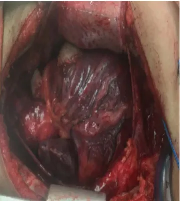
Beside the above mentioned procedure, special consideration was taken regarding the preservation of the pulmonary valve and annulus. The main pulmonary artery (MPA) is opened longitudinally (Figure 2). The first step of our technique is a precise division of the commissural fusion if there is any. The next step is extensive infundibulectomy through the small ventriculotomy incision and tricuspid valve. Now we start the smallest Hegar that the pulmonary valve can accept and start to dilate using the following Hegar size till we reach the size appropriate to the patient BSA. No excessive force should be used to dilate the valve, only by rolling movement of the Hegar aiming not to cause any traumatic injury to the valve or disruption of the annulus itself, Figure[1].
Post-operative:
All the patients who were transferred to intensive care unit, were managed as follow:
1. Continuous monitoring of:
• E.C.G. rate and rhythm
• Arterial blood pressure
• Venous blood pressure
• Peripheralbody temperature, core temperature and oxygen saturation
2. Regular checking of:
• The drains output per hour
• Urine output per hour
• Arterial blood gases and electrolytes
Journal of Advanced Pharmacy Education & Research | Jan-Mar 2018 | Vol 8 | Issue1 3
Figure 1:preservation of pulmonary valve with patch over RVOT and
MPA
3. Mechanical ventilation:
Regimen was maintained till they became hemodynamically and respiratory stable. Then gradual weaning off the mechanical ventilator is done till extubation.
4. Inotropic support:
The regimen of cardiac support was done with the use of inotropes, after load reducing agents, preload reducing agents and renal perfusion enhancement.
All doses were recorded in terms of mic/kg/hour to get average hours of inotrope used and the total infused dose. 5. Fluid and electrolyte balance was adjusted
depending on:
• C.V.P and cardiac output
• Urine output
• Body temperature
• Presence of anemia or heart failure
• Electrolyte monitoring and replacement 6. Echocardiography was done routinely
post-operative to assess:
• Left and right ventricular function
• Pulmonary and RVOT pressure gradient
• Residual pulmonary stenosis
• Grade of pulmonary regurge
• Other abnormalities or residual VSD 7. Chest X-Ray:
Routinely done to assess any collection post-operative and assess lung congestion, infection or atelectasis
8. E.C.G:
Twelve lead ECG for arrhythmias or any type of conduction abnormalities
9. Length of ICU stay:
In term of days, it was measured, and is always related to the average number of inotropes used and time of mechanical ventilation
10. Length of post-operative hospital stay:
In term of days, it was measured, and during this phase, the patient is observed closely for follow up medications, wound care and management of any postoperative heart rate arrhythmia.
11. 2nd look echocardiography before discharge: During this phase a second echocardiography was done to assess the final pressure gradient of RVOT and the grade pulmonary regurge before the final discharge from the hospital.
Results
Preoperative assessment:
In group (A) the median age was 12-month range (8-90month), while in group (B) it was 16 months (8-72 months). This difference was statistically insignificant (P>0.5).
In group (A), the median weight was 9 kilograms (5-40), while in group (B) it was 9 kilograms (6-22kg). This difference was statistically insignificant (P value>0.5).
In group (A), the median BSA was 0.50 mm2, while in group (B) it was 0.45 mm2. This difference was statistically insignificant (P value >0.05).
In group (A), the median Z score of pulmonary annulus measured by echocardiography was -3.22 range (-1.17 to -4.22), while in group (B) it was -3.89 range (-2.91 to -4.81). This difference was statistically significant (P value =0.001) (Figure 2).
Figure 2:Z score value between 2 groups
In group (A), the median pressure gradient across RVOT measured by echocardiography was 80mmhg (47-100 mmhg), while in group (B), it was 80 mmhg (60 -100 mmhg). This difference was statistically significant (P value >0.05).
In group (A), the mean cross clamp time was 48.76 with SD 11.65, while in group (B), the mean cross clamp time was 67.92 with SD 12.44. This difference was statistically significant (P value =0.0001) (Figure 3).
4 Journal of Advanced Pharmacy Education & Research | Jan-Mar 2018 | Vol 8 | Issue1 Figure 3:Relation between techniques and cross clamp time and
cardiopulmonary bypass time
To confirm the pulmonary annulus size measured pre-operative by echocardiography, we used to pass the smallest Hegar that can pass smoothly through the valve after completing the valvotomy and compare it to the previously measured MPA size
Post-operative assessment:
Regarding the mechanical ventilation in group (A) the mean was 10.20 hours SD 4.50, while in group (B), it was 18.48 hours SD 13.43. This difference was statistically significant (P value =0.0053). Mean ICU stay in group (A) was 4.76 days SD 2.00 while in group (B), it was 6.44 days SD 3.30. This difference was statistically significant (p value =0.03). Mean postoperative stay in group (A) was 4.66 days SD 3.45; while in group (B), it was 6.4 days SD 5.27. This difference was statistically insignificant (Table 1).
Table 1: postoperative mechanical ventilation, ICU stay and Post ICU stay between 2 groups
Group A Group B P value
Mean SD Mean SD
Mechanical ventilation
Hours 10.2 4.5 18.48 13.43 0.0053 ICU
Days 4.76 2.00 6.44 3.30 0.03 Post –ICU
Days 4.66 3.45 6.4 5.27 NS
Serial echocardiography was routinely done in the ICU to follow up the results. In our study, the echocardiography was done in the postoperative stay before discharge, and after the withdrawal of all inotropes. We recorded the pressure gradient across the RVOT, the degree of pulmonary stenosis, and pulmonary regurge.
Regarding RVOT pressure gradient in group (A), the median was 30 mmhg range (15-50 mmhg) while in group (B), it was 27 mmhg and range was (15-76 mmhg). This difference was statistically insignificant.
In our study, we categorized the RVOT pressure gradient into 3 groups; group 1 PG less than 20 mmhg, group 2 from 20-40 mmhg and group 3more than 40 mmhg. In group A, we had 2 (8%) patients with pressure gradient less than 20, 19 (76%) patients were between 20-40 mmhg, 4 (16%) patients were
more than 40mmhg, while in group B we had 5 (20%) patients with pressure gradient less than 20, 17 (68%) patients were between 20-40 mmhg, 3 (12%) patients were more than 40mmhg. This difference was statistically insignificant.
Regarding pulmonary regurge, there were 3 grades of pulmonary regurge. Grade 1 was considered to be non to mild PR, grade 2 was considered to be moderate, and group 3 was severe (Table 3). In group A, 20 patients 80%) were grade 1, 5 patients (20%) were grade 2, none of our patients had severe PR in this group; while in group B, 6 patients (24%) were in grade 1, 8 patients (32%) in grade 2, and 11 patients (44%) in grade 3. This was found to be statistically significant between 2 groups (P value 0.01) (Figure 4).
Figure 4:Pulmonary regurge between 2 groups
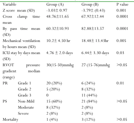
Postoperative echocardiography assessing pulmonary stenosis was done classifying the PS into 3 grades mild, moderate and severe. In group A, 15 patients (60%) had mild PS, 8 patients (32%) had moderate PS, and 2 patients (8%) had severe PS; while in group B, 21 patients (84%) had mild PS, 2 patients (8%) had moderate PS and 2 patients (8%) had severe PS. This difference was statistically insignificant (Table 2).
Table 2: Residual pulmonary stenosis between 2 groups
Group P value
A B
PS
Mild % within Count group
15
60.0% 84.0% 21
NS Mod % within Count
group
8
32.0% 8.0% 2
severe % within Count group
2
8.0% 8.0% 2
Only one patient in group B was reopened and entered on cardiopulmonary bypass again to do further excision of the infundibular muscle due to the presence of high RVOT pressure gradient, and there was failure to wean from by pass on the second attempt of repair.
Journal of Advanced Pharmacy Education & Research | Jan-Mar 2018 | Vol 8 | Issue1 5 cause that needed further intervention, rather the last arterial
blood gases before arresting showed hyperkalemia; while in group B, there were 3 patients (12 %) mortality, one of them previously mentioned before as failed to wean from cardiopulmonary bypass, 2 other patients had severe PR and RV failure. This difference was statistically insignificant.
Table 3: comparison between 2 groups as regard postoperative echocardiography, ICU stay, Z score and
mortality
Variable Group (A) Group (B) P value Z score mean (SD) -3.03± 0.97 -3.79± (0.43) 0.001 Cross clamp time
mean
48.76±11.65 67.92±12.44 0.0001
By pass time mean (SD)
60.32±10.93 82.88±13.57 0.0001
Mechanical ventilation by hours mean (SD)
10.2± 4.50 hr 18.48± 13.43hr 0.005
ICU stay by days mean (SD)
4.76 ± 2.0 days 6.44± 3.30 days 0.03
RVOT pressure gradient median (range)
30(15-50)mmhg 27 (15-76)mmhg >0.05
PR Grade 1 20 (20%) 6 (24%) 0.01 Grade 2 5 (20%) 8 (32%)
Grade 3 0 11 (44%)
PS Non-Mild 15 (60%) 21 (84%) >0.05 Moderate 8 (32%) 2 (8%)
Severe 2 (8%) 2 (8%)
Mortality 1 (4%) 3 (12%) >0.05
Discussion
In TOF surgery, the pulmonary valve anatomy, as it is related to surgery, has been historically neglected. Recently, most of the surgeons are paying attention to the valve anatomy and trying to push the boundaries in terms of valve preservation or valve orifice available. Thus, relying only on z scores can be misleading, and studies passed strictly on z scores can be confusing. Z scores must be part of the overall picture, but intraoperative evaluation of the valve is important as well[8]. In consistence with[9] and[10] who found that increased BSA those with favorable anatomy and good-sized branch pulmonary arteries should have primary repair and primary attempt to preserve the annulus, the results of the present study and our recent accumulation of experience in pediatric cardiac surgery and advances in cardiopulmonary bypass techniques may provide more promising results after primary repair in such situations even when dealing with neonates with low BSA.
During the early phase of our study, we aimed to preserve annulus with patients’ Z score -3, but the promising outcomes in our first case series allowed us to go far and preserve patients with annulus as small as -4 that was similar to what was found by Dr Vida and his group (2012). In his more recent experience, when the PV annulus is severely hypoplastic (PV z-score less than -4), the valve delamination and commissurotomy can be freely done[4].
We found that the degree of RVOT pressure gradient doesn’t merely reflect the decision of whether to preserve the annulus or
not, but supports the idea of generous removal of muscle bands obscuring the infundibulum and relieving the obstruction along pulmonary outflow tract, rather than whether the valve itself is dysplastic or not[11].
So it has to be stressed out from the previously mentioned data that the technique of pulmonary valve preservation which offers in most cases a competent pulmonary valve usually gives our patients a smooth postoperative course with mechanical ventilation period less than that underwent TAP. This all come along with results found by many authors who support that any TOF patient with adequate Z score should have a trail of preservation first[12]. In our institute, patients who were offered pulmonary valve sparing technique 80%, had grade 1 PR ranging from non to mild PR, 20% grade 2 PR with median Z score -3.22, even more we could preserve valves with z score reaching -4. Results were considered to be overwhelming after enlargement of the RVOT by single patch and other patch over the main pulmonary artery.
In light of the deleterious effect of chronic PV regurgitation, interest in preserving PV function has stimulated a few surgeons to apply valve-sparing techniques during TOF repair with the aim of preserving long-term RV function. During the last decade, with the aim of preserving PV function, we embarked on a new surgical approach that combines our standard early transatrial trans-pulmonary repair with an intraoperative systematic assessment of PV. Furthermore, in more severe cases, additional surgical maneuvers on the PV were adopted for avoiding a transannular patch and to promote PV competence[11].
In our center, along with other centers, we have found that the only true anatomical contraindication to these techniques is the presence of a unicuspid PV, where a single commissure and the dysplastic valve tissue limit our maneuvers[13].
Thus, virtually every surgeon would agree that pulmonary valve preservation is beneficial in this setting. The question is: at what price? In other words, how do you negotiate the two extremes of pulmonary stenosis (PS) and PR? In an ideal setting, one would have no regurgitation and no residual RVOT stenosis following TOF repair. Because this is rarely possible except in ideal anatomic circumstances, how much residual PS is too much to leave behind, and how much PR is acceptable.
In fact, as we became more familiar with the pulmonary valve preservation technique, we were able to preserve most pulmonary valves during early repair of tetralogy of Fallot, especially in the last few years; this contributed to the shorter follow-up time in the preservation group. In spite of the shorter postoperative follow-up time, and because we are still in a learning phase with this new technique, our results are encouraging. In fact, most patients in the preservation group, almost 80%, were discharged home with none trivial pulmonary valve regurgitation. Only a few of them, all with severe forms of tetralogy of Fallot, had a moderate degree of pulmonary valve regurgitation.
6 Journal of Advanced Pharmacy Education & Research | Jan-Mar 2018 | Vol 8 | Issue1
operation demonstrated a shorter time to extubation than patients who underwent TAP repair
We continue to recommend the earlier repair of children afflicted with this lesion. Importantly, the ability to preserve the pulmonary valve in these children should prevent the deleterious effects of free pulmonary insufficiency.
We highly recommend the PV preservation technique over the TAP, because it has proven to be more reliable in preserving the valve. The integrity and function of the PV can be preserved in selected patients during early repair of TOF, leading to a better mid-term right ventricular function. Besides, this technique has proven to be more adequate for patients who are more vulnerable for many risk factor as it has proven to:
• be applied to Z score -4
• decrease in cross clamp time
• decrease mechanical ventilation
• decrease the use on inotropes stay
• decrease ICU stay
• decrease the use of inotropes
• better pulmonary valve function up to non- mild regurge
References
1. StensenN: Quoted by Goldstein HI. Bull Hist Med 1945; 29:526.
2. Fallot A. Contribution to the pathological anatomy of the blue disease (cardiac cyanosis), Marseille Med. 1888; 25:77138, 207, 270, 341, 403.
3. Karamlou T, Silber I, LaoR: Outcomes after late reoperation in patients with repaired tetralogy of Fallot: the impact of arrhythmia and arrhythmia surgery. Ann Thorac Surg 2006; 81:1786-1793.
4. Vida VL, Angelini A, Guariento A, et al: Preserving the pulmonary valve during early repair of tetralogy of Fallot: Anatomic substrates and surgical strategies. J ThoracCardiovasc Surg 2015; 149: 1979-1984.
5. Bacha E, Danielle Gottlieb Sen, Marc Najjar, Betul Yimaz, Ste´phanie M. Levasseurm Bindu Kalessan.Jan M. Quaegebeur: Aiming to PreservePulmonary Valve Function in Tetralogy of Fallot Repair: Comparing a New Approach to Traditional Management Pediatr Cardiol, 2016 37:818–825.
6. Koestenberger M, Nagel B, Ravekes W, Avian A, Burmas A, Grangl G, Cvirn G, GamillschegA Reference values and calculation of z-scores of echocardiographic measurements of the normal pediatric right ventricle. Am J Cardiol. 2014Nov 15;114(10):1590-8.
7. Zilberman MV, Khoury PR, Kimball RT. Two-dimensional echocardiographic valve measurements in healthy children: gender-specific differences Pediatr Cardiol. 2008 Mar;29(2):475-480.
8. Bacha E. Valve-Sparing or Valve Reconstruction Options in Tetralogy of Fallot Surgery, Semin ThoracCardiovasc Surg PediatrCardSurgAnn 2017 20:79-83.
9. Vida VL, Padalino MA, Maschietto N, Biffanti R, Anderson RH, Milanesi O, et al. The balloon dilation of the pulmonary valve during early repair of tetralogy of Fallot. Catheter Cardiovasc Interv. 2012; 80:915-21.
10. Bacha E, Danielle Gottlieb Sen,Marc Najjar, Betul Yimaz, Ste´phanie M. Levasseurm Bindu Kalessan. Jan M. Quaegebeur: Aiming to Preserve Pulmonary Valve Function in Tetralogy of Fallot Repair: Comparing a New Approach to Traditional Management Pediatr Cardiol, 2016 37:818–825.
11. Vida VL, Guariento A, Castaldi B. Evolving strategies for preserving the pulmonary valve during early repair of tetralogy of Fallot: mid-term results. J Thorac Cardiovasc Surg 2014; 147:687-694.
12. Bacha EValve-Sparing or Valve Reconstruction Options in Tetralogy of Fallot Surgery, Semin ThoracCardiovasc Surg PediatrCardSurgAnn 2017 20:79-83.