R
OBERTŚ
MIGIEL1, M
AŁGORZATAP
IOTROWICZ2, I
ZABELAŁ
ACZMAŃSKA1,
I
ZABELAM
AKOWSKA1, A
NNAB
ŁOŃSKA3, K
RISTINAH
OFFMANN4, L
UCJUSZJ
AKUBOWSKI2,
N
IKOLAUSB
LIN4, M
ARIAM. S
ĄSIADEK1New Bacterial Artificial Chromosome
and Commercial FISH Probes for the 22q11.2 Region
in Patients with Congenital Heart Defect
and with Phenotype Resembling DiGeorge
and Velocardiofacial Syndromes*
Badania FISH z użyciem nowych sztucznych chromosomów
bakteryjnych oraz komercyjnych sond dla regionu 22q11.2
u pacjentów z wrodzoną wadą serca oraz fenotypem
zespołu DiGeorge’a oraz zespołu podniebienno−sercowo−twarzowego
1Department of Genetics, Silesian Piasts University of Medicine in Wrocław, Poland
2Department of Genetics, Polish Mother’s Memorial Hospital Research Institute, Łódź, Poland 3Department of Pathophysiology, Silesian Piasts University of Medicine in Wrocław, Poland
4Department of Molecular Genetics, Institute of Human Genetics, University of Tuebingen, Tuebingen, Germany
Adv Clin Exp Med 2007, 16, 6, 717–723 ISSN 1230−025X
EDITORIAL
© Copyright by Silesian Piasts University of Medicine in Wrocław
Abstract
Background.The majority of patients with DiGeorge syndrome (DGS) and velocardiofacial syndrome (VCFS) have a microdeletion in 22q11.2. The minimal DiGeorge critical region (MDGCR) has been narrowed down to 250 kb using FISH analysis. The construction of bacterial artificial contigs is an essential step towards the identification of deletions of smaller size.
Objectives.A set of bacterial artificial chromosomes (BACs) was used in a FISH assay in patients with congeni− tal heart defect and phenotype resembling DGS/VCFS to determine new, specific, deleted regions not encompas− sed by commercial probes.
Material and Methods.The study group comprised 69 patients with congenital malformations, including heart defects and dysmorphic features. The patients were divided into three subgroups. Group I comprised patients diagnosed with DGS/VCF syndrome by the detection of a 22q11.2 deletion using FISH applying the commercial probes TUPLE1 and N25. All the patients in this group also had a FISH study with seven BAC probes (115F6, 678G6, 770H11, 201C11, 919E7, 219G6, 431E9) comprising the critical region 22q11.2. Group II was made up of patients with clinical features of DGS/VCFS but without a deletion detected by FISH using the commercial FISH probes. FISH with BAC probes was also performed in this group. Group III was patients with clinical features suggesting DGS/VCFS with no deletion detected by the TUPLE1 and N25 probes. FISH with the BAC probes was not performed in the group.
Results.Within group I, deletions in the regions for BACs 770H11, 201C11, 919E7, 219G6, and 431E9 were de− tected in all 14 children with DGS/VCFS. FISH study with the 115F6 and 678G6 probes revealed the correct two signals in all patients of group I. No deletions were detected by any of the seven BACs tested in group II involving the patients with clinical DGS/VCFS features, nor was a deletion detected by the commercial probes. The clinical symptoms of the patients of the three clinically heterogeneous groups with diagnosed and suspected DGS/VCFS were compared. Palate insufficiency, hypocalcemia, and recurrent infections were significantly more frequent in patients with a 22q11.2 microdeletion confirmed cytogenetically by FISH.
Conclusions. These results strongly suggest that strict diagnostic criteria for DGS/VCFS are needed (Adv Clin Exp Med 2007, 16, 6, 717–723).
Key words:DiGeorge syndrome, velocardiofacial syndrome, phenotype, FISH, microdeletion.
DiGeorge syndrome (DGS) is a developmen− tal anomaly of the derivatives of the third and fo− urth pharyngeal pouches. It is associated with a spectrum of malformations, including absence or hypoplasia of the thymus and parathyroid glands, cardiovascular anomalies, and mild facial dysmor− phism. It has been proposed that the primary de− fect in DGS is the failure of cephalic neural crest cells to migrate properly during early embryonic development [1, 2]. Previously, cytogenetic stu− dies demonstrated that 20% of patients with DGS have chromosomal abnormalities, with the majori− ty of these chromosomal rearrangements involving the loss of the proximal long arm of chromosome 22 [3]. Subsequently, more detailed cytogenetic studies demonstrated that microdeletions of one copy of the region 22q11.2 are involved in the etiology of DGS [4].
Velocardiofacial syndrome (VCFS) is a com− mon autosomal dominant disorder characterized by cleft palate, cardiac anomaly, characteristic fa− cial features, and learning disability. Due to the phenotypic overlap between VCFS and DGS, it was postulated that both diseases might share a common pathogenesis or be etiologically related [5]. Using DNA markers for the region of 22q11.2 deleted in patients with DGS, it was possible to de− monstrate that the majority of VCFS patients are hemizygous for the same region [6]. Currently,
over 85% of the patients with a clinical diagnosis of VCFS are diagnosed with microdeletions of the 22q11.2 region [7]. These findings indicate that haplo−insufficiency of the critical region is a major factor in the development of this disorder [4].
The majority of DGS/VCFS patients (about 90%) have a large deletion which includes a com− mon set of markers in 22q11.2 [6, 8]. The size of the “commonly deleted region” (DGCR, the Di− George critical region) has been estimated to be about 3 Mb (megabases) and encompasses appro− ximately 30 genes [4, 9]. About 8% of patients, however, have a smaller deletion of 1.5 Mb which encompasses 24 genes [6, 9]. Moreover, individu− al patients can have deletion or low copy repeat si− tes which flank either proximally or distally the “commonly deleted region” [6]. Analyzing the translocation breakpoints and applying fluorescen− ce in situ hybridization (FISH) analysis, the region critical to DGS/VCFS has been narrowed down to a 250−kb (kilobase) area in the proximal fragment of the commonly deleted region (MDGCR, mini− mal DG critical region) [9, 10, 11]. This region in− cludes two markers, D22S75 (N25) and TUPLE1 (Fig. 1), which are the most consistently deleted markers in DGS/VCFS patients [4, 6, 9–11]. Ho− wever, only in a part of the patients clinically dia− gnosed as DGS/VCFS were microdeletion of the critical region confirmed by FISH assay [12, 13].
Streszczenie
Wprowadzenie.Delecja 22q11.2 jest jedną z najczęstszych mikrodelecji u człowieka i jest przyczyną kilku zespo− łów genetycznych, m.in.: zespołu DiGeorge’a (DGS) oraz podniebienno−sercowo−twarzowego (VCFS – velo−car− dio−facial syndrome). Rutynowa diagnostyka cytogenetyczna zespołów obejmuje fluorescencyjną hybrydyzację in situ(FISH) z użyciem sond charakterystycznych dla obszaru krytycznego. Ostatnie doniesienia wykazały, że w je− go obrębie mogą występować submikrodelecje obejmujące znacznie mniejsze obszary.
Cel.Analiza submikrodelecji w obszarze 22q11.2 metodą FISH z użyciem sztucznych chromosomów bakteryjnych (BAC) u pacjentów z wrodzonymi wadami serca i cechami dysmorficznymi.
Materiał i metody. Badania obejmowały 69 pacjentów z wadami wrodzonymi serca oraz cechami dysmorficznymi sugerującymi zespoły mikrodelecji 22q11.2. Pacjentów podzielono na 3 grupy: grupa I – pacjenci ze zdiagnozowa− nym zespołem DG/VCF za pomocą badania FISH z użyciem sond komercyjnych (TUPLE1, N25). W tej grupie wy− konano ponadto badanie FISH z użyciem siedmiu sond bakteryjnych (BAC – 115F6, 678G6, 770H11, 201C11, 919E7, 219G6, 431E9), obejmujących krytyczny region 22q11.2; grupa II – pacjenci z klinicznymi cechami suge− rującymi DGS/VCFS, ale bez zdiagnozowanej mikrodelecji sondami komercyjnymi. W tej grupie również wykona− no badanie FISH z użyciem sond bakteryjnych, charakterystycznych dla regionu 22q11.2, izolowanych ze sztucz− nych chromosomów bakterii; grupa III – pacjenci z cechami klinicznymi sugerującymi DGS/VCFS, bez zdiagnozo− wanej mikrodelecji sondami komercyjnymi, u których nie wykonano badania FISH z użyciem sond bakteryjnych.
Wyniki.W grupie pierwszej u wszystkich pacjentów ze stwierdzoną mikrodelecją 22q11.2 wykazano również de− lecję w regionie wyznaczonym przez sondy bakteryjne (770H11, 201C11, 919E7, 219G6, 431E9). Badanie FISH, wykonane z sondami 115F6, 678G6, nie wykazało nieprawidłowości (brak delecji). W drugiej grupie u żadnego pac− jenta nie stwierdzono mikrodelecji za pomocą sond bakteryjnych. Potwierdziło to swoistość sond komercyjnych. W pracy przeprowadzono ponadto analizę porównawczą danych klinicznych pacjentów z trzech grup ze zdiagnozo− wanym DGS/VCFS oraz z podejrzeniem ww. zespołów mikrodelecji. Wykazano istotne różnice w częstości wystę− powania niewydolności podniebienno−gardłowej, zmniejszonego stężenia wapnia w surowicy krwi oraz nawracają− cych zakażeń związanych z zaburzeniami odporności komórkowej u pacjentów z potwierdzonym zespołem mikro− delecji 22q11.2 w porównaniu z grupą II i III bez wykazanej delecji.
Wnioski. Powyższe dane sugerują, aby podejrzewając DGS/VCFS, kierować się pewnymi kryteriami diagnostycz− nymi (Adv Clin Exp Med 2007, 16, 6, 717–723).
It is now suggested that DGS/VCFS is caused by alternations of more genes in the critical deleted region, with different extents of the deletions [14]. Thus the identification of new cytogenetic and/or molecular markers is an essential step towards the diagnosis of cases which present the typical clini− cal features of DGS/VCFS but without the critical 22q11.2 microdeletion diagnosed by FISH analy− sis with commercial probes (N25 and TUPLE1). In the present study a set of bacterial artificial chromosomes (BACs) for the FISH assay was used in patients with congenital heart defect and phenotype resembling DGS/VCFS to determine new, specific, deleted regions not encompassed by commercial probes.
Material and Methods
Patients
The study group comprised 69 patients with congenital malformations, including heart defects and dysmorphic features. The patients were divi− ded into three subgroups. The first group (group I) consisted of 14 children diagnosed with DGS/VC− FS by the detection of 22q11.2 deletion using FISH applying the commercial probes TUPLE1 and N25. The second group (group II) encompassed 33 children with clinical features of DGS/VCFS but with no deletion detected in FISH by the commer− cial FISH probes TUPLE1 and N25. Based on the− ir clinical features, the children of groups I and II were selected for further investigation and FISH with seven BAC probes (115F6, 678G6, 770H11, 201C11, 919E7, 219G6, and 431E9) (Fig. 1). The third group (group III) included 22 children with clinical features suggesting DGS/VCFS but sho− wing no deletion detected by the TUPLE22 and N25 probes. This group was used as a control gro− up for clinical features.
Fluorescent
in situ
Hybridization (FISH)
with BAC Probes
The 194−kbp 115F6, 125−kbp 678G6, 140−kbp 770H11, 189−kbp 201C11, 95−kbp 919E7, 220− kbp 219G6, and 130−kbp 431E9 BAC probes, re− ceived from the Department of Molecular Gene− tics, University of Tuebingen, Germany, were la− beled using the CGH Nick Translation Kit (Vysis) according to the protocol (Tab. 1). Twenty µl (2 µg) of purified BAC clone and 5 µl of nick translation enzyme were used. The incubation time was four hours. The labeled BAC clones were purified by
Name of BAC Size (kb) Locus Distance (Nazwa BAC) (Rozmiar) (Lokus) from 22q (Odległość od 22q)
DD05−115F6 194 D22S420 16,233
DD06−678G6 125 D22S181 16,050
DD02−770H11 140 D22S111 17,850 DD09−201C11 189 D22S553 17,690
DD11−919E7 95 D22S941 17,784
DD18−431E9 130 D22S163 19,458
DD19−219G6 220 HCF2 19,466
Table 1.List of bacterial artificial chromosomes (BACs) for the FISH assay used in this study
Tabela 1.Lista sztucznych chromosomów bakteryjnych (BAC) do badania FISH użytych w pracy
Cardiac defect Number of patients (%) (Wada serca) (Liczba pacjentów – %)
Group I Group II Group III (Grupa I) (Grupa II) (Grupa III) ToF 10 (71.43) 8 (24.3) 4 (18.2)
VSD 2 (57.2) 7 (21.2) 9 (40.9)
ASD 1 (7.14) 1 (3) 3 (13.6)
TGA 0 5 (15.2) 0
HLHS 0 3 (9.1) 1 (4.6)
AVC 0 4 (12.1) 0
CoA 0 3 (9.1) 0
PDA 0 1 (3) 0
DORV 0 1 (3) 0
PFO 0 0 4 (18.2)
TA 0 0 1 (4.6)
No cardiac 1 (7.14) 0 0
defect
(Bez wady serca)
Total = 69 14 33 22
(Razem = 69)
Table 2.Phenotype of patients (cardiac defects) with DGS/VCFS in the three groups
Tabela 2. Fenotyp pacjentów (wada serca) z zespołem DG/VCF w trzech grupach
overnight precipitation with ethanol and sodium acetate according to the standard protocol. DNA was dissolved in 28 µl of Hybridization Solution (Vysis) and 12 µl of nuclease−free water and mixed at 40°C for four hours.
FISH analysis was performed using standard protocols. Three µl of BAC probe were applied at
each hybridization point on freshly prepared sli− des. After denaturation for 2 min at 73°C and over− night incubation at 37°C, the slides were washed for 2 min in 0.4 SSC/0.3% NP−40 wash solution in a 73°°C water bath and for 20 sec in 2 x SSC/0.1% NP−40 at room temperature. Ten µl of DAPI coun− terstain (Vysis) was applied to each hybridization
Clinical data Number of patients Statistics
(Dane kliniczne) (Liczba pacjentów) (Statystyka)
Group I Group II Group III Statistically (Grupa I) (Grupa II) (Grupa III) significance (Znaczenie statystyczne) p< 0.05 * Cardiac defect 13 (92.8) 33 (100) 22 (100) > 0.005 (Wada serca)
Multiple cardiac defect 8 (57.2) 18 (54.5) 10 (45.5) > 0.005 (Złożona wada serca)
Palate insufficiency, including 11 (78.6) 11 (45.5) 2 (9.09) < 0.005
cleft palate 2 (14.3) 4 (12.2) 0 > 0.005
(Niewydolność podniebienia, w tym rozszczep podniebienia)
Other congenital defect 4 (28.6) 14 (45.5) 2 (9.09) > 0.005 (Dodatkowe wady wrodzone)
Dysmorphic facial features 14 (100) 33 (100) 22 (100) > 0.005 (Cechy dysmorficzne twarzy)
Hypocalcemia 11 (78.6) 0 0 < 0.005
(Hipokalcemia)
Recurrent infection, cellular 9 (64.3) 8 (24.2) 4 (19.2) < 0.005 immunity deficit
(Nawracające infekcje, zaburzenia odporności komórkowej)
Psychomotor delay 6 (42.7) 7 (21.2) 5 (22.8) > 0.005 (Opóźnienie psychoruchowe)
Total = 69 patients
(Razem = 69 pacjentów) 14 33 22
Table 3. Clinical data of the three groups of patients
Tabela 3.Dane kliniczne pacjentów w trzech grupach
* Spearman’s correlation coefficient test and chi squared (χ2) Pearson’s test.
Group I – patients diagnosed with DGS/VCFS by detection of 22q11.2 deletion using FISH apply− ing the commercial probes TUPLE1 and N25.
Group II – patients with clinical features of DGS/VCFS without deletions detected in FISH by the commercial FISH probes TUPLE1 and N25 or by the seven BAC probes 115F6, 678G6, 770H11, 201C11, 919E7, 219G6, and 431E9.
Group III – patients with clinical features suggesting the diagnosis of DGS/VCFS with no dele− tions detected by TUPLE1 and N25 probes, without applying FISH using the BAC probes. * test Spearmana oraz Chi2(χ2) Pearsona.
Grupa I – pacjenci ze zdiagnozowanym zespołem DGS/VCF przez wykrycie delecji 22q11.2 w badaniu FISH z użyciem sondy komercyjnej TUPLE1 oraz N25.
location. A Nicon Eclipse microscope (USA) with appropriate optical filters (DAPI/FITC/TRICT) and ISIS Metasystems Software (Germany) were used for visualization and documentation (Figs. 2a and 2b). For each hybridization, 100 nuclei/me− taphase were examined.
The study design was accepted by Wroclaw Medical University’s Ethics Committee.
Results
Congenital heart defects within group I were diagnosed in 13 of the 14 patients (92.8%). Tetra− logy of Fallot (ToF) was observed in 10 cases, ventricule septal defect (VSD) in two, and atrial septal defect (ASD) in one case (Tab. 2). Multiple congenital heart defects were detected in 8 of the 14 cases (57.2%). Palatoschisis (cleft palate) was observed in only two cases, but palate insufficien− cy (regurgitation) in 11 cases (together: 93%). All children presented dysmorphic facial features. Hy− pocalcemia (and parathyroid insufficiency) was diagnosed and treated in 11 patients (93%). Recur− rent infections and deficient cellular immunity we− re observed in 9 patients (64.3%). An additional defect was observed in 4 cases (28.6%), these be− ing omphalocele, cryptorchismus, eyeball defect, and liver defect, and a delay in psychomotoric and neurological development was found in 6 patients (Tab. 3).
All the patients in group II had a congenital heart defect: 9 children had ToF, 5 transposition of great arteries (TGA), 3 hypoplastic left heart syn− drome (HLHS), 4 complete atrio−ventricular septal defect (AVC), 1 ASD, 7 VSD, 3 coarctation of aor− ta (CoA), 1 patent ductus arteriosus (PDA), and 1 with double outlet right ventricle (DORV) (Tab. 2). Multiple congenital heart defects were detected in
18 of the 33 cases (54.5%). Palatoschisis was ob− served in four cases and palate insufficiency (re− gurgitation) in 11 (together: 45.5%). All the chil− dren also presented dysmorphic facial features. No hypocalcemia (or parathyroid insufficiency) was diagnosed in this group. Recurrent infections and
Fig. 1. Human 22q11.2 region with the sizes of the deletions described in the text
Ryc. 1. Region 22q11.2 u człowieka z obszarami delecji opisanymi w tekście
Fig. 2a. Fluorescence in situhybridization (FISH) with the TUPLE1 Region Probe (22q11.2) and Control Probe (22qter). Microdeletion of 22q11.2 is diagnosed in this material. Two green signals are present repre− senting the 22qter region and one red signal represen− ting the TUPLE1 probe in the 22q11.2 region
Ryc. 2a. Badanie metodą fluoroscencyjnej hybrydyza− cji in situ(FISH) z użyciem sondy DiGeorge/VFCS TUPLE1 Region Probe (22q11.2) with Control Probe (22qter) – Cytocell. W badanym materiale stwierdzo− no obecność mikrodelecji badanego regionu 22q11.2. Widoczne są dwa sygnały zielone odpowiadające re− gionom 22qter oraz jeden sygnał czerwony odpowia− dający regionowi TUPLE1 (w obszarze 22q11.2)
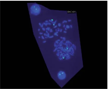
Fig. 2b. Fluorescence in situhybridization (FISH) with the BAC probe 431E9. Two correct green signals are present in two metaphases and interphases
deficient cellular immunity were observed in 8 pa− tients (24.2%). In this group, a additional defect was observed in 14 cases (42.3%), such as colobo− ma of the retina, deficit in the retina and choroidea of both eyes, cataracta, brain defect, esophageal atresia, omphalocele, cryptorchismus, anal atresia, urethral reflux, and skeletal defect. Delayed psy− chomotoric development or mild learning disabili− ty was diagnosed in 7 cases (Tab. 3).
In group III the following heart defects were diagnosed: 1 had hypoplastic left heart syndrome (HLHS) with interrupted aortic arch, hypoplastic aortic valve, and aneurysm of the interventricular septum and PDA, 9 had VSD, 4 patent foramen ovale (PFO), 1 tricuspidal valve insufficiency (TA), 3 ASD, and 4 children had ToF (Tab. 2). Congenital multiple heart defects were observed in 10 cases (45.5%). Within this group, two children had a cleft palate and two presented skeletal de− fects (polydactyly). There was no hypocalcemia in this group. Psychomotoric delay was observed in 5 cases and recurrent infection in 4 (Tab. 3).
FISH analysis using the seven above−mentio− ned BAC probes was performed in the patients of the first and second groups, all together 47 pa− tients. Within group I, deletions in the regions for BACs 770H11, 201C11, 919E7, 219G6, and 431E9 were detected in all 14 children with DGS/VCFS (Fig. 2). FISH with the remaining BACs, i.e. 115F6 and 678G6, revealed two correct signals in all the patients of group I. No deletions were detected by any of the seven BACs tested in group II, involving the patients with clinical DGS/VCFS features, nor was deletion detected by the commercial probes.
Discussion
The region of 22q11.2 deleted in the majority of patients with DGS or VCFS is greater than 1.5 Mb (DGCR). Using a limited number of patients with smaller deletions, it has been possible to narrow down the critical region to 250 kb (MDGCR) [9–12, 14]. Although several genes, such as TU− PLE1, COMT, and ZNF74, have been described within the commonly deleted region for DGS/VCFS, more genes beyond the MDGCR were recently identified [6, 9, 14]. The construction of a detailed deletion map of chromosome 22q11.2 is particu− larly important in DGS/VCFS because a number of patients present the phenotypic features and de− fects characteristic of DGS/VCFS, but no detecta− ble deletion at the MDGCR. This observation sug− gests that the size of the deletion varies among pa− tients. This was a FISH study in patients with the DGS/VCFS phenotype performed using seven dif−
ferent bacterial artificial chromosomes, i.e. 115F6, 678G6, 770H11, 201C11, 919E7, 431E9, and 219G6 (Tab. 1). Owing to their small size, BACs have remained a diagnostic challenge. Seven bac− terial chromosomal markers used in this study ha− ve a molecular size between 95 kb and 220 kb and flank the minimal commonly deleted region (MDGCR) either proximally or distally (Fig. 1). The complete chromosomal size encompassing all seven BAC regions was 3.230 Mb and was located at a distance from 16.230 Mb to 19.460 Mb from the 22−pter region.
The FISH analyses with the BACs were per− formed in 47 patients from the first and second groups. Monosomy 22q11.2 was disclosed for four BACs representing the DGCR (770H11, 201C11, 919E7, 431E9) in all patients with clinical and cytogenetic diagnoses of DGS/VCFS (microdele− tion diagnosed in FISH assay with the probes N25 and TUPLE1). The specific region for these BACs extends to 3 Mb. One marker, 219G6, located dis− tally to DGCR, was also deleted in all patients of the first group (Fig. 1). A similar finding in one multigenerational family was obtained by Rauch et al. [15]. This means that the critical region for DGS/VCFS deleted in our patients is considerably larger than the MDGCR diagnosed by commercial markers. Most DGS/VCFS deletions extend over a distance of more than 2 Mb, including a chro− mosomal fragment from the D22S111 to D22S163 position, as was shown in our patients (Fig. 1). This suggestion requires additional investigation.
There was no deletion identified in these pa− tients for two other BAC markers, 115F6 and 678G6, located proximally to the centromere of chromosome 22 and on the border of DGCR (Fig. 1). Moreover, there was also no deletion detected by any of the BACs in 22 patients of the second gro− up. This suggests that FISH results showing no de− letion at MDGCR sites in patients with clinical fe− atures of DGS/VCFS (assessed by the commercial markers TUPLE1 and N25) were reliable.
criteria for DGS/VCFS are needed [16, 17]. Each additional study concerning the etiology of DGS/VCFS, especially regarding non−deleted DGS/VCFS patients, presented in the literature will contribute to a better understanding of the phenotype of patients with 22q11.2 microdeletion.
These additional studies will also be necessary to determine how the length of the deletion and the kinds of the deleted chromosomal regions contri− bute to the various phenotypic abnormalities asso− ciated with these disorders.
References
[1] Kirby ML, Bockman DE: Neural crest and normal development: A new perspective. Anat Rec 1984, 209, 1–6.
[2] Lammer EJ, Opitz JM:The DiGeorge anomaly as a developmental field defect. Am J Med Genet 1986, 29, 113–127.
[3] Greenberg F, Elder F, Haffner P, Northup H, Ledbetter D: Cytogenetic findings in a prospective series of patients with DiGeorge anomaly. Am J Hum Genet 1988, 43, 605–611.
[4] Driscoll DA, Salvin J, Sellinger B, Budarf ML, McDonald−McGinn DM, Zackai EH, Emanuel BS:
Prevalence of 22q11 microdeletions in DiGeorge and velocardiofacial syndromes: implications for genetic coun− seling and prenatal diagnosis. J Med Genet 1993, 30, 813–817.
[5] Kelly D, Goldberg R, Wilson D, Lindsay E, Carey AH, Goodship J, Burn J, Cross I, Shprintzen R, Scambler PJ:Confirmation that the velo−cardio−facial syndrome is associated with haplo−insufficiency of genes at chromo− some 22q11. Am J Med Genet 1993, 45, 308−312.
[6] Lindsay EA: Chromosomal microdeletions: dissecting del22q11 syndrome. Nat Genet 2001, 11, 858−868.
[7] Carlson C, Sirotkin H, Pandita R, Goldberg R, McKie J, Wadey R, Patanjali SR, Weissman SM, Anyane− −Yeboa K, Warburton D, Scambler P, Shprintzen R, Kucherlapati R, Morrow BE: Molecular definition of 22q11 deletions in 151 Velo−Cardio−Facial Syndrome Patients. Am J Hum Genet 1997, 61, 620−629.
[8] Driscoll DA, Budarf ML, Emanuel BS: A genetic etiology for DiGeorge syndrome: consistent deletions and microdeletions of 22q11. Am J Hum Genet 1992a, 50, 924–933.
[9] Budarf ML, Collins J, Gong W, Roe B, Wang Z, Sellinger B, Michaud D, Driscoll D, Emanuel BS:Cloning a balanced translocation associated with DiGeorge syndrome and identification of a disrupted candidate gene. Nature Genet 1995, 10, 269–288.
[10] Li M, Budarf ML, Sellinger B, Jaquez M, Matalon R, Ball S, Pagon RA, Rosengren SS, Emanuel BS, Driscoll DA:Narrowing the DiGeorge region (DGCR) using DGS−VCFS associated translocation breakpoints. Am J Hum Genet 1994, 55, A10.
[11] Shaikh TH, Kurahashi H, Saitta SC, O’Hare AM, Hu P, Roe BA, Driscoll DA, McDonald−McGinn DM, Zackai EH, Budarf ML, Emanuel BS:Chromosome 22−specific low copy repeats and the 22q11.2 deletion syn− drome: genomic organization and deletion endpoint analysis. Hum Mol Genet 2000, 9, 4, 489−501.
[12] Ryan AK, Goodship JA, Wilson DI, Philip N, Levy A, Seidel H, Schuffenhauer S, Oechsler H, Belohradsky B, Prieur M, Aurias A, Raymound FL, Clayton−Smith J, Hatchwell E, McKeown C, Beemer FA, Dallapiccola B, Novelli G, Hurst JA, Jgnatius J, Green AJ, Winter RM, Brueton L, Brondum−Nielsen K, Scambler PJ:
Spectrum of clinical features associated with interstitial chromosome 22q11 deletions: a European collaborative study. J Med Genet 1997, 34, 789−804.
[13] Bocian E, Stankiewicz P, Jakubów−Druska K, Helias−Rodziewicz Z, Obersztyn E, Kutkowska−Kaźmierczak A, Szpecht−Potocka A, Mazurczak T: Diagnostyka kliniczna zespołów mikrodelecji – ocena przydatności metod cytogenetyki molekularnej. Ped Pol 2000, 75, 557−563.
[14] Scambler PJ, Mari A, Diglio MC, Mingarelli R, Marino B, Giannotti A, Novelli G, Dallapiccola B:
The 22q11 deletion syndromes. Hum Mol Genet 2000, 9, 2421−2426.
[15] Rauch A, Pfeiffer RA, Leipold G, Singer H, Tigges M, Hofbeck M: A novel 22q11.2 microdeletion in DiGeorge syndrome. Am J Med Genet 1999, 64, 659–666.
[16] Brunet A, Gabau E, Perich RM, Valdesoiro L, Brun C, Caballín MR, Guitart M: Microdeletion and microdu− plication 22q11.2 screening in 295 patients with clinical features of DiGeorge/Velocardiofacial syndrome. Am J Med Genet 2006, 15, 140, 22, 2426−2432.
[17] Oh AK, Workman LA, Wong GB: Clinical correlation of chromosome 22q11.2 fluorescentin situhybridization analysis and velocardiofacial syndrome. Cleft Palate Craniofac J 2007, 44, 1, 62−66.
Address for correspondence:
Robert Śmigiel Department of Genetics
Silesian Piasts University of Medicine Marcinkowskiego 1
50−368 Wrocław Tel.: +48 71 784 12 56
E−mail: [email protected]
Conflict of interest: None declared