IRANIAN JOURNAL OF BLOOD AND CANCER | Volume 2 | Number 1 | Autumn 2009 1 IJBC 2009; 5: 1-5
Methaemoglobin Content and
NADH-Methaemoglobin Reductase Activity of Three
Human Erythrocyte Genotypes
Paul Chidoka Chikezie
Department of Biochemistry, Imo State University, Owerri, Imo State, Nigeria.
Corresponding author: Paul Chidoka Chikezie, Department of Biochemistry, Imo State University, Owerri, Imo State, Nigeria. (Phone: +2348038935327, E-mail: [email protected])
Abstract
Background: To study methaemoglobin content and NADH-methaemoglobin reductase activity of three human
erythrocyte genotypes (HbAA, HbAS and HbSS).
Materials and Methods: Studies to ascertain methaemoglobin concentration and level of NADH-methaemoglobin reductase activity of three human erythrocyte genotypes (HbAA, HbAS and HbSS) were carried out in forty-three (43) healthy male participants of confirmed erythrocyte genotypes: HbAA (N=15), HbAS (N=15) and HbSS (N=13). Spectrophotometric method was used for determination of the two erythrocyte parameters.
Results: Whereas methaemoglobin concentrations in the three erythrocyte genotypes was in the order:
HbAS<HbAA<HbSS, levels of NADH-methaemoglobin reductase activity was: HbSS<hbAS<HbAA. There was no statistically significant difference (p>0.05) in methaemoglobin concentration and NADH-methaemoglobin reductase activity between HbAA and HbAS erythrocytes.
Conclusion: The results showed a relationship between erythrocyte NADH-methaemoglobin reductase activity and methaemoglobin concentration.
Keywords: Methaemoglobin, Erythrocyte, Genotype, NADH-methaemoglobin Reductase.
Introduction
Methaemoglobin (MHb) is an altered state of haemoglobin in which the ferrous (Fe2+) irons of heme are oxidized to the ferric (Fe3+) state1. The ferric haem moities of MHb are unable to bind to oxygen molecules.
NADH-Methaemoglobin Reductase (NADH-MR) (EC: 1.6.2.2) is a 300-amino acid, membrane-bound enzyme localized in the endoplasmic reticulum of all cells2.This enzyme transfers electrons from NADH to cytochrome b5 viaits flavin adenine dinucleotide (FAD) prosthetic group3. Under normal conditions, this NADH-dependent enzyme also referred to as ferricyanide reductase, NADH-diaphorase or cytochrome b5 reductase is the only system within the erythrocytethat maintains hemoglobin in its oxygen-carrying reduced state 4. It is noteworthy that a second form of thisenzyme, which is NADPH dependent, is less active in mammalsin converting
MHb back to hemoglobin than is the NADH-dependentform 5.
Generally, celluar activity of NADH-MR reflectsan organism’s capacity to reduce MHb and is therefore an important factor in evaluating MHbformation and etiology of methaemoglobinemia. From a toxicological standpoint, NADH-MR is the rate-limiting enzyme controllingthe toxicokinetics of the reduction of MHb4. Prevoius report by Borgese et al. 6 stated that the activity of NADH-MR is generally reduced in red cells of patients with recessive hereditary methaemoglobinemia. Deficiencies in the reductase systems and resulting clinical manifestations have been described 7.
NADH-MR activity has been measured in a variety of non-humanspecies8,9,10 and these studies have revealed that species with lower NADH-MR activities convert MHb back to haemoglobin at slower rates than do species with higheractivities 5.
IRANIAN JOURNAL OF BLOOD AND CANCER 2
The present study seeks to establish whether variability in NADH-MR activity exists amongst human erythrocyte genotypes, namely HbAA, HbAS and HbSS, in relation to their methaemoglobin contents. The findings of the present study will provide insights into the oxidant status of the three erythrocyte genotypes and deduce the degree of tolerance to toxic methaemoglobinemia by individuals who express these genotypes.
Materials and Methods
Selection of Volunteers/Collection of Blood Samples:
Forty-three healthy male (61-73kg) participants of confirmed HbAA (N=15), HbAS (N=15) and HbSS (N=13) genotypes between the ages of 20-28 years were enrolled for this study. Five milliliters (5.0ml) of human venous blood were obtained from participants by venipuncture and stored in EDTA anticoagulant tubes. Blood samples of HbSS genotype were from patients who attended clinics at the Federal Medical Center (FMC), Imo State University Teaching Hospital (IMSUTH), Orlu, St. John Clinic / Medical Diagnostic Laboratories, Avigram Medical Diagnostic Laboratories, and Qualitech Medical Diagnostic Laboratories. These centers are located in Owerri, Imo State, Nigeria.
Ethics:
The institutional review board of the Department of Biochemistry, University of Port Harcourt, Port Harcourt, Nigeria, granted approval for this study and all participants signed an informed consent form. This study was in accordance with the ethical principles of the Helsinki Declaration. Individuals were enrolled from Imo State University, Owerri, Nigeria and environs. The research protocols were in collaboration with registered and specialized clinics and medical laboratories.
Preparation of Erythrocyte Haemolysate:
The erythrocytes were washed by methods as described by Tsakiris et al.11 and Chikezie12. Within 2hours of collection of blood samples, portions of 1.0 ml of the samples were introduced into centrifuge test tubes containing 3.0ml of buffer solution pH=7.4: 250mM tris-HCl (Tris-HCl)/140mMNaCl/1.0mMMgCl2/10mMglucose. The erythrocytes were separated from plasma by
centrifugation at 1200g for 10minutes, washed three times by three similar centrifugations with the buffer solution. The erythrocytes re-suspended in 1.0ml of this buffer were stored at 40C. The washed erythrocytes were lysed by freezing/thawing as described by Galbraith and Watts 13 and Kamber et al.14. The erythrocyte haemolysate was used for the determination of NADH-methaemoglobin reductase activity and methaemoglobin concentration.
Determination of Methaemoglobin Concentration of Erythrocyte Lysate:
Determination of methaemoglobin content of red cell lysate was by modification of the method of Evelyn and Malloy15 as described by Akomopong et al.16 and Chikezie12. A total of 400μl of 0.5M Phosphate buffer (pH 6.5) was added to 600μl of the cell lysate and the mixture was centrifuged at 16,000g for 5minutes to sediment debris. A total of 700μl of the supernatant fraction was used to measure the absorbance at λmax=630nm (the absorbance maximum for methaemoglobin) and the reading was recorded as S1. A total of 50 μl of 10gpercentageKCN was added and after 5minutes at room temperature (240C), a second reading (S2) was recorded. KCN converts methaemoglobin to cyanomethaemoglobin, which does not absorb at 630nm; hence, the difference between absorbance readings of S1 and S2 represents the absorbance due to methaemoglobin.
To measure total hemoglobin levels, all of the hemoglobin was converted to methaemoglobin, the absorbance of the sample at λmax=630nm was recorded and then KCN was added to form cyanomethaemoglobin. Specifically, 70 μl of the supernatant fraction was diluted 10-fold into 600 μl of 0.1M phosphate buffer (pH=6.5). Next, 30μl of freshly prepared 20g% K3Fe(CN)6 was added and incubated for 5minutes at room temperature (240C) and an initial reading (T1) was recorded. A total of 50 μl of 10% KCN was subsequently added, and a second reading (T2) was recorded. The percent methaemoglobin in the sample was calculated as
[100(S1-S2)] / [10(T1-T2)].
Determination of Erythrocytes Haemolysate Haemoglobin Concentration:
A modified method of Baure17 described by Chikezie et al.18 based on cyanomethaemoglobin
Volume 2 | Number 1 | Autumn 2009 3 reaction was used for the determination of
haemolysate haemoglobin concentration. The expressed values were in grams per deciliter (g/dl). A 0.05ml portion of human red blood cell haemolysate was added to 4.95ml of Drabkin reagent (100mgNaCN and 300mg K4Fe(CN)6 per liter). The mixture was left to stand for 10minutes at room temperature and absorbance read at
λmax=540 nm against a blank. The absorbance was used to evaluate for haemolysate haemoglobin concentration by comparing the values with the standards.
Determination of NADH-Methaemoglobin Reductase Activity of Erythrocyte Lysate:
The enzyme activity represents the rate of oxidation of NADH and is described as follows:
NADH-MR activity of erythrocyte lysate was assayed according to the method of Board 19. A mixture of 0.2ml tris-HCl/EDTA buffer pH=8.0, 0.2ml NADH and 4.35ml of distilled water was introduced into a test tube and incubated for 10minutes at 300C. The whole content was transferred into a cuvette and the reaction above started by adding 0.2ml of K3Fe (CN)6/0.05ml erythrocyte haemolysate. The increase in absorbance of the medium was followed spectrophotometrically at
λmax=340nm per minute for 10 minutes at 300C against a blank solution.
Calculation of Enzyme Activity:
The equation below was used to evaluate erythrocyte NADH-MR activity in international unit per gram haemoglobin (iu/gHb).
Where,
EA =Enzyme activity in iu/gHb
[Hb] = Haemolysate haemoglobin concentration (g/dl)
0. D/min =Change per minute in absorbance at 340nm.
∑ =Millimolar extinction coefficient = 6.22, in reaction in which 1mole of NADH + H+ is oxidized.
VC =Cuvette volume (total assay volume) = 1.0ml.
VH = Volume of haemolysate in the reaction system (0.05ml).
Statistical Analysis:
The experiments were designed in a completely randomized method and data collected were analyzed by the analysis of variance procedure while treatment means were separated by the Least Significance Difference (LSD) incorporated in the Statistical Analysis System (SAS) package ver. 9.1.
Results
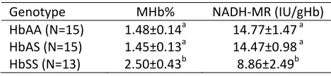
The mean (±S.D) of methaemoglobin concentration, expressed as percentage (MHb%) of total haemoglobin concentration of three erythrocyte genotypes (HbAA, HbAS and HbSS) was in the order: HbAS<HbAA<HbSS. Wheras there was no significant difference (p>0.05) in MHb% between HbAA and HbAS erythrocytes, level of methaemoglobin in sickle cell genotype exhibited significantly higher (p<0.05) concentration compared with the HbAA and HbAS genotypes. (Table 1).
The three human erythrocyte genotypes showed
varied levels of NADH-MR activity that was in the order: HbSS<HbAS<HbAA. Similar to methaemoglobin concentrations, there was no significant difference (p>0.05) in NADH-MR activity between HbAA and HbAS erythrocytes (Table 1). Therefore, there was an inverse relationship between MHb% and NADH-MR activity amongst the three human erythrocytes genotypes.
Discussion
Previous reports have revealed an association between dysfunctional erythrocytes (HbSS genotype) and raised levels of oxidized haemoglobin12, 20. They noted that the primary
Table 1: Methaemoglobin Concentration (MHb %) and NADH-Methaemoglobin Reductase Activity of Male Erythrocyte Haemolysate.
Genotype MHb% NADH-MR (IU/gHb) HbAA (N=15) 1.48±0.14a 14.77±1.47a HbAS (N=15) 1.45±0.13a 14.47±0.98a HbSS (N=13) 2.50±0.43b 8.86±2.49b
Means+S.D in the column with the same letter are not significantly different at p< 0.05 according to LSD. n= number of blood samples.
IRANIAN JOURNAL OF BLOOD AND CANCER 4
reason for the relatively raised concentration of oxidized haemoglobin (methaemoglobin) in HbSS erythrocytes was higher production of superoxide ion by these erythrocytes compared to those of HbAA and HbAS erythrocyte genotypes. Specifically, sickle erythrocytes spontaneously generate approximately twice as much superoxide, peroxide and hydroxyl radicals when compared with normal erythrocytes. In addition, HbSS erythrocytes contain increased amounts of malondialdehyde, a by-product of lipid peroxidation and evidence of abnormal amino-group cross-linking by malondialdehyde has been demonstrated in lipid extract of HbSS erythrocytes membranes21. This phenonmenone confirms the susceptibilty of sickle erythrocytes to endogenous free radical mediated oxidative damage that correlates with the proportion of irreversible sickled erythrocytes22.
The relative lower activity of NADH-MR of sickle erythrocytes compared with the HbAA and HbAS erythrocytes suggests an inverse relationship between the methaemoglobin concentration of human erythrocytes and NADH-MR activity. In concordance with this finding, previous reports by Agar and Harley 8 Lo and Agar9 and Whittington et al.10 showed that the capacity of non human species to revert oxidized haemoglobin-Fe3+ (MHb) to the reduced form haemoglobin-Fe2+ is inextricably connected with the NADH-MR activity of the corresponding erythrocytes. In addition, Geetha et al. 23 averred that high level of methaemoglobin concentration in cirrhotic erythrocytes might be partly due to low level of methaemoglobin reduction engendered by low level of NADH-MR activity. Therefore, this study suggests that persons who express the sickle erythrocytes will exhibit more susceptibilty and slower rate of recovery when challenged with agents that causes toxic methaemoglobinemia. This suggestion agrees with the findings of Daly et al. 24 who reported that several members of a family, who showed decreased activities of erythrocyte NADH-cytochrome b5 reductase, were predisposed to the development of clinically significant methaemoglobinaemia when they were administered oxidant drugs.
Conclusion
The present study showed that in the absence of exogenous oxidants, there was an inverse relationship between erythrocyte methaemoglobin concentration and NADH-MR activity.
References
1.Murray RK, Granner DK, Rodwell VW. Harper’s Illustrated Biochemistry 27th Edition. Asia: McGraw Hill Companies; 2006.
2.Dekker J, Eppink MH, van Zwieten R, Rijk T, Remacha AF, Law LK, et al. Seven new mutations in the nicotinamide adenine dinucleotide reduced-cytochrome b (5) reductase gene leading to methemoglobinemia type I. Blood. 2001; 97: 1106.
3. Yubisui T, Takeshita M. Reduction of methaemoglobin through flavin at the physiological concentration by NADPH-flavin reductase of human erythrocytes. J Biochem. 1980; 87: 1715-20.
4. Rockwood GA, Armstrong KR, Baskin SI. Species Comparison of Methemoglobin Reductase. Exp Biol Med. 2003; 228: 79–83.
5. Smith RP. Toxic responses of the blood. In: Amdur MO, Doull J, Klassen CD, Eds. Casarett and Doull’s Toxicology. New York: Pergamon Press; 1991. pp. 257– 81.
6.Borgese N, Pietrini G, Gaetani S. Concentration of NADH-cytochrome b5 reductase in erythrocytes of normal and methemoglobinemic individuals measured with a quantitative radioimmunoblotting assay. J Clin Invest. 1987; 80: 1296–302.
7.Jaffee ER. Methemoglobin pathophysiology. In: Wallach DFH. Ed. The Function of Red Blood Cells: Erythrocyte Pathobiology. New York: Alan R. Liss; 1981. pp. 133–51.
8.Agar NS, Harley JD. Erythrocyte methaemoglobin reductases ofvarious mammalian species. Experientia. 1972; 28: 1248–9.
9.Lo SCL, Agar NS. NADH-methemoglobin reductase activity in the erythrocytes of newborn and adult mammals. Experientia. 1986; 42: 1264–5.
10.Whittington AT, Parkinson AL, Spencer PBS, Grigg G, Hinds L, Gallagher CH, et al. Comparative study of the antioxidant defence systems in the erythrocytes of Australian marsupials and monotremes. Comp Biochem Physiol. 1995; 10C: 267–72.
11.Tsakiris S, Giannoulia-Karantana A, Simintzi I, Schulpis KH. The effect of aspartame metabolites on human erythrocyte membrane acetylcholinesterase activity. Pharmaco Res. 2005; 53: 1-5.
12.Chikezie PC. Comparative methaemoglobin concentrations of three erythrocyte genotypes (HbAA, HbAS and HbSS) of male participants administered with
Volume 2 | Number 1 | Autumn 2009 5
five antimalarial drugs. Afri J Biochem Res. 2009; 3: 266– 71.
13.Galbraith DA, Watts DC. Changes in some cytoplasmic enzymes from red cells fractionated into age groups by centrifugation in FicollTM/TriosilTM gradients. Comparison of normal human and patients with Duchenne muscular dystrophy. Biochem J. 1980; 191: 63-70.
14.Kamber K, Poyiagi A, Delikonstantinos G. Modifications in the activities of membrane-bound enzymes during in vivo ageing of human and rabbit erythrocytes. Comp Biochem Physiol. 1984; B.77B: 95-9.
15.Evelyn K, Malloy H. Micro determination of oxyhaemoglobin, methaemoglobin and sulfhaemoglobin in a single sample of blood. J Biol Chem. 1938; 126: 655-62.
16.Akomopong T, Ghori N, Haldar K. In vitro activity of riboflavin against the human malaria parasite Plasmodium falciparum. Antimicrob Agents Chemothera. 2000; 44: 88-96.
17.Baure JD. Laboratory investigation of hemoglobin. In: Sonnenwirth AC, Jarett L, St. Louis MO (Eds). Gradwohl’s Clinical Laboratory Methods and Diagnosis. Mosby; 1980. pp. 809-902.
18.Chikezie PC, Uwakwe AA, Monago CC. Glutathione S-transferase activity of erythrocyte genotypes HbAA,
HbAS and HbSS in male volunteers administered with fansidar and quinine. Afri J Biochem Res. 2009; 3: 210-14.
19.Board P, Coggan M, Johnston P, Ross V, Suzuki T, Webb G. Genetic heterogeneity of the human glutathione transferases; a complex of gene families. Pharmacol Ther. 1990; 48: 357-69.
20.Van Kiujk FJ, Savannah A, Handelman GI, Dratz EA. A new role for phospholipase A2: Peroxidation damage. TIBS. 1987; 12: 31-4.
21.Das SK, Nair RC. Superoxide dismutase, gluthatione peroxidase, catalase and lipid peroxidation of normal and sickled erythrocytes. Br J Haematol. 1980; 44: 87-92.
22.Rice-Evans C, Omorphos SC, Baysal E. Sickle cells membranes and oxidative damage. Biochem J. 1986; 237: 265-9.
23.Geetha A, Lakshmi Priya MD, Annie Jeyachristy S, Surendran R. Level of oxidative stress in the red blood cells of patients with liver cirrhosis. Indian J Med Res. 2007; 126: 204-10.
24.Daly JS, Hultquist DE, Rucknagel DL. Phenazopyridine induced methaemoglobinaemia associated with decreased activity of erythrocyte cytochrome b5 reductase. J Med Genetics. 1983; 20: 307-9.