Iranian Journal of Virology, Volume 4, Number 1, 2010 17
Original Article
Identification and Partial Characterization of Viral Agent of
Lettuce Big Vein in Tehran Province
Heidari F¹*, Koohi-Habibi M1, Mosahebi GH¹
1. Department of plant protection, Faculty of Agricultural Sciences and Engineering, College of Agriculture and Natural Resources, University of Tehran, Karaj, Iran.
Abstract
Background and Aims: The causal agents of viral lettuce big vein disease are two viruse,
Lettuce big vein associated virus (Varicosavirus) and Mirafiori lettuce virus (Ophiovirus). These viruses have coat proteins of similar size but have different morphologies and serologically unrelated.The purpose of this study was to distinguish and detect LBVaV and MiLV in lettuce fields in Tehran Province.
Patients and Methods: A total 344 samples with mosaic and big vein, head stunt, leaf deformation and motteling symptoms were collected from lettuce fields in Tehran Province.Using DAS – ELISA and specific antiserum for MiLV (DSMZ, AS-0798) and RT-PCR for LBVaV. Positive samples in ELISA and RT-RT-PCR were inoculated on index plants,including Chenopodium quinoa, Chenopodium amaranticolor, Lactuca sativa and
Nicotiana occidentalisp1.
Results: The results of ELISA and RT-PCR about MiLV showed that, virus is transmitted on
C.quinoa and produced chlorotic local lesion but about LBVaV, RT-PCR showed that
C.quinoa and C.amaranticolor were infected and the virus caused chlorotic local lesion. Extraction of total RNA with three methods using RNAWIZ buffer, Guanidium isothiosianat buffer and Qiagen kit showed that exteraction with RNeasy plant minikit (Qiagen company) is better for RT-PCR. RT-PCR with LBVaV and MiLV specific primer pairs (were designed with Navaro et al. 2004) were performed and the fragment length were amplified for LBVaV and MiLV respectively 296bp and 469bp. The sequence nucleotides of CP of LBVaV was determined and had high similarity with other isolates in gene bank.
Conclusion: This is the first report of occurrence of these viral diseases in lettuce in Iran (Tehran Province).
Keywords: Reverse Transcription Polymerase Chain Reaction; Enzyme-Linked Immunospot Assay; Province
Introduction
ettuce big vein disease (LBVD) was first found in California. LBVD is associated with a complex of two
viruses. Lettuce big vein associated virus (LBVaV) and Mirafiori lettuce virus (MiLV).
Lettuce big vein associated virus is the type species of the genus Varicosavirus and virions are rod-shaped with modal length of 320-360 nm and diameter of about 18 nm and contain a single coat protein (CP), with MW of about 48 kd (1). Lettuce big vein associated virus was reported to be a two segmented double – stranded RNA (dsRNA) virus, with contained two dsRNAs of approximately 6.5 and 7 Kbp
L
*Corresponding author: Fatemeh Heidari Ms.C,
Department of plant protection, Faculty of
Agricultural Sciences and Engineering, College of Agriculture and Natural Resources, University of Tehran, Karaj, IRAN
Tel: (+98) 261 222 40 22
Email: [email protected]
18 Iranian Journal of Virology, Volume 4, Number 1, 2010 (2). However, we recently demonstrated that LBVaV is not a dsRNA virus but is a single – stranded negative – sense RNA virus with a bipartite genome. Viral (negative - sense) and virus – complementary (positive – sense) RNAs are separately encapsidated in the virions and the viral RNA is predominant (3, 4). The genome is 12.9 Kb in size. LBVaV is serologically related to isolates of Tobacco stunt virus. Mirafiori lettuce virus has been included by specific morphology, serology and other characteristics into the genus of
Ophiovirus (5). The virions are naked filamentous nucleocapsids of about 3 nm in diameter. MiLV is a multicomponent ssRNA virus with a CP of approximately 48 kd (5) and particles contain four single stranded negative sense RNAs (6). The genome is 11.3 -12.5 Kb in size. Both LBVaV and MiLV are transmitted in soil by zoospores of the chytrid fungus Olpidium brassicae. The viruses are also transmitted experimentally, sometimes with difficulty, by mechanical inoculation. This disease has not been reported in Iran. The purpose of this study was the identification and partial characterization of viral agent of lettuce big vein in Tehran Province using serological, molecular and host range tests.
Methods
Sample collection
During 2005 and 2006, a total of 344 samples with mosaic, big vein, head stunt, leaf deformation and mottling symptoms were collected from lettuce fields in different areas of the province. The presence of MiLV and LBVaV were tested by RT-PCR using specific primers introduced by JA Navaro et al. (6), for both viruses. DAS-ELISA, Dot blot and Tissue print test were done using specific antiserum (DSMZ- AS, 0798) for MiLV. Because the antiserum against LBVaV was not available only RT- PCR was used for this virus.
Serological analysis of MiLV
Infected leaf samples were tested for MiLV by DAS-ELISA (Double Antibody Sandwich ELISA) as described previously. A polyclonal antibody to MiLV1 was used as the first antibody and a MiLV2 (AS-0798, prepared in DSMZ, Germany) was used as the second antibody. P-nitrophenyl phosphate was used as the substrate. All samples, antibody solutions, and substrate solutions were used at a 100 µl vol. By DAS-ELISA as described by Clark and Adams (1977). Samples were tested by Dot Immunobinding Assay and Tissue Print Immuno Assay as described by Huth (1997) using specific antibody (DSMZ, AS-0798) of MiLV.
Host Range studies
During 2005 and 2006, leaves with viral symptoms (especially big vein) were collected from lettuce fields in Tehran Province. The positive samples in DAS-ELISA and RT-PCR were used for mechanical inoculation. The sap (one volume (w/v) of 0.03 M, PH 8.3, Sodium Phosphat buffer containing 0.2% DIECA and activated charcoal at 100 mg/ml) was rubbed on the leaves of test plants, Chenopodium quinoa, Chenopodium amaranticolor, Lactuca Sativa and Nicotiana Occidentalis P1.
Total RNA exteraction
Total RNA from LBVaV and MiLV infected plants were extracted using RNeasy plant minikit (Qiagen Company).
Reverse Transcription Polymerase Chain Reaction (RT-PCR)
Total RNA from LBVaV and MiLV infected plants which were extracted with RNeasy plant minikit (Qiagen Company) were submitted to reverse transcription in a final volume of 20 μl, using 3 µl of RNA , 4 µl RT buffer 5x, 1 µl DTT (100 mM/µl), 1 µl dNTPs (10 mmol/µl), 0.5 µl RNase inhibitors ( 40 u/µl), 2 µl reverse primer of MiLV (5'-TATCAGCTCACATA CTCCCTATCG-3') and reverse primer of
LBVaV (5'-CGCCAGGATCTTTGATCC
ATCTG-3'), 100 Pmol / µl) and 8 µl H2O for 45 minutes at 42˚C with 0.5 µl MMULV reverse transcriptase (200 u/µl). 5 µl of the RT
Iranian Journal of Virology, Volume 4, Number 1, 2010 19 Fig. 2. TPIA assay on the lettuce infected with MiLV using the MiLV antiserum colored with fast red solution. Absence of the red dots is indicative of lack of the reaction.
Fig. 1. DIBA assay on the lettuce infected with MiLV using the MiLV antiserum colored with fast red solution. Absence of the red dots is indicative of lack of the reaction.
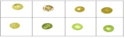
Fig. 3. Chlorotic local lesions on
Chenopodium quinoa after inoculated with the infected samples to both MiLV and LBVaV.
Fig. 4. Chlorotic local lesions on
Chenopodium amaranticolor after inoculated with the infected sample of LBVaV.
reaction were used, for PCR using a 2.5 µl PCR buffer 10x, 1 µl MgCl2 (50 mmol/µl), 0.5
µl dNTPs (10 mmol/ µl), 0.3 µl Taq DNA polymerase (5 u/ µl), 14.6 H2O, 0.5 µl reverse primer (100 pmol/ µl) and 0.6 µl forward primer (5'-TGCGACATGTTCCTCCTCATCG -3') of LBVaV, 0.5 µl reverse primer and 0.6 µl forward primer (5'-CAACTAGCTCAGAA TACATGCAG-3') of MiLV (6). PCR reaction was performed by a first denaturation of the samples at 95˚C for 30 seconds followed by 35 cycles at 94˚C for 3 minutes, 62˚C for 30 seconds and 72˚C for 40 seconds and a final elongation step at 72˚C for 10 minutes. PCR products were analyzed by electrophoresis on 1% agarose gel.
Nucleotide sequence of the coat protein gene of LBVaV
Nucleotide sequence of the coat protein gene of LBVaV isolate from Tehran was determined and compared with the other isolates in gene bank using Blast, DNAMAN and Vector NTI programs.
Results
Percentage of infected samples
From 344 lettuce samples, 1.74%, 0.87% and 0.53% were infected, respectively, with both viruses, LBVaV and MiLV alone. Samples were considered positive when their absorbance in DAS – ELISA tests, was twice of thr negative samples. DIBA and TPIA tests also confirmed the results of DAS – ELISA test. Positive samples had deep red color
change as compared to the colorless negative samples (Fig. 1, 2).
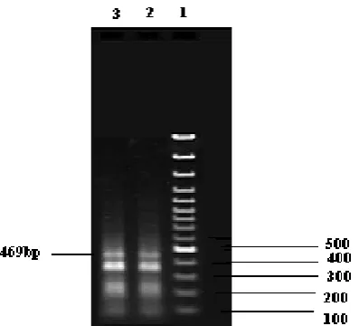
20 Iranian Journal of Virology, Volume 4, Number 1, 2010 Fig. 5. Electrophoresis of the product of RT-PCR using specific primers of LBVaV Line 1: 1 kb Marker (Gene Ruler TM 1Kb DNA ladder)
Line 2 and 3: healthy plant
Line 4: Lettuce Sativa infection LBVaV the amplified 296bp segment.
Fig. 6. Electrophoresis of the product of RT-PCR using specific primers of MiLV
Line 1: 1 kb Marker (Gene Ruler TM 1Kb DNA ladder)
Line 2 and 3: Lettuce Sativa infection MiLV the amplified 469bp segment.
R(Reverse) =248
ATTGCCCCTTCAGACAAAATACTGTGC AGATACCGTTGCCATCTTACCCAACAT CTTTTCGATGGGAAAGCTATCtACAGC AGACTAGCAATCCTCTGAACATTGCTG TGCTAAAACAGATGGCCCCGGAGAGA AAGAGGTACACAAGACAGGTGGCGAA GAACATCTACCATCACTTCATGGTGGT TGCCAGGGCCCTGAACAACGACATGTT CGACACAGACAAATATAAGTTTGTGG AATCCGACGATGAGGAGGAACATGTC GCAA
Fig. 7. The sequence of LBVaV isolated from letuuce.
Fig. 8. The dendrogram related to the isolate LBVaV collected from Tehran, along with the other isolates existed in the gene bank (using DNA MAN software). Host Range studies
The results of DAS-ELISA and RT-PCR of MiLV showed that the virus was transmitted
on C.quinoa and produced colored local lesions, LBVaV, RT-PCR showed that
C.quinoa and C.amaranticolor were infected by virus as colored lesions were developed (Fig. 3, 4).
RT-PCR
RT-PCR with LBVaV and MiLV specific primer pairs were performed for LBVaV 296 bp and for MiLV 469 bp were amplified (Fig. 5, 6).
Sequence nucleotide of coat protein gene of LBVaV
Sequence nucleotid of coat protein gene of LBVaV was determined and had %93-97% similarity with other isolates in gene bank (Fig. 7, 8).
LBVaVGL LBVaVMU
98
LBVaVSO LBVaVUK
LBVaVIra
95 0.05
Iranian Journal of Virology, Volume 4, Number 1, 2010 21 The isolates LBVaV – MUR2 (AY581691.1)
and LBVaV – GAL1 (AY366412.1) had the high and the least similarity with Iranian isolate.
Discussion
Big vein disease of lettuce (Lactuca Sativa L.) is an important problem in most established production areas. Young plants that are affected remain small and unmarketable (7). The disease is commonly present at temperatures below 20°C (8, 9). In recent years, the sensitivity of plant virus detection in a great number of virus host combinations has improved due to use of nucleic acid – based techniques compared with serological techniques. Indeed, such techniques are now considered reliable fast and inexpensive. Confirmation of the findings of Roggero et al., (2000), that MiLV as well as LBVaV occure in lettuce crops has been briefly noted (10, 11). Lettuce big vein is associated with a complex of two viruses, Lettuce big vein associated virus and Mirafiori lettuce virus.
RT-PCR using LBVaV and MiLV specific
primer pairs (12) were performed
approximately for LBVaV and MiLV 296bp and 469bp which were compatible with Navaro et al., reports. Sequence nucleotide of coat protein of LBVaV was determined it has high similarity with other isolates in gen bank. Both viruses were mechanically transmissible from lettuce to herbaceous hosts and to lettuce, although with variable success. The results of DAS-ELISA and RT-PCR about MiLV showed that the virus was transmitted to
Chenopodium quinoa. RT-PCR test showed that Chenopodium quinoa and Chenopodium amaranticolor were infected by LBVaV. But with repeated inoculation on lettuce, big vein symptom did not appear. Reports showed that with repeated mechanical transfers, both viruses appeared to lose the ability to be vector transmitted, and MiLV, when transmitted, appeared to lose the ability to cause big vein (13). LBVaV and MiLV are transmitted by the zoospores of the soil born fungus Olpidium brassicae (Wor) Dang (13). In this regard, both viruses MiLV and LBVaV are existent in
lettuce fields of Tehran, Thus separately and mix infections of two viruses were determined in samples.
Virus is existent in both the irrigation water and the soil, which may be infested with the resting spores of the vector (14). Thus, virus inoculum does not increase unless the fungus infects susceptible host cells. This is the first report of occurrence of these viruses on lettuce in Iran (Tehran Province).
In this regard, crop rotation could represent a feasible measure of control. Olpidium brassicae can infect a great variety of host plants including weeds, which may act as a reservoir. Rigorous weed control is an important stop to take in order to reduce the incidence of viruses infecting lettuce crops.
Acknowledgement
We sincerely thank Dr. S. Winter, DSMZ Plant Virus Department (Germany) for providing MiLV antiserum.
References
1. Kuwata,S, Kubo S, Yamashita S, and Doi Y. Rod-shaped particles, a probable entity of lettuce big vein virus. Annals of Phytopath-ology Society of Japan. 1983; 49(2):246-251. 2. Mayo MA. Virus Taxonomy: Seventh Report of the International Committee on Taxonomy of Viruses. Academic Press. 2000; 521-523.
3. Sasaya T, Ishikawa K, and Koganezawa H. Nucleotide sequence of the coat protein gene of Lettuce big-vein virus. Journal of General Virology. 2001; 82: 1509–1515.
4. Sasaya T, Ishikawa K, and Koganezawa H. The nucleotide sequence of RNA1 of Lettuce big-vein virus, genus Varicosavirus reveals its relation to nonsegmented negative strand RNA viruses. Virology. 2002; 297:289–297.
5. Roggero P, Ciuffo M, Vaira AM, Accotto GP, Masenga V, and Milne RG. An Ophiovirus isolated from lettuce with big-vein symptoms. Archives of Virology. 2000; 145:2629–2642.
6. Navarro JA, Torok VA, Vetten HJ, and Pallas,V. Genetic variability in the coat protein
22 Iranian Journal of Virology, Volume 4, Number 1, 2010 genes of lettuce big-vein associated virus and Mirafiori lettuce big-vein virus. Archives of Virology. 2005; 150:681–694.
7. Navarro JA, Botella F, Maruhenda A, Sastre P, S´anchez-Pina MA, and Pallas V. Comparative infection progress analysis of Lettuce big-vein virus and Mirafiori lettuce virus in lettuce crops by developed molecular diagnosis techniques. Phytopathology. 2004; 94:470-477.
8. Zink FW, and Grogan R.G. The interrelated effects of big vein and market price on the yield of head lettuce. Plant Disease Reporter. 1954; 38:844-846.
9. Bos L, and Huijberts N. Screening for resistance to big-vein disease of lettuce (Lactuca sativa). Crop Protection. 1990; 9(6):446-452.
10. Verbeek M, Van Der Wilk F, Vetten HJ, and Van Der Heuvel JFJM. Viruses associated with the lettuce big-vein syndrome. Abstract
Arbeitskreis Viruskrankheiten der Pflanzen Tagung .2001; 29–30.
11. Roggero P, Lot H, Souche S, Lenzi R, and Milne RG. Occurrence of Mirafiori lettuce virus and Lettuce big-vein virus in relation to development of big-vein symptoms in lettuce crops. European Journal of Plant Pathology. 2003; 109: 261–267.
12. Navarro JA, Torok VA, Vetten HJ, and Pallas V. Genetic variability in the coat protein genes of lettuce big-vein associated virus and Mirafiori lettuce big-vein virus. Archives of Virology. 2005; 150: 681–694.
13. Lot H, Campbell RN, Souche S, Milne RG, and Roggero P. Transmission by Olpidium brassicae of Mirafiori lettuce virus and Lettuce vein virus, and their roles in lettuce big-vein etiology. Phytopathology. 2002; 92: 288-293.
14. Campbell RN. Weeds as reservoir hosts of the lettuce big-vein virus. Canadian Journal of Botany. 1965; 43:1141-1149.